Evaluation of in-house PCR for diagnosis of smear-negative pulmonary tuberculosis in Kampala, Uganda doc
Bạn đang xem bản rút gọn của tài liệu. Xem và tải ngay bản đầy đủ của tài liệu tại đây (269.81 KB, 8 trang )
RES E A R C H A R T I C L E Open Access
Evaluation of in-house PCR for diagnosis of
smear-negative pulmonary tuberculosis in
Kampala, Uganda
Lydia Nakiyingi
1,2*†
, David P Kateete
3†
, Ponsiano Ocama
1,2
, William Worodria
3
, Joseph B Sempa
1
,
Benon B Asiimwe
3
, Fred A Katabazi
3
, Achilles Katamba
2
, Laurence Huang
4
, Moses L Joloba
3
and Harriet Mayanja-Kizza
2
Abstract
Background: Nucleic acid amplification tests (NAATs) have offered hope for rapid diagnosis of tuberculosis (TB).
However, their efficiency with smear-negative samples has not been widely studied in low income settings. Here,
we evaluated in-house PCR assay for diagnosis of smear-negative TB using Lowenstein-Jensen (LJ) culture as the
baseline test. Two hundred and five pulmonary TB (PTB) suspects with smear-negative sputum samples, admitted
on a short stay emergency ward at Mulago Hospital in Kampala, Uganda, were enrolled. Two smear-negative
sputum samples were obtained from each PTB suspect and processed simultaneously for identification of MTBC
using in-house PCR and LJ culture.
Results: Seventy two PTB suspects (35%, 72/205) were LJ culture positive while 128 (62.4%, 128/205) were
PCR-positive. The sensitivity and specificity of in-house PCR for diagnosis of smear-negative PTB were 75%
(95% CI 62.6-85.0) and 35.9% (95% CI 27.2-45.3), respectively. The positive and negative predictive values were 39%
(95% CI 30.4-48.2) and 72.4% (95% CI 59.1-83.3), respectively, while the positive and negative likelihood ratios
were 1.17 (95% CI 0.96-1.42) and 0.70 (95% CI 0.43-1.14), respectively. One hundred and seventeen LJ culture-
negative suspects (75 PCR-positive and 42 PCR-negative) were enrolled for follow-up at 2 months. Of the
PCR-positive suspects, 45 (60%, 45/75) were still alive, of whom 29 (64.4%, 29/45) returned for the follow-up visit; 15
(20%, 15/75) suspects died while another 15 (20%, 15/75) were lost to follow-up. Of the 42 PCR-negative suspects,
22 (52.4%, 22/42) were still alive, of whom 16 (72.7%, 16/22) returned for follow-up; 11 (26.2%, 11/42) died while
nine (21.4%, 9/42) were lost to follow-up. Overall, more PCR-positive suspects were diagnosed with PTB during
follow-up visits but the difference was not statistically significant (27.6%, 8/29 vs. 25%, 4/16, p = 0.9239).
Furthermore, mortality was higher for the PCR-negative suspects but the difference was also not statistically
significant (26.2% vs. 20% p = 0.7094).
Conclusion: In-house PCR correlates poorly with LJ culture for diagnosis of smear-negative PTB. Therefore, in-house
PCR may not be adopted as an alternative to LJ culture.
Keywords: Pulmonary tuberculosis, Smear-negative TB, HIV-infected, HIV-TB co-infection, CD4 cell counts, Nucleic
acid amplification tests, In-house PCR, Lowenstein-Jensen culture, Sensitivity, Specificity, Resource limited settings
* Correspondence:
†
Equal contributors
1
Infectious Diseases Institute, Makerere University College of Health Sciences,
Mulago Hospital Complex, Kampala, Uganda
2
Department of Medicine, School of Medicine, Makerere University College
of Health Sciences, Kampala, Uganda
Full list of author information is available at the end of the article
© 2012 Nakiyingi et al.; licensee BioMed Central Ltd. This is an Open Access article distributed under the terms of the Creative
Commons Attribution License ( which permits unrestricted use, distribution, and
reproduction in any medium, provided the original work is properly cited.
Nakiyingi et al. BMC Research Notes 2012, 5:487
/>Background
The genetically homogeneous subspecies of the Myco-
bacterium tuberculosis complex (MTBC; M. tuberculosis,
M. bovis, M. bovis BCG, M. africanum, M. caprae and
M. cannetti) cause tuberculosis (TB) [1,2], a global dis-
ease that affects one third of the human population
[3,4]. TB and HIV co-infection affects many in sub-
Saharan Africa [5-7]; Uganda has a high HIV prevalence
and is also among the world’s 22 high TB-burdened
countries with an estimated incidence of 402 cases per
100,000 individuals [3]. Kampala, the capital of Uganda
has approx. 2 million inhabitants and accounts for
approx. 30% of the nation’s TB burden [4].
Accurate diagnosis is crucial for efficient management
of TB patients [3]; however, TB diagnosis remains a chal-
lenge particularly in resource limited settings (RLS)
where the disease is complicated by HIV co-infection.
Conventional approaches to TB diagnosis in RLS still
rely on methods that have major limitations [8-10].
Smear microscopy is the most widely available method
but has varying sensitivity (30 to 60%) particularly in
TB-HIV co-infected patients. The chest X-ray, often a
supplementary test for diagnosis of smear-negative pul-
monary TB (PTB) also has low specificity. Solid cultures
are used as confirmatory tests but are expensive, lengthy
(up to 8 weeks) and not widely available in RLS [11].
The World Health Organization (WHO) recommends
liquid cultures in high TB burdened countries due to the
advantage of rapid detection and incremental yield in
comparison with solid media [12]. However, liquid cul-
ture systems are expensive, prone to contamination and
usually support the growth of non-tuberculous mycobac-
teria (NTM).
Nucleic acid amplification tests (NAATs) are promis-
ing new methods for rapid detection of M. tuberculosis
(MTB) directly in samples or TB culture and are being
considered as cost-effective alternatives in RLS [13,14].
The latest development was the WHO’s endorsement of
the GeneXpert (Xpert MTB/Rif) for use in TB endemic
countries, declaring the system a major milestone for
global TB diagnosis. The high cost notwithstanding [15],
some sub-Saharan African countries (e.g. South Africa,
Morocco, etc.) have introduced the Xpert MTB/Rif sys-
tem for routine TB diagnostics. Even then, research on
the optimal use of NAATs for TB diagnosis is still want-
ing in sub-Saharan Africa where there is high burden of
HIV/TB co-infection.
An in-house PCR assay for rapid identification of
MTBC in smear-positive sputum samples and acid fast
bacilli (AFB) positive cultures was previously introduced
in this setting [16], but it has never been evaluated for
the diagnosis of smear-negative PTB in the same setting.
Using LJ culture as the base-line test, this study evalu-
ated in-house PCR for rapid diagnosis of smear-negative
PTB in a low income setting with high burden of TB/
HIV co-infection.
Methods
Setting, participants and specimen collection
This study was conducted between September 2007 to
February 2008, on a short stay emergency medical ward
at Mulago National Referral and Teaching Hospital in
Kampala, Uganda. The emergency ward temporarily
admits an d triages patients before transfer to specialized
medical units. Approx. 30 patients per day are admitted,
of whom one third have respiratory symptoms. Patients
with respiratory symptoms were examined by specialists
who identified PTB suspects. PTB suspects were defined
as patients with cough for ≥2 weeks with or without any
of the following; sputum production, haemoptysis, chest
pain, shortness of breath, loss of appetite, weight loss,
fatigue, night sweat and fever.
Two sputum samples (one on spot and another early
morning) were collected from each PTB suspect and
examined by Ziehl-Neelson (ZN) microscopy for identi-
fication of AFB [17]. Sputum induction (using 7% hyper-
tonic saline inhaled by nebulization) was used for
patients who were unable to expectorate sputum.
Inclusion/exclusion criteria
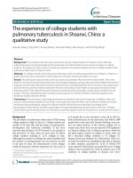
A total 320 PTB suspects were screened; patients with
smear-positive sputum samples were excluded but started
on TB treatment according to the Uganda National TB
guidelines. To be enrolled in the study, a PTB suspect
ought to have consecutively produced at least two AFB
smear-negative sputum samples upon ZN microscopy.
Overall, sputum samples for culture and DNA extraction
were obtained from a total of 205 PTB suspects who met
the above criteria (i.e. two smear-negative sputum sam-
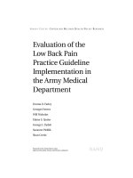
ples); these were recruited as study participants (Figure 1).
In addition, demographic and clinical data were obtained
from the 205 enrolled participants.
HIV testing
HIV testing was performed for all the enrolled patients
following the algorithm for the ministry of health, Uganda
[18]. CD4+ cell counts by BD FACS calibur (Becton and
Dickinson, Franklin Lakes, NJ, USA) were performed, as
well as chest X-rays.
Sputum processing
Sputum processing and culture were performed in biosaf-
ety l evel 3 facility at the national TB reference laboratory
(NTRL) in Kampala, Uganda. The sputum samples were
processed by digestion and decontamination in a bio-
safety cabinet class II as previously described [16,19].
Briefly, 200 μl of digestion buffer (2.9% sodium citrate, N-
Acetyl L-cysteine [NALC] and 6% NaOH) was added to an
Nakiyingi et al. BMC Research Notes 2012, 5:487 Page 2 of 8
/>equal volume of sputum, vortexed and incubated at room
temperature for 15 min. The digested sample was then
diluted to 50 ml with phosphate buffer (pH 6.8), mixed
thoroughly and centrifuged at 4000 g for 15 min; the sedi-
ment was then suspended in 2 ml phosphate buffer.
MTBC cultures
LJ culture, widely used for TB diagnosis in RLS [20], was
used as a baseline test to assess the diagnostic accuracy of
in-house PCR. LJ culture was chosen as the gold standard
because all the mycobacterial colonies on LJ-positive sam-
ples in this setting are predominantly MTBC [21,22]. For
cultures, 100 μl each of the processed sputum (see above)
was inoculated into L J culture bottles and incubated
at 37°C for up to 3 months. Cultures were considered
positive only if mycobacterial colonies appeared within
8 weeks following inoculation. Colonies from culture-
positive LJ bottles were confirmed for presence of AFB by
ZN microscopy and 16 s rRNA PCR.
16S rRNA PCR was performed on LJ-positive cultures
to confirm mycobacteria (which were presumptively
regarded MTBC [21,22]) and rule out subtle growth from
other acid fast bacilli organisms on LJ media (such as
Nocardia, Corynebacteria and Frankia). PCRs were per-
formed on all LJ-positive cultures using the following pri-
mers: 5'-ACG GTG GGT ACT AGG TGT GGG TTT C-
3', forward and 5'-TCT GCG ATT ACT AGC GAC TCC
GAC TTC A-3', reverse. The amplification program was
as follows: initial denaturation at 94°C for 4 min, followed
by 31 cycles each consisting of denaturation at 94°C for
30s; annealing at 63°C for 30s and extension at 72°C for
45 s. Then, there was a final extension at 72°C, for 10 min.
Amplicons were analyzed by agarose gel electrophoresis in
which a 600 bp fragment was detected in positive samples.
Chromosomal DNA extraction
DNA extraction and molecular assays were performed at
the Molecular Biology Laboratory, Department of Med-
ical Microbiology, Makerere University College of Health
Sciences. Approx. 0.5 ml each of the processed sputum
samples (see sputum processing) in screw-capped cryo-
vials (Nalgene, Thermo Fisher Scientific, Rochester,
USA) were incubated at 80°C for 2 h to heat kill the ba-
cilli. Then, chromosomal DNA was extracted with the
Master pure™ purification kit (Epicentre Biotechnologies,
Madison, USA) following the manufacturer’s guidelines.
PCR positive,
culture positive
N = 48
PCR positive,
culture negative
N = 75
PCR negative,
culture positive
N = 16
PCR negative,
culture negative
N = 42
Both In-house PCR &
LJ culture results
available: N=181
5 cultures
contaminated
In-house PCR results: N=186
LJ culture results: N=200
320 PTB suspects screened for AFB using ZN microscopy
ZN-Positive
N=115
ZN-Negative on 2 smears
N=205 (Recruited)
LJ culture and In-house PCR
N= 205
Treated
5 PCRs with no culture
results, excluded
Figure 1 Study flow chart.
Nakiyingi et al. BMC Research Notes 2012, 5:487 Page 3 of 8
/>In-house PCR assays
The IS6110 insertion sequence, which is unique to the
MTBC members [23,24] was the target for the in-house
PCR assay. Amplification reactions were performed with
primers, P43 (forward, 5'-TCAGCCGCGTCCACGCCG
CCA-3'), and P53 (Reverse, 5'-CCGACCGCTCCGACC
GACGGT-3') [16], which amplify 521 bp of IS6110. Each
reaction contained 20 pmoles of the forward and reverse
primer, 1 μl of custom PCR-Master mix (10 mM Tris-
HC1, pH 9.0, 2 mM MgCl
2
, 50 mM KCl, 200 μM dNTPs
and 5% DMSO), 0.5U Taq polymerase and 2 μlof
chromosomal DNA template in a reaction volume of
10 μl.
Amplifications were performed in the PTC-200 Peltier
thermocycler (MJ Research, Waltham, USA) under the
following conditions: initial denaturation at 94°C for
5 min; followed by 34 cycles each consisting of 94°C,
30 s; 65°C, 30 s; and 72°C, 45 s; and a final extension
step at 72°C for 10 min. Then, amplicons were electro-
phoretically analyzed using 1% agarose gel in TBE (Tris-
Borate EDTA) buffer stained with ethidium bromide and
visualized under ultraviolet (UV) light in a UV transillu-
minator. Positive control reactions included template
DNA purified from MTB strain H
37
Rv, while negative
controls included reactions with only pure nuclease free
water or DNA extracted from M. smegmatis and Escheri-
chia coli. Presence of an approx. 500 bp fragment in the
test lanes indicated presence of MTBC in the sample
provided controls were valid.
Patient follow-up
To determine survival status and confirm diagnosis of
TB, follow-up at 2 months (window 8 to 16 weeks) was
done for PCR-positive/culture negative as well as PCR-
negative/culture negative study participants and the
outcomes of both categories compared. ZN-sputum
microscopy was performed during the follow-up visits.
Additionally, medical records and additional test-results
were also reviewed. Information wa s obtained by tele-
phone interviews for participants who were unable to re-
turn for follow-up vis its. Patients were classified as
having PTB ba sed on any of the following: MTB C
isolated in at least one culture; positive ZN sputum
smear; granulomas on histopathology; and clinical re-
sponse to TB treatment in absence of a non-TB alternative
diagnosis.
Quality control
Cross contamination of cultures was minimized through
use of sterile disposable aerosol resistant tips. For each
sample, separate tubes with decontamination/phosphate
buffers were used to avoid cross-transfer of specimens.
Samples with only phosphate buffer were always included
in the batch being processed and these remained negative
upon culture. For molecular assays, separate rooms were
used for sample preparation, reaction mixes, DNA amplifi-
cation and detection. After use, UV hoods were deconta-
minated by turning on UV light. Negative controls were
included in each PCR batch to detect cross-contamination
during premixes. To determine the effect of PCR inhibi-
tors, reactions were spiked with 500 ng of MTB chromo-
somal DNA from the reference strain H
37
Rv (ATCC
27294) and ran in parallel; amplification of the IS6110
fragment implied absence of or minimal PCR inhibition.
All laboratory personnel were blinded to the clinical and
culture data.
Data analysis
The data wer e analyzed with STATA version 10.0 (Stata-
Corp LP, College Station, TX, USA). The sensitivity, spe-
cificity, positive and negative predictive values as well as
diagnostic likelihood ratios for in-house PCR assay were
calculated using LJ culture as the base line test. To com-
pare clinical outcomes (TB diagnosis and mortality) at
2 months of follow-up between P CR-positive/culture
negative and P CR-negative/culture negative partici-
pants , a Z-test was used to test for differences in pro-
portions. A p value of < 0.05 was considered statistically
significant.
Ethical considerations
The study wa s approved by the Makerere University Fac-
ulty of Medicine Research and Ethics Committee. Writ-
ten informed consent was obtained from all the patients
who participated in this study.
Results
Two hundred and five smear-negative PTB suspects
were recruited, with a mean age of 34.7 years (±10.4
standard deviation) and an equal gender distribution.
There were few smokers and most patients were HIV-
infected (85.9%, 176/205), of whom 72.2% (127/176) had
advanced immunosuppression (CD4+ cell count of ≤ 200
cells/μL). Furthermore, many patients had abnormal
chest findings (76.1%, 156/2 05). Although many patient s
reported fever, only 41% (84/205) had a body
temperature of ≥ 37.5°C at enrolment (Table 1).
Of the 205 cultured samples, 72 (35.1%) grew myco-
bacterial colonies on LJ media. Since LJ culture method
has been found non-conducive for growth of non-
tuberculous mycobacteria in our setting [21,22] all the
LJ culture-positive samples were regarded as MTBC.
Furthermore, 128 (62.4%) samples had no visible growth
while five (2.4%) cultures were contaminated (Figure 1).
Of the 205 sputum samples analyzed by in-house PCR,
19 (9.3%, 19/205) results were not available leaving 186
(90.7%, 186/205) PCR results for analysis. Of the 186,
128 (68.8%, 128/186) were confirmed as MTBC while
Nakiyingi et al. BMC Research Notes 2012, 5:487 Page 4 of 8
/>five (5/186) had the corresponding LJ culture results
contaminated hence excluded (Figure 1).
Performance of the in-house PCR in diagnosing
smear-negative PTB
Five PCR samples had culture results contaminated and
were excluded from the analysis leaving 181corresponding
PCR and LJ culture results (Figure 1 and Table 2). The
sensitivity and specificity of in-house PCR in diagnosing
smear-negative PTB was 75% (95% CI 62.6-85.0) and
35.9% (95% CI 27.2-45.3), respectively. The positive and
the negative predictive values were 39% (95% CI 30.4-
48.2) and 72.4% (95% CI 59.1-83.3), respectively. The posi-
tive and negative likelihood ratios were 1.17, 95% CI
(0.96-1.42) and 0.7, 95% CI (0.43-1.14) respectively. Details
of these performance indices are shown in Tables 2 and 3.
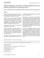
Clinical outcomes
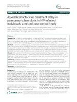
One hundred and seventeen culture-negative suspects
(75 PCR-positive and 42 PCR-negative) were enrolled
for the 2 months follow-up visit. Of the 75 PCR-positive
ones, 45 (60%) were still alive of whom 29 (64.4%, 29/
45) returned for the follow-up visit; 15 (20%) suspe cts
died while another 15 (20%) were lost to follow-u p
(Figure 2). Of the 42 PCR-negative suspects, 22 (52.4%)
were still alive of whom 16 (72.7%, 16/22) returned for
follow-up (Figure 2); 11 (26.2%, 11/42) suspects died
while nin e (21.4%, 9/42) were lost to follow-up.
Overall, more PCR-positive suspects were diagnosed
with TB during follow-up but the difference was not sta-
tistically significant (27.6%, 8/29 vs. 25%, 4/16,
p = 0.9239). On the other hand, mortality was higher for
PCR-negative suspects but the difference was also not
statistically significant (26.2% vs. 20% p = 0.7094).
Discussion
In this study, in-house PCR correlated poorly with LJ
culture for diagnosis of smear-negative PTB. Although
reported previously in other settings [25,26], this is
among the few studies evaluating the performance of in-
house PCR on smear-negative PTB suspects in Uganda,
a low income country with high rates of HIV/TB co-in-
fection. The sensitivity for the in-house PCR in the
Table 1 Baseline characteristics of smear-negative PTB
suspects (n = 205)
Characteristics Frequency Percentage
Socio-demographics
Females 105 51.2
Never smoked 140 68.2
Smoked 65 31.8
Clinical parameters as reported
by patients
Cough (>2 weeks) 205 100
Fever and/or night sweats 193 94.1
Weight loss 191 93.2
Wheezing 42 20.5
Difficulty in breathing 130 63.4
Blood in sputum 57 30.1
Antibiotic exposure in previous 2 weeks 84 41.0
Clinical examination (physical)
Body temperature (°C)*
<37.5 113 57.4
≥37.5 84 42.6
≤20 45 23.6
>20 146 76.4
Oxygen saturation (% measured by
a pulseoximeter)*
≤90 47 23.1
>90 156 76.9
Pulse rate, beats/min (measured by
a pulseoximeter)
Median rate (percentile range) 106 (88 to119)
Lung examination
Normal (clear chest) 49 23.9
Abnormal (Rhonchi, crepitation, bronchial
breathing, absent breath sounds)
156 76.1
HIV status
Positive 176 85.9
Negative 29 14.1
CD4 Cell Count* (cell/ml)
≤ 200 127 76
>200 40 24
*Some measurements missing.
Table 2 Performance indices for In-house PCR using LJ
Culture as the base line test
Culture Positive Culture Negative Total
PCR Positive 48 75 123
PCR Negative 16 42 58
Total 64 117 181
Table 3 More performance indices for In-house PCR
Performance Indices Index value 95% CI
Sensitivity 75% 62.6-85.0
Specificity 35.9% 27.2-45.3
Positive Predictive Value 39% 30.4-48.2
Negative Predictive Value 72.4% 59.1-83.3
Diagnostic Likelihood ratio (Positive) 1.17 0.96-1.42
Diagnostic Likelihood ratio (Negative) 0.70 0.43-1.14
Nakiyingi et al. BMC Research Notes 2012, 5:487 Page 5 of 8
/>current study was higher than that reported in an earlier
African stud y on ZN-negative sputum samples (i.e., 75%
vs. 40%) [26]. Although we endeavored to control for
PCR inhibitors, the sensitivity of the DNA polymerase
can be affected by the paucibacillary nature of specimens
[27], which could also have affected the PCR assay sensi-
tivity in our study.
Although the performance indices in this study were
estimated with LJ culture which is not the gold standard
for MTBC identification, virtually all LJ-positive cultures
in this setting are MTBC and speciation tests to confirm
MTB are deemed unnecessary since colonies are pre-
sumptively MTBC [22]. LJ culture also is widely used as
a gold standard for TB diagnosis in RLS [20].
Since our concern was mostly TB-diagnosis (i.e. pa-
tient-care) of which LJ is the gold standard for MTBC
culture, we thus did not speciate cultures but confirmed
the presumptive MTBC as mycobacteria through 16S
rRNA PCR.
Specificity for the in-house PCR in the current study
was also low (35.9%). Although a couple of PCR studies
have achieved high specificity with smear-negative PTB,
they mostly worked with commercial tests [28,29] that
are expensive for many in RLS. Otherwise, most studies
with in-house PCR on smear-negative PTB have revealed
substantial variability in specificity [28,30].
It is still possible that the many false positives in the
current study could have resulted from the low sensitiv-
ity of LJ culture [22,31]. Indeed, the culture-positive/
PCR-negative isolates could have been NTM, which are
known to cause severe disease in immunocompromised
HIV-positive patients with low CD4+ counts. Moreover,
majority of the subjects in this study were HIV-infected
with low CD4+ cell counts. We hope future studies will
consider these omissions (i.e. speciating NTM among
AFB smear-negative PTB suspe cts).
Furthermore, while the Flores et al. 2005, meta-
analysis for in-house PCR accuracy [28] found the
IS6110 amplification target highly accurate, this was not
shown in the current study, meaning that IS6110 alone
may not be adequate for increased diagnostic yield. The
diagnostic likelihood ratio (DLR) for a positive in-house
PCR was 1.17 [95% CI (0.96-1.42)] implying that a posi-
tive in-house PCR test may not indicate presence of
MTBC. Likewise, the negative DLR was 0.7 [95% CI
(0.43-1.14)] implying that a negative in-hou se PCR test
is not indicative of absence of MTBC. Therefore, with
in-house PCR in this setting, a clinician will need add-
itional diagnostic methods to confirm PTB in smear-
negative suspects.
There was no significant difference in mortality and
diagnosis of TB at follow-up between PCR-positive/cul-
ture negative and PCR-negative/culture negative PTB sus-
pects, although mortality was higher for PCR-negative
suspects. However, this could be due to other factors that
were not addressed, for instance co-morbidities.
Limitations
Due to limited funding, we did not use biochemical tests
or DNA sequencing which methods are considered gold
standards for MTBC identification; probably these would
have provided higher accuracy estimates for the in-
house PCR. Species-confirmation of the LJ-positive cul-
tures was not done in light of recent findings in parallel
studies in this setting, in which AFB growth on LJ
medium is virtually MTBC [21,22].
LJ culture negative participants
N=117
In-house PCR Positive
N=75
Alive
N=45
Dead
N=15
Lost to follow-up
N= 15
Returned for Follow-
up N=29
Returned for follow-up
N=16
TB diagnosed
N=4
TB
diagnosed
N=8
In-house PCR Negative
N=42
Alive
N=22
Dead
N=11
Lost to follow-up
N= 9
Figure 2 Clinical outcomes for LJ-culture negative patients comparing PCR-positive and PCR-negative groups at 2 months follow-up.
Nakiyingi et al. BMC Research Notes 2012, 5:487 Page 6 of 8
/>Few participants returned for the follow-up visits and
we were unable to establish the possible cause of death
in the participants who died. Although predictive values
are reported, these cannot be accurately interpreted in
this pooled population. Lastly, this study does not repre-
sent the general use of in-house PCR in a real world set-
ting, since PCR methods vary widely with setting and
the data herein may not be generalizable.
Conclusions
In-house PCR is inefficient for diagnosis of smear-negative
PTB. Its diagnostic accuracy is low and it may not be used
as an alternative for LJ culture in this setting.
Abbreviations
AFB: Acid fast bacilli; BSL-3: Biosafety level 3; DMSO: Dimethyl sulfoxide;
dNTPs: Deoxyribonucleotide triphosphates; EDTA: Ethylenediaminetetraa cetic
acid; ELISA: Enzyme linked immunosorbent assays; HIV: Human
immunodeficiency virus; LJ: Lowenstein-Jensen media; MTB: Mycobacterium
tuberculosis; MTBC: Mycobacterium tuberculosis complex; NAAT: Nucleic acid
amplification tests; NALC: N-Acetyl L-cysteine; NPV: Negative predictive value;
NTRL: National tuberculosis reference laboratory; NTM: Non tuberculous
mycobacteria; PCR: Polymerase chain reaction; PPV: Positive predictive value;
PTB: Pulmonary tuberculosis; RLS: Resource limited settings; TB: Tuberculosis;
TBE: Tris-Borate EDTA; UV: Ultraviolet light; WHO: World health organization;
ZN: Ziehl-Neelson.
Competing interests
The authors declare that they have no competing interests.
Authors’ contributions
LN, PO, MLJ, HMK, LH conceived and designed the study. LN and FAK
performed the molecular assays. LN, JBS, WW, AK, MLJ and HMK analyzed
the data. LN, DPK and PO wrote the manuscript. All authors read and
approved the manuscript.
Acknowledgements
The authors thank the Fogarty International Clinical Research Scholars and
Fellows Program (FIRCS-F) and Makerere University Infectious Diseases
Institute (IDI) for the scientific and financial support; the staff at Mulago
Hospital, Ward 3BEM; the laboratory technicians; the MIND- IHOP study team;
the Makerere University Department of Medical Microbiology (Molecular
biology laboratory) and the NTRL for laboratory support; and the study
participants. The authors also thank Professor Walter Schlech of Dalhousie
University, Canada, for proof reading the manuscript.
Author details
1
Infectious Diseases Institute, Makerere University College of Health Sciences,
Mulago Hospital Complex, Kampala, Uganda.
2
Department of Medicine,
School of Medicine, Makerere University College of Health Sciences, Kampala,
Uganda.
3
Department of Medical Microbiology, School of Biomedical
Sciences, Makerere University College of Health Sciences, Kampala, Uganda.
4
HIV/AIDS Division and Division of Pulmonary and Critical Care Medicine, San
Francisco General Hospital, University of California-San Francisco, San
Francisco, CA, USA.
Received: 12 December 2011 Accepted: 31 August 2012
Published: 5 September 2012
References
1. Cole ST: Comparative and functional genomics of the Mycobacterium
tuberculosis complex. Microbiology 2002, 148(Pt 10):2919–2928.
2. Gagneux S, Burgos MV, DeRiemer K, Enciso A, Muñoz S, Hopewell PC, Small
PM, Pym AS: Impact of Bacterial Genetics on the Transmission of
Isoniazid-Resistant Mycobacterium tuberculosis. PLoS Pathog 2006,
2(6):e61.
3. WHO: Global tuberculosis control: epidemiology, strategy, financing.
WHO report. 2009, Available from: />WHO_TB_report_without_annexes_2009.pdf.
4. USAID: Uganda Tuberculosis Country Profile. [www.usaid.gov/our_work/
global_health/id/tuberculosis/…/uganda.pdf]
5. The deadly synergy of HIV and tuberculosis. Lancet Infect Dis 2010, 10
(7):441. doi:10.1016/S1473-3099(10)70124-9.
6. El-Sadr WM, Tsiouris SJ: HIV-associated tuberculosis: diagnostic and
treatment challenges. Semin Respir Crit Care Med 2008, 29(5):525–531.
7. Tsiouris SJ, Gandhi NR, El-Sadr WM, Friedland G: Tuberculosis and HIV-
needed: a new paradigm for the control and management of linked
epidemics. MedGenMed 2007, 9(3):62.
8. Nahid P, Pai M, Hopewell PC: Advances in the diagnosis and treatment of
tuberculosis. Proc Am Thorac Soc 2006, 3(1):103–110.
9. Foulds J, O’Brien R: New tools for the diagnosis of tuberculosis: the
perspective of developing countries. Int J Tuberc Lung Dis 1998,
2(10):778–783.
10. Perkins MD: New diagnostic tools for tuberculosis. Int J Tuberc Lung Dis
2000, 4(12 Suppl 2):S182– 188.
11. Reid MJA, Shah NS: Approaches to tuberculosis screening and diagnosis
in people with HIV in resource-limited settings. Lancet Infect Dis 2009,
9(3):173–184.
12. Shen G-H, Chiou C-S, Hu S-T, Wu K-M, Chen J-H: Rapid Identification of the
Mycobacterium tuberculosis Complex by Combining the ESAT-6/CFP-10
Immunochromatographic Assay and Smear Morphology. J Clin Microbiol
2011, 49(3):902–907.
13. Desmond EP, Loretz K: Use of the Gen-Probe amplified mycobacterium
tuberculosis direct test for early detection of Mycobacterium
tuberculosis in BACTEC 12B medium. J Clin Microbiol
2001,
39(5):1993–1995.
14. Ryang DW, Ryang DH, Shin MG, Shin JH, Kee SJ, Suh SP: Alternative use of
polymerase chain reaction instead of rho-nitro-alpha-acetylamino-beta-
hydroxypropiophenone test for the early detection of Mycobacterium
tuberculosis in BACTEC 12B cultures. APMIS 1996, 104(6):444–450.
15. Evans CA: GeneXpert—a game-changer for tuberculosis control? PLoS
Med 2011, 8(7):e1001064. doi:10.1371/journal.pmed.1001064.
16. Muhumuza J, Asiimwe BB, Kayes S, Mugyenyi P, Whalen C, Mugerwa RD,
Boom H, Eisenach KD, Joloba ML: Introduction of an in-house PCR for
routine identification of M. tuberculosis in a low-income country. Int J
Tuberc Lung Dis 2006, 10:1262–1267.
17. Harries A, Maher D, WHO: TB/HIV, a clinical manual. 1996, http://www.
uphs.upenn.edu/bugdrug/antibiotic_manual/TB-HIVclinicalmanual.pdf.
18. The Ministry of Health, Uganda: Uganda National Policy Guidelines for HIV
Counselling and Testing. www.who.int/hiv/pub/guidelines/uganda_art.pdf.
19. Kent L, McHugh T, Billington O, Dale J, Gillespie S: Demonstration of
homology between IS6110 of Mycobacterium tuberculosis and DNAs of
other Mycobacterium spp.? [published erratum appears in J Clin
Microbiol 1995 Nov;33(11):3082]. J Clin Microbiol 1995, 33(9):2290–2293.
20. Zhu C, Cui Z, Zheng R, Yang H, Jin R, Qin L, Liu Z, Wang J, Hu Z: A multi-
center study to evaluate the performance of phage amplified
biologically assay for detecting TB in sputum in the pulmonary TB
patients. PLoS One 2011, 6(9):e24435.
21. Lukoye D, Cobelens FG, Ezati N, Kirimunda S, Adatu FE, Lule JK, Nuwaha F,
Joloba ML: Rates of anti-tuberculosis drug resistance in Kampala-Uganda
are low and not associated with HIV infection. PLoS One 2011,
6(1):e16130.
22. Worodria W, Anderson J, Cattamanchi A, Davis JL, den Boon S, Andama A,
Yoo SD, Joloba M, Huang L, Kato-Maeda M: The role of speciation in
positive lowenstein-jensen culture isolates from a high tuberculosis
burden country. PLoS One 2011, 6(11):e27017.
23. Cave MD, Eisenach KD, McDermott PF, Bates JH, Crawford JT: IS6110:
conservation of sequence in the Mycobacterium tuberculosis complex
and its utilization in DNA fingerprinting. Mol Cell Probes 1991, 5(1):73–80.
24. Hellyer TJ, DesJardin LE, Assaf MK, Bates JH, Cave MD, Eisenach KD:
Specificity of IS6110-based amplification assays for Mycobacterium
tuberculosis complex. J Clin Microbiol 1996, 34(11):2843–2846.
25. Sarmiento OL, Weigle KA, Alexander J, Weber DJ, Miller WC: Assessment by
meta-analysis of PCR for diagnosis of smear-negative pulmonary
tuberculosis. J Clin Microbiol 2003, 41(7):3233–3240.
26. Kambashi B, Mbulo G, McNerney R, Tembwe R, Kambashi A, Tihon V,
Godfrey-Faussett P: Utility of nucleic acid amplification techniques for the
Nakiyingi et al. BMC Research Notes 2012, 5:487 Page 7 of 8
/>diagnosis of pulmonary tuberculosis in sub-Saharan Africa. Int J Tuberc
Lung Dis 2001, 5(4):364–369.
27. Amicosante M, Richeldi L, Trenti G, Paone G, Campa M, Bisetti A, Saltini C:
Inactivation of polymerase inhibitors for Mycobacterium tuberculosis
DNA amplification in sputum by using capture resin. J Clin Microbiol 1995,
33(3):629–630.
28. Flores LL, Pai M, Colford JM Jr, Riley LW: In-house nucleic acid
amplification tests for the detection of Mycobacterium tuberculosis in
sputum specimens: meta-analysis and meta-regression. BMC Microbiol
2005, 5:55.
29. Brodie D, Schluger NW: The diagnosis of tuberculosis. Clin Chest Med 2005,
26(2):247–271. vi.
30. Rattan A: PCR for diagnosis of tuberculosis: where are we now? Ind J Tub
2000, 47:79–82.
31. Negi SS, Khan SF, Gupta S, Pasha ST, Khare S, Lal S: Comparison of the
conventional diagnostic modalities, bactec culture and polymerase chain
reaction test for diagnosis of tuberculosis. Indian J Med Microbiol 2005,
23(1):29–33.
doi:10.1186/1756-0500-5-487
Cite this article as: Nakiyingi et al.: Evaluation of in-house PCR for
diagnosis of smear-negative pulmonary tuberculosis in Kampala,
Uganda. BMC Research Notes 2012 5:487.
Submit your next manuscript to BioMed Central
and take full advantage of:
• Convenient online submission
• Thorough peer review
• No space constraints or color figure charges
• Immediate publication on acceptance
• Inclusion in PubMed, CAS, Scopus and Google Scholar
• Research which is freely available for redistribution
Submit your manuscript at
www.biomedcentral.com/submit
Nakiyingi et al. BMC Research Notes 2012, 5:487 Page 8 of 8
/>