Báo cáo khoa học: Thermosynechoccus elongatus DpsA binds Zn(II) at a unique three histidine-containing ferroxidase center and utilizes O2 as iron oxidant with very high efficiency, unlike the typical Dps proteins ppt
Bạn đang xem bản rút gọn của tài liệu. Xem và tải ngay bản đầy đủ của tài liệu tại đây (717.72 KB, 15 trang )
Thermosynechoccus elongatus DpsA binds Zn(II) at a
unique three histidine-containing ferroxidase center and
utilizes O
2
as iron oxidant with very high efficiency, unlike
the typical Dps proteins
Flaminia Alaleona*, Stefano Franceschini*, Pierpaolo Ceci, Andrea Ilari and Emilia Chiancone
C.N.R. Institute of Molecular Biology and Pathology, Department of Biochemical Sciences ‘A. Rossi-Fanelli’, University of Rome
‘La Sapienza’, Italy
Introduction
The widely expressed bacterial Dps proteins (DNA-
binding proteins from starved cells) are part of the
complex defense system that bacteria use to combat
stress conditions. The family prototype was identified
in stationary-phase Escherichia coli cells, where it binds
DNA and protects it from DNase cleavage, and also
renders cells resistant to hydrogen peroxide stress [1].
Later observations established that E. coli Dps is also
expressed during exponential growth in cells exposed
to oxidative stress [2], and that it protects DNA from
Keywords
Dps proteins; ferroxidase center;
ferroxidation reaction; protection from;
reactive oxygen species;
Thermosynechococcus elongatus
Correspondence
E. Chiancone, Department of Biochemical
Sciences ‘A. Rossi-Fanelli’, University of
Rome ‘La Sapienza’, 00185 Rome, Italy
Fax: +39 06 4440062
Tel: +39 06 49910761
E-mail:
Database
The atomic coordinates for DpsA-Te have
been deposited in the RCSB Brookhaven
Protein Data Bank ()
under accession code PDB ID 2VXX
*These authors contributed equally to this
work
(Received 13 October 2009, revised 20
November 2009, accepted 4 December
2009)
doi:10.1111/j.1742-4658.2009.07532.x
The cyanobacterium Thermosynechococcus elongatus is one the few bacteria
to possess two Dps proteins, DpsA-Te and Dps-Te. The present character-
ization of DpsA-Te reveals unusual structural and functional features that
differentiate it from Dps-Te and the other known Dps proteins. Notably,
two Zn(II) are bound at the ferroxidase center, owing to the unique substi-
tution of a metal ligand at the A-site (His78 in place of the canonical
aspartate) and to the presence of a histidine (His164) in place of a hydro-
phobic residue at a metal-coordinating distance in the B-site. Only the
latter Zn(II) is displaced by incoming iron, such that Zn(II)–Fe(III) com-
plexes are formed upon oxidation, as indicated by absorbance and atomic
emission spectroscopy data. In contrast to the typical behavior of Dps pro-
teins, where Fe(II) oxidation by H
2
O
2
is about 100-fold faster than by O
2
,
in DpsA-Te the ferroxidation efficiency of O
2
is very high and resembles
that of H
2
O
2
. Oxygraphic experiments show that two Fe(II) are required to
reduce O
2
, and that H
2
O
2
is not released into solution at the end of the
reaction. On this basis, a reaction mechanism is proposed that also takes
into account the formation of Zn(II)–Fe(III) complexes. The physiological
significance of the DpsA-Te behavior is discussed in the framework of a
possible localization of the protein at the thylakoid membranes, where
photosynthesis takes place, with the consequent increased formation of
reactive oxygen species.
Structured digital abstract
l
MINT-7312099: DpsA (uniprotkb:Q8DL82) and DpsA (uniprotkb:Q8DL82) bind (MI:0407)
by x-ray crystallography (
MI:0114)
Abbreviations
H-FtHu, recombinant human H-ferritin; ICP-AES, inductively coupled plasma atomic emission spectroscopy.
FEBS Journal 277 (2010) 903–917 ª 2010 The Authors Journal compilation ª 2010 FEBS 903
UV and gamma irradiation, and acid and base shock
[3]. Furthermore, it was established that the DNA-
binding capacity is shared only by those members
of the family that possess a flexible N-terminus or
C-terminus rich in positively charged residues or a
positively charged molecular surface [4–8]. In contrast,
all Dps proteins have iron oxidation⁄ uptake capacity
[9] and are characterized by a shell-like assembly
[10–13], in both respects resembling ferritin. They were
thus assigned to the ferritin superfamily. There are,
however, several different structural and functional
features between the two protein families.
The ferritin oligomer has 432 symmetry, and in ani-
mals is built from 24 highly similar subunits, the
L-chains and H-chain, with the latter harboring intra-
subunit catalytic centers, whereas Dps proteins are
formed from 12 identical subunits assembled with 23
tetrahedral symmetry, and contain unusual intersubunit
ferroxidase centers, located at the dimer interfaces [9].
Importantly, whereas purified ferritins use O
2
as iron
oxidant, with the production of H
2
O
2
, Dps proteins
typically prefer H
2
O
2
, which is about 100-fold more effi-
cient than O
2
[14]. The simultaneous consumption of
Fe(II) and H
2
O
2
reduces their potential toxicity, as it
inhibits hydroxyl radical production via Fenton chemis-
try. It follows that Dps proteins are able to protect bio-
logical macromolecules from Fe(II)-mediated and
H
2
O
2
-mediated stress more efficiently than ferritins.
This functional disparity manifests itself in the different
sensitivity of ferritin and Dps deletion mutants to
O
2
-generated and peroxide-generated oxidative stress
[15,16]. In turn, differences in the physiological roles of
ferritins and Dps proteins are likely to underlie the
significant variability in the type and number of ferritin-
like proteins expressed in different bacteria. Thus,
E. coli and Salmonella enterica possess two ferritins,
one heme-containing ferritin (bacterioferritin) and a
Dps protein [17,18], whereas Porphyromonas gingivalis
[16] and Campylobacter jejuni [15] each contain one fer-
ritin and a Dps protein. Only a few bacterial species
express two Dps proteins, such as the radiation-resistant
mesophilic eubacterium Deinococcus radiodurans [19,20]
and several bacilli [12,21]. The presence of two dps genes
appears to be more frequent in cyanobacteria, on the
basis of the known genomes sequenced (http://genome.
kazusa.or.jp/cyanobase/). Thermosynechococcus elonga-
tus [22,23], Anabaena variabilis, Gloeobacter violaceus,
Nostoc punctiforme, Prochlorococcus marinus, Synecho-
coccus sp. and Trichodesmium erythraeum belong to this
category. The coexistence of ferritins and Dps proteins
is most intriguing, as the structural and functional prop-
erties of the Dps family members characterized to date
appear to be very conserved.
Key to the physiological activity of all of these pro-
teins is the ferroxidase center, which is highly con-
served in both ferritins and Dps proteins. In ferritins,
the center is bimetallic, as in all known proteins with
ferroxidase activity; the two iron atoms are at a dis-
tance of about 3 A
˚
, and are connected by an oxo-
bridge. The so-called A-site typically uses a histidine
and carboxylates as iron-coordinating ligands, and
binds iron with higher affinity than the so-called B-site,
where the metal is coordinated only by means of carb-
oxylates [24]. Among Dps proteins, the ferroxidase
center was identified in Listeria innocua Dps, where it
contains one strongly bound iron coordinated by
Glu62 and Asp58 from one subunit, by His31 from
the symmetry-related subunit, and by a water molecule
that is located about 3 A
˚
from the iron and forms a
hydrogen bond with His43 from the same monomer
[11]. Ilari et al. [11] proposed that a second iron atom
could replace the water molecule and give rise to a
canonical bimetallic ferroxidase center. In the known
X-ray structures of Dps proteins, the occupancy of the
ferroxidase center with iron varies despite the conser-
vation of the iron ligands, a fact that points to a sig-
nificant influence of residues in the second ligation
sphere. Thus, in E. coli Dps the center contains two
water molecules, a fact ascribed to the presence of a
lysine (Lys48) engaging Asp78, one of the iron ligands,
in a salt bridge interaction [25].
For investigation of the physiological basis of the
coexistence of two Dps proteins within a single bacte-
rium, those expressed by T. elongatus appeared to be
of special interest. T. elongatus is a thermophilic, uni-
cellular, rod-shaped cyanobacterium that lives in hot
springs at 55 °C. The occurrence of oxygenic photo-
synthesis entails increased formation of reactive oxygen
species as a result of the photosynthetic transport of
electrons, such that, besides photosystems I and II,
which are the main targets of photodamage, other cel-
lular components are at risk. The T. elongatus genome
contains the genes encoding for two Dps proteins,
Dps-Te and DpsA-Te (IDs of the respective genes,
tll2470 and tll0614), and one ferritin, but lacks cata-
lase ⁄ peroxidase genes. Thus, Dps-Te and DpsA-Te,
together with ferritin, must play an important role in
alleviating the toxic effects of reactive oxygen species.
The most interesting of the two T. elongatus Dps pro-
teins is DpsA-Te. A sequence alignment (Fig. 1) shows
that it is the only member of the family among those
known that carries a substitution at the ferroxidase cen-
ter, where a histidine (His78) replaces the canonical
aspartate (Asp58 in L. innocua). Near the ferroxidase
center, His164 replaces a hydrophobic residue (phenyl-
alanine or methionine), and a phenylalanine (Phe52)
The unusual Thermosynechoccus elongatus DpsA F. Alaleona et al.
904 FEBS Journal 277 (2010) 903–917 ª 2010 The Authors Journal compilation ª 2010 FEBS
replaces the highly conserved tryptophan (Trp32 in
L. innocua).
The structural and functional properties of DpsA-Te
described here show features, such as the presence of
two Zn(II) bound at the ferroxidase center and the high
efficiency of O
2
as iron oxidant, that render this protein
unique among the Dps proteins characterized to date,
and point to a distinct physiological role of DpsA-Te
relative to the previously studied Dps-Te [23].
Results
Sequence analysis of T. elongatus DpsA
The DpsA-Te sequence was compared with those of
the Dps family members of known three-dimensional
structure (Fig. 1). A sequence similarity search
performed with blast ( />Blasy.cgi) showed the highest identity (36%, 64 ⁄ 175
residues) with Halobacterium salinarum DpsA, 29%
identity with Dps-Te (46 ⁄ 158 residues), 28% identity
with Bacillus brevis Dps (40 ⁄ 139 residues), and 27%
with Bacillus anthracis Dps2 (40 ⁄ 139 residues). The
sequence identity with the prototypic E. coli Dps and
L. innocua Dps was about 22%.
DpsA-Te possesses a long N-terminal extension that
has a partially hydrophobic character and lacks the
DNA-binding signature characteristic of the E. coli
Dps N-terminus, namely the positively charged lysines
and arginines that interact with the negatively charged
DNA backbone. On this basis, and given the lack of a
long, positively charged C-terminal extension as in
Mycobacterium smegmatis Dps [7], DpsA-Te is not
predicted to bind DNA.
Fig. 1. Alignment of representative
sequences of Dps proteins. DpsA-Te from
T. elongatus, Dps from H. salinarum
(Dps-Hs), Dps from E. coli (Dps-Ec), Dps
from B. brevis (Dps-Bb), Dps1 from B.
anthracis (Dps1-Ba), Dps2 from B. anthracis
(Dps2-Ba), MrgA from Bacillus subtilis
(MrgA-Bs), Dps from L. innocua (Dps-Li),
Dps-Te from T. elongatus (Dps-Te), and Nap
protein from Helicobacter pylori (Nap-Hp).
The residues at the ferroxidase center are
indicated by arrows, the cysteines are in
gray, and DpsA-Te His164 (see text) is in
bold and underlined.
F. Alaleona et al. The unusual Thermosynechoccus elongatus DpsA
FEBS Journal 277 (2010) 903–917 ª 2010 The Authors Journal compilation ª 2010 FEBS 905
The most striking features emerging from the
sequence comparison concern, as expected, the replace-
ment of the otherwise conserved aspartate at the ferr-
oxidase center with a histidine (His78), and the
absence of tryptophans. Typically, Dps proteins con-
tain two conserved tryptophans, one near the ferroxi-
dase center (Trp52 in E. coli Dps, present in 90% of
the known sequences) and the other (Trp160 in E. coli
Dps, present in the majority of the known sequences)
located at the three-fold interface. These two residues
are replaced, respectively, by a phenylalanine and a
tyrosine. A further unusual feature of DpsA-Te is the
presence of five cysteines (Cys30, Cys69, Cys102,
Cys103, and Cys114), as the other Dps sequences con-
tain a maximum of one cysteine per monomer (e.g.
E. coli Dps and H. salinarum DpsA).
X-ray crystal structure of T. elongatus DpsA
DpsA-TeHis yielded X-ray quality crystals, whereas all
attempts to crystallize DpsA-Te failed. DpsA-TeHis
forms cubic I23 crystals with the following cell dimen-
sions: a = b = c = 174.504 A
˚
, a = b = c = 90.00°.
The best crystal diffracted at 2.4 A
˚
resolution
(Table 1). The dataset collected from this crystal was
used to determine the protein structure by molecular
replacement, using as search model the H. salinarum
DpsA tetramer (Protein Data Bank entry: 1MOJ),
which displays 36% sequence identity with DpsA-Te.
The final model contains four identical subunits that
represent the asymmetric unit and are related by a
two-fold and a three-fold symmetry axis. The coordi-
nates and structure factors have been deposited in the
Protein Data Bank (ID: 2VXX).
As for the other members of the family, the DpsA-
TeHis monomer is folded into a four-helix bundle and
assembles into a shell-like dodecamer characterized by
tetrahedral 23 symmetry, with external and internal
diameters of about 90 A
˚
and 45 A
˚
, respectively.
However, upon superimposition of the DpsA-TeHis
monomer with those of Dps-Te and L. innocua Dps
(rmsd values of 1.18 A
˚
and 1.15 A
˚
, respectively), the
N-terminal part of the DpsA-TeHis D-helix appears to
be slightly bent (about 5°) towards the B-helix, a fea-
ture that has important ramifications at the interfaces
(see below). The DpsA-TeHis N-terminal extension
(1–15) is long and flexible as in E. coli and H. salina-
rum Dps. It is in a random coil conformation, and is
visible apart from the first two residues. The next six
amino acids of the extension assume a different con-
formation with respect to H. salinarum Dps, whereas
the last seven have the same disposition. The five char-
acteristic cysteines are located in the A-helix and
B-helix (Cys30 and Cys69, respectively) and in the BC-
loop (Cys102, Cys103, and Cys114). The X-ray crystal
structure clearly shows that Cys30, Cys69 and Cys114
are completely buried in the monomer, and that the
side chains of Cys102 and Cys103 are oriented towards
the core of the protein and therefore cannot interact
directly with solvent. The C-terminal extension (six res-
idues long) assumes an extended conformation and is
completely visible, whereas the 13 residues belonging
to the His-tag are not.
The symmetry of the dodecamer defines two non-
equivalent interfaces and pores along the three-fold
axes that have been named ‘Dps-type’ and ‘ferritin-
like’, as the first are typical of Dps proteins, and the
second resemble the trimeric interfaces of canonical
ferritins with octahedral 432 symmetry [11].
In DpsA-Te, the subunits forming the pores at the
ferritin-like interfaces have a slightly different orienta-
tion with respect to the three-fold symmetry axes than
in the other Dps structures (Fig. 2A). This fact, taken
together with the slight bending of the N-terminal part
of the D-helix towards the C-helix, leads to a rear-
rangement of the ferritin-like interfaces that results in
Table 1. Crystal parameters, data collection and refinement statis-
tics of DpsA-TeHis. Values in parentheses are for the highest-reso-
lution shell.
Data reduction and crystal parameters
Space group I23
a = b = c (A
˚
) 174.504
No. of molecules in asymmetric unit 4
Solvent content (%) 52.7
Matthews coefficient (A
˚
3
.Da
)1
) 2.62
Resolution range (A
˚
) 100–2.4 (2.46–2.39)
Unique reflections 34 749
Completeness (%) 99.9 (98.3)
R
merge
a
0.18 (0.50)
v
2
0.9 (0.6)
<I ⁄ r(I)> 10.8 (2.5)
Refinement
Resolution range (A
˚
) 100–2.4 (2.46–2.4)
Reflections used for refinement 32 937 (2426)
R
crys
(%) 16.5 (21.3)
R
free
(%) 21.6 (28.8)
Correlation coefficient, F
o
– F
c
0.952
Correlation coefficient, F
o
– F
c
free 0.914
Geometry
rmsd bonds (A
˚
) 0.007
rmsd angles (°) 0.987
Ramachandran plot
Residues in core region of
Ramachandran plot (%)
99.3
Residues in most allowed region (%) 0.7
Residues in generously allowed
region (%)
0
The unusual Thermosynechoccus elongatus DpsA F. Alaleona et al.
906 FEBS Journal 277 (2010) 903–917 ª 2010 The Authors Journal compilation ª 2010 FEBS
the loss of the typical funnel shape of the pores and in
an increase in their cross-section (Fig. 2B). Further-
more, the nature and spatial arrangement of the resi-
dues lining the pore change with respect to the other
Dps family members. On the side facing the inner cav-
ity, tyrosines (Tyr149) replace the three-fold symmetry-
related aspartes that typically form the ‘bottleneck’ of
the pore. Furthermore, the orientation of the Tyr149
hydroxyl groups is such that the aromatic rings hinder
access to the inner cavity. The opening of the pores on
the external surface of the dodecamer is lined by
Glu140, Arg145, Thr137, and Leu155. These amino
acids replace the aspartates and glutamates that give
rise to the negative electrostatic gradient characteristic
of Dps proteins [10–13] and ferritins [24]. Interestingly,
the entrance of the DpsA-Te ferritin-like pores is occu-
pied by an ion (Fig. 2A,C) coordinated by the three
symmetry-related Glu140 residues that is considered to
be iron, given the presence in the X-ray fluorescence
emission spectrum of a peak at 6500 eV typical of iron
ions and the high affinity of glutamates for iron.
Other distinctive features of the DpsA-Te ferritin-like
interfaces concern the nature of the stabilizing interac-
tions, which are mainly hydrophilic and comprise
hydrogen bonds and a large number of salt bridges.
The involvement of four arginines (Arg8, Arg83,
Arg133, and Arg145) in establishing these interactions
is noteworthy: Arg83, a conserved residue among the
Dps family members, forms a salt bridge with Glu159
of a three-fold symmetry-related subunit (NH1–OE1 =
2.97 A
˚
) and with Asp144 of the same subunit (NH2–
OD1 = 3.0 A
˚
). Arg133, another conserved residue,
interacts with the Ile19 and Leu20 carbonyl groups (O
Leu–NH1 = 3.1 A
˚
), Arg8 interacts with the Asn171
carbonyl group (O Asn–NH1 = 2.76 A
˚
), and Arg145
forms salt bridges with Asp152 (OD1–NH1 = 3.25 A
˚
,
OD2–NH1 = 3.0 A
˚
) and Glu140 (OE–NH2 = 2.77
A
˚
). The other residues that participate in hydrogen
bond formation at the ferritin-like interfaces are:
Tyr149 interacting with Gln153, His164 interacting
with Glu82, and His167 interacting with Asn85. In
A
B
C
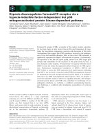
Fig. 2. Ferritin-like pore of DpsA-Te. (A) View of the pore perpen-
dicular to the three-fold symmetry axis. The residues lining the pore
are shown as sticks and colored according to atom type: N, blue;
O, red; C, yellow, azure and green in the different three-fold sym-
metry-related subunits. (B) Schematic representation of the pore.
View perpendicular to the three-fold symmetry axis. The residues
lining the pore of a single subunit are indicated. (C) View of
the pore in the dodecamer along the three-fold symmetry axis
containing an iron ion (colored gray). Pictures were generated using
PYMOL [41].
F. Alaleona et al. The unusual Thermosynechoccus elongatus DpsA
FEBS Journal 277 (2010) 903–917 ª 2010 The Authors Journal compilation ª 2010 FEBS 907
addition, the ferritin-like interface is stabilized by two
hydrophobic patches: one formed by Ala162, Val18,
Ile19, Leu122, and Ile129, and the other by the Ala146,
Leu150, Leu155 and Leu156 side chains.
The pores at the so-called Dps-type interfaces show
marked variability in their dimensions and chemical
nature among the Dps family members. In DpsA-Te,
the external perimeter of the pore is lined by Asn171
and Val176 placed on the flexible C-terminal tail, the
bottleneck by Glu58, Pro61, Asp75, and the internal
perimeter by Gln64.
The DpsA-Te ferroxidase center is unique, owing to
the presence of a histidine (His78) in place of the
canonical aspartate metal ligand (Asp58 in L. innocua).
Furthermore, there is Phe52 in place of the nearby,
highly conserved tryptophan (Trp32 in L. innocua), as
shown in Fig. 1. The electron density map clearly
shows that the ferroxidase center A-site and B-site are
both occupied by a metal ion (Fig . 3A,B). The two
ions are at a distance of about 3.0 A
˚
, and are coordi-
nated tetrahedrally by two histidines, a water molecule,
and a bridging glutamate (Glu82). In particular, the
A-site ion is coordinated by His78, His51 (His31 in
L. innocua Dps), a water molecule, and Glu82 (Glu62
in L. innocua Dps), and the B-site ion is coordinated
by Glu82, His63 (His43 in L. innocua Dps), a water
molecule, and His164 belonging to the three-fold sym-
metry-related monomer (Fig. 3A,B). His164 is not
conserved among the Dps family members, with the
exception of H. salinarum DpsA, in which, however,
the B-site does not contain a metal ion. The two
strong peaks in the difference Fourier map, F
obs
–
F
calc
, that identify the two metals at the A-site and the
B-site disappear when the map is contoured at 10r
and 7r, respectively. The bound metal ions were
assigned to Zn(II) on the basis of the presence of two
strong peaks at 8800 eV and 10 300 eV in the X-ray
fluorescence emission spectrum, and on inductively
coupled plasma atomic emission spectroscopy (ICP-
AES) measurements on the soluble protein that
AB
CD
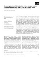
Fig. 3. Ferroxidase center of DpsA-Te. (A) Overall view of the ferroxidase center. The residues of the first and the second Zn(II) coordination
shell are shown as sticks and colored according to atom type: N, blue; O, red; C, yellow. The carbon atoms and the three different subunits
are colored gray, blue, and yellow. Water molecules are shown as spheres and depicted in red; zinc ions are shown as spheres and depicted
in gray. (B) Electron density map 2F
o
– F
c
of the ferroxidase center contoured at 1r. (C) Comparison between the DpsA-Te ferroxidase cen-
ter (light blue), the G. intestinalis flavodiiron protein iron-binding site (dark blue), and the catalytic site of the Th. thermophilus RNA degrada-
tion protein (orange). (D) The two-fold symmetry interface. The tyrosines lining the interface are shown as sticks and colored according to
atom type: N, blue; O, red. The carbon atoms of the tyrosines and the different subunits are colored gray, blue, and yellow. Pictures were
generated using
PYMOL [41].
The unusual Thermosynechoccus elongatus DpsA F. Alaleona et al.
908 FEBS Journal 277 (2010) 903–917 ª 2010 The Authors Journal compilation ª 2010 FEBS
indicate a zinc content of 24 per dodecamer. Assuming
an occupancy of 1.0, the Zn(II) refinement gives rea-
sonable mean thermal parameters of 30 and 48 A
˚
2
in
the A-site and the B-site, respectively, and thus points
to tighter binding of the metal to the former site.
Accordingly, the distances between Zn(II) and the pro-
tein ligands range between 2.0 and 2.2 A
˚
for His51,
His63, and His78, whereas those pertaining to Zn(II)
at the B-site and His164 range between 2.2 and 2.5 A
˚
in the four monomers present in the asymmetric unit.
Interestingly, three tyrosines (Tyr60, Tyr70, and
Tyr163) are placed in the second Zn(II) coordination
shell with the hydroxyl groups oriented towards the
internal cavity. Tyr60 and Tyr163 are, respectively, at
6.2 and 7.1 A
˚
from the B-site Zn(II), and Tyr70 is at
6.4 A
˚
from the A-site Zn(II). In some monomers, the
phenol ring of Tyr60 displays an alternative conforma-
tion, with the side chain rotated about 30° in the direc-
tion of the Zn(II)-binding sites (Fig. 3A,B,D).
The DpsA-Te ferroxidase center bears a striking
similarity to the catalytic sites of the Thermus thermo-
philus RNA degradation protein and of the Giardia
intestinalis flavodiiron protein (Fig. 3C). The first
belongs to the metallo-b-lactamase superfamily and
contains two Zn(II) in the catalytic site [26], whereas
the second, which is believed to act as an oxygen scav-
enger, binds two irons in the catalytic site [27].
Structural characterization in solution
As in all known Dps proteins, the DpsA-Te dodecam-
er is characterized by a sedimentation coefficient, s
20,w
,
of 10.5 S. The CD spectrum in the near-UV region has
major positive peaks around 280 nm that are attribut-
able to tyrosines, and positive ellipticity in the 260–
270 nm region that can be assigned to phenylalanines
(Fig. S1). Importantly, DpsA-Te and DpsA-TeHis
show very similar spectra, an indication that the His-
tag at the C-terminus does not change the protein
structure in solution.
The ellipticity in the far-UV region was used to
study DpsA-Te thermostability in comparison with
that of Dps-Te. For both T. elongatus Dps proteins,
the transition from the native to the denatured state
could not be monitored over the pH range 7.0–3.0,
owing to the extremely high protein stability even at
100 °C. Thermal unfolding was followed at pH 2.0, a
condition under which both DpsA-Te and Dps-Te pre-
serve their native quaternary structure at room temper-
ature (Fig. S2). At this pH, the denaturation process
of both proteins was complete at 75–80 °C (Fig.
S2). As the transitions are irreversible, the midpoint of
the denaturation process, T
m
, was taken as a measure
of thermostability. This value is 20 °Cor30°C higher
than those measured for the mesophilic L. innocua and
E. coli Dps proteins under the same experimental con-
ditions [23].
Iron oxidation and incorporation kinetics
The efficiency of O
2
and H
2
O
2
as Fe(II) oxidants was
assessed by following the kinetics of the oxidation
reaction spectrophotometrically at 350 nm and pH 7.0
in parallel experiments on DpsA-Te, DpsA-TeHis, and
Dps-Te.
Dps-Te, like nearly all Dps proteins so far character-
ized and as reported by Franceschini et al. [23], prefers
H
2
O
2
to O
2
as an iron oxidant (Fig. 4A, inset). Thus,
Fig. 4. Kinetics of iron oxidation ⁄ incorporation by DpsA-Te (A),
using O
2
or H
2
O
2
as oxidant, and corresponding UV–visible spectra
(B). (A) Oxidant, O
2
(o), and H
2
O
2
(
•
). Traces were measured at
350 nm, which enables monitoring of the formation of the ferric
core. Fe(II) was added to an Fe(II) ⁄ dodecamer ratio of 24 : 1. The
inset depicts the behavior of Dps-Te. (B) Oxidant, O
2
( ), and
H
2
O
2
(—). The two spectra at the bottom were recorded at 1.5 s
after addition of the oxidant.
F. Alaleona et al. The unusual Thermosynechoccus elongatus DpsA
FEBS Journal 277 (2010) 903–917 ª 2010 The Authors Journal compilation ª 2010 FEBS 909
after the addition of 24 Fe(II) per dodecamer, the half-
time of the reaction in the presence of H
2
O
2
(0.5 : 1
molar ratio with respect to iron) was 2.5 s, as com-
pared with 250 s in the presence of O
2
. Quite unex-
pectedly, in the parallel experiment on DpsA-Te
containing 24 Zn(II) per dodecamer, ferroxidation by
O
2
was about 20-fold faster (t
1 ⁄ 2
= 11 s). When the
experiment was repeated on a DpsA-Te sample treated
with 6 mm EDTA and containing only 12 Zn(II) per
dodecamer on the basis of ICP-AES determinations,
the same t
1 ⁄ 2
value was obtained, and the rate of fer-
roxidation by H
2
O
2
was only two-fold higher
(t
1 ⁄ 2
= 6 s; Fig. 4A). The DpsA-Te oxidation kinetics
followed at different temperatures yielded the same
results, in that H
2
O
2
was approximately two-fold more
efficient than O
2
over the whole range studied. The
activation energy, E
a
, calculated from the Arrhenius
plot, corresponded to 18.6 and 12.1 kcalÆmol
)1
when
H
2
O
2
and O
2
were used as oxidant, respectively (Fig.
S3).
The unusual reactivity of DpsA-Te called for a
more extensive characterization of the ferroxidation
reaction. As Fe–Zn complexes are known to display
charge transfer absorption bands between 300 and
400 nm, the possible formation of oxidation interme-
diates was followed over the range 300–600 nm. Dur-
ing oxidation of 24 Fe(II) per dodecamer, similar
bands at about 320 and 370 nm were observed 1.5 s
after admission of O
2
or H
2
O
2
, and persisted at the
end of the reaction (Fig. 4B). In addition, to establish
the reaction stoichiometry and the possible presence
of H
2
O
2
in solution at the end of the reaction, oxy-
graphic experiments were employed. Fe(II) solutions
were added to 4 lm DpsA-Te or recombinant human
H-ferritin (H-FtHu) [respective molar ratios: Fe(II) ⁄
docecamer, 12 : 1; or Fe(II) ⁄ 24mer, 14 : 1], and oxy-
gen consumption was measured. When the Fe(II)⁄
oligomer ratio was £ 24 : 1 for DpsA-Te or £ 48 : 1
for H-FtHu, the addition of Fe(II) to the protein
resulted in fast oxygen consumption, according to an
O
2
⁄ Fe(II) molar ratio of 1 : 2.0 to 1 : 2.1, in three
different experiments (Fig. 5). This ratio shifted pro-
gressively towards 1 : 4 when the Fe(II) ⁄ protein ratio
increased, and reached values of 1 : 3.8 to 1 : 4.0
(n = 3) at and beyond 96 Fe(II) per dodecamer (inset
to Fig. 5). In the case of DpsA-Te, the addition of
catalase at the end of the reaction did not cause O
2
production, indicating that H
2
O
2
was not released
into solution. In contrast, O
2
is produced in the pres-
ence of H-FtHu, where the ferroxidation reaction
characterized by a 2 : 1 Fe(II) ⁄ O
2
stoichiometry is
known to result in the quantitative production of
H
2
O
2
[9].
The formation of a ferric core by DpsA-Te and
Dps-Te was followed in parallel at pH 7.0 in 50 mm
Mops by using O
2
as oxidant, as precipitation occurs
in the presence of H
2
O
2
when the added iron exceeds
about 150 atoms per dodecamer. An Fe(II) ⁄ dodecamer
molar ratio of 250 : 1 was achieved by adding five suc-
cessive increments of 100 lm Fe(II) to 2 lm DpsA-Te
or Dps-Te; the intervals between the iron additions
were 60 min or 5 min, respectively. The increase in
absorbance at 350 nm and analytical ultracentrifuga-
tion experiments indicated that all of the iron added
was oxidized and incorporated. Thus, the sedimenta-
tion coefficient, s
20,w
, of apoDpsA-Te increased from
10.5 to 12.9 S after incorporation of 250 Fe(III) per
dodecamer, as compared with an increase from 10.1 to
12.8 S in the case of apoDps-Te (Fig. S4). A minor
component at 14.6 S and at 18.7 S, present
respectively in apoDpsA-Te and mineralized DpsA-Te,
can be assigned to dimers of dodecamers, as the pro-
tein is ‡ 99% pure upon SDS gel electrophoresis.
DNA-binding assay and DNA protection against
hydroxyl radical formation
The possible interaction between DpsA-Te and DNA
was assessed in agarose gel mobility shift assays,
using supercoiled pET-11a DNA as a probe. Under
the conditions employed, E. coli Dps forms Dps–
Fig. 5. Oxygen consumption during the DpsA-Te and H-FtHu Fe(II)
oxidation reaction. A solution of Fe(II) was added (at about 1.5 min)
to 4 l
M apoDpsA-Te (—) or H-FtHu ( ) at an Fe(II) ⁄ protein molar
ratio of 12 : 1 or 24 : 1, respectively. Buffer: 50 m
M Mops ⁄ NaOH
(pH 7.0), at 25 °C. The addition of Fe(II) to both DpsA-Te and H-
FtHu results in fast oxygen consumption, according to an O
2
⁄ Fe(II)
molar ratio of 1 : 2. The subsequent addition of catalase (light
arrows) results in oxygen production only in the case of H-FtHu.
The inset shows oxygen consumption when Fe(II) is added to apo-
DpsA-Te at an atom ⁄ protein molar ratio of 96 : 1.
The unusual Thermosynechoccus elongatus DpsA F. Alaleona et al.
910 FEBS Journal 277 (2010) 903–917 ª 2010 The Authors Journal compilation ª 2010 FEBS
DNA complexes that are too large to migrate into
the gel matrix [4]. The reaction between DpsA-Te
(3 lm) and DNA (20 nm) was allowed to proceed for
5 min in BAE or TAE (pH 6.5 or pH 7.5, respec-
tively). At both pH values, no interaction was
observed (data not shown). Dps-Te, analyzed in par-
allel as a control, likewise does not bind DNA, as
reported in [20].
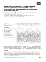
The ability to prevent hydroxyl radical-mediated
DNA cleavage was determined by means of an
in vitro damage assay [13]. Plasmid pET-11a DNA in
30 mm Tris ⁄ HCl (pH 7.3) (Fig. 6, lane 1) was fully
degraded by the hydroxyl radicals formed by the
combined effect of 50 lm Fe(II) and 1 mm H
2
O
2
via
a Fenton reaction (Fig. 6, lane 4). The efficient DNA
protection resulting from the presence of Dps-Te
(Fig. 6, lane 1) or DpsA-Te (Fig. 6, lane 2) is
indicated by the essentially unaltered pattern of the
plasmid bands.
Discussion
DpsA-Te is the sole known Dps protein carrying a sub-
stitution at the ferroxidase center, where a histidine
(His78) replaces the highly conserved metal-coordinat-
ing aspartate at the A-site (Asp58, Listeria numbering).
This aspartate fi histidine replacement is the basis for
the unforeseen binding of Zn(II) at the ferroxidase
center, and most likely for the high efficiency of O
2
as
Fe(II) oxidant. These properties differentiate DpsA-Te
with respect to almost all characterized Dps proteins,
and are suggestive of a distinctive role in the bacterium.
Although the exceptionality of DpsA-Te can be traced
back principally to the aspartate fi histidine replace-
ment at the ferroxidase center, the possible effects of
the few other substitutions of nearby, conserved resi-
dues cannot be discounted, although they are difficult
to pinpoint in the absence of site-specific mutagenesis
studies, e.g. Phe52 replacing Trp32 (Listeria number-
ing), Tyr163 replacing the other tryptophan at the
three-fold symmetry axis (Trp144, Listeria numbering),
and His164 replacing a hydrophobic residue (methio-
nine in Listeria Dps) near the metal-binding B-site.
The aspartate fi histidine replacement at the ferrox-
idase center impacts on the most intriguing characteris-
tic of the DpsA-Te X-ray crystal structure, namely the
presence of Zn(II) in both metal-binding sites. The two
Zn(II) are coordinated tetrahedrally by two histidines,
a water molecule, and a bridging glutamate. In partic-
ular, the A-site ion is coordinated by His78 and His51
(Asp58 and His31, respectively, in L. innocua Dps),
Glu82 (Glu62 in L. innocua Dps), and a water mole-
cule. The B-site ion is coordinated by Glu82, His63
(His43 in L. innocua Dps), and a water molecule, a
fourth protein ligand being furnished by His164
belonging to the three-fold symmetry-related mono-
mer. Among the known Dps family members, His164
is present only in H. salinarum DpsA, where, however,
the B-site does not contain a metal ion. The coordina-
tion bond lengths between Zn(II) and the histidine
ligands belonging to the two-fold symmetry-related
subunits (His51, His63, and His78) are all in the range
2.0–2.2 A
˚
, whereas the distance between His164 and
the B-site Zn(II) is 2.2–2.5 A
˚
. This observation indi-
cates that Zn(II) is bound less strongly at the latter
site, in accordance with the mean thermal parameters
of the two metal ions [30 A
˚
2
and 48 A
˚
2
, respectively,
for Zn(II) bound at the A-site and the B-site]. In full
agreement with the X-ray data, ICP-AES measure-
ments showed that the zinc content of the sample used
for determination of the X-ray structure corre-
sponds to 24 Zn per dodecamer, and decreases to
12 Zn per dodecamer upon dialysis against 6 mm
1234
Fig. 6. DNA protection by DpsA-Te and Dps-Te. Lane 1: plasmid
DNA with 1 m
M H
2
O
2
,50lM Fe(II), and 3 lM Dps-Te. Lane 2: plas-
mid DNA with 1 m
M H
2
O
2
,50lM Fe(II), and 3 lM DpsA-Te. Lane
3: plasmid DNA. Lane 4: plasmid DNA with 1 m
M H
2
O
2
and 50 lM
Fe(II).
F. Alaleona et al. The unusual Thermosynechoccus elongatus DpsA
FEBS Journal 277 (2010) 903–917 ª 2010 The Authors Journal compilation ª 2010 FEBS 911
EDTA. Importantly, upon exposure of the 12 Zn per
dodecamer sample to 24 Fe(II) per dodecamer under
air, rapid ferroxidation takes place that does not
involve removal of the bound Zn(II).
From a functional viewpoint, DpsA-Te stands out
for the unusual efficiency of O
2
as iron oxidant, such
that the rates of ferroxidation by H
2
O
2
and O
2
are
comparable (Fig. 4A). Thus, H
2
O
2
is about two-fold
more efficient than O
2
, in marked contrast to the
100-fold difference that characterizes Dps proteins,
with the sole exception of B. anthracis Dps2 (also
named Dlp2). B. anthracis Dps2 has canonical metal
ligands at the ferroxidase center, but reacts with Fe(II)
and H
2
O
2
three-fold faster than with O
2
[28]. However,
the absolute rates are about 10-fold slower than in the
case of DpsA-Te.
To unravel the mechanism underlying DpsA-Te
catalysis, two approaches were used: the ferroxidation
rates of the proteins containing 24 or 12 Zn(II) were
compared, and oxygraphic experiments were per-
formed to establish the stoichiometry of the ferroxida-
tion reaction. No differences ascribable to the Zn(II)
content were detected. At an Fe(II) ⁄ dodecamer ratio
of £ 24 : 1, the oxygraphic data showed that the pro-
tein uses two Fe(II) to reduce O
2
and that H
2
O
2
is not
released into solution (Fig. 5). At higher Fe(II) ⁄ dode-
camer ratios, H
2
O
2
is likewise undetectable at the end
of the reaction, but the number of Fe(II) required to
reduce O
2
increases progressively to reach a value of 4.
This indicates that crystal growth, whose contribution
increases progressively with increases in the
Fe(II) ⁄ dodecamer ratio, leads to the production of
water, as in all Dps proteins and ferritins [9,14].
The findings just described can be rationalized on
the basis of the following overall scheme:
2Fe(II) þ O
2
þ 2H
þ
! 2Fe(III) þ H
2
O
2
ð1Þ
H
2
O
2
þ 2Fe(II) þ 2H
þ
! 2Fe(III) þ 2H
2
O ð2Þ
Several comments are in order. The similarity of the
rate of ferroxidation by O
2
and H
2
O
2
suggests that
reaction (2) is rate-limiting. Furthermore, the fact that
H
2
O
2
is produced, as shown by the observed Fe ⁄ O
2
stoichiometry, but is undetectable is related to its
reduction to water, although its entrapment by the
protein moiety cannot be excluded.
The most intriguing aspect, however, concerns the
mechanism that allows reduction of one O
2
by two
Fe(II) at a ferroxidase center that contains a perma-
nently bound Zn(II) at the A-site. After entry of Fe(II)
via the ferritin-like pores (Fig. 2A,C), the Fe(II)-binding
step involves the B-site, with the concomitant displace-
ment of Zn(II) and the formation of Zn–Fe complexes,
as indicated by the ICP-AES and optical absorbance
data. Thus, upon addition of oxygen or H
2
O
2
, absorp-
tion bands at 320 and 370 nm appear, and persist dur-
ing the course of the reaction (Fig. 4B). These bands
can be assigned to Fe–Zn charge transfer [29], with a
possible contribution of charge transfer between oxy-
gen and either metal at 320 nm [30]. Two different sce-
narios can be envisaged for the subsequent iron
oxidation step, which must entail the successive oxida-
tion of two Fe(II) bound either to the same ferroxidase
center or to two distinct centers located at the same
dimeric interface. The first hypothesis requires forma-
tion of an oxygen radical intermediate, and the second
that the two ferroxidase centers be connected by an
efficient electron transfer pathway along the dimeric
interface, a task that can probably be performed by the
Tyr44 and Tyr70 lining it (Fig. 3D). The significant
ferroxidase activity of DpsA-Te despite the concomi-
tant presence of iron and zinc at the catalytic center is
yet another manifestation of its uniqueness. Thus, in
other members of the Dps family, notably L. innocua
Dps [31] and Streptococcus suis Dpr [32], binding of
Zn(II) at the ferroxidase center leads to inhibition of
the iron oxidation ⁄ uptake reaction.
Significantly, despite the distinctive ferroxidation
mechanism and the lack of DNA-binding capacity,
DpsA-Te protects this macromolecule against Fe(II)-
mediated and H
2
O
2
-mediated damage just as efficiently
as the previously characterized Dps-Te (Fig. 6).
At this point of the discussion, the question arises of
the physiological relevance of the present data
obtained with recombinant DpsA-Te. Given the resem-
blance between the zinc uptake systems in bacteria
[33], DpsA-Te is expected to be saturated with Zn(II)
also in its physiological environment, and O
2
is
expected to act as the preferred Fe(II) oxidant. The
long hydrophobic N-terminal tail may be indicative of
DpsA-Te localization at the thylacoid membranes,
where photosynthesis takes place and O
2
is produced.
If so, the specific role of DpsA-Te would be to protect
photosystems I and II from this oxidant. In contrast,
Dps-Te would have the canonical Dps function of
inhibiting the Fe(II)-mediated and H
2
O
2
-mediated pro-
duction of hydroxyl radicals via Fenton chemistry.
These ideas will be verified in ad hoc immune-localiza-
tion experiments, using antibodies directed against
DpsA-Te.
The possible binding of substrates other than O
2
could occur, and DpsA-Te could catalyze other types
of reaction, as water is a metal ligand, as in all cata-
lytic zinc sites [34,35]. This possibility is suggested by
The unusual Thermosynechoccus elongatus DpsA F. Alaleona et al.
912 FEBS Journal 277 (2010) 903–917 ª 2010 The Authors Journal compilation ª 2010 FEBS
the similarity between the DpsA-Te ferroxidase center
and those of the Th. thermophilus RNA degradation
protein and the G. intestinalis flavodiiron protein, and
could account for the unusual features of the ferritin-
like pores, which remain unexpected, namely their size,
shape, and the distinct nature of the lining residues
(Fig. 2B).
In conclusion, the present work on DpsA-Te has
disclosed unique structural and functional properties
that point to a different physiological role than that of
Dps-Te and warrant further investigation. Priority will
be given to the localization of the protein in the bacte-
rium, as it will allow us to validate the suggestion that
the unusual efficiency of O
2
as iron oxidant is related
to the occurrence of photosynthesis.
Experimental procedures
Strains and media
E. coli strain BL21(DE3) was grown at 37 °C on liquid LB
medium (10 gÆL
)1
Tryptone, 5 gÆL
)1
yeast extract, and
5gÆL
)1
NaCl) or LB plates containing 50 lgÆmL
)1
ampicillin.
Cloning, overexpression, and purification
The dpsA-Te gene was amplified by PCR from the genome
of T. elongatus BP1, kindly provided by T. Kaneko
(Kazusa DNA Research Institute), using primers DpsA-Te1
(5¢-GGAGTATCGT
CATATGACGACCAGTGCATTG-3¢)
and DpsA-Te2 (5¢-CAGACGACACA
AAGCTTCACC
TTG-3¢). The NdeI and HindIII restriction sites are under-
lined. The amplified fragment (530 bp) was digested with
NdeI and HindIII, purified using the QIAquick PCR
purification kit (Qiagen, Hilden, Germany), and cloned
into the expression vector pET-22b (Novagen, Darmstadt,
Germany) digested with NdeI and HindIII.
Expression of the dpsA-Te gene yields an insoluble protein
product. Therefore, a construct containing a C-terminal His-
tag was designed, and a stop codon was introduced down-
stream of the His-tag sequence. The resultant plasmid (dpsA-
TeHis) was introduced into E. coli BL21(DE3) and
sequenced by dideoxy sequencing to confirm the presence of
the correct gene. E. coli BL21(DE3) cells harboring the
recombinant dpsA-TeHis plasmid were grown to A
600 nm
=
0.6 at 37 °C in 1 L of ampicillin-containing liquid LB med-
ium. Gene expression was induced by addition of 0.5 mm
isopropyl thio-b-d-galactoside, and the culture was incu-
bated for a further 3–4 h. Cells were harvested by centrifuga-
tion (15 000 g for 20 min), suspended in 50 mm Tris ⁄ HCl
(pH 7.5), 0.5 m m dithiothreitol, 1 mm EDTA, and 500 mm
NaCl, and disrupted by sonication. The lysate was centri-
fuged at 15 000 g for 45 min, and the supernatant was
treated for 30 min at 37 °C with 0.1 mgÆmL
)1
DNase
(Sigma-Aldrich, St Louis, MO, USA) was supplied with
10 mm MgCl
2
, heated to 75 °C for 10 min, cooled on ice,
and then centrifuged at 10 000 g for 15 min to remove dena-
tured proteins. The recovered supernatant was dialyzed over-
night against 30 mm Tris ⁄ HCl (pH 7.8), and loaded onto a
HiTrap Q-Sepharose HP cellulose column (GE Healthcare,
Uppsala, Sweden) equilibrated with the same buffer. DpsA-
TeHis was eluted with 100 mm NaCl. The relevant fraction
was dialyzed overnight against 30 mm Tris ⁄ HCl (pH 7.8),
10 mm imidazole, and 300 mm NaCl, and loaded onto a
HisTrap HP column (GE Healthcare) equilibrated with the
same buffer. DpsA-TeHis was eluted with 350 mm imidazole;
it was dialyzed against 30 mm Tris ⁄ HCl (pH 7.8), with or
without 6 mm EDTA, and stored at )75 °C. The use of
either DpsA-Te or DpsA-TeHis is specified throughout.
The purity of all preparations was assessed by means of
Coomassie brilliant blue staining of 15% SDS ⁄ PAGE gels.
Protein concentration was determined spectrophotometri-
cally at 280 nm, using a molar extinction coefficient (on a
dodecamer basis) of 2.03 · 10
5
m
)1
Æcm
)1
, calculated with
protparam ().
Removal of the His-tag
A factor Xa cleavage site was created between the last
amino acid (valine) and the His-tag. Cleavage by factor Xa
occurs after an arginine, and the preferred cleavage site is
Asp (or Glu or Ile)-Gly-Arg. Factor Xa was chosen as pro-
tease instead of the more common thrombin, because there
is a thrombin cleavage site at position 8 (MTTSALPR) of
the DpsA-Te sequence. The factor Xa cleavage site was cre-
ated using the QuikChange Site-Directed Mutagenesis Kit
(Stratagene La Jolla, CA, USA). Removal of the His-tag
was achieved by incubating DpsA-TeHis overnight at room
temperature with bovine factor Xa protease (GE Health-
care) at 10 units per mg DpsA-TeHis. The reaction was
performed in 50 mm Tris ⁄ HCl (pH 8.0), 1 mm CaCl
2
, and
0.1 m NaCl, at 25 °C. DpsA-Te was obtained after applica-
tion of the digested protein to a Superdex 200 size exclusion
column (GE Healthcare).
CD spectroscopy
CD measurements were performed with a Jasco J-715 spec-
tropolarimeter. Near-UV spectra (250–330 nm) were
recorded at 20 °C, using a 1 cm optical quartz cuvette. The
protein concentration was 0.5 mgÆmL
)1
in 50 mm Mops (pH
7.0). Spectra were taken after averaging 16 accumulations.
Thermal unfolding experiments were monitored in the far-
UV region (220 nm) in a stoppered 0.1 cm quartz cell. The
protein concentration was 0.5 mgÆmL
)1
at pH 7.0 (50 mm
Mops buffer), pH 3.0 (50 mm glycine ⁄ HCl), and pH 2.0
(10 mm HCl). The parameter chosen to compare the transi-
tion curves of DpsA-Te and Dps-Te was the melting temper-
ature (T
m
), defined as the midpoint of the denaturation
F. Alaleona et al. The unusual Thermosynechoccus elongatus DpsA
FEBS Journal 277 (2010) 903–917 ª 2010 The Authors Journal compilation ª 2010 FEBS 913
process and calculated by plotting the first derivative of the
molar ellipticity values as a function of temperature.
Protein crystallization, data collection, and data
processing
Crystallization experiments, performed at 298 K by the
hanging drop vapor diffusion method, yielded X-ray-quality
crystals only with DpsA-TeHis. The crystallization trials
were performed with a protein sample concentrated to
about 10 mgÆmL
)1
. After dialysis against 20 mm Tris ⁄ HCl
at pH 7.5, 1 lL aliquots of the protein solution were mixed
with an equal amount of reservoir solution containing 12%
w ⁄ v poly(ethylene glycol) 8000 in 0.1 m Mes at pH 6.0.
Well-shaped crystals grew in 2 weeks, and reached dimen-
sions of 0.1 · 0.1 · 0.2 mm
3
. The DpsA-TeHis crystals
were cryoprotected in a solution containing the reservoir
solution (75% v ⁄ v) and poly(ethylene glycol) 200 (25%
v ⁄ v). The crystals were mounted in nylon loops and flash-
frozen by quick submersion in nitrogen liquid for transport
to the synchrotron radiation source.
A single-wavelength dataset (k =1A
˚
) was collected from
a DpsA-TeHis crystal at the synchrotron radiation source
ELETTRA (Trieste, Italy), using a MAR CCD detector at a
temperature of 100 K. The dataset was processed with
denzo and scaled with scalepack [36]. The autoindexing
procedure indicates that the crystals are cubic. On the basis
of the scaling procedure, the crystals belong to the I23 space
group, with cell parameters a = b = c = 174.504 A
˚
. The
data are 99.9% complete, with an R
merge
value of 16% at
2.4 A
˚
resolution. All of the data statistics are reported in
Table 1.
Structure solution and refinement
On the basis on the calculated monomer molecular mass of
20.9 kDa and the presence of 24 asymmetric units in the
I23 cell with dimensions a = b = c = 174.504 A
˚
, the
value of V
M
(the unit cell volume divided by the total
molecular mass of the protein in the unit cell) is
11 ⁄ n A
˚
3
ÆDa
)1
, where n is the number of molecules per
asymmetric unit. Given that the protein in solution is a
dodecamer, the value of n can only be 4. Assuming the
presence of a tetramer in the asymmetric unit, the value of
V
m
is 2.62 A
˚
3
ÆDa
)1
, with a solvent content of 52.7%.
The structure was solved by molecular replacement, using
as search model the tetramer of H. salinarum DpsA (Pro-
tein Data Bank entry: 1MOJ), which displays 36%
sequence identity with DpsA-Te (calculated using the
ncbi-blast server). The rotational and translational
searches performed with molrep [37,38] in the resolution
range 10.0–3.0 A
˚
produced a clear solution.
Refinement was performed using the maximum likelihood
method with refmac
5 [39], and model building was
performed with coot [40] (Table 1). The final DpsA-Te
model is a tetramer containing 686 residues (171 for
each monomer), eight Zn(II) with full occupancy, one
iron with full occupancy, one iron with 0.33 occupancy,
four poly(ethylene glycol) molecules, and 114 water
molecules. The final R
crys
for all resolution shells (100–
2.4 A
˚
) calculated using the working set reflections
(32 937) is 16.5%, and the R
free
calculated using the test
set reflections (1742) is 21.6%. The final R
crys
calculated
for the highest-resolution shell (2.46–2.4 A
˚
) using the
working set reflections (2426) is 21.3%, and the R
free
value calculated using the test set of reflections (116) is
28.8%. The most favored regions of the Ramachandran
plot contain 99.3% nonglycine residues.
The chemical identity of the metal ions bound to the pro-
tein was assessed by means of X-ray fluorescence emission
spectra collected at the ELETTRA synchrotron radiation
source in an energy range between 2000 and 10 000 eV.
Iron oxidation and incorporation kinetics
Kinetic experiments of Fe(II) oxidation by H
2
O
2
or O
2
in the
presence of DpsA-Te, DpsA-TeHis or Dps-Te were
performed on a Hewlett-Packard diode array spectropho-
tometer at 10, 15, 17.5, 20, 25, 35, 40 and 50 °C. Ferrous
ammonium sulfate solutions were prepared freshly in Thun-
berg tubes prior to the experiments, and kept under nitrogen
gas. In experiments on iron oxidation by O
2
,48lm Fe(II)
was added to 2 lm protein in 50 mm Mops (pH 7.0). During
the course of the reaction, the protein solutions were main-
tained in air under stirring. As a control, Fe(II) autoxidation
was measured in parallel (Fig. S3). In investigations on the
kinetics of iron oxidation by H
2
O
2
,48lm Fe(II) was added
to degassed 2 lm protein in 50 mm Mops buffer at pH 7.0.
The solution was kept under nitrogen gas until addition of
24 lm H
2
O
2
in the same Mops buffer [molar ratios:
Fe(II) ⁄ dodecamer, 24 : 1; Fe ⁄ H
2
O
2
, 2 : 1]. All of the time-
dependent absorbance traces were collected at 350 nm and
analyzed with origin 7.5 (Originlab Corporation, North-
ampton, MA, USA).
In the experiments performed as a function of temperature,
the rate constant, k, and temperature, T (in Kelvin), were cor-
related by means of the Arrhenius plot k = A exp()E
a
⁄ RT),
where E
a
is the activation energy, A the pre-exponential fac-
tor, and R the gas constant.
Formation of a ferric core was achieved with O
2
as oxi-
dant by adding six increments of 50 Fe(II) per dodecamer
to Dps-Te and DpsA-Te solutions (2 lm)in50mm Mops
(pH 7.0) equilibrated in air at 20 °C. Ferrous ammonium
sulfate solutions were prepared in Thunberg tubes and kept
under nitrogen until use. The iron incorporation reaction
was followed at 350 nm, where the ferric iron micelles
absorb. After a constant absorbance had been reached,
the samples were centrifuged at 10 000 g for 15 min, passed
through a HiTrap Q-Sepharose HP cellulose column, and
analyzed by analytical ultracentrifugation.
The unusual Thermosynechoccus elongatus DpsA F. Alaleona et al.
914 FEBS Journal 277 (2010) 903–917 ª 2010 The Authors Journal compilation ª 2010 FEBS
Oxygraphic measurements
Oxygen consumption assays were performed in a gas-tight
vessel using Clark-type selective electrodes (Oxygraph-2K;
Oroboros Instruments, Innsbruck, Austria). A ferrous
ammonium sulfate solution was added to 4 lm DpsA-Te or
to H-FtHu to achieve an Fe(II) ⁄ protein molar ratio of
12 : 1 or 24 : 1, respectively. At the end of the reaction, the
possible presence of H
2
O
2
was monitored by addition of
2mgÆmL
)1
bovine liver catalase (Sigma-Aldrich). Measure-
ments were performed at 25 °C in air-equilibrated 50 mm
Mops (pH 7.0). The software datlab 4.2, furnished by the
manufacturers, was used for data acquisition and analysis.
Analytical ultracentrifugation
Sedimentation velocity studies were performed on a Beck-
man-Coulter XLI analytical ultracentrifuge, using absor-
bance optics. Experiments were conducted at 30 000 r.p.m.
and 20 °Con4lm DpsA-Te in 20 mm Tris ⁄ HCl buffer
(pH 7.5) and 0.2 m NaCl.
Radial absorbance scans were obtained at 280 and
350 nm at a spacing of 0.003 cm with three averages in
continuous scan mode. Sedimentation coefficients were cal-
culated using sedfit (provided by P. Schuck, National
Institutes of Health), and were reduced to water and 20 °C
(s
20,w
) using standard procedures. The buffer density and
viscosity were calculated by sednterp.
DNA-binding and protection assays
The DNA-binding abilities of DpsA-Te and Dps-Te were
assessed in gel shift experiments, using supercoiled pUC9-
5S (3115 bp, 20 nm) as probe. DNA was purified using the
Qiaquick Gel Extraction kit (Qiagen), which ensures
removal of impurities and salts. DNA was incubated for
5 min at room temperature with the Dps proteins (3 lm)in
TAE at pH 7.5 or BAE at pH 6.5. To resolve the Dps–
DNA complexes, electrophoresis was performed on 1%
agarose gels in 0.04 m TAE or BAE. The gels were stained
with ethidium bromide and imaged using ImageMaster
VDS (Amersham Biosciences, Uppsala, Sweden).
DNA protection from oxidative damage was assessed
in vitro using 20 nm supercoiled pET-11a DNA (5600 bp).
The assay was performed in 12 lLof30mm Tris ⁄ HCl
(pH 7.2). Plasmid DNA was allowed to interact with Dps-
Te or DpsA-Te (3 lm) for 5 min prior to the addition of
50 lm FeSO
4
. Simultaneously, H
2
O
2
was added (final con-
centration of 1 mm), and the mixture was incubated for
5 min at room temperature to allow complete consump-
tion of Fe(II). Thereafter, the reaction mixture was incu-
bated at 100 °C for 10 min. Plasmid DNA was resolved
by electrophoresis on 1% agarose gel in TAE. The gel
was stained with ethidium bromide and imaged using
ImageMaster VDS.
Acknowledgements
The valuable help of E. Forte with the oxygraphic
experiments and of G. Forte with the ICP-AES experi-
ments are gratefully acknowledged. The work was
supported by local grants from the Ministero dell’Uni-
versita
`
e Ricerca, MiUR, to E. Chiancone. We thank
the ELETTRA Synchrotron Light Source, Basovizza
(Trieste), Italy, where the data were obtained.
References
1 Almiron M, Link AJ, Furlong D & Kolter R (1992)
A novel DNA-binding protein with regulatory and
protective roles in starved Escherichia coli. Genes Dev 6,
2646–2654.
2 Martinez A & Kolter R (1997) Protection of DNA dur-
ing oxidative stress by the nonspecific DNA-binding
protein Dps. J Bacteriol 179, 5188–5194.
3 Nair S & Finkel SE (2004) Dps protects cells against
multiple stresses during stationary phase. J Bacteriol
186, 4192–4198.
4 Ceci P, Cellai S, Falvo E, Rivetti C, Rossi GL &
Chiancone E (2004) DNA condensation and self-aggre-
gation of Escherichia coli Dps are coupled phenomena
related to the properties of the N-terminus. Nucleic
Acids Res 32, 5935–5944.
5 Bhattacharyya G & Grove A (2007) The N-terminal
extensions of Deinococcus radiodurans Dps-1
mediate DNA major groove interactions as well as
assembly of the dodecamer. J Biol Chem 282,
11921–11930.
6 Stillman TJ, Upadhyay M, Norte VA, Sedelnikova SE,
Carradus M, Tzokov S, Bullough PA, Shearman CA,
Gasson MJ, Williams CH et al. (2005) The crystal
structures of Lactococcus lactis MG1363 Dps proteins
reveal the presence of an N-terminal helix that is
required for DNA binding. Mol Microbiol 57,
1101–1112.
7 Roy S, Saraswathi R, Gupta S, Sekar K, Chatterji D &
Vijayan M (2007) Role of N and C-terminal tails in
DNA binding and assembly in Dps: structural studies
of Mycobacterium smegmatis Dps deletion mutants.
J Mol Biol 370, 752–767.
8 Ceci P, Mangiarotti L, Rivetti C & Chiancone E (2007)
The neutrophil-activating Dps protein of Helicobacter
pylori, HP-NAP, adopts a mechanism different from
Escherichia coli Dps to bind and condense DNA.
Nucleic Acids Res 35, 2247–2256.
9 Chiancone E, Ceci P, Ilari A, Ribacchi F & Stefanini S
(2004) Iron and proteins for iron storage and detoxifica-
tion. Biometals 17, 197–202.
10 Grant RA, Filman DJ, Finkel SE, Kolter R & Hogle
JM (1998) The crystal structure of Dps, a ferritin
F. Alaleona et al. The unusual Thermosynechoccus elongatus DpsA
FEBS Journal 277 (2010) 903–917 ª 2010 The Authors Journal compilation ª 2010 FEBS 915
homolog that binds and protects DNA. Nat Struct Biol
5, 294–303.
11 Ilari A, Stefanini S, Chiancone E & Tsernoglou D
(2000) The dodecameric ferritin from Listeria innocua
contains a novel intersubunit iron-binding site. Nat
Struct Biol 7, 38–43.
12 Papinutto E, Dundon WG, Pitulis N, Battistutta R,
Montecucco C & Zanotti G (2002) Structure of two
iron-binding proteins from Bacillus anthracis. J Biol
Chem 277, 15093–15098.
13 Ceci P, Ilari A, Falvo E & Chiancone E (2003) The
Dps protein of Agrobacterium tumefaciens does not
bind to DNA but protects it toward oxidative cleav-
age: x-ray crystal structure, iron binding, and hydro-
xyl-radical scavenging properties. J Biol Chem 278,
20319–20326.
14 Zhao G, Ceci P, Ilari A, Giangiacomo L, Laue TM,
Chiancone E & Chasteen ND (2002) Iron and hydrogen
peroxide detoxification properties of DNA-binding
protein from starved cells. A ferritin-like DNA-binding
protein of Escherichia coli. J Biol Chem 277, 27689–
27696.
15 Ishikawa T, Mizunoe Y, Kawabata S, Takade A,
Harada M, Wai SN & Yoshida S (2003) The iron-
binding protein Dps confers hydrogen peroxide stress
resistance to Campylobacter jejuni. J Bacteriol 185,
1010–1017.
16 Ueshima J, Shoji M, Ratnayake DB, Abe K, Yoshida
S, Yamamoto K & Nakayama K (2003) Purification,
gene cloning, gene expression, and mutants of Dps from
the obligate anaerobe Porphyromonas gingivalis. Infect
Immun 71, 1170–1178.
17 Andrews SC (1998) Iron storage in bacteria. Adv
Microb Physiol 40, 281–351.
18 Velayudhan J, Castor M, Richardson A, Main-Hester
KL & Fang FC (2007) The role of ferritins in the physi-
ology of Salmonella enterica sv. Typhimurium: a unique
role for ferritin B in iron–sulphur cluster repair and
virulence. Mol Microbiol 63, 1495–1507.
19 Romao CV, Mitchell EP & McSweeney S (2006) The
crystal structure of Deinococcus radiodurans Dps protein
(DR2263) reveals the presence of a novel metal centre
in the N terminus. J Biol Inorg Chem 11, 891–902.
20 Grove A & Wilkinson SP (2005) Negative cooperativity
of uric acid binding to the transcriptional regulator
HucR from Deinococcus radiodurans. J Mol Biol 347,
495–508.
21 Antelmann H, Engelmann S, Schmid R, Sorokin A,
Lapidus A & Hecker M (1997) Expression of a stress-
and starvation-induced dps ⁄ pexB-homologous gene is
controlled by the alternative sigma factor sigmaB in
Bacillus subtilis. J Bacteriol 179, 7251–7256.
22 Nakamura Y, Kaneko T, Sato S, Ikeuchi M, Katoh H,
Sasamoto S, Watanabe A, Iriguchi M, Kawashima K,
Kimura T et al. (2002) Complete genome structure of
the thermophilic cyanobacterium Thermosynechococcus
elongatus BP-1.
DNA Res 9, 123–130.
23 Franceschini S, Ceci P, Alaleona F, Chiancone E &
Ilari A (2006) Antioxidant Dps protein from the
thermophilic cyanobacterium Thermosynechococcus
elongatus. FEBS J, 273, 4913–4928.
24 Lawson DM, Treffry A, Artymiuk APJ, Harrison PM,
Yewdall SJ, Luzzago A, Cesareni G, Levi S & Arosio P
(1989) Identification of the ferroxidase centre in ferritin.
FEBS Lett 254, 207–210.
25 Ilari A, Ceci P, Ferrari D, Rossi GL & Chiancone E
(2002) Iron incorporation into Escherichia coli Dps
gives rise to a ferritin-like microcrystalline core. J Biol
Chem 277, 37619–37623.
26 Ishikawa H, Nakagawa N, Kuramitsu S & Masui R
(2006) Crystal structure of TTHA0252 from Thermus
thermophilus HB8, a RNA degradation protein of the
metallo-beta-lactamase superfamily. J Biochem 140,
535–542.
27 Di Matteo A, Scandurra FM, Testa F, Forte E, Sarti P,
Brunori M & Giuffre
`
A (2008) The O
2
-scavenging flav-
odiiron protein in the human parasite Giardia intestinal-
is. J Biol Chem 283, 4061–4068.
28 Liu X, Kim K, Leighton T & Theil EC (2006) Paired
Bacillus anthracis Dps (mini-ferritin) have different reac-
tivities with peroxide. J Biol Chem 281, 27827–27835.
29 Yogo T, Nakafuku T, Sakamoto W & Hirano S (2005)
Synthesis of Fe-doped ZnO particle ⁄ polymer hybrid
from metalorganics. J Mat Res 20, 1470–1475.
30 He YP, Miao YM, Li CR, Wang SQ, Cao L, Xie SS,
Yang GZ & Zou BS (2005) Size and structure effect on
optical transitions of iron oxide nanocrystals. Phys Rev
B 71, 125411–125419.
31 Stefanini S, Cavallo S, Montagnini B & Chiancone E
(1999) Incorporation of iron by the unusual dodeca-
meric ferritin from Listeria innocua. Biochem J 338,
71–75.
32 Havukainen H, Haataja S, Kauko A, Pulliainen AT,
Salminen A, Haikarainen T, Finne J & Papageorgiou
AC (2008) Structural basis of the zinc- and terbium-
mediated inhibition of ferroxidase activity in Dps ferri-
tin-like proteins. Protein Sci 9, 1513–1521.
33 Hantke K (2001) Bacterial zinc transporters and regula-
tors. Biometals 14, 239–249.
34 Vallee BL & Auld DS (1993) Cocatalytic zinc motifs in
enzyme catalysis. Proc Natl Acad Sci USA 90, 2715–
2718.
35 Vallee BL & Auld DS (1995) Zinc metallochemistry in
biochemistry. EXS 3, 259–277.
36 Otwinowski Z & Minor W (1997) Processing of X-ray
diffraction data collected in oscillation mode. Macromol
Crystallogr A, 276, 307–326.
37 Vagin A & Teplyakov A (2000) An approach to multi-
copy search in molecular replacement. Acta Cryst D
Biol Crystallogr 56, 1622–1624.
The unusual Thermosynechoccus elongatus DpsA F. Alaleona et al.
916 FEBS Journal 277 (2010) 903–917 ª 2010 The Authors Journal compilation ª 2010 FEBS
38 Collaborative Computational Project, Number 4 (1994)
The CCP4 suite: programs for protein crystallography.
Acta Crystallogr D Biol Crystallogr 50, 760–763.
39 Murshudov GN, Vagin AA & Dodson EJ (1997)
Refinement of macromolecular structures by the maxi-
mum-likelihood method. Acta Crystallogr D Biol Crys-
tallogr 53, 240–255.
40 Emsley P & Cowtan K (2004) Coot: model-building
tools for molecular graphics. Acta Crystallogr D Biol
Crystallogr 60, 2126–2132.
41 DeLano WL. The PyMOL Molecular Graphics System.
DeLano Scientific LLC, San Carlos, CA.
Supporting information
The following supplementary material is available:
Fig. S1. Near-UV CD spectra of DpsA-Te and DpsA-
His.
Fig. S2. Thermal denaturation of Dps-Te and DpsA-
Te.
Fig. S3. Iron oxidation kinetics of DpsA-Te as a func-
tion of temperature.
Fig. S4. Sedimentation velocity of Dps-Te and DpsA-
Te before and after oxidation ⁄ incorporation of 250 Fe
per dodecamer.
This supplementary material can be found in the
online version of this article.
Please note: As a service to our authors and readers,
this journal provides supporting information supplied
by the authors. Such materials are peer-reviewed and
may be re-organized for online delivery, but are not
copy-edited or typeset. Technical support issues arising
from supporting information (other than missing files)
should be addressed to the authors.
F. Alaleona et al. The unusual Thermosynechoccus elongatus DpsA
FEBS Journal 277 (2010) 903–917 ª 2010 The Authors Journal compilation ª 2010 FEBS 917