Botulinum Toxin in Aesthetic Medicine_2 pptx
Bạn đang xem bản rút gọn của tài liệu. Xem và tải ngay bản đầy đủ của tài liệu tại đây (15.47 MB, 45 trang )
Chapter 6
6
e following three chapters will focus on ad-
vanced indications and techniques. Some of
these indications and techniques may have been
discussed before. However, the following chap-
ters will oer a dierent view on these topics.
6.1 Facial Asymmetries
Mauricio de Maio
6.1.1 Introduction
Facial paralysis triggers aesthetic and functional
changes, with physical and psychological reper-
cussions. Static and dynamic imbalances can
aect, in a striking manner, a person’s ability to
express emotions. e physical aspects can bring
disastrous results to a patient’s self- image as well
as emotional state.
A smile can express such feelings as those re-
lated to pleasure, friendship, acceptance, embar-
rassment, happiness, delight and/or agreement.
We communicate through our smiles. Not being
able to smile would be to deprive ourselves of
one of our most basic tools for communication
in a social environment.
Upon analyzing the half of the face not aect-
ed by facial paralysis, one can perceive the great
variations in static and dynamic patterns of ad-
aptation that the mimetic muscle tissues suer in
the absence of movement in the other hemiface.
Gaining knowledge regarding the facial
nerve, the mimetic muscle tissues and the types
Contents
6.1 Facial Asymmetries . . . . . . . . . . 93
6.1.1 Introduction . . . . . . . . . . . . . 93
6.1.2 Anatomy . . . . . . . . . . . . . . 94
6.1.3 Aim of Treatment . . . . . . . . . . . 97
6.1.4 Patient Selection . . . . . . . . . . . 97
6.1.5 Technique . . . . . . . . . . . . . . 97
6.1.6 Results . . . . . . . . . . . . . . . . 99
6.1.7 Complications . . . . . . . . . . . . 99
6.1.8 Conclusions . . . . . . . . . . . . . 99
6.1.9 Tips and Tricks . . . . . . . . . . . 101
6.1.10 References . . . . . . . . . . . . . 101
6.2 Facial Liing with Botulinum Toxin . 102
6.2.1 Introduction . . . . . . . . . . . . 102
6.2.2 Anatomy: Antagonists and Synergists . 103
6.2.3 Aim of Treatment . . . . . . . . . . 105
6.2.4 Patient Selection . . . . . . . . . . 105
6.2.5 Technique . . . . . . . . . . . . . 109
6.2.6 Complications . . . . . . . . . . . 114
6.2.7 Tips and Tricks . . . . . . . . . . . 114
6.2.8 References . . . . . . . . . . . . . 114
6.3 Treatment with Microinjections . . . 115
6.3.1 Introduction . . . . . . . . . . . . 115
6.3.2 Microinjections of the Crow’s Feet Area 115
6.3.3 Microinjections of the Longitudinal
Lines of the Cheeks
. . . . . . . . . 115
6.3.4 Doses to be Used . . . . . . . . . . 116
6.3.5 Combination of Macro- and Microin-
jections . . . . . . . . . . . . . . 116
6.3.6 Disadvantages of the Microinjection
Technique . . . . . . . . . . . . . 116
6.3.7 Tips and Tricks . . . . . . . . . . . 116
Advanced Indications
and Techniques
Mauricio de Maio, Berthold Rzany
94 Mauricio de Maio, Berthold Rzany
6
of smiles that can be produced is of vital impor-
tance for professionals who deal with this quite
complex group of patients. e expertise that
derives from treating patients with asymmetries
enables any practitioner to inject any cosmetic
patient with excellence and condence.
Forehead asymmetries are easily treated
and are very similar to the cosmetic tech
-
niques that may be found in the specic section.
Other asymmetries require more anatomical
knowledge.
6.1.2 Anatomy
e facial nerve (cranial nerve pair VII) is re-
sponsible for stimulating the mimic muscles, cre-
ating a balance between the synergic and antago-
nistic forces that act upon the facial structures. It
is also responsible for the muscular tonus when
a person is in a relaxed state, and the voluntary
and involuntary contraction of the muscles of
each side of the face.
e facial nerve emerges in the stylomastoid
foramen and gives origin to its many ramica-
tions. e rst ramication is the posterior au-
ricular branch, the second is the temporal-facial
branch that divides into the temporal, zygomatic
and buccal ramications and the third is the cer-
vical-facial branch that divides itself up into the
marginal mandibular and cervical ramications
(Table .).
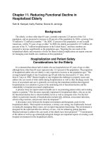
e most complex group of mimetic muscles
is the one that controls the movements of the lips
and cheeks. It is very important to know each
muscle action and the respective synergists and
antagonists when injecting patients with asym-
metries in the peribucal area. e interaction of
these muscles creates an almost unlimited num-
ber of facial movements and individual expres-
sions (Fig. .). ere are dierent patterns for
the smiles, depending on the muscles which are
dominant. e smile may be classied into three
types: ‘Mona Lisa’, in which the m. zygomaticus
major is dominant; ‘canine’, when the m. levator
labii superioris is dominant and ‘full denture’, the
smile in which all of the elevators and depres-
sors are involved. e shape of a person’s smile is
the result of the dynamic action of the forces that
act upon the mouth, and it varies from patient to
patient. A smile may also be classied as a com-
mon smile, in which the teeth are not shown, or
a ‘square’ smile, in which the upper and lower
teeth are displayed. In the former type, the m.
zygomaticus major is predominant, whereas in
the latter, the both the elevators and depressors
of the lip are predominant.
ere are ve elevators for the upper lip;
three of them act more on the upper lip (m.
levator labii superioris alaeque nasi, m. levator
labii superioris and m. zygomaticus minor) and
the other two act on the angle of the mouth (m.
levator anguli oris and m. zygomaticus major)
(Table .).
e muscles that act on the lower lip may be
divided into one levator and three depressors.
e m. mentalis is the levator and the depressors
include the m. depressor labii inferioris, m. de-
pressor anguli oris and platysma (Table .).
ere are other muscles that inuence the
balance of the mouth which include the m. or-
bicularis oris, m. risorius and m. buccinator
(Table .).
Table .. Specic facial regions and the corresponding
ramications of the facial nerve
Area Facial Nerve
Frontal Temporal branch
Orbital Zygomatic branch
Upper lip Buccal branch
Lower lip Marginal mandibular
branch
Neck Cervical branch
Chapter 6 95Advanced Indications and Techniques
Fig. .. Muscles responsible
for severe facial asymmetries
Table .. Description of the elevators of the lip, their actions and the synergists and antagonists. NB: the modiolus
is the area where the muscles that elevate and depress the lip interdigitate, laterally to the oral commissure
Muscle Action Synergists Antagonists
M. levator labii superi-
oris alaeque nasi
Medial part: dilates the
nostril
Lateral part: raises and
everts the upper lip
Medial part: M. dilator nasi
Lateral part: m. levator labii
superioris,
m. zygomaticus major and mi
-
nor and m. levator anguli oris
M. depressor anguli oris
and m. orbicularis oris
M. levator labii supe
-
rioris
Elevates and everts the
upper lip
Lateral part of m. levator
labii superioris alaeque nasi,
m. levator anguli oris and m.
zygomaticus major and minor
M. depressor anguli oris
and m. orbicularis oris
M. zygomaticus minor Elevates the upper lip
and assists in elevating
the intermediate part of
the nasolabial fold
Lateral part of the m. levator
labii superioris alaeque nasi,
m. levator labii superioris, M.
levator anguli oris, m. zygo
-
maticus major
M. orbicularis oris and
m. depressor anguli oris
M. levator anguli oris
(caninus)
Raises the angle of the
mouth and xes the
modiolus
All the other four elevators M. depressor anguli
oris, platysma and m.
orbicularis oris
M. zygomaticus major Retracts and elevates the
modiolus and the angle
of the mouth
All the other four elevators M. orbicularis oris, m.
depressor anguli oris
and platysma
96 Mauricio de Maio, Berthold Rzany
6
Table .. Description of the muscles that act on the lower lip
Muscles Action Synergists Antagonists
M. mentalis Raises the mental tissue,
mentolabial sulcus and base
of the lower lip
M. levator anguli oris and
zygomaticus major
M. depressor labii infe-
rioris and m. depressor
anguli oris
M. depressor labii
inferioris
Depresses the lower lip later
-
ally and assists in eversion
Platysma pars labialis and
m. depressor anguli oris
M. orbicularis oris
M. depressor anguli
oris
Depresses the modiolus and
angle of the mouth
Platysma pars modiolus
and m. depressor labii
inferioris
M. levator anguli oris and
m. zygomaticus major
Platysma Anterior bers: assist man-
dibular depression
Intermediate bers: pars la
-
bialis – depress the lower lip
Posterior bers: pars mo
-
diolaris – depress the buccal
angle
M. depressor anguli oris M. levator anguli oris
Table .. Other muscles inuencing the balance of the mouth
Muscle Action Synergists Antagonists
M. orbicularis oris Deep bers: direct closure
of lips
Supercial and decussat
-
ing bers: lip protrusion
M. incisivus labii superi
-
oris and inferioris*
m. mentalis
e ve upper lip
levators, the m. depressor
anguli ori and m. labii
inferioris and the m. buc
-
cinator
M. buccinator Compresses the cheek
against the teeth and
draws the angle of the
mouth laterally
M. risorius M. orbicularis oris
M. risorius Retracts the angle of the
mouth
M. zygomaticus major
and m. buccinator
M. orbicularis oris
* ese muscles assist the action of the orbicularis oris in protruding the lip.
Chapter 6 97Advanced Indications and Techniques
6.1.3 Aim of Treatment
e goals of treatment of facial asymmetries
include static balance with correction of facial
deviations and rotations, and reduction or to-
tal control of facial deviation during animation
while avoiding any functional impairment.
6.1.4 Patient Selection
Damage suered to the facial nerve may produce
deformities of varying degrees, resulting in aes-
thetic and functional disorders in such patients.
e side of the face aected by facial paralysis
presents common characteristics among all pa-
tients. On the surface of the skin, there are fewer
wrinkles, due to the lack of muscular traction on
the dermis; the nasolabial fold becomes less evi-
dent, and there is a drooping of both the corner
of the mouth and the brow. Depending on the
extent of facial paralysis, and the time of onset,
the aesthetic aspects may be aected to a greater
or lesser extent (Fig. .).
e ‘normal’ side or the side opposite to
that aected by facial paralysis replies with a
hyperkinetic reaction of the muscle tissues due
to the lack of tonus on the paralyzed side. is
imbalance of vector forces creates facial devia-
tions. e dynamic deviations to the ‘normal’
side are less evident in paralyses that have lasted
a short time. With longer periods, there are also
static deviations in the labial, nasal and orbital
regions, leading to shortening of the face (Fig.
.). It is on this hyperkinetic or hypertonic side
of the face that botulinum toxin plays the most
important role.
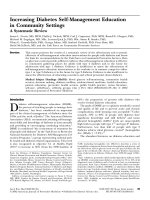
6.1.5 Technique
For best results and facial balance, all the main
muscles on the hyperkinetic side should be treat-
ed (Fig. .). e botulinum toxin should be ad-
ministered through intramuscular injection with
a -gauge needle. e needle should be inserted
at an angle of ° from the skin’s surface, with the
patient lying on his back. It is advisable to avoid
contact with the periosteum.
e botulinum toxin should be distributed
in the perioral muscles to enable the coordina-
tion of the muscles that act upon both the upper
Fig. .. e muscle over-contraction on the hypertonic
side (right) may provoke facial deviations and shortening
due to a long period of lack of muscle antagonism on the
le side. e longer the paralysis, the more muscle over-
contraction on the opposite side
Fig. .. Note the dierences in skin wrinkling. On the
hyperkinetic side (le) the muscle hyperactivity produces
evident and numerous wrinkles. e lack of muscle ac
-
tivity results in a younger-looking skin on the paralyzed
side (right)
98 Mauricio de Maio, Berthold Rzany
6
Table .. Suggested injection point and doses
Site Botox Dose
Range
Dysport Dose
Range
M. zygomaticus major at its point of origin – U – U
M. zygomaticus minor at its point of origin – U – U
M. levator labii superioris alaeque nasi – U – U
M. levator labii superioris at the orbital
margin
– U – U
e modiolus, at a distance of . cm from
the corner of the mouth
– U – U
M. risorius cm from the corner of the
mouth
– U – U
M. depressor labii inferioris at . cm from
the corner of the mouth
– U – U
M. depressor labii inferioris at a distance of
cm from the white line transition
– U – U
Fig. .. Injection points for facial asymmetries
Chapter 6 99Advanced Indications and Techniques
Fig. .. Schematic portrayal of the vector forces that
act upon the side aected by facial paralysis, the hyperki
-
netic side. It should be noted that there are both straight
and curved vectors, which represent the traction and
rotation that the perioral region suers due to muscle
hyperkinesis
Fig. .. Schematic representation of the vectors of forces
that act upon the perioral area
and lower lips (Table ., Figs. . and .). It is
important to point out that the dose may vary
according to the type of muscular contraction. It
is advisable to start with half of the dose initially
and aer days to add an extra dose depending
on the muscular response.
6.1.6 Results
With the decrease of hyperkinesis aer the
injection of botulinum toxin, improvement in
both static and dynamic positions is found. In
static analysis, it is very common to achieve
an excellent symmetry and correction of
deviations and rotation of the face (Fig. .a,b).
In animation, the reduction in the hyperkinesis
controls the excessive muscular excursion and
corrects the excessive labial distortion and teeth
show (Fig. .a,b).
6.1.7 Complications !
e adverse events with the use of botulinum
toxin are generally linked to high doses of the
drug. Aer the injection of botulinum toxin
there is an abrupt change in the mimetic mus
-
cle behavior and, consequently, in the patients’
learning and adaptation patterns. Despite an en-
hanced aesthetic appearance, these changes may
lead to functional impairment. Usually, there
may be mild diculty in speaking, chewing and
swallowing. Oral incontinence for liquids and
solids may happen with a high dose and mis-
placed injections.
6.1.8 Conclusions
In the treatment of patients suering from facial
paralysis, botulinum toxin may be considered
as a single treatment, as a pre-operative test or
as a complementary measure in post-surgical
treatments. It may reduce facial deviations and
rotations, minimizing aesthetic sequelae. Yet, its
most important feature seems to be the poten
-
tial for use in children and adolescents, who will
greatly benet from the treatment during mus
-
cular and skeletal development.
100 Mauricio de Maio, Berthold Rzany
6
Fig. .a,b. Before treatment, under static analysis, the patient presented a common hyperkinetic reaction on her
right-hand side: a deep nasolabial fold, with nasal are and lip deviations. Aer treatment, a static balance of the face
is obtained. e patient reported social re-integration and an improvement in self-esteem
Fig. .a,b. On animation, the patient presented excessive teeth show with distortion of the smile. Aer injection,
there is a balance of all muscles that act upon the hyperkinetic side, resulting in an improved smile
Chapter 6 101Advanced Indications and Techniques
6.1.9 Tips and Tricks
■
Focus the treatment of facial asymmetries
on the muscle vectors and distribute the
botulinum toxin in an even manner. Re
-
member that blocking one single muscle
may unbalance the others. When starting
to treat facial asymmetries do not try to aim
for a single treatment session; be cautious
and use at least a two-step treatment with
lower doses to minimize complications.
6.1.10 References
Adant, JP () Endoscopically assisted suspension in
facial palsy. Plast Reconstr Surg :
Arden RL, Sunhat PK () Vertical suture placation of
the orbicularis oris muscle: a simple procedure for
the correction of unilateral marginal mandibular
nerve paralysis. Facial Plast Surg :
Armstrong MW et al. () Treatment of facial synkine
-
sis and facial asymmetry with Botulinum toxin type
A following facial nerve palsy. Clin Otolaryngol :
Aviv JE, Urken ML () Management of the paralyzed
face with microneurovascular free muscle transfer.
Arch Otolaryngol Head Neck Surg :
Badarny S et al. () Botulinum toxin injection eec
-
tive for post-peripheral facial nerve palsy synkinesis.
Harefuah :
Bento RF et al. () Treatment comparison between
dexamethasone and placebo for idiopathic palsy. Eur
Arch Otolaryngol Dec: S
Bernardes DFF et al. () Functional prole in patients
with facial paralysis treated in a myofunctional ap
-
proach. Pro Fono :
Bikhazi NB, Maas CS () Renement in the rehabili
-
tation of the paralyzed face using Botulinum toxin.
Otolaryngol Head Neck Surg :
Bleicher JN et al. () A survey of facial paralysis: etiol
-
ogy and incidence. Ear Nose roat J :–
Boerner M, Sei S () Etiology and management of
facial palsy. Curr Opin Ophthalmol :
Boroojerdi B et al. () Botulinum toxin treatment of
synkinesia and hyperlacrimation aer facial palsy.
J Neurol Neurosurg Psychiatr :
Brans, JW et al. () Cornea protection in ptosis in
-
duced by Botulinum injection. Ned Tijdschr Ge
-
neeskd. :
Burres SA, Fisch U () e comparison of facial grad-
ing systems. Arch. Otolaryngol. Head Neck Surg
:
Burres SA () Facial biomechanics: e standards of
normal. Laryngoscope :
Burres SA () Objective grading of facial paralysis.
Ann Otol Rhinol Laryngol :
Burres SA () e qualication of synkinesis and fa
-
cial paralysis. Eur Arch Otolaryngol Dec:S
Carruthers A, Carruthers J () Botulinum toxin type
A: history and current cosmetic use in the upper face.
Sem Cut Med Surg :
Clark RP, Berris CE () Botulinum toxin: a treatment
for facial asymmetry caused by facial nerve paralysis.
Plast Reconstr Surg :
Dawidjan B () Idiopathic facial paralysis: a review
and case study. J Dent Hyg :
Dobie RA, Fisch U () Primary and revision surgery
(selective neurectomy) for facial hyperkinesia. Arch
Otorhinolaringol Head Neck Surg :
Dodd SL et al. () A comparison of the spread of three
formulations of botulinum neurotoxin A as deter
-
mined by eects on muscle function. Eur J Neurol
():–
Dressler D, Schonle PW () Hyperkinesias aer hy
-
poglossofacial nerve anastomosis – treatment with
Botulinum toxin. Eur Neurol :
Faria JCM () A critical study of the treatment of fa
-
cial palsy through a gracilis transfer. Doctoral thesis,
Medical College, University of the State of Sao Paolo.
Farkas LG () Anthropometry of the head and face.
Second edition. New York: Raven Press pp –
Fine NA et al. () Use of the innervated platysma ap
in facial reanimation. Ann Plast Surg :
Guereissi JO () Selective myectomy for postparetic
facial synkinesis. Plast Reconstr Surg :
Harii K et al. () One-stage transfer of the latissiumus
dorsi muscle for reanimation of a paralyzed face: a
new alternative. Plast Reconstr Surg :
102 Mauricio de Maio, Berthold Rzany
6
Kermer C et. al. () Muscle-nerve-muscle neurotiza-
tion of the orbicularis oris muscle. J Craniomaxillo
-
fac Surg :
Kozak J et al. () Contemporary state of surgical treat
-
ment of facial nerve paresis. Preliminary experience
with new procedures. Acta Chir Plast :
Kukwa A et al. () Reanimation of the face aer facial
nerve palsy resulting from resection of a cerebello
-
pontine angle tumor. Br J Neurosurg :
Kumar PA () Cross-face reanimation of the paralysed
face with a single stage microneurovascular gracilis
transfer without nerve gra: a preliminary report. Br
J Plast Surg :
Labbe D () Lengthening temporalis myoplasty. Rev
Stomatol Chir Maxillofac :
Laskawi R () Combination of hypoglossal-facial
nerve anastomosis and Botulinum toxin injections
to optimize mimic rehabilitation aer removal of
acoustic neurinomas. Plast Reconstr Surg :
May M et al. () Bell’s palsy: management of sequelae
using EMG rehabilitation, Botulinum toxin, and sur
-
gery. Am J Otol :
Moser G, Oberascher G () Reanimation of the para
-
lyzed face with new gold weight implants and goretex
so-tissue patches. Eur Arch Otorhinolaryngol :S
Muhlbauer W et al. () Mimetic modulation for prob
-
lem creases of the face. Aesthet. Plast. Surg. :
Neuenschwander MC et al. () Botulinum toxin in
otolaryngology: a review of its actions and opportu
-
nity for use. Ear Nose roat J :
Riemann R et al. () Successful treatment of crocodile
tears by injection of Botulinum toxin into the lacri
-
mal gland: a case report. Ophthalmology :
Rubin LR () Anatomy of facial expression. In Rubin
LR (Ed.) Reanimation of the paralysed face. New Ap
-
proaches. St. Louis: Mosby pp –
Sadiq SA, Downes RN () A clinical algorithm for the
management of facial nerve palsy from an oculoplas
-
tic perspective. Eye :
Samii M, Matthies C () Indication, technique and re
-
sults of facial nerve reconstruction. Acta Neurochir
:
Shumrick KA, Pensak ML () Early perioperative use
of polytef suspension for the management of facial
paralysis aer extirpative skull base surgery. Arch Fa
-
cial Plast Surg :
Sulica L () Botulinum toxin: basic science and clini-
cal uses in otolaryngology. Laryngoscope :
Terzis JK, Kalantarian B () Microsurgical strategies
in patients for restoration of dynamic depressor
muscle mechanism: a neglected target in facial reani
-
mation. Plast Reconstr Surg :
Tulley P et. al. () Paralysis of the marginal mandibu
-
lar branch of the facial nerve: Treatment options. Br
J Plast Surg :
Ueda K et. al. () Evaluation of muscle gra using fa
-
cial nerve on the aected side as a motor source in
the treatment of facial paralysis. Scand J Plast Recon
-
str Surg Hand Surg :
Wong GB et. al. () Endoscopically assisted facial sus
-
pension for the treatment of facial palsy. Plast Recon
-
str Surg :
6.2 Facial Lifting
with Botulinum Toxin
Maurício de Maio
6.2.1 Introduction
e aging process causes a variety of changes in
skin, muscles and bones. Volumetric loss of fat
tissue in the face produces a saggy appearance
which is worsened by the gravitational forces
that tend to pull the facial tissues down. Muscles
respond dierently depending on their position
in the face: the elevators are more important than
the depressors in youth and the depressors over-
contract during the aging process. e elevators
get weaker and weaker with time and, as a re-
sult, the vectors of forces which were antagonist
to gravitational forces and were able to maintain
the facial structures in an upward position, sim-
ply invert (Fig. .). e depressors corroborate
with gravitational forces and tend to drop the fa-
cial structures.
Understanding muscular behavior with its
synergistic and antagonistic eects has enabled
the development of new techniques such as
‘BNT-A liing’. When blocking the correct mus-
Chapter 6 103Advanced Indications and Techniques
cular group, we can again invert the vectors to
an upward position, and a faceli eect can be
obtained.
6.2.2 Anatomy:
Antagonists and Synergists
To understand how the mimetic muscles act
on the face, it is important to understand the
denition of the prime movers, antagonists and
synergists.
Prime movers are the principal muscles ac-
tively generating the movement. Antagonists
are dened as the muscles that act in opposition
to the prime movers and by their contraction
are capable of preventing or reversing the move-
ment. If the prime movers contract, the antago-
nists relax to assist their movement. It must be
highlighted that this relaxation is as important
as the prime movers’ contraction. For example,
in the forehead, the brow elevation is only pos
-
sible because the frontalis contracts AND the
brow depressors relax! When promoting eye-
brow elevation, we block the mm. corrugatores,
the m. procerus and the lateral bers of the mm.
orbiculares oculi. Partial eyebrow elevation oc-
curs if only partial blocking of the depressors
is undertaken. Major eyebrow elevation occurs
with the maximum depressor blocking possible
and the frontalis with its all strength is main-
tained. e inability to relax the opponents will
prevent the execution of the prime mover total
action.
e antagonists are also important for assist-
ing and modulating the prime movers’ action.
e stronger the action of prime movers and the
greater the resistance encountered, the more re-
laxed the antagonists are. If the prime movers are
involved in a precise movement, the antagonists
are relaxed but immediately ready to steady or
moderate the movement. Prime movers and an
-
tagonists may act at the same time. is is found
in isometric contraction, for example, the con-
traction of the mm. corrugatores and m. fronta
-
lis when expressing concern and surprise.
Synergists are dened as xation muscles,
which are those muscles that provide a rm base
for movements executed by other muscles. ey
are also important for providing precision and
avoiding exhaustion of the prime movers. In the
glabella, the procerus acts as a synergist to the
mm. corrugatores for the movement of the me-
dial portion of the eyebrows.
ere are various systems for dividing the
face. e classic system divides the face into
three thirds: upper, mid and lower thirds. e
upper third is from the hairline to the brow; the
mid third is from the brow to the base of the nose
and the lower third is from the base of the nose
to the chin. e platysma inuences the lower
and mid thirds.
e upper third has only one levator which
is the frontalis, whose medial bers are stron-
ger than the lateral bers. In contrast, there are
three or four depressors that tend to lower the
eyebrow. e medial bers of the m. frontalis
Fig. .. With the aging process there is an inversion of
vectors which, together with gravitational forces, pull the
facial so tissue down
104 Mauricio de Maio, Berthold Rzany
6
have the mm. corrugatores, m. procerus and
mm. depressores supercilii as the main oppo-
nents. Although the mm. orbiculares oculi may
also counteract the frontalis medial bers, it is
the mm. depressores supercilii that inuences
this level. e mm. orbiculares oculi lateral -
bers (the ones that produce the crow’s feet) tend
to depress the lateral aspect of the eyebrow.
(Table . and Figs. . and .a–c) Please note
that the mm. depressores supercilii is considered
by some as only a thickening of the orbicularis
oculi and not as a separate muscle.
e mid third, as described above, is the area
from the brow down to the base of the nose. Di-
dactically speaking, for BNT-A liing, elevators
will be described according to their ability to act
in opposition to gravitational forces. From me
-
dial to lateral, we may nd the m. levator labii su
-
perioris alaeque nasi, m. levator labii superioris,
m. zygomaticus minor, m. zygomaticus major
and m. levator labii superioris in a deeper plane.
It is also important to point out that the contrac
-
tion of the lower bers of the orbicularis oculi
pars orbitalis elevates the cheek area. e eleva-
tors at this level obey the same rule as found in
the frontalis: when the medial frontalis bers are
blocked, the lateral bers tend to elevate more
for a compensatory balance. e same happens
with the elevators at the upper lip level: if over
blocking at the m. levator labii superioris alae-
que nasi and m. levator labii superioris occurs,
over contraction of the zygomaticus major and
the ‘joker smile’ may result.
e depressors are those muscles that
supplement the eect of gravitational forces.
ey aggravate the descent of facial structures.
ere are three depressors: the m. depressor
labii inferioris and the m. depressor anguli oris
(from medial to lateral) (Figs. .a,b). e most
important one is the platysma (Figs. .a,b).
Although the vast majority of the bers of the
platysma are located in the neck, its bers blend
with the m. depressor labii inferioris and m.
depressor anguli oris: some authors even regard
the m. risorius as simply a thickening of the
platysma at the level of the lips (Table .).
Table .. Antagonist and synergists in the upper third
Function Muscle Action Synergists Antagonists
Elevator M. frontalis Elevates the eye-
brow
M. occipitalis M. procerus, m.
corrugator super
-
cilii, m. orbicularis
oculi and m. depres
-
sor supercilii
Depressor M. corrugator
supercilii
Draws eyebrows
medially and down
M. orbicularis oculi,
m. procerus and m.
depressor supercilii
M. frontalis
Depressor M. procerus Depresses the
medial aspect of the
eyebrow
M. corrugator, m. or
-
bicularis oculi and m.
depressor supercilli
M. frontalis
Depressor M. orbicularis oculi Orbital part: lowers
and protrudes the
eyebrows
M. corrugator, m.
procerus and m.
depressor supercilii
M. frontalis
Depressor M. depressor su-
percilii
Pulls down medial
eyebrow
M. corrugator, m.
procerus, m. orbicu-
laris oculi
M. frontalis
Chapter 6 105Advanced Indications and Techniques
6.2.3 Aim of Treatment
e target of the treatment for BNT-A liing is
the complete blocking of the depressors of the
upper, mid and lower face and neck as well as
the subtle blocking of the medial elevators and
no block of the lateral elevators. With the depres-
sors blocked, the elevators will strengthen with
time (Fig. .).
6.2.4 Patient Selection
Patients must be evaluated at rest and during
animation. Static evaluation should be directed
to important landmarks of the face: eyebrows,
cheeks, oral commissure, mandible and neck.
e status of these structures should be analyzed
(Table .).
As mentioned above, the best candidates for
BNT-A liing are those who do not present sig-
nicant saggy skin in the mid and lower face
and neck. ey are from to years of age
and present no important asymmetries during
animation. ey are precisely those patients who
are too young for a surgical faceli, even a minor
one, but would benet from a mild non-surgical
face li. e ideal patient for BNT-A liing usu-
ally presents the typical signs (Table .):
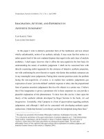
Fig. .. a,b Weakening the medial portion of the m.
frontalis and the depressors using BNT-A will make the
lateral part of the eyebrow li and erase the horizontal
line in the forehead.
c Split photograph of the patient in
a,b, showing the eect of BNT-A aer injections in the
central forehead region
Fig. .. Contraction of the depressors of the eyebrow
provokes drooping of the forehead. It will gradually pro
-
duce an aged appearance. In younger patients the elevator
(m. frontalis) is stronger than the depressors
106 Mauricio de Maio, Berthold Rzany
6
Fig. .. a Contraction of the depressor anguli oris, depressor labii inferioris and mentalis cause drooping of the
mid third, resulting in at cheekbones.
b Aer treatment of the mid third by blocking the depressor anguli oris and
mentalis there is improvement of the malar projection and oral commissure
Fig. .. a Hypertonic lateral platysmal bands distorting the mandible shape. ey are pulling down the lower face,
worsening the jowls. e black spots are the sites of BNT-A injection.
b Aer treatment with BNT-A. Note the weaken-
ing of the lateral platysmal bands which do not distort the mandible shape. Using this method, a liing of the lateral
aspect of the face is achieved
Chapter 6 107Advanced Indications and Techniques
Table .. Antagonists and synergists in the middle and lower third
Function Muscle Action Synergists Antagonists
Elevator M. levator labii
superioris alaeque
nasi
Medial part: dilates
the nostril
Lateral part: raises and
everts the upper lip
Medial part: m.
dilator nasi
Lateral part: m.
levator labii supe
-
rioris,
m. zygomaticus ma
-
jor and minor and
m. levator anguli
oris
M. depressor anguli
oris and m. orbicu
-
laris oris
Elevator M. levator labii
superioris
Elevates and everts the
upper lip
Lateral part of the
m. levator labii
superioris, alaeque
nasi, m. levator
anguli oris and m.
zygomaticus major
and minor
M. depressor anguli
oris and m. orbicu
-
laris oris
Elevator M. zygomaticus
minor
Elevates the upper lip
and assists in elevating
the intermediate part
of the nasolabial fold
Lateral part of the
m. levator labii
superioris alaeque
nasi, m. levator labii
superioris, m. leva
-
tor anguli oris, m.
zygomaticus major
M. orbicularis oris
and m. depressor
anguli oris
Elevator M. levator anguli
oris (caninus)
Raises the angle of the
mouth and xes the
modiolus
All the other four
elevators
M. depressor anguli
oris, platysma and
m. orbicularis oris
Elevator Zygomaticus major Retracts and elevates
the modiolus and the
angle of the mouth
All the other four
elevators
M. orbicularis oris,
m. depressor anguli
oris and platysma
Depressor M. depressor labii
inferioris
Depresses the lower
lip laterally and assists
in eversion
Platysma pars labia
-
lis and m. depressor
anguli oris
M. orbicularis oris
Depressor M. depressor anguli
oris
Depresses the modio
-
lus and angle of the
mouth
Platysma pars mo
-
diolus and depres
-
sor labii inferioris
M. levator anguli
oris and m. zygo-
maticus major
Depressor Platysma Anterior bers: assist
mandibular depres
-
sion
Intermediate bers:
pars labialis – depress
the lower lip
Posterior bers: pars
modiolaris – depress
the buccal angle
M. depressor anguli
oris
M. levator anguli
oris
108 Mauricio de Maio, Berthold Rzany
6
Table .. Desired outcomes and indications for BNT-A liing and/or surgery
Structure Eyebrow Cheekbones Oral Commis-
sure
Mandible Neck
Desirable Elevated and
curved shaped
with its lateral
aspect slightly
higher
Projected with
fullness
Upward line at
the corner of
the mouth with
a slightly pro
-
jected modiolus
Well dened
with no jowls
and no saggy
skin
No bands or
saggy skin
BNT-A Liing Downwards
mainly at its
lateral aspect
Flat with no
projection and
no laxity
Horizontal or
mild descent
line with no
saggy skin
Mild presence of
jowls
Medial and
lateral platysma
bands with no
saggy skin or fat
deposit
Surgery Downwards
with excessive
skin excess at
upper eyelid
Flat with laxity
and saggy skin
Very deep mari
-
onette lines with
saggy skin
Saggy skin and
evident jowls
deforming the
mandible shape
Evident saggy
skin with signi
-
cant laxity and
fat content
Fig. .. e aim of the BNT-A liing is to weaken the
depressors and strengthen the elevators to promote a
more refreshed look
Chapter 6 109Advanced Indications and Techniques
6.2.5 Technique
6.2.5.1 Upper Third Treatment
e frontalis plays the most important role in
eyebrow liing. Its medial bers are stronger
than the lateral bers and that is one of the rea-
sons why the lateral part of the eyebrow drops
with time. e opposite muscles to the frontalis
are the depressors of the eyebrows. e mm. cor-
rugatores and the m. procerus lower the medial
part of the eyebrow while the lateral bers of the
mm. orbiculares oculi pars orbitalis lower the
lateral eyebrow when it contracts.
e eyebrow liing results from the blocking
of the superior medial bers of the frontalis and
the blocking of the eyebrow depressors: mm. cor-
rugatores supercilli, m. procerus, and the lateral
bers of the mm. orbiculares oculi. e blocking
of the m. procerus plays an important role for
the liing of the medial portion of the eyebrows.
Only the medial bers of the m. frontalis should
be blocked to enable the liing of the lateral por-
tion of the eyebrow. e mm. corrugatores, m.
procerus and the upper bers of the mm. orbicu-
lares oculi pars orbitalis should be fully blocked.
e m. frontalis bers should only be partially
blocked so that the elevating bers are still able
to promote eyebrow liing.
6.2.5.2 Mid and Lower Thirds
Treatment
Crow’s feet should be treated with regular doses,
observing that it is advisable to block the inferior
medial extension of the orbicularis oculi bers
only very supercially.
Table .. Signs indicating a good patient for a BNT-A faceli
Structure Signs
Forehead Horizontal lines especially during animation and none or mild lines at rest
Glabella Vertical line between eyebrows mainly at frown and strong horizontal line
at the nasal radix at frown. Lines can be evident at rest, but not deep
Eyebrow e medial aspect at normal position or slightly low and the lateral aspect
evidently low
Upper Eyelid No or mild skin excess with no eye bags
Lower Eyelid Evident crow’s feet with no eye bags
Nose Presence of bunny lines and tip droop when smiling
Nasolabial Fold Prominent due to muscle hyperactivity especially at its upper position. No
evident saggy skin or fat deposit
Cheekbones Flat or with mild projection with no saggy skin
Upper And Lower Lip Perioral wrinkling when pursing
Oral Commissure Downwards with mild marionette lines at rest
Chin Mild wrinkling
Mandible Mild jowls presence but evident down traction with the platysma lateral
band contraction
Platysma Evident medial and even stronger lateral platysma bands with no or minor
saggy skin and no fat deposit in the neck area
110 Mauricio de Maio, Berthold Rzany
6
It must be veried whether there is a promi-
nent nasolabial fold and whether this is due to
the hyperactivity of the mm. levatores labii su-
perioris alaeque nasi and/or the mm. levatores
labii superioris. Otherwise, injecting BNT-A
into these muscles will promote no eect at all
and may lead to complications. e blocking of
these muscles soens the nasolabial groove. e
correct indication together with precise dosing
may also produce an interesting li of the lateral
malar zone: blocking the medial elevators of the
upper lip will synergistically make the lateral el-
evators contract, liing the lateral part of the mid
third of the face, and project the cheekbones.
Patients with a short distance between the
upper lip and the base of the nose are the best
candidates for nasal tip liing. If the tip of the
nose drops during a smile, the blocking of the m.
depressor septi nasi will produce a delicate eleva-
tion of the nose and a younger appearance.
Perioral wrinkling in the upper and lower lips
should also be treated to smooth the skin in this
area. If wrinkling appears only during pursing,
major improvement is obtained with BNT-A.
Deep wrinkling should be treated with the com-
bination of other methods such as peels and ll-
ers. Injecting into the upper lip medially, close to
the philtrum and into the skin and mucosa tran-
sition line is advisable and the dose should be as
low as possible.
6.2.5.3 Lower Third and Neck
e lower third is the part of the face that oen
shows the most undesirable aging signs, such as
deep oral commissure, loss of denition of the
mandible arch, and platysma bands. Blocking
the mm. depressores anguli oris will li the cor-
ner of the mouth because the opposite muscles,
the elevators of the oral commissure, will enable
this area to li. e sad look around the mouth
will be improved.
Injecting into the platysma may produce a bet-
ter neck contour. e over-contraction of the lat-
eral platysma bands usually pulls down the lateral
part of the face and alters the mandible shape. To
obtain an improvement at the mandible arch, one
must block the lateral platysma bands beginning
with the very upper bers that interdigitate with
the facial muscular bers. Major liing of the face
is achieved when the lower third of the masseter
bers are blocked. As a result, the upper bers
will contract, pulling up the zygomatic zone and
thinning the lower part of the face.
e dose to be used for BNT-A liing will de-
pend on the needs of each patient. As mentioned
before, the goal is to promote full blocking of the
depressors and mild or no blocking of the eleva
-
tors. Below you may nd suggested initial doses,
which however does not mean that all the listed
muscles should be injected in the rst treatment.
Proper physical examination at rest and during
animation will identify the injection sites and
muscles to be treated (Fig. ., Table .).
Fig. .. e injection sites for BNT-A liing. e initial
doses should be low during the rst treatment session.
e second treatment should be viewed as an opportu
-
nity to improve the performance of the liing eect
Chapter 6 111Advanced Indications and Techniques
Table .. Doses for the treatment of dierent muscles for a BNT-face li
Function Muscle Botox Dysport Comments
Elevator M. frontalis – U – U e blocking should be
only on the most super
-
cial bers to remove the
wrinkling and NOT its
liing eect
Depressor M. corrugator
supercilii
– U – U Full blocking is desirable
Depressor M. procerus – U .– U Full blocking is desirable
Depressor M. orbicularis oculi – U – U e lower bers should be
injected at a very super
-
cial level
Depressor M. depressor septi
nasi
– U – U Into the nasal base, prefer
-
ably in patients with short
upper lip
Elevator M. levator labii
superioris alaeque
nasi
– U – U Very supercially, prefer-
ably into its medial part
Elevator M. levator labii
superioris
- - Not usually injected for
this purpose
Elevator M. zygomaticus
minor
- - Not usually injected for
this purpose
Elevator M. levator anguli
oris (caninus)
- - Not usually injected for
this purpose
Elevator M. zygomaticus
major
– U – U Only if cheek lines are
present and very super-
cially (intradermal)
Depressor M. depressor labii
inferioris
- - SHOULD NOT BE
BLOCKED
Depressor M. depressor anguli
oris
– U – U Very important for cor
-
recting ‘sad mouth’
Depressor Platysma medial
bands
– U – U Supercial if no fat con
-
tent and deeper with fat
deposits in the neck
Depressor Platysma lateral
bands
– U – U Same as above and the
most important depressors
that drop the face
All dosages are given for the total area, e.g. both sides if applicable. e dosages are for some indications sometimes lower as
described in Section to avoid overtreatment.
112 Mauricio de Maio, Berthold Rzany
6
e evaluation of results should be completely
dierent from a surgical approach. A natural, re-
freshed look should be the target in the upper,
mid and lower third and in the neck area. is
treatment is quite suitable when patients do not
have a formal surgical indication and are willing
to have a quick, eective and minimally invasive
non-surgical procedure (Figs. ..a,b–.a,b).
Fig. .a,b. e ideal patient should present a tired ap-
pearance with only mildly saggy skin. Aer the treatment,
the patient presents a natural and refreshed look
Fig. .a–c. Before and aer the treatment. Eyebrow
liing is evident as can been seen clearly in the split
photograph
Chapter 6 113Advanced Indications and Techniques
Fig. .a,b. Aer the treatment, there is an improvement in the jawline and the skin seems to be tighter. ere is also
improvement in the neck area
Fig. .a,b. e cheek bone area is more projected and has a fuller appearance aer the treatment
Fig. .a,b. Aer the treatment there is an overall improvement in skin quality. e eyebrow is lied, the crow’s feet
reduced. e zygoma area is less at and more projected, the jawline is better shaped and the platysma bands have
disappeared. Note that the result should be subtle and the procedure should not lead to a frozen appearance
114 Mauricio de Maio, Berthold Rzany
6
6.2.6 Complications !
BNT-A liing is considered the most challeng-
ing treatment to achieve with botulinum toxin,
not only because of the use of a considerable
number of units, but also because of the areas
involved. If proper static and dynamic evalua-
tion is not conducted, unnecessary muscles may
be injected and more probability of complica
-
tions results.
By far the most common complication is
asymmetry, due to the inexperience of practi-
tioners in evidencing them before the treatment
and injecting the site symmetrically. Another
common complication is imbalance of the syn-
ergists and antagonists due to inexperience with
vector forces that act upon the mimetic muscles.
Other complications such as eyelid ptosis,
forehead pseudo ptosis, eye dryness, upper lip
ptosis, ‘joker smile’ and swallowing problems
are not direct complications of BNT-A liing,
but could be found in any case of improper tech
-
nique to any single area. erefore, BNT-A li
-
ing should only be tried by experienced injectors
of upper, mid and lower face and neck.
6.2.7 Tips and Tricks
■
To ensure that no complication may result
from injecting BNT-A for a faceli eect,
a two-step treatment is advisable until
the exact dose is dened for each patient.
Generally, the depressors should be treated
with a full dose and the elevators should
get stronger with the absence of their an
-
tagonists’ forces.
■
e younger the patient is, the stronger the
elevators are, and as a consequence, the
easier it is to obtain a better result. With
older patients, the rule is not to let the
drooping muscles (the depressors) recover.
In this way the elevators will become stron
-
ger and the depressors will not pull down
the facial structures.
6.2.8 References
Ahn MS et al. () Temporal brow li using botulinum
toxin A. Plast Reconstr Surg. ():–; discus
-
sion pp –
Atamoros FP () Botulinum toxin in the lower one
third of the face. Clin Dermatol ():–
Balikian RV, Zimbler MS () Primary and adjunctive
uses of botulinum toxin type A in the periorbital re
-
gion. Facial Plast Surg Clin North Am ():–
Bulstrode NW, Grobbelaar AO () Long-term pro
-
spective follow-up of botulinum toxin treatment for
facial rhytides. Aesthetic Plast Surg ():–
Carruthers J, Carruthers A () Botox: beyond wrin
-
kles. Clin Dermatol ():–
Carucci JA, Zweibel SM () Botulinum A exotoxin
for rejuvenation of the upper third of the face. Facial
Plast Surg ():–
Chen AH, Frankel AS () Altering brow contour with
botulinum toxin. Facial Plast Surg Clin North Am
():–
Cook BE Jr et al. () Depressor supercilii muscle: anat
-
omy, histology, and cosmetic implications. Ophthal
Plast Reconstr Surg ():–
de Almeida AR, Cernea SS () Regarding browli
with botulinum toxin. Dermatol Surg ():
de Maio M () e minimal approach: an innovation
in facial cosmetic procedures. Aesthetic Plast Surg
():–
Frankel AS, Kamer FM () Chemical browli. Arch
Otolaryngol Head Neck Surg ():–
Harrison AR () Chemodenervation for facial dysto
-
nias and wrinkles. Curr Opin Ophthalmol ():–
Huilgol SC et al. () Raising eyebrows with botulinum
toxin. Dermatol Surg ():–; discussion
Klein AW () Botox for the eyes and eyebrows. Der
-
matol Clin ():–
Koch RJ et al. () Contemporary management of the ag
-
ing brow and forehead. Laryngoscope ():–
Kokoska MS et al. () Modications of eyebrow po
-
sition with botulinum exotoxin A. Arch Facial Plast
Surg ():–
Le Louarn C () Botulinum toxin and facial wrinkles:
a new injection procedure. Ann Chir Plast Esthet
():–
Chapter 6 115Advanced Indications and Techniques
Le Louarn C () Botulinum toxin A and facial
lines: the variable concentration. Aesthetic Plast
Surg.():–
Le Louarn C () Functional facial analysis aer
botulin on toxin injection. Ann Chir Plast Esthet
():–
Lee CJ et al. () e results of periorbital rejuvenation
with botulinum toxin A using two dierent proto
-
cols. Aesthetic Plast Surg ():–
Matarasso A, Hutchinson O () Evaluating rejuve
-
nation of the forehead and brow: an algorithm for
selecting the appropriate technique. Plast Reconstr
Surg ():–
Mendez-Eastman SK () BOTOX: a review. Plast Surg
Nurs Summer; ():–
Michelow BJ, Guyuron B () Rejuvenation of the up
-
per face. A logical gamut of surgical options. Clin
Plast Surg ():–
Muhlbauer W, Holm C () Eyebrow asymmetry: ways
of correction. Aesthetic Plast Surg ():–
Ozsoy Z et al. () A new technique applying botuli
-
num toxin in narrow and wide foreheads. Aesthetic
Plast Surg ():–
Redaelli A, Forte R () How to avoid brow ptosis aer
forehead treatment with botulinum toxin. J Cosmet
Laser er (–):–
Sadick NS () e cosmetic use of botulinum toxin
type B in the upper face. Clin Dermatol ():–
Sclafani AP, Kwak E () Alternative management
of the aging jawline and neck. Facial Plast Surg
():–
6.3 Treatment with Microinjections
Berthold Rzany
6.3.1 Introduction
e microinjection technique has always been
the favorite technique for some doctors. In re-
cent years, more and more doctors are starting
to use this technique in addition to the standard
technique. e advantage of the microinjection
technique lies in the decreased risk of adverse
reactions as very small doses are injected quite
supercially. is allows the treatment of areas
like the cheeks, which for a long time had been
thought to be untreatable.
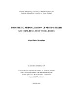
6.3.2 Microinjections of the Crow’s
Feet Area
One of the rst areas where the microinjection
technique was used was the crow’s feet area (Fig.
.; see also Sect. .). Here the most caudal
point might be very close to the bres of the zy-
gomaticus major, which is not a perfect candi-
date for the treatment with botulinum toxin A,
as treatment might result in a longer upper lip.
6.3.3 Microinjections
of the Longitudinal Lines
of the Cheeks
Another example of a good indication for micro-
injections are the longitudinal lines of the cheeks
Fig. .. Injection points for the crow’s feet area using
the microinjection technique
116 Mauricio de Maio, Berthold Rzany
6
that appear when the patients smiles (Figs. .,
.a,b). Here the muscles responsible, the m.
risorius and the m. zygomaticus major are tar
-
geted. Too deeply placed microinjections might
as well act like macroinjections and can cause
unwanted asymmetry (Fig. .a,b).
6.3.4 Doses to be Used
e doses to be used are the doses for the mac-
roinjection. e only dierence is that instead of
three injection points, the dose will be distrib-
uted in – injection points.
6.3.5 Combination of Macro- and
Microinjections
e combination of macro– and microinjections
can be very rewarding. A good example is again
the crow’s feet area. Here two macroinjections
cm lateral to the orbital rim will eectively in-
hibit the activity of the m. orbicularis oculi. e
more caudal area might be treated with four to
ve supercial microinjections, thereby reducing
the risk of an unwanted ptosis of the upper lip.
6.3.6 Disadvantages
of the Microinjection
Technique
e main disadvantage of the microinjection
technique lies in the multiple injections which
increase the risk of punctual hematoma and the
intensity of real or perceived injection pain. As
the existing needles are not made for repeated
injections, the bevel of the needle may become
dull quite easily.
6.3.7 Tips and Tricks
■
If you use the microinjection technique,
please remember the total dose you are us
-
ing in this area. Otherwise you might be
prone to over– or underdose.
Fig. .. Injection points for the cheek area using the
microinjection technique
Chapter 6 117Advanced Indications and Techniques
Fig. .a,b. Cheek area before and aer microinjections with BNT-A. Light decrease of longitudinal wrinkles on
both sides
Fig. .a,b. Asymmetry in the cheek area aer treatment with microinjections. On the le side, one of the injec-
tion points was placed too close to the modiolus, leading to an impairment of the m. risorius and the m. zygomaticus
major