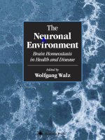
The Neuronal Environment: Brain Homeostasis in Health and Disease pot
Bạn đang xem bản rút gọn của tài liệu. Xem và tải ngay bản đầy đủ của tài liệu tại đây (3.67 MB, 428 trang )
Humana Press
Brain Homeostasis
in Health and Disease
Edited by
Wolfgang Walz
The
Neuronal
Environment
The Neuronal Environment
Contemporary Neuroscience
The Neuronal Environment: Brain
Homeostasis in Health and Disease,
edited by Wolfgang Walz, 2002
Neurotransmitter Transporters:
Structure, Function, and Regulation,
2/e, edited by Maarten E. A. Reith,
2002
Pathogenesis of Neurodegenerative
Disorders, edited by Mark P. Mattson,
2001
Stem Cells and CNS Development, edited
by Mahendra S. Rao, 2001
Neurobiology of Spinal Cord Injury,
edited by Robert G. Kalb and
Stephen M. Strittmatter, 2000
Cerebral Signal Transduction: From
First to Fourth Messengers, edited by
Maarten E. A. Reith, 2000
Central Nervous System Diseases:
Innovative Animal Models from Lab to
Clinic, edited by Dwaine F. Emerich,
Reginald L. Dean, III,
and Paul R. Sanberg, 2000
Mitochondrial Inhibitors and
Neurodegenerative Disorders, edited
by Paul R. Sanberg, Hitoo Nishino,
and Cesario V. Borlongan, 2000
Cerebral Ischemia: Molecular and
Cellular Pathophysiology, edited by
Wolfgang Walz, 1999
Cell Transplantation for Neurological
Disorders, edited by
Thomas B. Freeman and
Håkan Widner,1998
Gene Therapy for Neurological
Disorders and Brain Tumors, edited
by E. Antonio Chiocca and
Xandra O. Breakefield, 1998
Highly Selective Neurotoxins: Basic and
Clinical Applications, edited by
Richard M. Kostrzewa, 1998
Neuroinflammation: Mechanisms and
Management, edited by Paul L.
Wood, 1998
Neuroprotective Signal Transduction,
edited by Mark P. Mattson, 1998
Clinical Pharmacology of Cerebral
Ischemia, edited by Gert J. Ter Horst
and Jakob Korf, 1997
Molecular Mechanisms of Dementia,
edited by Wilma Wasco and
Rudolph E. Tanzi, 1997
Neurotransmitter Transporters:
Structure, Function, and Regulation,
edited by Maarten E. A. Reith, 1997
Motor Activity and Movement Disorders:
Research Issues and Applications,
edited by Paul R. Sanberg,
Klaus-Peter Ossenkopp, and
Martin Kavaliers, 1996
Neurotherapeutics: Emerging
Strategies, edited by Linda M. Pullan
and Jitendra Patel, 1996
Neuron–Glia Interrelations During
Phylogeny: II. Plasticity and
Regeneration, edited by Antonia
Vernadakis and Betty I. Roots, 1995
Neuron–Glia Interrelations During
Phylogeny: I. Phylogeny and
Ontogeny of Glial Cells, edited by
Antonia Vernadakis and
Betty I. Roots, 1995
The Biology of Neuropeptide Y and
Related Peptides, edited
by William F. Colmers and
Claes Wahlestedt, 1993
The Neuronal
Environment
Brain Homeostasis
in Health and Disease
Edited by
Wolfgang Walz
Department of Physiology,
University of Saskatchewan, Saskatoon,
Saskatchawan, Canada
Humana Press
Totowa, New Jersey
© 2002 Humana Press Inc.
999 Riverview Drive, Suite 208
Totowa, New Jersey 07512
www.humanapress.com
All rights reserved. No part of this book may be reproduced, stored in a retrieval system, or transmitted in
any form or by any means, electronic, mechanical, photocopying, microfilming, recording, or otherwise
without written permission from the Publisher.
The Humana Press Inc.
The content and opinions expressed in this book are the sole work of the authors and editors, who have
warranted due diligence in the creation and issuance of their work. The publisher, editors, and authors are
not responsible for errors or omissions or for any consequences arising from the information or opinions
presented in this book and make no warranty, express or implied, with respect to its contents.
This publication is printed on acid-free paper. ∞
ANSI Z39.48-1984 (American Standards Institute) Permanence of Paper for Printed Library Materials.
Production Editor: Diana Mezzina
Cover Illustration: Figure 9 from Chapter 4, “Transmitter-Receptor Mismatches in Central Dopamine,
Serotonin, and Neuropeptide Systems,” Further Evidence for Volume Transmission, by A. Jensson, L. Descarries,
V. Cornea-Hébert, M. Riad, D. Vergé, M. Bancila, L. F. Agnati, and K. Fluxe.
Cover design by Patricia Cleary.
For additional copies, pricing for bulk purchases, and/or information about other Humana titles, contact
Humana at the above address or at any of the following numbers: Tel.: 973-256-1699; Fax: 973-256-8341;
E-mail: ; or visit our Website: www.humanapress.com.
Photocopy Authorization Policy:
Authorization to photocopy items for internal or personal use, or the internal or personal use of specific
clients, is granted by Humana Press Inc., provided that the base fee of US $10.00 per copy, plus US $00.25 per
page, is paid directly to the Copyright Clearance Center at 222 Rosewood Drive, Danvers, MA 01923. For
those organizations that have been granted a photocopy license from the CCC, a separate system of payment
has been arranged and is acceptable to Humana Press Inc. The fee code for users of the Transactional Report-
ing Service is: [0-89603-882-3/02 $10.00 + $00.25].
Printed in the United States of America. 10 9 8 7 6 5 4 3 2 1
Library of Congress Cataloging in Publication Data
The neuronal environment: brain homeostasis in health and diease/edited by
Wolfgang Walz
p. cm (Contemporary neuroscience)
Includes bibliographical references and index.
ISBN : 0-89603-882-3 (alk. paper)
1. Neurons Physiology. 2. Homeostasis. 3. Neuroglia. 4. Brain Metabolism. 5. Blood-brain barrier.
I. Walz, Wolfgang. II. Series.
QP363.N47758 2002
612.8’2 dc21
2001039827
Preface
To function properly, neurons cannot tolerate fluctuations of their local environ-
mental variables. This mainly results from their high degree of specialization in synap-
tic integration and action potential conduction. Even small changes of certain
extracellular ion concentrations, as well as in the dimensions of the extracellular space,
alter ion channel kinetics in such a way as to distort the information represented by the
nerve impulses. Another potential problem is the huge consumption of glucose and
oxygen by neurons caused by the heavy compensatory ion pumping used for counter-
acting passive ion flux. This problem is compounded by the low glucose storage capac-
ity of the neurons. A complicated structure surrounds the neurons to sustain the required
level of metabolites and to remove waste products.
The Neuronal Environment: Brain Homeostasis in Health and Disease
examines the function of all the components involved, including their perturbation dur-
ing major disease states, and relates them to neuronal demands. The two introductory
chapters focus on neuronal requirements. The dependence of their excitability on
external factors that accumulate in the extracellular space, as well as their varying
demands for energy metabolites, are described. Following that, the close interaction of
neurons with elements of their microenvironment is illustrated. The extracellular space
is no longer seen as a passive constituent of the CNS, but as a separate compartment in
its own right, as a communication channel, and an entity that reacts with plastic changes
in its size that will affect the concentrations of all its contents. Astrocytes participate in
many neuronal processes, particularly in the removal of excess waste and signal sub-
stances, the supply of energy metabolites, and the modulation of synaptic transmission.
In addition to their homeostatic role, astrocytes are now seen as an active partner
involved in synaptic transmission between neurons. The classical example of a close
relationship of neurons with a component of their environment is, of course, their rela-
tionship with the surrounding myelin sheath. This speeds up action potential conduc-
tion, but is itself a potential source of problems in various disease states. In the last few
years new imaging techniques have demonstrated a close coupling between local blood
flow and neuronal activity, and several theories have been put forward to explain these
interactions. The special status of the brain in having its own insulated circulation
system—the cerebrospinal fluid contained in the ventricles and ducts—is also under-
lined. The brain is the only organ that is protected from fluctuations of blood-borne
chemicals by the existence of the blood–brain barrier. However, windows exist in this
barrier in the form of the circumventricular organs that allow direct two-way commu-
nication between neurons and blood constituents. Finally, despite their protection and
insulation, the neurons are accessible to the immune system. Resident macrophages
and invasion by blood-borne immune cells that cross the endothelial cell barrier enable
v
an immune reaction to take place. This complex interaction of neurons with their
immediate environment is integral to the tasks that the neurons must perform to ensure
that the organism can cope with its environmental challenges. Most diseases originat-
ing in the brain start in these accessory systems of the neuronal microenvironment and
affect neurons only second hand. Therefore, understanding the elements of the neu-
ronal environment and the interactions with neurons, and with each other, is crucial in
understanding the development and impact of most brain diseases.
All the authors contributing to The Neuronal Environment: Brain Homeostasis in
Health and Disease have made an attempt not only to explain the normal functioning
of these accessory elements, but also their involvement in major diseases. Therefore,
this book not only addresses researchers, graduate students, and educators who want to
understand the complex environment of neurons, but also health professionals who
need to know more about the normal homeostatic role of the neuronal environment to
follow disease patterns.
Wolfgang Walz
vi Preface
Contents
vii
Preface v
Contributors ix
I. NEURONAL ACTIVITY AND ITS DEPENDENCE ON THE MICROENVIRONMENT
1 Central Nervous System Microenvironment
and Neuronal Excitability 3
Stephen Dombrowski, Imad Najm, and Damir Janigro
2 Neuronal Energy Requirements 25
Avital Schurr
II. BRAIN MICROENVIRONMENT
3 Plasticity of the Extracellular Space 57
Eva Syková
4 Transmitter–Receptor Mismatches in Central Dopamine,
Serotonin, and Neuropeptide Systems: Further Evidence
for Volume Transmission 83
Anders Jansson, Laurent Descarries, Virginia Cornea-Hébert,
Mustapha Riad, Daniel Vergé, Mircea Bancila,
Luigi Francesco Agnati, and Kjell Fuxe
5 The Extracellular Matrix in Neural Development, Plasticity,
and Regeneration 109
Jeremy Garwood, Nicolas Heck, Franck Rigato,
and Andreas Faissner
6 Homeostatic Properties of Astrocytes 159
Wolfgang Walz and Bernhard H. J. Juurlink
7 Glutamate–Mediated Astrocyte–Neuron Communication
in Brain Physiology and Pathology 187
Micaela Zonta and Giorgio Carmignoto
8 Axonal Conduction and Myelin 211
Jeffrey D. Kocsis
9 Coupling of Blood Flow to Neuronal Excitability 233
Albert Gjedde
III. BRAIN MACROENVIRONMENT
10 Choroid Plexus and the Cerebrospinal–Interstitial
Fluid System 261
Roy O. Weller
viii Contents
11 The Blood–Brain Barrier 277
Richard F. Keep
12 Circumventricular Organs 309
James W. Anderson and Alastair V. Ferguson
13 Glial Linings of the Brain 341
Marc R. Del Bigio
IV. I
MMUNE SYSTEM-NEURON INTERACTIONS
14 Microglia in the CNS 379
Sophie Chabot and V. Wee Yong
15 Invasion of Ischemic Brain by Immune Cells 401
Hiroyuki Kato and Takanori Oikawa
Index 419
Contributors
LUIGI FRANCESCO AGNATI, Department of Human Physiology,
University of Modena, Modena, Italy
JAMES W. ANDERSON, Department of Physiology, Queen’s University,
Kingston, Ontario, Canada
MIRCEA BANCILA, Laboratoire de Neurobiologie de Signaux Intercellulaires,
Institut des Neurosciences, Université Pierre et Marie Curie, Paris, France
GIORGIO CARMIGNOTO, Department of Experimental Biomedical Sciences,
University of Padova, Padova, Italy
SOPHIE CHABOT, Department of Oncology and Clinical Neurosciences,
University of Calgary, Calgary, Canada
VIRGINIA CORNEA-HÉBERT, Département de Pathologie et Biologie Cellulaire,
Université de Montréal, Montréal, Canada
MARC DEL BIGIO, Department of Pathology, Health Sciences Centre and
University of Manitoba, Winnipeg, Canada
LAURENT DESCARRIES, Département de Pathologie et Biologie Cellulaire,
Université de Montréal, Montréal, Canada
STEPHEN DOMBROWSKI, Department of Neurosurgery,
Cleveland Clinic Foundation, Cleveland, OH
ANDREAS FAISSNER, Laboratoire de Neurobiologie du Developpment et de la
Regeneration, Strasbourg, France
ALASTAIR V. F ERGUSON, Department of Physiology, Queen's University,
Kingston, Ontario, Canada
KJELL FUXE, Department of Neuroscience, Karolinska Institute, Stockholm, Sweden
JEREMY GARWOOD, Laboratoire de Neurobiologie du Developpment et de la
Regeneration, Strasbourg, France
ALBERT GJEDDE, The Pathophysiology and Experimental Tomography Center,
Aarhus General Hospital, Aarhus C, Denmark
NICOLAS HECK, Centre National De la Recherche Scientifique, Strasbourg, France
D
AMIR JANIGRO, Division of Cerebrovascular Research,
Department of Neurosurgery, Cleveland Clinic Foundation, Cleveland, OH
ANDERS JANSSON, Department of Neuroscience, Division of Cellular
and Molecular Neurochemistry, Karolinska Institute, Stockholm, Sweden
ix
x Contributors
B
ERNHARD H.J. JUURLINK, Department of Anatomy and Cell Biology,
University Saskatchewan, Saskatoon, Canada
HIROYUKI KATO, Department of Neurology and Neuroendovascular Therapy,
Tohoku University School of Medicine, Sendai, Japan
RICHARD F. KEEP, Departments of Surgery and Physiology,
University of Michigan, Ann Arbor, MI
JEFFERY D. KOCSIS, Neuroscience Research Center, Department of Veterans Affairs
Medical Center, Yale University School of Medicine, West Haven, CT
IMAD NAJM, Department of Neurosurgery, Cleveland Clinic Foundation,
Cleveland, OH
TAKANORI OIKAWA, Department of Neurology, Tohoku University School of
Medicine, Sendai, Japan
M
USTAPHA RIAD, Departement de Pathologie et Biologie Cellulaire,
Universite de Montreal, Montreal, Canada
FRANCK RIGATO, Centre Natioanl De la Recherche Scientifique, Strasbourg, France
AVITAL SCHURR, Department of Anesthesiology, University of Louisville,
School of Medicine, Louisville, KY
EVA SYKOVÁ, Department of Neuroscience, Institute of Experimental Medicine,
Academy of Sciences, Prague, Czech Republic
D
ANIEL VERGÉ, Laboratoire de Neurobiologie de Signaux Intercellulaires,
Institut des Neurosciences, Université Pierre et Marie Curie, Paris, France
WOLFGANG WALZ, Department of Physiology, University of Saskatchewan,
Saskatoon, Canada
ROY O. WELLER, Department of Microbiology and Pathology,
Southhampton General Hospital, Southampton, UK
V. WEE YONG, Departments of Oncology and Clinical Neurosciences,
University of Calgary, Calgary, Canada
M
ICAELA ZONTA, Department of Experimental Biomedical Sciences,
University of Padova, Padova, Italy
CNS Microenvironment and Excitability 1
I
NEURONAL ACTIVITY AND ITS DEPENDENCE
ON THE MICROENVIRONMENT
CNS Microenvironment and Excitability 3
3
From: The Neuronal Environment: Brain Homeostasis in Health and Disease
Edited by: W. Walz © Humana Press Inc., Totowa, NJ
1
Central Nervous System Microenvironment
and Neuronal Excitability
Stephen Dombrowski, Imad Najm, and Damir Janigro
1. INTRODUCTION
The biological cell membrane, the interface between the cell and its environment, is
a complex biochemical entity, one of whose major jobs is to allow or impede transport
of specific substances in one direction or another. A related major job of the cell mem-
brane is the maintenance of chemical gradients, particularly electrochemical gradients,
across the plasma membrane. These gradients can be of high specificity (e.g., sodium
vs. potassium ions), and of great functional significance (e.g., in the production of
action potentials in nerve and muscle cells) (1).
The separation of intra- and extracellular compartments by lipidic bilayers is one of
the crucial steps in evolution. One of the consequences of this partition is the signifi-
cant difference in the cytosol and extracellular contents of cells. Furthermore, cells
with different functions tend to have different intracellular composition, and cellular
elements from different tissues are exposed to extracellular media of different chemi-
cal nature. In addition to a variety of nutrients and growth factors, the extracellular
milieu also contains molecules that either promote cell differentiation (e.g., adhesion
molecules) or survival (growth factors), as well as ions constituting the basis of electri-
cal activity (or silence) of mammalian cells. Granting that appropriate control of the
composition of the extracellular space significantly impacts the cytosolic content, and
vice versa, change in the intracellular components of central nervous system (CNS)
cells impacts the composition of extracellular fluids. The dynamic process involved in
the maintenance of the composition of intra- and extracellular ingredients is called
“homeostasis.”
The general design used for the separation of intracellular and extracellular space
has also been used during the evolution to maintain the nervous system of vertebrates,
isolated, at least in part, from systemic influences. Therefore, a double bilayer, similar
to the lipophilic barrier isolating the cytoplasm from the external milieu and formed by
brain microvascular endothelial cells [the blood–brain barrier (BBB)], separates the
CNS from the blood, in vertebrates.
From a neuroscientist’s point of view, the fact that the neuronal extracellular milieu
composition is controlled by such a complex cascade of serially occurring events best
illustrates the relevance of controlled neuronal activity to ensure the organism’s
4 Dombrowski, Najm, and Janigro
Table 1
Examples of Homeostatic Mechanisms in CNS and Their Possible Involvement in Pathogenesis
Mechanisms involved Cell types involved Pathology Refs.
Barrier function BBB Endothelium Brain tumors
Choroid plexus Neuroepithelium Stroke
Brain–CSF barrier Pia–glia Hypertension (90)
Alzheimer’s (91)
P-glycoprotein Endothelium Epilepsy
Transport of nutrients Glucose transport Endothelium GLUT1 deficiency
and neurotransmitters GLUT1–GLUT3 Astrocytes Epilepsy
Alzheimer’s
Amino acid transport Neurons (92–96)
Glia (43,45,46,97,98)
GLAST Endothelium
Ion homeostasis Na
+
/K
+
-ATPase Neurons Epilepsy
Glia Vascular dementia
Endothelium
Inward rectifier Astrocytes
Metabolic control Autoregulation Vascular smooth muscle; Head injury (99,100)
of CNS function Systemic influences glia, neurons
4
CNS Microenvironment and Excitability 5
survival (Table 1). The following paragraphs summarize some of the most relevant
mechanisms involved in the regulation of neuronal excitability by factors present in the
extracellular milieu.
2. CELLULAR CORRELATES OF BRAIN HOMEOSTASIS
2.1. Neuroglia
The necessity for tight control of the composition of brain extracellular fluids is in
part a consequence of the evolutionary push for miniaturization of the cellular compo-
nents of the CNS (neurons and glia), paralleled by the need to produce ultrafast signal-
ing at the neuronal synapse, and to allow comparably fast neural transmissions along
axons. Functional compromise between a high velocity of neuronal computation and
reduced size of the neuron-to-neuron axonal wiring has been reached, in vertebrates,
by ensheathing the axons by a myelin isolator produced by oligodendroglia, allowing
for so-called “saltatory conductance” (2). One of the clear advantages of this design is
that the myelin sheath occupies much less volume than an equally conductive axon
with a much larger diameter would occupy (for mathematical modeling and other bio-
physical considerations, see ref. 3).
Miniaturization of the vertebrate CNS occurred as a consequence of the necessity to
protect the brain and spinal cord with a bony structure, limiting the overall volume
available for cellular expansion. A consequence of this limiting factor is that the extra-
cellular space in the brain is very small, amplifying the concentration changes occur-
ring across the plasmalemma surrounding the cells (4). The size of the extracellular
space is not homogeneous, and regional differences have been found, even within the
contiguous CA1 and CA3 hippocampal regions (5). The possibility that these regional
variations also relate to different glial subpopulations within the hippocampus has been
proposed (6).
Finally, in an attempt to further minimize the cellular number and volume of the
CNS, the lymphatic drainage apparatus has been sacrificed, leaving the composition of
extracellular fluids in the brain at the mercy of the brain cells themselves. The subse-
quent necessity to shield the central nervous system from uncontrolled systemic influ-
ences, and in order to minimize the extravasation of potentially noxious or osmotically
active molecules from the blood, is perhaps the best-understood reason for the creation
of the blood–brain-barrier (7–9). Similarly, the requirement for an extralymphatic
mechanism of clearance and homeostasis constitutes the teleonomic reason for the
numeric preponderance of glial cells in the mammalian central nervous system. These
glia are directly responsible for the control of the composition of the extracellular space.
Glial cells themselves do not constitute a homogeneous population, and at least three
classes of glial cells have been described. Oligodendroglia are primarily responsible
for the production of myelin, which isolates axons, leaving unsheathed segments with
high densities of sodium and potassium channels (10,11). Astrocytes are present in
both gray and white matter of the CNS, and are perhaps the most numerous subpopula-
tion of glial cells. Astrocytes are involved in a number of different processes, including
the control of ionic homeostasis, control of neuronal metabolism, as well as mainte-
nance of blood–brain barrier integrity (12–19); recent evidence also suggests that they
may actively participate in synaptic transmission (20–23). Microglia are the cellular
6 Dombrowski, Najm, and Janigro
substrates of the neuroimmune response. Their possible role in the homeostasis of CNS
extracellular fluids is not known, but these cells express ion channels involved in the
control of potassium homeostasis performed by astrocytes (24,25).
2.2. Vascular Endothelium and Smooth Muscle
In addition to parenchymally located glial cells, at least two additional cell types
participate in the process of the control of the composition of the extracellular space in
the brain: the cellular elements constituting intraparenchymal vessels, the endothelial
cells lining the intraluminal portion of blood vessels, and only cellular element consti-
tuting the BBB at the capillary level; and vascular smooth muscle, the final effectors
responsible for the control of cerebral perfusion.
There are numerous ways by which these vascular elements may cooperate with
parenchymal glia toward the maintenance of a stable extracellular milieu. BBB endo-
thelial cells are believed to control ionic homeostasis, by preventing equalization of
plasma levels of ions with those present in the cerebral spinal fluid (26–28). Part of this
process is energy-dependent, and directly impacts the ionic homeostasis for potassium
ions (see Subheading 3.).
Vascular smooth muscle are also indirectly involved in the control of brain homeo-
stasis, since their powerful effect on the control of cerebral perfusion will be the final
determinant of the amount of oxygen and glucose delivered to the brain, as well as to
the level of “cleansing” by cerebral blood flow of potential noxious metabolites pro-
duced by neural activity. The control of cerebral circulation is mostly independent of
extrinsic neuronal influences (29). Both capillary function and the amount of blood
perfusing the brain parenchyma are directly proportional to the metabolic activity of
neuronal cells, a phenomenon called “autoregulation,” which appears to depend on a
number of different mechanisms, including nitric oxide, adenosine, potassium, and pH
(30–35).
Finally, vascular (endothelial cells and vascular smooth muscle) and parenchymal
(neurons and glia) cells cooperate closely, and directly influence each other’s develop-
ment. The best-understood mechanism of this tight cell-to-cell interaction is perhaps
the ontogenesis of the blood–brain barrier, a phenomenon directly dependent on the
presence of abluminal glial endfeet, which transmit as-yet unknown signals to neigh-
boring endothelial cells (17,36,37). This example clearly illustrates one of the unique
mechanisms by which the central nervous system parenchyma influences the cerebral
vasculature, without involvement of signals generated distally, a feature that is com-
mon in the systemic circulation, where barrier function is not crucial, because of the
presence of lymphatic drainage. Note that this general difference does not apply to
highly specialized peripheral systems, such as the testicle, where active barrier func-
tion is bestowed upon capillary endothelial cells (38).
3. BASIC ELECTROPHYSIOLOGY
AS RELEVANT TO EXTRACELLULAR SPACE (ECS) HOMEOSTASIS
Electrical phenomena occur whenever charges of opposite sign are separated or
moved in a given direction. Static electricity is the accumulation of electric charge:
An electric current results when these charges flow across a permissive material, called
a “conductor.” An ion current is a particular type of current carried by charges present
CNS Microenvironment and Excitability 7
on atoms or small molecules flowing in aqueous solution. Separation of charges in an
aqueous solution can be achieved by inserting an impermeable membrane in the solu-
tion itself. In mammalian cells, these membranes coincide with the plasma membrane,
and its lipophylic composition ensures a remarkable level of electrical isolation for
cells and tissues. Excitable cells, as well as most nonexcitable cells, are characterized
by an asymmetric distribution of electrical charges across the plasma membrane.
The control of the distribution of electrical charges across the plasma membrane is an
energy-consuming process. A significant portion of this homeostatic control involves the
tight regulation of sodium and potassium gradients. The molecular mechanism respon-
sible is the so-called “Na
+
/K
+
-adenosine triphosphatase (ATPase),” an ubiquitous enzyme
whose activity is highly dependent on intra-cellular levels of ATP. It is clear that even
minimal changes in the availability of energy substrates (ATP) will cause significant
changes in the resting potential of the cells. It is well understood that intracellular Na
+
concentrations are controlled by the exchange of three Na
+
against two K
+
, an electro-
genic mechanism that contributes substantially to the regulation of resting membrane
potential (RMP) in both neurons and glia. The activity of this enzyme depends, in addi-
tion to availability of ATP, on internal Na
+
and external K
+
concentrations, and, since
[K
+
] (and, to a lesser extent, [Na
+
]) are the main ionic mechanism of the generation of a
stable resting membrane potential (39), it becomes obvious that energy supply, ionic
homeostasis, and the control of RMP are closely interconnected mechanisms. Because
the probability of neuronal firing depends to a large extent on the transmembrane volt-
age, the link between ionic homeostasis and neuronal excitability becomes evident.
Neuronal cells use a single type of long distance signaling strategy, based on the
propagation of all-or-nothing action potentials. Sodium action potentials, such as those
recorded in axons or cell bodies, are relatively invariant in normal tissue, and thus the
shape and duration of these electrical signals does not vary significantly within the
nervous system. Calcium action potentials are similarly predictable, but the underlying
ionic mechanism can be complex, depending on the cell type, and on the topographic
location within the cell. The terms “sodium action potential” and “calcium action
potential” refer to the initial (depolarizing) phase of these rapid membrane polarity
changes, and, although genetic or molecular alteration of I
Na
and I
Ca
can significantly
affect neuronal firing and, ultimately, CNS/peripheral nervous system neurophysiol-
ogy, gross changes in neuronal excitability may also result by altering the repolariza-
tion phase of individual action potentials because of the dramatic changes in
extracellular potassium concentrations that accompany neuronal firing, and the con-
secutive feedback effect of [K
+
]
out
on neuronal resting membrane potential (Fig. 1).
From a functional standpoint, the genesis of fast, sodium action potentials is a hall-
mark of neuronal function, to the degree that during neurophysiological recordings,
presence or absence of Na
+
spikes is frequently used to determine the neuronal or glial
cell type (40–42). Recently, this notion has been challenged, and glial action potentials
have been reported with increasing frequency (43–46). These responses, however, usu-
ally appear to be associated with pathologic conditions (brain tumors, epilepsy), and
the old perception that neuronal cells are the exclusive tenants of sufficient I
Na
density,
to promote active responses, is still generally accepted.
Although it is obvious that any significant ionic flux across neuronal membranes
will invariably lead to changes in the extracellular/intercellular milieu composition,
8 Dombrowski, Najm, and Janigro
the following subheading describes in some detail only mechanisms involved in the
control of K
+
homeostasis, because K
+
ions have historically been linked to strict con-
trol of neuronal excitation by their profound effect on neuronal resting potential and
synaptic transmission. Recent evidence from the author’s laboratory also suggests that
failure of K
+
homeostasis by glial cells may lead to abnormal extracellular fluid com-
position and a propensity to seizures.
4. POTASSIUM HOMEOSTASIS
Potassium channels are present in virtually every animal cell type, and serve a vari-
ety of functions. Historically, these ubiquitous ionic mechanisms were associated with
Fig. 1. Diagrammatic representations of potassium fluxes into the CNS. This scheme is
based on original spatial buffering concepts described by Orkand (46a), as well as from results
inferred from experiments on isolated cells and BVs isolated from the brain (30). The depo-
larization of pre- and postsynaptic terminals depicted in the right side of the picture causes
opening of voltage-dependent potassium channels in neurons. Activation of outward potas-
sium currents causes large potassium fluxes from the cytoplasm to the ECS. Although a frac-
tion of excess potassium ions may directly return into the neuronal cell by active transport
via Na
+
/K
+
-ATPase (not shown in figure), additional uptake of potassium occurs, under most
conditions, by voltage-dependent uptake into astrocytic endfeet. Fluxes of potassium through
the glial syncytium may then lead either to return of K
+
into the ECS surrounding the neurons,
or, perhaps, under more extreme conditions, to release of excess potassium into the blood stream
by glial endfeet. The top part of the figure represents the passage of potassium across one single
astrocyte, characterized by a cell body and endfeet surrounding a BV, as well as an
ensheathment of synaptic terminals. The bottom part of the figure refers to a more common
situation, in which multiple glial cells are coupled by gap junctions (6). Gap junction expres-
sion is altered in epileptic tissue (45).
CNS Microenvironment and Excitability 9
control of cell resting potential and, after the discovery of sodium action potentials, the
repolarization phase leading to the recovery of pre-action potential RMPs. Potassium
channels belong to a large and complex group that can be divided into functional, struc-
tural, or molecular families. Voltage-dependent K channels (K
V
) are constituted of
six transmembrane regions (S1–S6) and a P or H5 segment between S5 and S6; the selec-
tivity filter contains a specific sequence (glycine-tyrosine-glycine); the voltage sensor
consists of positively charged amino acids in the S4 region. Inward rectifier potassium
channels (K
IR
) are distantly related to the voltage-dependent family, and are made of
four subunits, each consisting of two transmembrane segments (M1 and M2) and a P or
H5 segment located between. These channels do not allow passage of current at posi-
tive potentials. The voltage-dependency of the K
V
channels depends on the presence of
a voltage sensor, but inward rectification is achieved by voltage-dependent blockade
of the intracellular portion of the channel pore by cytosolic cations (47,48). Opening of
the channels may be achieved by G protein-coupled mechanisms (as in the GIRK sub-
family), or by metabolic changes (intracellular ATP, K
IR
6.1, or ATP-sensitive potas-
sium channels) (49–51).
4.1. Extracellular Space Composition
and Regulation of Neuronal Excitability
Central nervous system astrocytes are strategically located in proximity to excitable
neurons, and are sensitive to changes in extracellular ion composition that follow neu-
ronal activity (see diagram in Fig. 1). Several lines of evidence suggest that brain glial
cells support the homeostatic regulation of the neuronal microenvironment. In cortical
regions, glial cells participate in the genesis of the extracellular field potential changes
associated with neuronal depolarization and efflux of potassium in the extracellular
space (52–54).
Several mechanisms have been proposed to explain how astrocytes sense and react
to changes in extracellular potassium concentrations, following both normal and
abnormal neuronal activity. As summarized in the previous paragraphs, neuronal
excitability is regulated by a complex interaction of voltage-dependent ion currents
and synaptically mediated excitatory and inhibitory potentials. In principal neocortical
or hippocampal neurons, depolarizing ion conductances involved in action potential
generation, are regulated primarily by the voltage-dependent activation/inactivation
properties of Na
+
and Ca
2+
channels; inward Na
+
and Ca
2+
fluxes also underlie the
generation of excitatory postsynaptic potential (EPSPs). Termination of these depolar-
izing potentials occurs by the voltage- and calcium-dependent activity of intrinsic
potassium conductances, and by activation of interneurons, which release inhibitory
neurotransmitters to produce inhibitory postsynaptic potentials (IPSPs): The latter are
mediated by postsynaptic activation of chloride and potassium currents.
Although I
Na
, I
Ca
, and I
EPSP
are, under physiological conditions, independent of
modest changes in the driving force for the permeant ions (since E
Na
and E
Ca
are remote
with respect to cell resting potential), both repolarizing potassium and IPSP conductances
are critically affected by even modest changes in cell RMP, [K
+
]
out
and [Cl]
in
/[Cl]
out
.
Thus, ionic changes directly associated with excitatory (depolarizing) activity seem to
impact minimally ionic homeostasis, but repolarization and inhibition are powerful
modulators of [K
+
]
out
, [Cl
–
]
in/out
, and so on. As a consequence, failure to control potas-
10 Dombrowski, Najm, and Janigro
sium and chloride homeostasis is likely to promote neuronal excitability by decreasing
the efficacy of repolarizing K currents and IPSPs.
The concentration of potassium in the ECS (K
ECS
), in the mammalian CNS, increases
measurably (from 3 to ~4 mM) during physiological stimulation; to a larger extent (up
to 12 mM), during seizures or direct, synchronous stimulation of afferent pathways;
and to exceedingly high values (>30 mM), during anoxia or spreading depression
(6,41,55,56). Despite these rapid and large changes in K
ECS
, K
+
values return to normal
levels in a short period of time. Several mechanisms have been proposed to explain the
rapid clearance of K
+
from the ECS, including uptake by glia, passive diffusion, and
neuronal reuptake. Experiments have suggested that glial uptake plays a pivotal role
under conditions in which there is massive K
out
accumulation (41,53,55,57). [K]
out
can
also be redistributed through active removal by blood flow, or by passive diffusion
through the ECS (58); however, these mechanisms alone are not fast enough to account
for the rapid K
+
removal from the ECS seen under experimental conditions.
4.2. Astrocytes and Buffering of ECS Potassium
Glial involvement in CNS potassium homeostasis has been long suspected, but never
unequivocally demonstrated in the mammalian CNS. Two different hypotheses have
been formulated: K
+
may accumulate directly into astrocytes, and increased local con-
centrations of K
ECS
may be buffered through glial cells by current-carried transport
mechanisms. The combination of potassium uptake into glial cells, immediately fol-
lowed by redistribution through electronically coupled glial gap junctions (“spatial
buffering” [59–65]) provides a valid working hypothesis to explain some of the fea-
tures of K
+
movements in the extracellular space. The spatial buffering mechanism
rests on the following hypotheses: Glial RMP closely follows E
K
(i.e., glial cells are
selectively and exclusively permeant to K
+
); and glial cells form a topographically
complex syncytium, by virtue of their tight electrotonic coupling via gap junctions.
Both of these hypotheses have been experimentally challenged. A clear correlation
between astrocyte RMP and [K
+
]
out
has been described in virtually every glial cell type
studied, but RMP more positive than those predicted by a Nernstian behavior have
been frequently reported (for discussion, see ref. 65). The deviation of glial RMP from
those predicted by Nernstian behavior has been attributed to one or more of the follow-
ing: the electrogenicity of the Na
+
/K
+
-ATPase pump; a persistent sodium conductance
activated at cell resting potential (66); chloride currents.
A modification of spatial buffering has been described for retinal Müller cells
(potassium siphoning) (67). This process is characterized by a topographic segregation
of high conductance zones in the plasma membrane. Thus, a large density of potassium
channels is localized in the cell region where extracellular space accumulation occurs,
and distally, at the glial endfeet, where potassium excretion into the ECS occurs.
No significant K fluxes are possible in the central region of the cell, where no K
+
removal or excretion occurs. A similar mechanism could explain several features of
extracellular potassium dynamics in cortical structures, and preliminary evidence (65),
supporting a preferential distribution of potassium channels in cortical astrocyte mem-
brane has been recently provided; double recordings from neighboring astrocytes dem-
onstrated that a heterogeneous expression of inward rectifier and outward rectifier
channels is present in these cells. The proposed model of potassium movements in
CNS Microenvironment and Excitability 11
these syncytia of neocortical astrocytes shared characteristics of both spatial buffering
(influx of potassium driven by E
m
and E
K
) and siphoning (segregated expression of
inward rectifier and outward rectifier channels).
4.3. Astrocytic Ion Currents and Potassium Homeostasis
For the purpose of this minireview, we will describe in some detail the endowment
of potassium channels expressed by glial cells in the CNS. Their role in the control of
extracellular K homeostasis is also briefly summarized.
Glial cells express a variety of potassium currents, but the expression of these cur-
rents seems to depend on the glial cell type considered (microglia vs astrocyte vs oligo-
dendroglia), as well as on the experimental conditions used to determine potassium
channel expression. Culturing of glia in vitro dramatically changes the expression of a
variety of glial specific markers, including ion channels. The relevance of these
expression changes is presently unknown. However, since exposure to serum (or, con-
versely, serum deprivation) plays a major role in cell differentiation, it is possible that,
under physiological conditions, the serum-free CNS environment may act as a matura-
tion factor for astrocytic differentiation. When and where brain homeostasis fails, as
the result of opening of the blood–brain barrier, serum proteins will extravasate
into the CNS: This may have profound effects on glial ion channel expression, and
play a major pathogenetic role.
It has been known for a long time that the predominant ion channel mechanism
expressed in astrocytes is the so-called “inward rectifier potassium channel.” The prop-
erties of these channels are consistent with the mechanisms involved in the simul-
taneous control of RMP and voltage-dependent uptake of potassium from the
extracellular space. The coupling of these channels to this dual control mechanism
justifies, in part, the old spatial-buffering theory proposed many years ago by Orkand
(1986). The mechanism of potassium entry into the cell is consistent with the biophysi-
cal properties of inwardly rectifying potassium channels (68,69), but it is still unclear
how the spatial redistribution of potassium ions occurs, and what mechanisms are used
by astrocytes to return potassium ion to the extracellular space. The presence of both
inward and outward currents on astrocytes, if topographically segregated as shown in
retinal astrocytes (67,70), would account for both uptake and redistribution of potas-
sium across cortical structures.
4.3.1. Potassium Uptake via Voltage-Dependent Currents
Glial cells lack regenerative, AP-like responses. However, glial cells express a num-
ber of voltage-, second messenger-, and agonist-operated channels (71–73). Potassium
channels are the most common electrophysiological feature of both cultured and in situ
astrocytes, and can be categorized as follows: channels that allow inward, but not out-
ward, current flow (inward rectifiers [K
IR
]); channels that allow outward, but not
inward, current flow (delayed rectifier [I
DR
]; transient outward current [I
A
]); channels
that are opened by intracellular calcium (I
K(Ca)
). Glial potassium channels differ
in their sensitivity to blockers: Inward rectifiers are blocked by submillimolar con-
centrations of external Cs
+
and Ba
2+
; outward I
DR
and I
A
are both sensitive to tetraethyl-
ammonium and 4-aminopyridine, but I
A
blockade by tetraethylammonium requires high
concentrations. Recently, a mixed-cation channel (I
ha
), permeant to K
+
and Na
+
, has
12 Dombrowski, Najm, and Janigro
been described in cultured astrocytes (74). A cardiac-type outward potassium current
has been described in in situ glia (25); this current (I
HERG
) seems to be involved in
potassium homeostasis in cooperation with K
IR
. A direct demonstration that K
IR
, I
HERG
,
or I
ha
play a role in spatial K
+
buffering is still lacking, but evidence from both in vivo
and in vitro studies has demonstrated proepileptogenic neuronal changes after applica-
tion of mostly glia-specific potassium channel blockers ([6,25,41]; see also Subhead-
ing 4.3.2., and Figs. 2–6).
Voltage-dependent, tetrodotoxin-sensitive and -insensitive sodium channels are also
expressed in both cultured and in situ glial cells (75). Although astrocytes are inca-
pable of producing action potential-like responses (but see ref. 73), possibly because of
the low Na
+
current densities in these cells, a role for Na
+
channels in spatial buffering
has been proposed. According to this hypothesis Na
+
influx sustains the Na
+
/K
+
-ATPase
pump, resulting in net K
+
uptake. Finally, calcium channels are represented sparingly
in glial cells, and require either neuronal or otherwise-differentiating factors for
Fig. 2. Changes in neuronal activity, EC potassium, and glial resting potential, after chemi-
cal ablation of spatial buffering by cesium. The left panel shows the size of EC field potentials
recorded in the CA1 region of the hippocampus during a 1 Hz stimulation trial of 15 min in
duration. The traces below refer to the IC recording, by patch clamp, of glial cell resting poten-
tial during the same period of time, as well as changes in EC potassium. Note that decreased
field potential amplitude does not necessarily correlate with either glial or EC potassium
changes. After exposure of the cells to a saturating concentration of cesium, the EC field poten-
tial response was not significantly altered, suggesting that direct neuronal effects were absent.
However, profound changes in basal concentration of EC potassium (indicated by the milli-
mole values under the bottom traces), as well as glial resting potential, occurred. (Reproduced
with permission from ref. 41.)
CNS Microenvironment and Excitability 13
13
Fig. 3. Astrocytes lose inward K
+
currents after in vivo traumatic brain injury. Whole-cell voltage-clamped cells in control hippocampal slices
exhibited large Cs
+
-sensitive currents (A, top and bottom panel), and were characterized by a large Cs
+
-sensitive component. In contrast, cells in
post-FPI hippocampal slices displayed little Cs
+
-sensitivity (
B, top and bottom panel), and showed a decreased Cs
+
-sensitive component of the
whole-cell inward currents. (C) The percentage of Cs
+
-sensitive currents (I
Cs
) for glia in normal and post-FPI hippocampus is shown for membrane
potentials from –140 to –80 mV. Voltage commands consisted of ramps from –170 to +100 mV, over 750 ms, from holding potential o
f
–70 mV.
(Reprinted with permission from ref. 79.)
14 Dombrowski, Najm, and Janigro
expression (71,72); whether I
Ca
can be recorded from in situ hippocampal astrocytes is
still unknown, but release of calcium from intracellular stores, in response to neuro-
transmitters acting on astrocytes, has been clearly demonstrated. Relevant to spatial
buffering, micromolar [Ca
2+
]
i
can cause opening of I
K(Ca)
, and may thus participate in
the generation of outward potassium fluxes.
4.3.2. K
+
Buffering by Furosemide-Sensitive Na,K,2Cl Cotransporter
Under conditions involving high levels of neuronal activity (e.g., seizures), [K]
out
accumulation is accompanied by cell swelling. The swelling that accompanies epilep-
tiform neuronal discharge results from excess activity of ionic mechanisms normally
involved in the control of ECS homeostasis. One of several proposed mechanisms
associated with cell swelling, the Na,K,2Cl co-transporter, is also believed to partici-
pate in uptake of K
+
into glia. This transporter is blocked by the general diuretic, furo-
semide. Treatment of epileptic hippocampal slices (treated with bicuculline, 0 Ca
2+
, or
4-aminopyridine) with furosemide has been shown to inhibit spontaneous burst dis-
charge. It has been hypothesized (76) that this mechanism was related to furosemide
Fig. 4. Neuronal stimulation induces abnormal accumulation of EC K
+
and burst discharge
in slices from post-traumatic rats. Field electrode and KSM were placed in CA3 stratum
radiatum. A stimulating electrode was placed in CA2 stratum radiatum. K
+
activity recordings
were performed during 0.05- and 1-Hz antidromic stimulation. (A) Control slices (filled circles)
had a basal [K
+
]
out
similar to that of bathing a CSF. Antidromic stimulation, at 1 Hz for 4 min,
induced a transient elevation of [K
+
]
out
to about 5 mM, and its recovery toward baseline values
within the fourth minute. During the following 0.05 Hz, [K
+
]
out
transiently decreased to about
4 mM, then recovered. Post-FPI slices (empty circles) had elevated basal [K
+
]
out
during stimu-
lation at 0.05 Hz. When the high-frequency stimulation was performed, [K
+
]
out
transiently
increased to 5.4 mM, then decreased to 5 ± 0.05 mM, without reaching the baseline value
(asterisk, p < 0.001). During the following 0.05 Hz, [K
+
]
out
transiently decreased to ~4.7 mM.
(B) Post-FPI CA3 develops frequency-dependent afterdischarges for antidromic stimulation.
In control, only a small fraction of slices developed afterdischarges during antidromic 1-Hz
stimulation (28%, 2/7 slices). Post-FPI slices showed a higher excitability. They did not dis-
play afterdischarges during 0.05-Hz stimulation, but afterdischarges appeared during 1-Hz
stimulation (80%, 8/10 slices). (Reprinted with permission from ref. 79.)
CNS Microenvironment and Excitability 15
Fig. 5. Abnormal accumulation of EC potassium is caused by impaired glial homeostasis.
In naïve slices, 3-Hz antidromic stimulation induced a modest K rise to 4.9 ± 0.1 mM. At the
end of the 5-min period, [K
+
]
out
was 4.5 ± 0.05 mM. In the following 5 min of stimulation at
0.05 Hz, [K
+
]
out
reached the value of 3.9 ± 0.1 mM (B). (C) Cs
+
(1 mM), added to the control
bath solution, increased baseline [K
+
]
out
to 4.9 mM. As expected, blockade of potassium uptake
into glia caused exaggerated potassium transients. These were identical to those recorded from
Cs
+
-free post-traumatic slices.
blockade of the swelling induced by large ionic (and water) shifts that accompany
Na,K,2Cl co-transporter activity.
4.3.3. K
+
Buffering by Na
+
/K
+
-ATPase
Neuronal K
+
reuptake, and part of the hyperpolarizing undershoot that follows the
action potential is mediated in part by an energy-dependent process that requires
Na
+
/K
+
-ATPase activity. Similarly, extracellular potassium accumulation into glia may
depend on energy-dependent processes.
Na
+
/K
+
-ATPase activity is regulated by both [Na
+
]
i
and [K]
out
. Thus, extracellular
potassium increases, or Na
+
influx will cause activation of this electrogenic uptake
mechanism. As a result, glial cells will accumulate potassium and extrude Na
+
, the net
result being a hyperpolarization. Whether Na
+
/K
+
-ATPase-dependent potassium
uptake plays any role in K buffering is still controversial, partially because selective
pharmacological blockade of the glial pump has been unavailable.


