Báo cáo khoa học: Peroxisomes as dynamic organelles: peroxisome abundance in yeast docx
Bạn đang xem bản rút gọn của tài liệu. Xem và tải ngay bản đầy đủ của tài liệu tại đây (317.48 KB, 10 trang )
MINIREVIEW
Peroxisomes as dynamic organelles: peroxisome
abundance in yeast
Ruchi Saraya, Marten Veenhuis and Ida J. van der Klei
Molecular Cell Biology, Groningen Biomolecular Sciences and Biotechnology Institute (GBB), University of Groningen, Kluyver Centre for
Genomics of Industrial Fermentation, Haren, The Netherlands
Introduction
Eukaryotic cells are characterized by the presence of
specific compartments, the organelles. The advantages
of compartmentalization may include the creation of
unique microenvironments with specific (bio)chemical
properties to improve the efficiency of certain pro-
cesses or to provide additional pathways for regula-
tion. To form and maintain these compartments,
highly complex mechanisms exist in eukaryotic cells.
Peroxisomes represent an important class of organ-
elles that are present in almost all eukaryotes [1]. Their
function and significance varies with the organism in
which they occur, their developmental stage and envi-
ronmental conditions. They are generally involved in
the metabolism of reactive compounds, such as hydro-
gen peroxide or glyoxylate [1].
In yeast, peroxisomes are predominantly involved
in the metabolism of various unusual carbon and
nitrogen sources, such as oleic acid, methanol, d-
amino acids and purines [2]. Upon transfer of glucose-
grown yeast cells to media containing these com-
pounds, the number and size of peroxisomes shows a
strong increase.
The biogenesis of peroxisomes depends on the func-
tions of unique genes (termed PEX genes). At present,
over 30 PEX genes have been identified, most of which
are involved in the process of matrix protein import [3].
Two PEX genes (PEX3 and PEX19) have been impli-
cated in the targeting and insertion of peroxisomal
membrane proteins. The remaining PEX genes are
involved in regulating organelle size and numbers [4].
Conceptually, peroxisome abundance is a result of
the rate of development (fission, de novo synthesis) rel-
ative to the rate of (autophagic) degradation and
reduction via the segregation of organelles to daughter
Keywords
de novo synthesis; fission; organelle
inheritance; peroxisomes; yeast
Correspondence
I. J. van der Klei, Molecular Cell Biology,
Groningen Biomolecular Sciences and
Biotechnology Institute (GBB), PO Box 14,
NL-9750 AA Haren, The Netherlands
Fax: +31 (0)50 363 8280
Tel: +31 (0)50 363 2179
E-mail:
(Received 2 March 2010, revised 23 April
2010, accepted 17 May 2010)
doi:10.1111/j.1742-4658.2010.07740.x
Peroxisomes are cell organelles that are present in almost all eukaryotic
cells and involved in a large range of metabolic pathways. The organelles
are highly dynamic in nature: their number and enzyme content is highly
variable and continuously adapts to prevailing environmental conditions.
This review summarizes recent relevant developments in research on pro-
cesses that are involved in the regulation of peroxisome abundance and
maintenance. These processes include fission of the organelles, formation of
new peroxisomes from the endoplasmic reticulum, autophagic degradation
and segregation ⁄ inheritance during cell division.
Abbreviations
DRP, dynamin-related protein; PEX, peroxisome gene.
FEBS Journal 277 (2010) 3279–3288 ª 2010 The Authors Journal compilation ª 2010 FEBS 3279
cells during cell division (Fig. 1). The extent to which
these processes control peroxisome numbers in a spe-
cific cell is still largely unknown. In this review we
summarize the current knowledge of the various pro-
cesses regulating peroxisome abundance in yeast.
Modes of peroxisome formation
For decades, the classical view on peroxisome prolifer-
ation was that the organelles are autonomous and rep-
licate by fission. This growth-and-fission model was
supported by the finding that peroxisomal membrane
and matrix proteins are synthesized on free polysomes
and post-translationally incorporated into pre-existing
organelles. In this view the close connection of growing
peroxisomes with the endoplasmic reticulum was inter-
preted to mean the endoplasmic reticulum served as a
source of membrane lipids. More recently, evidence
has accumulated in support of a model suggesting that
peroxisomes can arise de novo from the endoplasmic
reticulum. This phenomenon was in observed par-
ticularly in specific peroxisome-deficient (pex) yeast
mutants (which lack any peroxisome membrane rem-
nants) upon re-introduction of the corresponding gene
[5–8]. Recently, it became clear that the two machiner-
ies – de novo synthesis from the endoplasmic reticulum
and fission of pre-existing peroxisomes – may occur
simultaneously, especially in higher eukaryotes and
in the yeast Yarrowia lipolytica [9–12]. However, in
wild-type strains of the yeasts Saccharomyces cerevisiae
and Hansenula polymorpha this is probably not true,
because in these species peroxisomes seem to prolifer-
ate exclusively by fission [13,14]. In these yeast species
de novo formation is only observed during conditions
when the cells lack peroxisomes, in which, by defini-
tion, peroxisomes cannot be formed by fission of a
pre-existing organelle.
Peroxisome formation from the
endoplasmic reticulum
De novo peroxisome formation has predominantly been
studied in cells with a defect in PEX3. Yeast pex3 cells
lack any peroxisome membrane structures, but intact
peroxisomes re-appear after re-introduction of the cor-
responding deleted gene. Upon re-introduction of the
PEX3 gene in pex3 cells, the Pex3 produced is first
sorted to the perinuclear or cortical endoplasmic retic-
ulum, after which it colocalizes with Pex19 in specific
compartments, termed pre-peroxisomes. In this sce-
nario, Pex3 is suggested to be essential for the forma-
tion of this initial vesicular subcompartment where it
serves as a docking site for Pex19–peroxisomal mem-
brane protein complexes that are essential to direct
Fig. 1. Hypothetical model of peroxisome abundance. In wild-type yeast cells peroxisome numbers may be maintained by a balance
between four processes. (1) Peroxisome formation from the endoplasmic reticulum (involving Pex3), during which a pre-peroxisomal struc-
ture is formed that grows by importing newly synthesized peroxisomal membrane and matrix proteins to form a mature peroxisome. (2) Per-
oxisome fission (involving Pex11 and DRPs), during which a mature peroxisome first elongates, and then divides, to form a new small
peroxisome that can grow to form a mature peroxisome. (3) Peroxisome inheritance (involving Myo2, Inp2, Inp1, Pex3 and Pex19), in which
peroxisomes are faithfully inherited into the newly formed bud (3a) and ⁄ or are retained in the mother cell (3b) during cell division. (4) Peroxi-
some degradation, when redundant ⁄ exhausted organelles are degraded in the vacuole.
Peroxisome abundance in yeast R. Saraya et al.
3280 FEBS Journal 277 (2010) 3279–3288 ª 2010 The Authors Journal compilation ª 2010 FEBS
various peroxisomal membrane proteins to this newly
formed structure. Once a functional peroxisomal
matrix protein import complex is formed, these pre-
peroxisomal structures will import matrix proteins (see
the review by Wolf et al. [15] in this miniseries), grow
and subsequently multiply by fission. However, the
molecular details of this pathway, for instance the
principles of Pex3 docking and subsequent vesicle for-
mation from the endoplasmic reticulum (which is pre-
dominantly resolved by advanced live-cell imaging
techniques), are still an enigma.
The endoplasmic reticulum, as a template for
de novo peroxisome formation during complementation
of yeast pex3 cells, is not debated. During this process,
Pex3 is first targeted to the endoplasmic reticulum and,
at a later stage, is present at the peroxisomal mem-
brane. However, whether Pex3 invariably traffics via
the endoplasmic reticulum to peroxisomes (i.e. also in
wild-type cells), is still uncertain. In fact, recent data in
mammalian cells [16,17] indicated that newly synthe-
sized Pex3 protein can also directly sort to pre-existing
peroxisomes.
In wild-type cells, peroxisomal membrane proteins
other than Pex3 have also been suggested to travel via
the endoplasmic reticulum to pre-existing organelles.
However, the transient localization of certain peroxi-
somal membrane proteins at the endoplasmic reticu-
lum (e.g. Pichia pastoris Pex30 and Pex31 [18] may
also be related to other processes. For instance, a
vesicular transport pathway has been suggested to exist
which transports endoplasmic reticulum-derived lipids,
together with certain peroxisomal membrane proteins,
to peroxisomes. However, recent data indicate that
phopholipids are probably directly transferred from
the endoplasmic reticulum to peroxisomes without
vesicular transport [19]. Hence, the physiological role
of the localization of certain peroxisomal membrane
proteinss at the endoplasmic reticulum needs further
analysis.
The peroxisome fission machinery
Based on studies of the function of Pex11b in mam-
malian cells, the process of peroxisome fission has
been proposed to involve four, partially overlapping,
consecutive steps, namely (a) the insertion of Pex11b
into the membrane, (b) the elongation of peroxisomes,
(c) the segregation of Pex11b and the formation of
Pex11b-enriched patches and (d) the division of per-
oxisomes [20,21] (see also Fig. 2). Pex11b (or its
homolog in other organisms) is important for the ini-
tial stages of peroxisome fission (steps a–c), whereas
the organelle fission machinery is responsible for the
final step (d).
The yeast homolog of mammalian Pex11b is Pex11.
Upon overexpression of S. cerevisiae PEX11, elongated
clusters of peroxisomes were observed and the cyto-
plasm of the cells was crowded with peroxisomes
[22,23]. In contrast, deletion of PEX11 resulted in a
strong decrease in peroxisome numbers, which was
paralleled by a strong increase in size. Very similar
observations have been made in many other organisms
(e.g. filamentous fungi, trypanosomes and human cells;
reviewed previously [21]), indicating that the role of
Pex11 in peroxisome elongation is highly conserved.
Of all the PEX genes known, the expression levels
of PEX11 are enhanced most when peroxisome prolif-
eration is induced. This is true upon shifting S. cerevi-
siae cells from glucose- to oleic acid-containing media
[24,25], as well as for H. polymorpha cells shifted from
glucose to methanol [26]. Hence, modulating Pex11
levels is an important mode to vary peroxisome abun-
dance. Unexpectedly, mammalian Pex11b is not
induced by peroxisome proliferators.
AB C
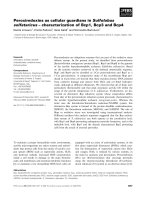
Fig. 2. Morphological stages of peroxisome fission. Ultrathin sections of KMnO
4
-fixed cells grown in a methanol-limited chemostat at
D = 0.12 h
)1
, demonstrating the three stages involved during peroxisome inheritance: (A) elongation into the bud; (B) separation of a small
organelle; and (C) the actual fission and migration of a small organelle into the bud. The bar represents 0.5 lm.
R. Saraya et al. Peroxisome abundance in yeast
FEBS Journal 277 (2010) 3279–3288 ª 2010 The Authors Journal compilation ª 2010 FEBS 3281
Recently, Knoblach & Rachubinski [27] showed that
in vivo S. cerevisiae Pex11 exists in two isoforms,
namely a phosphorylated form and a dephosphorylated
form. Interestingly, studies using PEX11 phosphomim-
icking mutants indicated that strains producing only
constitutively dephosphorylated Pex11 show a pheno-
type similar to that of pex11 cells, whereas strains
producing constitutive phosphorylated Pex11 show
enhanced peroxisome proliferation, similar to that of
Pex11-overproducing cells. This suggests that Pex11
phosphorylation may include a mechanism to regulate
Pex11 activation ⁄ inactivation.
A recent study of 249 S. cerevisiae kinase- and phos-
phatase-deletion strains [28] indeed indicated that
phosphorylation processes are crucial in regulating per-
oxisome abundance. In particular, deletion of PHO85,
a cyclin-dependent kinase, had a strongly negative
effect on peroxisome numbers. Interestingly, overex-
pression of PHO85 results in hyperphosphorylation of
Pex11 and peroxisome proliferation [27].
The second class of proteins essential for peroxisome
fission is the family of dynamin-related proteins
(DRPs). DRPs are large GTPases that are involved in
membrane fission and fusion events. In S. cerevisiae,
two DRPs – Vps1 and Dnm1 – play a role in peroxi-
some fission. Dnm1 also plays a role in mitochondrial
fission. Dnm1 is, in particular, essential for peroxisome
fission during conditions of peroxisome induction by
oleate [29], whereas Vps1 functions in peroxisome rep-
lication under repressing conditions (e.g. in the pres-
ence of glucose). Dnm1 is recruited to peroxisomes via
two homologus proteins, Mdv1 and Caf4, which are
associated with the peroxisomal membrane via the tail-
anchored protein, Fis1 [30]. Mdv1 is a WD repeat pro-
tein, which is absent in higher eukaryotes. Caf4 is an
Mdv1 paralog in S. cerevisiae that is absent in other
organisms.
In S. cerevisiae, Vps1 is involved in peroxisome fis-
sion [29]; however, in H. polymorpha, Vps1 does not
play a role in this process [14]. In this respect, H. poly-
morpha seems to be more similar to mammalian and
plant cells, where a single DRP (Dlp1 or DRP3A,
respectively) is involved in peroxisome fission. Interest-
ingly, in Arabidopsis thaliana, it has been shown that
the DRP 5B is responsible for the fission of chlorop-
lasts as well as of peroxisomes [31]. Additionally in
A. thaliana it has been shown that three out of five
PEX11 isoforms (PEX11c, PEX11d and PEX11e) are
important in the recruitment of Fis1b to the peroxi-
some membrane for the replication of pre-existing per-
oxisomes [32]. Similarly, in mammals, Fis1 interacts
with Pex11b [33]. As for other peroxisomal membrane
proteins, Pex19, a peroxin important for peroxisomal
membrane biogenesis, is also important for the target-
ing of Fis1 to peroxisomes in mammals [34].
Remarkably, the Fis1–DRP organelle fission
machinery was initially identified as being responsible
for mitochondrial fission [30]. Indeed, Fis1 and Dnm1
show a dual localization on peroxisomes and mito-
chondria. In contrast to peroxisomal Fis1, no proteins
involved in Fis1 targeting to mitochondria have yet
been identified.
Why both organelles share the same fission machin-
ery is unknown, but this may serve as a mechanism to
coordinate mitochondrial and peroxisome fission (e.g.
during the cell cycle). Fluorescence microscopy studies
in H. polymorpha revealed that green fluorescent pro-
tein (GFP)-conjugated Dnm1 is not evenly distributed
over the cytosol, but is present as multiple spots that
contain many GFP–Dnm1 molecules. Interestingly,
Mdv1 co-localizes with these Dnm1 spots. Live cell
imaging revealed that these spots dynamically associate
and disassociate from mitochondria and peroxisomes,
stressing the fact that the same protein molecules are
involved in the fission of both organelles [35].
Peroxisome fission in H. polymorpha is fully blocked
upon the deletion of DNM1 [14]. These cells contain a
single, enlarged peroxisome, which forms a long exten-
sion that protrudes into the developing bud. These
extensions are not observed in dnm1 pex11 cells, which
is in agreement with the model in which Pex11 plays a
role in peroxisome elongation. Notably, as in mamma-
lian cells [20], Pex11 is concentrated at the base of
these peroxisome extensions in dnm1 cells, indicating
that also in yeast the third step in peroxisome fission is
the segregation of Pex11 and the formation of Pex11-
enriched patches.
Other proteins implemented in
peroxisome development and
abundance
Besides Pex3, Pex11 and Fis1 ⁄ DRPs as key compo-
nents in determining organelle development and abun-
dance, other proteins have been identified as regulators
of these processes. These include components that were
initially identified in the secretory pathway and various
recently identified peroxins, and are discussed in more
detail below.
Components of the secretory pathway
Several proteins known to play a role in the secretory
pathway and localized to membranes of compartments
involved in this pathway (e.g. endoplasmic reticulum,
Golgi, COP vesicles) have been suggested to play a
Peroxisome abundance in yeast R. Saraya et al.
3282 FEBS Journal 277 (2010) 3279–3288 ª 2010 The Authors Journal compilation ª 2010 FEBS
role in peroxisome abundance. These proteins may be
important for de novo peroxisome formation or for the
delivery of endoplasmic reticulum-derived lipids to the
peroxisomal membrane.
A recent study indicated a possible role for S. cerevi-
siae SEC39, SEC21 and DSL in the trafficking of per-
oxisomal membrane proteins from the endoplasmic
reticulum to the peroxisome [36]. S. cerevisiae ARF1
and ARF3 were also proposed to work antagonistically
during peroxisome proliferation [37].
Emp24 is a protein of the p24 family of proteins
and localizes to the Golgi apparatus, endoplasmic
reticulum and COP vesicles [38]. However, a detailed
proteomics study in S. cerevisiae suggested that Emp24
is also localized to peroxisomes [39]. Moreover, in the
yeast H. polymorpha, Emp24 was localized to peroxi-
somes and the endoplasmic reticulum [40]. Interest-
ingly, deletion of EMP24 in H. polymorpha resulted in
a strong reduction in peroxisome number. Unexpect-
edly, this was not caused by a defect in the formation
of peroxisomes from the endoplasmic reticulum, but
by a defect in peroxisome fission. Possibly, p24 pro-
teins are required to bring various components
involved in peroxisome fission together at the peroxi-
somal membrane to allow organelle elongation at the
initial stage of peroxisome fission.
A similar function has recently been suggested for
caveolin-1 at peroxisomes in mammalian cells [41].
Caveolin-1 is crucial for the formation of caveolae,
subtypes of microdomains ⁄ rafts that are morphologi-
cally recognizable as flask-like invaginations in the
plasma membrane. Recent localization studies in rat
hepatocytes revealed that caveolin-1 is also enriched in
the peroxisomal membrane. A function for this protein
at the peroxisomal membrane, however, has not yet
been established.
Peroxins
Besides Pex11, two other members of the S. cerevisiae
Pex11 family – Pex25 and Pex27 – play a role in per-
oxisome proliferation [23]. Data obtained from the
analysis of overexpression strains suggest that both
peripheral membrane proteins function in organelle fis-
sion, in particular under conditions when proliferation
of the organelles is repressed. Also, proteins of the
Pex24 protein family (Pex24, Pex28 and Pex29) are
involved in regulating peroxisome numbers. All three
proteins are components of the peroxisomal mem-
brane, of which Pex24, but not Pex28 and Pex29, is
induced by growth conditions that promote peroxi-
some proliferation (i.e. oleate). Remarkably, deletion
of PEX28 and PEX29 in S. cerevisiae is accompanied
by increased numbers of reduced-size organelles [42].
In addition, three other oleate-inducible baker’s yeast
proteins (Pex30, Pex31 and Pex32), which show homol-
ogy towards Y. lipolytica Pex23, have been shown to
be involved in regulating peroxisome numbers [43].
Peroxisome inheritance
During vegetative reproduction of wild-type yeast cells,
organelle replication is essential for maintaining the
organelle population in the mother cells during multi-
ple rounds of budding. Upon division, part of the
organelle population is administered to the bud. In the
methylotrophic yeast H. polymorpha, this is accompa-
nied by asymmetrical peroxisome fission and subse-
quent migration of the newly formed, small organelle
to the developing bud. The number of organelles
migrating to the bud is dependent on the culture con-
ditions [44] (Fig. 3).
In yeast, peroxisome inheritance requires the func-
tion of Inp1, Inp2, the class V myosin motor (Myo2)
and the actin skeleton [45–47]. Of these, Inp1 has been
identified as the peroxisome-specific retention factor,
connecting peroxisomes that are retained in the mother
cells to a yet-unknown anchoring structure. Similarly,
Inp1 is also implemented in the retention of peroxi-
somes in developing buds [45,48]. Unexpectedly, in the
absence of Pex11, peroxisome retention is also defec-
tive in H. polymorpha, despite the fact that Inp1 is
properly localized to peroxisomes [48]. Hence, Pex11
may have a second function in organelle retention in
addition to its role in peroxisome fission.
Recently, a function in peroxisome inheritance was
also attributed to Pex3 [49]. In an elegant study,
Munck et al. [49] demonstrated that Pex3 also func-
tions in peroxisome retention. The authors showed
that Pex3 interacted directly with Inp1 at the peroxi-
somal membrane and suggested a role for Pex3 to
recruit Inp1 to the peroxisomal membrane. Impor-
tantly, the Inp1-binding region in the Pex3 protein
could be separated from the regions involved in
membrane formation during the de novo synthesis of
peroxisomes [49]. Hence, Pex3 is a multifunctional pro-
tein in peroxisome biology, implemented in formation
of the peroxisome membrane and organelle inheritance.
Inp2 is a peroxisomal membrane protein that acts as
the peroxisomal receptor for Myo2 and attaches the
globular tail of Myo2 to the peroxisome, thus allowing
transport of the organelle to the bud [46]. Recently,
the region of Myo2 involved in Inp2 binding was iden-
tified using mutant variants of Myo2 [50]. These stud-
ies also showed that Inp2 is a phosphoprotein whose
level of phosphorylation is coupled to the cell cycle.
R. Saraya et al. Peroxisome abundance in yeast
FEBS Journal 277 (2010) 3279–3288 ª 2010 The Authors Journal compilation ª 2010 FEBS 3283
Chang et al. [51] recently suggested that Inp2 is
unique for baker’s yeast and related species and pro-
vided evidence that Y. lipolytica Pex3 and its paralog,
Pex3B, function as the peroxisome-specific receptors of
Myo2. A similar function was attributed to baker’s
yeast Pex3. However, in a subsequent study, Saraya
et al. [52] demonstrated that Inp2, although weekly
conserved, is also present and functional in other yeast
species, including H. polymorpha. The finding that
H. polymorpha Inp2 interacted with Myo2 points to a
conserved function for this protein as a binding factor
for Myo2. Remarkably, in H. polymorpha, Myo2–Inp2
binding was dependent on Pex19. This is consistent
with the view that Pex19 may have a stabilizing role in
the interaction between Inp2 and Myo2, and also is in
line with the observed defect in peroxisome inheritance
in H. polymorpha pex19 cells [53].
Constitutive peroxisome degradation
Peroxisomal membrane proteins are generally post-
translationally incorporated into the organelle mem-
brane. This implies that the main quality-control
systems for these proteins reside outside the organelle
(i.e. in the cytosol). However, peroxisomes do contain
a few specific proteases that are implemented in the
removal of exhausted or nonfunctional matrix pro-
teins. Although different protease activities have been
detected in peroxisomes [54], so far only one gene
encoding a peroxisomal protease, a Lon protease, has
been identified in yeast, in contrast to mammals
where up to three proteases have been identified [55].
Peroxisomal Lon of H. polymorpha degrades short-
lived or nonfunctional components of the peroxisomal
lumen and therefore may participate in a housekeep-
ing process aimed at maintaining a functional peroxi-
some population. In the absence of Lon, protein
aggregates may accumulate in the organelle lumen.
Such protein aggregates are probably devastating for
organelle function and require removal of the entire
organelle to maintain cell vitality. Recent studies in
human cells suggested that the peroxisomal Lon pro-
tease is involved in accurate sorting, processing and
activation of the peroxisomal enzyme acyl CoA
oxidase [56].
Redundant organelles are removed by selective per-
oxisome autophagy (see the review in this miniseries
by Oku & Sakai [57] for details). However, constitutive
removal of peroxisomes is observed in H. polymorpha
when cultured under conditions that promote organelle
proliferation. Hence, under conditions of peroxisome
induction, development and degradation of the organ-
elles occurs simultaneously. The data from Bener
Aksam et al. [55] suggest that constitutive peroxisome
degradation suppresses the negative effects of deletion
of LON. This is indicated by the observation that in
an ATG1 deletion background, in which peroxisome
turnover is inhibited, deletion of the gene encoding
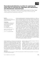
AB
Fig. 3. Peroxisome inheritance numbers vary with environmental conditions. In budding cells of methanol-limited cultures of Hansenula poly-
morpha cells grown at high dilution rates (A; D = 0.12 h
)1
), generally only a single peroxisome is inherited to the bud, whereas several small
organelles are inherited to buds in cultures grown at low dilution rates (B; D = 0.03 h
)1
). Electron micrographs of thin sections are shown.
Cells are fixed in KMnO
4
. The bar represents 1 lm.
Peroxisome abundance in yeast R. Saraya et al.
3284 FEBS Journal 277 (2010) 3279–3288 ª 2010 The Authors Journal compilation ª 2010 FEBS
peroxisomal Lon resulted in a decrease of cell viability.
This is consistent with the view that timely removal of
these organelles is essential for cell viability. Untimely
removal of peroxisomes may result in detrimental
effects (i.e. the accumulation of reactive oxygen spe-
cies, finally resulting in cell death) [55]. Constitutive
degradation of peroxisomes in H. polymorpha is an
autophagic process and thus requires the function of
ATG genes. However, the precise sequence of events
that mediate this constitutive degradation process is
still unknown and awaits further elucidation. One pos-
sibility is that, similarly to mitochondria, fission pro-
cesses may be involved that allow separation of
dysfunctional, aggregate-containing parts, which are
specifically recognized for degradation [58].
Perspectives
Peroxisomes are extremely flexible and dynamic organ-
elles. Several cues are known that cause rapid changes
in their abundance. During recent years much progress
has been made in the identification and analysis of
genes involved in changing organelle abundance. How-
ever, except for the proteins of the Fis1 ⁄ DRP organelle
fission machinery, the function of most other proteins
is still very speculative.
One problem that may have been underestimated so
far is that – unlike for genes involved in peroxisome
protein import – the underlying mechanism of mutants
displaying aberrant organelle numbers may be related
to two, basically opposite, machineries. Obviously, a
protein import defect results in cytosolic mislocaliza-
tion of matrix proteins. However, alterations in orga-
nelle abundance may, in fact, reflect either defects in
organelle formation or, alternatively, in organelle turn-
over by autophagy. Also, mutations that affect the rate
of the two opposite machineries of organelle formation
and degradation to the same extent, will result in an
unaltered steady-state number of peroxisomes. Thus,
the mere organelle steady-state number, which is gen-
erally used to determine peroxisome abundance, is not
sufficiently informative about the actual rates of the
different processes that determine organelle abundance
in a separate cell.
To understand in more detail the underlying reasons
for the presence of certain phenotypes there is an
urgent need to develop better techniques to establish
the phenotype of mutants more precisely. Using live
cell imaging techniques the rates of the processes that
affect peroxisome abundance should be quantitatively
determined in vivo. Such data could eventually be used
to develop mathematical models describing the kinetics
of these processes.
Organelle fission and de novo synthesis could be
studied using photoactivatable proteins or the HaloTag
technology, as successfully used in mammalian cells
[10,17]. The rate of degradation can easily be deter-
mined biochemically by determining protein half lives.
One way to determine the involvement of a gene in
de novo synthesis is to study the effect of mutations on
functional complementation of a pex3 mutant with the
PEX3 gene. Using this approach we showed that
DNM1, VPS1 and EMP24 are not required for peroxi-
some re-introduction from the endoplasmic reticulum
in H. polymorpha pex3 cells [14,40]. Interestingly, no
mutations have so far been described that result in a
defect in pex3 mutant complementation. Why such
genes ⁄ mutations have not yet been identified is
unknown. Possibly these genes are also involved in
other endoplasmic reticulum-related process and hence
the mutations may be lethal.
In addition to the detailed molecular mechanisms
of the various processes, their regulation is still lar-
gely unexplored. Important questions regarding this
include the following. What is the signal that triggers
the peroxisomes to divide? How are organelles that
should be degraded, retained or inherited, distin-
guished from the other organelles of the total orga-
nelle population?
Systems biology approaches have already been
shown to be very helpful in this respect, for example,
the analysis of kinase- and phosphatase-deficient
mutants [28], proteomics and transcriptomics.
Acknowledgements
We thank Rinse de Boer for preparing Figs 2 and 3.
This project was carried out within the research pro-
gramme of the Kluyver Centre for Genomics of Indus-
trial Fermentation, which is part of the Netherlands
Genomics Initiative ⁄ Netherlands Organization for Sci-
entific Research.
References
1 Gabaldon T (2010) Peroxisome diversity and evolution.
Philos Trans R Soc Lond B Biol Sci 365, 765–773.
2 van der Klei IJ & Veenhuis M (1997) Yeast peroxi-
somes: function and biogenesis of a versatile cell orga-
nelle. Trends Microbiol 5, 502–509.
3 Heiland I & Erdmann R (2005) Biogenesis of peroxi-
somes. Topogenesis of the peroxisomal membrane and
matrix proteins. Febs J 272, 2362–2372.
4 Kiel JA, Veenhuis M & van der Klei IJ (2006) PEX
genes in fungal genomes: common, rare or redundant.
Traffic 7, 1291–1303.
R. Saraya et al. Peroxisome abundance in yeast
FEBS Journal 277 (2010) 3279–3288 ª 2010 The Authors Journal compilation ª 2010 FEBS 3285
5 Tam YY, Fagarasanu A, Fagarasanu M & Rachubinski
RA (2005) Pex3p initiates the formation of a preperox-
isomal compartment from a subdomain of the endo-
plasmic reticulum in Saccharomyces cerevisiae. J Biol
Chem 280, 34933–34939.
6 Kragt A, Voorn-Brouwer T, van den Berg M & Distel
B (2005) Endoplasmic reticulum-directed Pex3p routes
to peroxisomes and restores peroxisome formation in a
Saccharomyces cerevisiae pex3Delta strain. J Biol Chem
280, 34350–34357.
7 Haan GJ, Baerends RJ, Krikken AM, Otzen M,
Veenhuis M & van der Klei IJ (2006) Reassembly of
peroxisomes in Hansenula polymorpha pex3 cells on
reintroduction of Pex3p involves the nuclear envelope.
FEMS Yeast Res 6, 186–194.
8 Hoepfner D, Schildknegt D, Braakman I, Philippsen P
& Tabak HF (2005) Contribution of the endoplasmic
reticulum to peroxisome formation. Cell 122, 85–95.
9 Mullen RT & Trelease RN (2006) The ER-peroxisome
connection in plants: development of the ‘‘ER semi-
autonomous peroxisome maturation and replication’’
model for plant peroxisome biogenesis. Biochim Biophys
Acta 1763, 1655–1668.
10 Kim PK, Mullen RT, Schumann U & Lippincott-Sch-
wartz J (2006) The origin and maintenance of mamma-
lian peroxisomes involves a de novo PEX16-dependent
pathway from the ER. J Cell Biol 173, 521–532.
11 Titorenko VI, Smith JJ, Szilard RK & Rachubinski RA
(2000) Peroxisome biogenesis in the yeast Yarrowia
lipolytica. Cell Biochem Biophys 32, 21–26.
12 Nagotu S, Veenhuis M & van der Klei IJ (2010) Divide et
impera: the dictum of peroxisomes. Traffic 11, 175–184.
13 Motley AM & Hettema EH (2007) Yeast peroxisomes
multiply by growth and division. J Cell Biol 178, 399–
410.
14 Nagotu S, Saraya R, Otzen M, Veenhuis M & van der
Klei IJ (2008) Peroxisome proliferation in Hansenu-
la polymorpha requires Dnm1p which mediates fission
but not de novo formation. Biochim Biophys Acta 1783,
760–769.
15 Wolf J, Wolfgang S & Erdmann R (2010) Peroxisomes as
dynamic organelles: peroxisomal matrix protein import.
Febs J 277, 3268–3278.
16 Matsuzaki T & Fujiki Y (2008) The peroxisomal
membrane protein import receptor Pex3p is directly
transported to peroxisomes by a novel Pex19p- and
Pex16p-dependent pathway. J Cell Biol 183, 1275–1286.
17 Huybrechts SJ, Van Veldhoven PP, Brees C,
Mannaerts GP, Los GV & Fransen M (2009) Peroxi-
some dynamics in cultured mammalian cells. Traffic 10,
1722–1733.
18 Yan M, Rachubinski DA, Joshi S, Rachubinski RA &
Subramani S (2008) Dysferlin domain-containing pro-
teins, Pex30p and Pex31p, localized to two compart-
ments, control the number and size of oleate-induced
peroxisomes in Pichia pastoris. Mol Biol Cell 19, 885–
898.
19 Raychaudhuri S & Prinz WA (2008) Nonvesicular phos-
pholipid transfer between peroxisomes and the endo-
plasmic reticulum. Proc Natl Acad Sci U S A 105,
15785–15790.
20 Schrader M, Reuber BE, Morrell JC, Jimenez-Sanchez
G, Obie C, Stroh TA, Valle D, Schroer TA & Gould SJ
(1998) Expression of PEX11beta mediates peroxisome
proliferation in the absence of extracellular stimuli.
J Biol Chem 273, 29607–29614.
21 Thoms S & Erdmann R (2005) Dynamin-related pro-
teins and Pex11 proteins in peroxisome division and
proliferation. Febs J 272, 5169–5181.
22 Marshall PA, Krimkevich YI, Lark RH, Dyer JM,
Veenhuis M & Goodman JM (1995) Pmp27 promotes
peroxisomal proliferation. J Cell Biol 129, 345–355.
23 Rottensteiner H, Stein K, Sonnenhol E & Erdmann R
(2003) Conserved function of pex11p and the novel
pex25p and pex27p in peroxisome biogenesis. Mol Biol
Cell 14, 4316–4328.
24 Koerkamp MG, Rep M, Bussemaker HJ, Hardy GP,
Mul A, Piekarska K, Szigyarto CA, De Mattos JM &
Tabak HF (2002) Dissection of transient oxidative
stress response in Saccharomyces cerevisiae by using
DNA microarrays. Mol Biol Cell 13, 2783–2794.
25 Smith JJ, Marelli M, Christmas RH, Vizeacoumar FJ,
Dilworth DJ, Ideker T, Galitski T, Dimitrov K, Rachu-
binski RA & Aitchison JD (2002) Transcriptome profil-
ing to identify genes involved in peroxisome assembly
and function. J Cell Biol 158, 259–271.
26 van Zutphen T, Baerends RJ, Susanna KA, de Jong A,
Kuipers OP, Veenhuis M & van der Klei IJ (2010)
Adaptation of Hansenula polymorpha to methanol:
a transcriptome analysis. BMC Genomics 11,1.
27 Knoblach B & Rachubinski RA (2010) Phosphoryla-
tion-dependent activation of peroxisome proliferator
protein PEX11 controls peroxisome abundance. J Biol
Chem 285, 6670–6680.
28 Saleem RA, Knoblach B, Mast FD, Smith JJ, Boyle J,
Dobson CM, Long-O’Donnell R, Rachubinski RA &
Aitchison JD (2008) Genome-wide analysis of signaling
networks regulating fatty acid-induced gene expression
and organelle biogenesis. J Cell Biol 181, 281–292.
29 Kuravi K, Nagotu S, Krikken AM, Sjollema K,
Deckers M, Erdmann R, Veenhuis M & van der Klei IJ
(2006) Dynamin-related proteins Vps1p and Dnm1p
control peroxisome abundance in Saccharomyces
cerevisiae. J Cell Sci 119, 3994–4001.
30 Mozdy AD, McCaffery JM & Shaw JM (2000) Dnm1p
GTPase-mediated mitochondrial fission is a multi-step
process requiring the novel integral membrane compo-
nent Fis1p. J Cell Biol 151, 367–380.
31 Zhang X & Hu J (2010) The Arabidopsis chloroplast
division protein DYNAMIN-RELATED PROTEIN5B
Peroxisome abundance in yeast R. Saraya et al.
3286 FEBS Journal 277 (2010) 3279–3288 ª 2010 The Authors Journal compilation ª 2010 FEBS
also mediates peroxisome division. Plant Cell 22, 431–
442.
32 Lingard MJ, Gidda SK, Bingham S, Rothstein SJ,
Mullen RT & Trelease RN (2008) Arabidopsis
PEROXIN11c-e, FISSION1b, and DYNAMIN-
RELATED PROTEIN3A cooperate in cell cycle-associ-
ated replication of peroxisomes. Plant Cell 20,
1567–1585.
33 Kobayashi S, Tanaka A & Fujiki Y (2007) Fis1, DLP1,
and Pex11p coordinately regulate peroxisome morpho-
genesis. Exp Cell Res 313, 1675–1686.
34 Delille HK & Schrader M (2008) Targeting of hFis1 to
peroxisomes is mediated by Pex19p. J Biol Chem 283,
31107–31115.
35 Nagotu S, Krikken AM, Otzen M, Kiel JA, Veenhuis
M & van der Klei IJ (2008) Peroxisome fission in Han-
senula polymorpha requires Mdv1 and Fis1, two pro-
teins also involved in mitochondrial fission. Traffic 9 ,
1471–1484.
36 Perry RJ, Mast FD & Rachubinski RA (2009) Endo-
plasmic reticulum-associated secretory proteins Sec20p,
Sec39p, and Dsl1p are involved in peroxisome biogene-
sis. Eukaryot Cell 8, 830–843.
37 Lay D, B LG, Heid H, Gorgas K & Just WW (2005)
Binding and functions of ADP-ribosylation factor on
mammalian and yeast peroxisomes. J Biol Chem 280,
34489–34499.
38 Carney GE & Bowen NJ (2004) p24 proteins, intracellu-
lar trafficking, and behavior: Drosophila melanogaster
provides insights and opportunities. Biol Cell 96,
271–278.
39 Marelli M, Smith JJ, Jung S, Yi E, Nesvizhskii AI,
Christmas RH, Saleem RA, Tam YY, Fagarasanu A,
Goodlett DR et al. (2004) Quantitative mass spectrome-
try reveals a role for the GTPase Rho1p in actin organi-
zation on the peroxisome membrane. J Cell Biol 167,
1099–1112.
40 Kurbatova E, Otzen M & van der Klei IJ (2009) p24
proteins play a role in peroxisome proliferation in yeast.
FEBS Lett 583, 3175–3180.
41 Woudenberg J, Rembacz KP, van den Heuvel FA,
Woudenberg-Vrenken TE, Buist-Homan M, Geuken M,
Hoekstra M, Deelman LE, Enrich C, Henning RH et al.
(2009) Caveolin-1 is enriched in the peroxisomal mem-
brane of rat hepatocytes. Hepatology 51, 1744–1753.
42 Vizeacoumar FJ, Torres-Guzman JC, Tam YY,
Aitchison JD & Rachubinski RA (2003) YHR150w and
YDR479c encode peroxisomal integral membrane pro-
teins involved in the regulation of peroxisome number,
size, and distribution in Saccharomyces cerevisiae. J Cell
Biol 161, 321–332.
43 Vizeacoumar FJ, Torres-Guzman JC, Bouard D,
Aitchison JD & Rachubinski RA (2004) Pex30p,
Pex31p, and Pex32p form a family of peroxisomal
integral membrane proteins regulating peroxisome size
and number in Saccharomyces cerevisiae. Mol Biol Cell
15, 665–677.
44 Veenhuis M, van Dijken JP, Pilon SA & Harder W
(1978) Development of crystalline peroxisomes in meth-
anol-grown cells of the yeast Hansenula polymorpha and
its relation to environmental conditions. Arch Microbiol
117, 153–163.
45 Fagarasanu M, Fagarasanu A, Tam YY, Aitchison JD
& Rachubinski RA (2005) Inp1p is a peroxisomal mem-
brane protein required for peroxisome inheritance in
Saccharomyces cerevisiae. J Cell Biol 169, 765–775.
46 Fagarasanu A, Fagarasanu M, Eitzen GA, Aitchison
JD & Rachubinski RA (2006) The peroxisomal mem-
brane protein Inp2p is the peroxisome-specific receptor
for the myosin V motor Myo2p of Saccharomyces
cerevisiae. Dev Cell 10, 587–600.
47 Hoepfner D, van den Berg M, Philippsen P, Tabak HF
& Hettema EH (2001) A role for Vps1p, actin, and
the Myo2p motor in peroxisome abundance and
inheritance in Saccharomyces cerevisiae. J Cell Biol 155,
979–990.
48 Krikken AM, Veenhuis M & van der Klei IJ (2009)
Hansenula polymorpha pex11 cells are affected in peroxi-
some retention. Febs J 276, 1429–1439.
49 Munck JM, Motley AM, Nuttall JM & Hettema EH
(2009) A dual function for Pex3p in peroxisome forma-
tion and inheritance. J Cell Biol 187, 463–471.
50 Fagarasanu A, Mast FD, Knoblach B, Jin Y, Brunner
MJ, Logan MR, Glover JN, Eitzen GA, Aitchison
JD, Weisman LS et al. (2009) Myosin-driven peroxi-
some partitioning in S. cerevisiae. J Cell Biol 186,
541–554.
51 Chang J, Mast FD, Fagarasanu A, Rachubinski DA,
Eitzen GA, Dacks JB & Rachubinski RA (2009) Pex3
peroxisome biogenesis proteins function in peroxisome
inheritance as class V myosin receptors. J Cell Biol 187,
233–246.
52 Saraya R, Cepinska MN, Kiel JA, Veenhuis M & van
der Klei IJ (2010) A conserved function for Inp2 in
peroxisome inheritance. Biochim Biophys Acta 1803,
617–622.
53 Otzen M, Krikken AM, Ozimek PZ, Kurbatova E,
Nagotu S, Veenhuis M & van der Klei IJ (2006) In the
yeast Hansenula polymorpha, peroxisome formation from
the ER is independent of Pex19p, but involves the func-
tion of p24 proteins. FEMS Yeast Res 6, 1157–1166.
54 Stewart MQ, van Dijk R, Veenhuis M & Goodman JM
(2002) Monomeric alcohol oxidase is preferentially
digested by a novel protease from Candida boidinii.
Biochim Biophys Acta 1542, 160–172.
55 Aksam EB, Koek A, Kiel JA, Jourdan S, Veenhuis M
& van der Klei IJ (2007) A peroxisomal lon protease
and peroxisome degradation by autophagy play key
roles in vitality of Hansenula polymorpha cells.
Autophagy 3, 96–105.
R. Saraya et al. Peroxisome abundance in yeast
FEBS Journal 277 (2010) 3279–3288 ª 2010 The Authors Journal compilation ª 2010 FEBS 3287
56 Omi S, Nakata R, Okamura-Ikeda K, Konishi H &
Taniguchi H (2008) Contribution of peroxisome-specific
isoform of Lon protease in sorting PTS1 proteins to
peroxisomes. J Biochem 143, 649–660.
57 Oku M & Sakai Y (2010) Peroxisomes as dynamic
organelles: autophagic degradation. FEBS J 277,
3289–3294.
58 Twig G, Elorza A, Molina AJ, Mohamed H, Wikstrom
JD, Walzer G, Stiles L, Haigh SE, Katz S, Las G et al.
(2008) Fission and selective fusion govern mitochondrial
segregation and elimination by autophagy. EMBO J 27,
433–446.
Peroxisome abundance in yeast R. Saraya et al.
3288 FEBS Journal 277 (2010) 3279–3288 ª 2010 The Authors Journal compilation ª 2010 FEBS