Báo cáo khoa học: Proteoglycans in health and disease: novel regulatory signaling mechanisms evoked by the small leucine-rich proteoglycans ppt
Bạn đang xem bản rút gọn của tài liệu. Xem và tải ngay bản đầy đủ của tài liệu tại đây (1004.72 KB, 12 trang )
MINIREVIEW
Proteoglycans in health and disease: novel regulatory
signaling mechanisms evoked by the small leucine-rich
proteoglycans
Renato V. Iozzo
1
and Liliana Schaefer
2
1 Department of Pathology, Anatomy and Cell Biology, and the Cancer Cell Biology and Signaling Program, Kimmel Cancer Center, Thomas
Jefferson University, Philadelphia, PA, USA
2 Pharmazentrum Frankfurt, Institut fu
¨
r Allgemeine Pharmakologie und Toxikologie ⁄ ZAFES, Klinikum der JW Goethe-Universita
¨
t Frankfurt am
Main, Germany
Introduction
The small leucine-rich proteoglycans (SLRPs) were
originally grouped on the basis of their relatively
small protein core (36–42 kDa), compared with the
larger aggregating proteoglycans such as aggrecan
Keywords
biglycan; cancer; decorin; EGFR; IGF-IR;
inflammation; lumican; Met; signal
transduction; Toll-like receptor
Correspondence
R. V. Iozzo, Department of Pathology,
Anatomy and Cell Biology, Thomas Jefferson
University, 1020 Locust Street, Room 249
JAH, Philadelphia, PA 19107, USA
Fax: +1 215 923 7969
Tel: +1 215 503 2208
E-mail:
or
L. Schaefer, Pharmazentrum Frankfurt
Institut fu
¨
r Allgemeine Pharmakologie und
Toxikologie, Klinikum der JW Goethe-
Universita
¨
t Frankfurt am Main Haus 74,
Z.3.108a, Theodor-Stern-Kai 7, 60590
Frankfurt am Main, Germany
Fax: +49 69 6301 83027
Tel: +49 69 6301 7899
E-mail:
(Received 15 April 2010, revised 10 July
2010, accepted 27 July 2010)
doi:10.1111/j.1742-4658.2010.07797.x
The small leucine-rich proteoglycans (SLRPs) are involved in many aspects
of mammalian biology, both in health and disease. They are now being rec-
ognized as key signaling molecules with an expanding repertoire of molecu-
lar interactions affecting not only growth factors, but also various
receptors involved in controlling cell growth, morphogenesis and immunity.
The complexity of SLRP signaling and the multitude of affected signaling
pathways can be reconciled with a hierarchical affinity-based interaction of
various SLRPs in a cell- and tissue-specific context. Here, we review this
interacting network, describe new relationships of the SLRPs with tyrosine
kinase and Toll-like receptors and critically assess their roles in cancer and
innate immunity.
Abbreviations
BMP, bone morphogenetic protein; EGFR, epidermal growth factor receptor; IGF-IR, insulin-like growth factor receptor type 1;
IL-1, interleukin-1; LRR, leucine-rich repeat; Met, hepatocyte growth factor receptor; NLR, nucleotide-binding oligomerization domain-like
receptor; P2X, purinoreceptor; RTK, receptor tyrosine kinase; SLRP, small leucine-rich proteoglycan; TLR, Toll-like receptor.
3864 FEBS Journal 277 (2010) 3864–3875 ª 2010 The Authors Journal compilation ª 2010 FEBS
and versican, and on their unique structural organiza-
tion composed of tandem leucine-rich repeats (LRRs)
[1,2]. It also became evident that at least three SLRP
classes could be distinguished based upon additional
unique features such as the organization of disulfide
bonds at their N- and C-termini, with the cysteine
residues following a class-specific topology, and on
the basis of their genomic organization, with each
individual class harboring an almost identical number
and size of exons, often positioned in a similar
sequential pattern within chromosomes [3,4]. More
recently, five distinct classes of SLRPs have been pro-
posed based on shared biological activity and func-
tions, albeit some of SLRPs are not classical
proteoglycans [5]. SLRP biology and function are further
complicated by their post-translational modifications
including substitution with sugars and glycosamino-
glycan side chains of various types. For example, the
canonical class I members decorin and biglycan con-
tain chondroitin or dermatan sulfate side chains, with
the exception of asporin. By contrast, all class II
members harbor polylactosamine or keratan sulfate
chains in their LRRs and sulfated tyrosine residues in
their N-termini. Class III members contain chondroi-
tin ⁄ dermatan sulfate (epiphycan), keratan sulfate (os-
teoglycin) or no glycosaminoglycan (opticin) chain.
Finally, noncanonical class IV and class V members
lack any glycosaminoglycan chain, with the exception
of chondroadherin which is substituted with keratan
sulfate [6]. Thus, the presence of finite sugar chains,
together with further post-translational refinements,
including modification in their degree of sulfation or
epimerization, endows this class of proteoglycans with
an extra layer of structural complexity.
Initially thought to act exclusively as structural com-
ponents, SLRPs are now recognized as key players in
cell signaling, capable of influencing a host of cellular
functions such as proliferation, differentiation, sur-
vival, adhesion, migration and inflammatory responses.
All of these functions are mediated by the intrinsic
SLRP ability to interact with both cytokines and
ligands as well as with surface receptors. This minire-
view critically assesses recent advances on the modula-
tion of various signaling pathways that are affected by
SLRPs, including signaling through receptor tyrosine
kinase such as the epidermal growth factor receptor
(EGFR), hepatocyte growth factor receptor (Met) and
insulin-like growth factor receptor type 1 (IGF-IR), as
well as receptors involved in innate immunity and
inflammation such as Toll-like receptors and purinergic
P2X receptors. We focus specifically on decorin, bigly-
can and lumican, the best-studied SLRP members to
date. More extensive and specialized reviews on the
subject have been published covering other aspects of
SLRP biology [6–13].
Antiproliferative effects on cancer
cells via EGFR and Met suppression
The first demonstration of an antiproliferative effect
of decorin, at that time called PG40 to reflect its
apparent size, was achieved over two decades ago
when Yamaguchi & Ruoslahti [14] discovered that
stable transfection of decorin causes growth arrest in
Chinese hamster ovary cells. They subsequently dis-
covered that this growth inhibition was due to deco-
rin’s ability to bind and block TGFb [15], a property
also shared by other SLRPs [16]. This original obser-
vation has led to a large number of studies focusing
on decorin’s ability to inhibit fibrosis, the main path-
ogenetic mechanism of which involves overactivation
of the TGFb signaling pathway. However, other stud-
ies using a variety of transformed cells have shown
that de novo decorin expression causes severe growth
retardation in vitro [17] and suppression of tumorige-
nicity in animal models of human tumor xenografts
[18]. Because most of these transformed cells are not
dependent on TGFb for their growth, it was hypothe-
sized that another receptor system had to be
involved, insofar as decorin is a soluble proteoglycan.
One of the key observations that emerged from these
studies was that decorin-expressing tumor cells
become arrested in the G
1
phase of the cell cycle and
overproduce the cyclin-dependent kinase inhibitor
p21
WAF1
[19], supporting earlier observations that
decorin gene expression is markedly induced during
quiescence [20,21]. Indeed, both the mouse and
human decorin structural organization of their gene
and promoter are quite complex [22–24] and subject
to an intricate transcriptional regulation [1,25,26]. It
was soon discovered that decorin directly interacts
with the EGFR with a K
D
value of 87 nm [27].
This interaction evokes a transient activation [28,29]
followed by a profound downregulation of the recep-
tor and inhibition of its downstream signaling activity
[30,31]. Subsequent studies using the yeast two-hybrid
system revealed that decorin binds to a narrow region
within ligand-binding domain L2 of the EGFR, over-
lapping with the EGF-binding domain [32]. The
structural constraints of the EGFR binding region
support a stochiometry of 1 : 1 for the decorin pro-
tein core and EGFR, suggesting that decorin is bio-
logically active as a monomer [33]. This interaction
prevents receptor dimerization and targets the EGFR
to a sustained internalization via caveolin-mediated
endocytosis [34], eventually leading to its degradation
R. V. Iozzo and L. Schaefer Novel signaling mechanisms triggered by SLRPs
FEBS Journal 277 (2010) 3864–3875 ª 2010 The Authors Journal compilation ª 2010 FEBS 3865
(Fig. 1). Notably heparanase induces EGFR phos-
phorylation [35], using similar Tyr residues that are
activated by decorin. However, the results are quite
different because heparanase leads to EGFR activa-
tion [35], whereas decorin leads to EGFR downregu-
lation [36]. Another effect of decorin is its activation
of caspase 3, one of the key enzymes involved in pro-
grammed cell death, thereby increasing decorin’s an-
tioncogenic activity [37]. Similar effects are also
observed in normal mesangial cells where overexpres-
sion of decorin activates caspase 3, induces apoptosis
and arrests the cells in the G
0
⁄ G
1
phase of the cell
cycle via EGFR downregulation [38]. Also, caspase 8
activation has been detected in a wide variety of
transformed cells when decorin is overexpressed using
adenoviral vectors [39].
The consequences of decorin signaling through
receptor tyrosine kinases (RTKs) are exemplified by
several observations using decorin-null animals. First,
crossing decorin-null mice, which exhibit a skin fragil-
ity phenotype [40], with p53-null mice causes an early
lethality of the double-mutant animals with massive
organ infiltration by a T-cell lymphoma [41]. This is in
contrast to p53-null mice, which show a wide variety
of tumor types, including carcinomas and sarcomas,
and a prolonged survival compared with the double-
mutant mice. The second key observation is that
approximately one-third of decorin-deficient mice
develop intestinal adenomas that eventually develop
into adenocarcinomas, and this process is accelerated
and amplified by subjecting decorin-null mice to a
western diet enriched in lipids and low in calcium and
MAPK
Caspase 3
Receptor
internalization
Receptor
internalization
Proteasomal
degradation
β-catenin
Receptor
downregulation
Met EGFR
IGFIR
Cell motility
invasion
metastasis
↓
Apoptosis
Tumor
growth
↓
PI3K
Akt/PKB
p21
Apoptosis
↓
mTOR
p70S6K
Fibrillin-1
synthesis
↑
Ectodomain
shedding
Tumor
growth
Anti-proliferative effects
(Cancer cells)
Proliferative effects
(Normal cells)
↑
↓
Lysosomal
degradation
Decorin
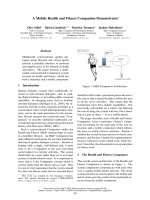
Fig. 1. Schematic representation of decorin effects as an antiproliferative (left) and proliferative (right) molecule. In most cancer cells investi-
gated to date, decorin causes a downregulation of EGFR and Met with consequent activation of p21 and caspase 3, which leads to apopto-
sis. Decorin also interferes with the noncanonical b-catenin pathway via the Met receptor. In normal cells such as renal tubular epithelial
cells, decorin evokes a prosurvival and proliferative response via the IGF-IR and downstream signaling. Please, refer to the text for additional
information.
Novel signaling mechanisms triggered by SLRPs R. V. Iozzo and L. Schaefer
3866 FEBS Journal 277 (2010) 3864–3875 ª 2010 The Authors Journal compilation ª 2010 FEBS
vitamin D [42]. Notably, tumorigenesis in decorin-defi-
cient mice is associated with a downregulation of both
cyclin-dependent kinase inhibitors p21
WAF1
and p27
Kip1
and a concurrent upregulation of b-catenin. Together,
these in vivo observations suggest that decorin defi-
ciency is permissive for tumorigenesis.
Adenovirus-mediated gene delivery or systemic
administration of the decorin gene in various tumor
xenograft models has revealed an effective inhibition
of tumor growth, downregulation of both EGFR and
ErbB2, and an inhibitory effect on metastatic spread-
ing [39,43–48]. Some of these in vivo effects might be
mediated by decorin’s ability to inhibit the endogenous
tumor cell production of vascular endothelial growth
factor A [49].
In an animal model of prostate carcinoma generated
by a targeted deletion of the tumor suppressor PTEN
in the prostate, systemic delivery of decorin causes a
marked downregulation of the EGFR in the treated
tumors with an associated reduction in tumor growth
[50]. Notably, decorin also interferes with cross-talk
between the EGFR and the androgen receptor in pros-
tate carcinoma cells [50]. The interplay between deco-
rin and the EGFR is further underscored by
osteosarcoma cells which escape the decorin-suppress-
ing activity via protracted expression and activation of
their endogenous EGFR [51,52].
The complex binding repertoire of decorin would
predict that this SLRP might modulate the bioactivity
of other RTKs. Indeed, decorin binds directly and
with high affinity (K
D
1.5 nm) to Met, the receptor
for hepatocyte growth factor [53]. Notably, binding
of decorin to Met can be efficiently displaced by
hepatocyte growth factor, and less efficiently by in-
ternalin B, a known bacterial ligand of Met with
structural homology to decorin LRRs. The interaction
between decorin and Met induces transient activation
of the receptor, recruitment of the E3 ubiquitin ligase
c-Cbl, followed by rapid intracellular degradation of
Met. Tumor growth is further suppressed through
caspase 3-mediated apoptosis. Notably, signaling
through Met leads to the phosphorylation of b-cate-
nin, a known downstream Met effector, directing it
to proteosomal degradation, thereby decreasing cellu-
lar motility, tissue invasion and metastasis (Fig. 1).
These findings indicate that decorin exerts its antipro-
liferative activity by antagonistically targeting multiple
tyrosine kinase receptors, thereby contributing to
reduction in primary tumor growth and metastastic
spreading. The role of decorin as a marker for prog-
nosis, as well as an anticancer therapeutic, is reviewed
in the accompanying minireview by Theocharis et al.
[54].
Proliferative effects on normal cells via
the IGF-IR
By contrast, in normal cells, decorin signaling
through IGF-IR exerts antiapoptotic and proliferative
effects, favoring cellular growth. Decorin binds IGF-
IR with affinity in the low nanomolar range
(K
D
1–2 nm) in endothelial cells [55], renal fibro-
blasts [56] and human tubular epithelial cells [57]. In
addition, decorin binds to and sequesters the IGF-I
(K
D
18 nm), the natural ligand of this RTK [55].
By binding to the IGF-IR, decorin triggers phosphor-
ylation and downstream activation of phosphoinosi-
tide-3 kinase, Akt ⁄ protein kinase B and p21
WAF1
,
inducing an antiapoptotic effect [55,57,58] (Fig. 1).
The relevance of decorin in the IGF-IR pathway is
reinforced in two experimental animal models of
inflammatory angiogenesis and unilateral ureteral
obstruction. In both cases, decorin deficiency causes a
significant increase in IGF-IR levels compared with
controls [55,56]. Moreover, lack of decorin promotes
renal tubular epithelial cell apoptosis in experimental
diabetic nephropathy [57,58] and in a renal obstruc-
tion model with interstitial inflammation and fibrosis
[55,57]. In renal fibroblasts, decorin activates the
mammalian target of rapamycin and p70S6 kinase
(p70S6K) downstream of IGF-IR ⁄ phosphoinositide-
3 kinase ⁄ Akt signaling [58]. This ultimately results in
increased translation and synthesis of fibrillin-1,
thereby indirectly promoting cell proliferation [59].
These pathways might represent intricate regulatory
mechanisms, whereby decorin modulates IGF-IR sig-
naling in a cell type-specific manner, thereby giving
rise to different biological outcomes. In contrast to
the well-characterized interactions of decorin with the
EGFR family, the biological necessity for decorin-
triggered activation of the canonical IGF signaling
cascade is not well characterized. Decorin appears to
mimic the effects of IGF-I and stimulates the IGF-IR
without inhibiting signaling, as has been shown for
its interaction with receptors of the ErbB family.
However, the significance of the decorin ⁄ IGF-IR
interaction is not clear. In endothelial cells, decorin
promotes transient receptor phosphorylation and acti-
vation and subsequent degradation, but it also pro-
motes adhesion and migration on fibrillar collagen
[55,60]. In extravillus trophoblasts, instead, decorin
inhibits migration by affecting the IGF-IR pathway
[61]. All of these studies were performed with ‘nor-
mal’ cells. Thus, there are no published data on the
role of decorin in modulating cancer growth via the
IGF-IR in transformed cells or in tumor models.
Further studies are needed to elucidate the role of
R. V. Iozzo and L. Schaefer Novel signaling mechanisms triggered by SLRPs
FEBS Journal 277 (2010) 3864–3875 ª 2010 The Authors Journal compilation ª 2010 FEBS 3867
decorin in the regulation of IGF-IR and to clarify
whether decorin ⁄ IGF-IR signaling might be operative
in carcinoma cells as well.
The complexity of decorin signaling is further
expanded by additional degradative pathways involved
in decorin catabolism. The endocytosis and lysosomal
degradation of decorin comprises multiple pathways
including those mediated by the EGFR [34] and
low-density lipoprotein receptor-related protein [62].
Interestingly, lipid-raft-dependent EGFR signaling also
modulates decorin uptake, a process that may consti-
tute a regulatory mechanism for desensitization of
decorin-evoked signaling [63]. Thus, there are numerous
opportunities for feedback control of decorin activity
and its efficiency for signaling. The ability of decorin to
bind to more than one RTK suggests that decorin is
directly involved in the intricate cross-talk between
receptors and their downstream signaling cascades.
Biglycan, a danger signal that induces
cooperativity of innate immunity
receptors
Biglycan, a class I SLRP structurally related to deco-
rin, serves as an agonist of different cell-surface recep-
tors, thereby giving rise to diverse biological outcomes
[64]. The initial observation was made during studies
of a renal obstruction model caused by pressure injury.
In these studies, biglycan was markedly overexpressed
in resident renal tubular epithelial cells prior to the
infiltration of macrophages, suggesting that biglycan
might be involved in the initiation of the inflammatory
response [58]. More recently, several reports have
firmly established that biglycan, in analogy to decorin,
acts as a signaling molecule especially important in the
innate immune system [65,66]. Under physiological
conditions, biglycan is sequestered in the extracellular
milieu, acting as a structural component with no
apparent immunological function. Upon tissue stress
or injury, biglycan is released from the extracellular
matrix by a proteolytic processing that is not yet char-
acterized. In contrast to the sequestered proteoglycan,
soluble biglycan turns into an endogenous ligand of
innate immunity receptors and interacts with the Toll-
like receptors (TLR)-2 and -4 on macrophages, thereby
triggering a robust inflammatory response. It is intrigu-
ing that both TLRs and biglycan contain LRR-motifs
with the potential to interact with each other. Down-
stream of TLRs, biglycan signaling involves MyD88,
p38, extracellular signal-regulated kinase and nuclear
LPS
MyD88
NF-κB
ASC
NLRP3
Caspase-1
pro-IL-1β
IL-1β
TNF-α
LRR
LRR
pro-IL-1β
TNF-α
BIGLYCAN
LUMICAN
P2X7
TLR2
TLR4
LRR
M
a
c
r
o
p
h
a
g
e
Fig. 2. Schematic representation of biglycan
and lumican effects on the innate immune
system. Please, refer to the text for detailed
information.
Novel signaling mechanisms triggered by SLRPs R. V. Iozzo and L. Schaefer
3868 FEBS Journal 277 (2010) 3864–3875 ª 2010 The Authors Journal compilation ª 2010 FEBS
factor jB and results in the synthesis and secretion of
tumor necrosis factor a and macrophage inflammatory
protein 2. Consequently, additional neutrophils and
macrophages are recruited to the site of tissue injury.
This initial step does not require de novo synthesis of
the proinflammatory agents and therefore generates a
fast response to tissue damage. Moreover, macrophag-
es stimulated by proinflammatory cytokines can syn-
thesize biglycan de novo [65], thereby boosting the
inflammatory response in an autocrine and paracrine
manner (Fig. 2). Thus, soluble biglycan appears to rep-
resent a ‘danger’ motif (danger-associated molecular
pattern) in analogy to pathogen-associated molecular
patterns in pathogen-driven inflammation. Besides its
interaction with TLRs [65], biglycan also acts as a
ligand for selectin L ⁄ CD44 and is thus directly
involved in the recruitment of CD16(-) natural killer
cells [67].
Soluble biglycan, as a pivotal danger-associated
molecular pattern, is not only secured by its interaction
with TLR-2 ⁄ 4 but is also involved in signaling through
the cytoplasmic nucleotide-binding oligomerization
domain-like receptors (NLRs) (Fig. 2). This is due to
an interaction with and clustering of membrane-bound
Toll-like and purinergic P2X receptors, whereby bigly-
can induces receptor cooperativity within these newly
formed multireceptor complexes. By signaling through
TLR-2 ⁄ 4, biglycan stimulates the expression of NLRP3,
a member of the NLRs, and pro-IL-1b mRNA.
Importantly, biglycan is simultaneously capable of
interacting with P2X
4
⁄ P2X
7
receptors which will
activate the NLRP3 ⁄ ASC inflammasome in a reactive
oxygen species- and heat shock protein 90-dependent
manner. These combined signaling events culminate in
the activation of caspase 1 and in the processing of
pro-IL-1b into its mature form, without the need for
additional costimulatory factors [66]. Collectively,
these findings provide solid evidence for the multifunc-
tional involvement of biglycan within the innate
immune system. In particular, biglycan appears to spe-
cifically interact with two classes of receptors, thereby
providing cross-talk between their downstream signal-
ing, a function that might be facilitated by the pres-
ence of tandem LRRs and glycosaminoglycan side
chains. Notably, a recent report has shown that bigly-
can gene expression is specifically upregulated in
human aortic valve stenosis and that the enhanced
accumulation of biglycan within the stenotic valves
contributes to the production of phospholipid transfer
protein, a key factor in atherosclerotic aortic valve
development, via TLR-2 [68]. Thus, biglycan is well
suited to serve as a cross-linker for different cell-sur-
face receptors.
In a model of noninfectious inflammation in the kid-
ney, the so-called unilateral ureteral obstruction model,
biglycan-deficient mice display lower levels of active
caspase 1 and mature interleukin (IL)-1b, resulting in
reduced infiltration of mononuclear cells and less kid-
ney damage. In a prototypical innate immune process
such as lipopolysaccharide-induced sepsis, lack of
biglycan results in a clear survival benefit associated
with lower levels of circulating tumor necrosis factor a
and IL-1b, reduced activation of the NLRP3 inflam-
masome and less infiltration in the lung, a major target
organ of sepsis in mice [65,66]. These findings have led
to a new understanding of the regulation of pathogen-
independent (‘sterile’) inflammation. Sterile inflamma-
tion appears to be driven by soluble biglycan as an
endogenous agonist for two crucial TLRs acting as an
autonomous trigger of the innate immunity system. By
contrast, in pathogen-associated molecular pattern-
mediated conditions, biglycan would serve as an ampli-
fier of the inflammatory response by signaling through
the second TLR, which is not involved in pathogen
sensing. This concept describes a fundamental para-
digm of how tissue injury is monitored by innate
immune receptors detecting the release of minute
amounts of components from the extracellular matrix
and turning such a signal into a robust inflammatory
response. This clearly implicates biglycan as a novel
target of anti-inflammatory strategies.
In addition to being a strong trigger of proinflam-
matory signaling within the innate immune system,
biglycan can also affect bone morphogenetic protein
(BMP) signaling, thereby influencing the differentiation
of tendon stem ⁄ progenitor cells and subsequent tendon
formation [69]. Biglycan forms complexes with BMP-4
and modulates osteoblast differentiation [70] as well as
enhancing its binding to chordin [71]. The latter, in
turn, leads to BMP-4 inactivation by the chordin–
twisted gastrulation complex [71].
Lumican signaling in cell growth and
inflammation
The role of lumican in the regulation of cell signaling
has not been studied in great detail. In analogy to
decorin, lumican inhibits tumor cell growth in soft
agar by increasing the expression of the cyclin-depen-
dent kinase inhibitor p21
WAF1
[72]. Again, similar to
decorin, these growth inhibitory effects of lumican
occur in a variety of cell types including fibrosarcoma,
carcinoma and normal embryonic cells [72]. Notably,
expression of membrane-type metalloprotease 1
reduces lumican secretion and abrogates lumican-medi-
ated p21
WAF1
induction [72]. Also decorin is cleaved
R. V. Iozzo and L. Schaefer Novel signaling mechanisms triggered by SLRPs
FEBS Journal 277 (2010) 3864–3875 ª 2010 The Authors Journal compilation ª 2010 FEBS 3869
by membrane-type metalloprotease 1 [72] suggesting
that protease processing is important in SLRP biology.
The role of shedding of cell-surface syndecans is
reviewed in the accomapnying minireview by Manon-
Jensen et al. [73].
Lumican reduces colony formation in soft agar and
tumorigenicity in nude mice of cells transformed by
v-src and K-ras oncogenes [74]. In mouse embryonic
fibroblasts, lumican-evoked upregulation of p21
WAF1
occurs through a p53-mediated mechanism with a sub-
sequent decrease in the cyclins A, D1 and E [75].
Lumican deficiency is associated with proliferation of
stromal keratinocytes and embryonic fibroblasts [76].
Its inhibitory effects on cell growth have also been
observed in tumor cells, with some of these cells secret-
ing lumican in an autocrine manner [77]. In melanoma
cells, lumican regulates vertical growth, suppresses
anchorage-independent proliferation and inhibits
cyclin D1 expression [78,79]. A recent study has fur-
ther shown that lumican not only inhibits melanoma
invasion and metastasis, but also induces tumor cell
apoptosis and inhibits angiogenesis [80]. Thus, lumican
might contribute as a therapeutic agent to combat mel-
anoma metastasis.
Lumican can interact with b1-containing integrin
receptors and this signaling leads to inhibition of mela-
noma cell migration by enhancing cell adhesion [81].
Indeed, several components of the focal adhesion com-
plex are modulated by lumican-evoked signaling,
including vinculin and focal adhesion kinase [82].
Lumican alters the relationship between actin filaments
and b1 integrin, which in turn would affect focal adhe-
sion formation, thereby explaining the anti-invasive
effects of this SLRP [82]. A commonality of signaling
between lumican and decorin is also supported by
recent studies showing the involvement of decorin in
modulating various integrins in controlling prolifera-
tion, adhesion and migration [60,83]. Notably, lumican
manufactured by endothelial cells binds to the cell
surface of extravasated neutrophilic leukocytes via
b2-containing integrin receptors and promotes migra-
tion during the inflammatory response [84]. Thus, there
is a possible endothelial-dependent lumican expression
that might mediate in a paracrine fashion neutrophil
recruitment and migration. Lumican also is involved in
Fas–FasL-induced apoptosis by upregulating Fas
(CD95) in mouse embryonic fibroblasts [75].
In terms of TLR signaling, lumican presents patho-
gen-associated molecular patterns to the receptor com-
plex. The protein core of lumican is capable of binding
and presenting lipopolysaccharide to CD14, thereby
activating TLR4 signaling [85] (Fig. 2). Lumican also
binds to and signals through the FasL, it increases the
synthesis and secretion of proinflammatory cytokines
and accelerates the recruitment of macrophages and
neutrophils [76,86]. Via its protein core, lumican inter-
acts with the CXC-chemokine KC (CXCL1), thereby
creating a chemokine gradient in the tissue along
which neutrophil will infiltrate the site of injury [87].
Conclusions and perspectives
Undoubtedly, SLRPs are structural components espe-
cially important during development and the matura-
tion of various tissues enriched in mesenchyme.
Utilization of animal models including the mouse
[7,40,88–101] and zebrafish [102], or cellular systems
with finite SLRP deficiencies [83,103–105], has revealed
fundamental roles for SLRPs in embryonic life and
disease progression. The past decade has further wit-
nessed many members of the SLRP gene family emerg-
ing as signaling molecules. The discovery that soluble
SLRPs engage various cell-surface receptors, resulting
in a triggering of downstream signaling events, has
shed a new light on how SLRPs might regulate cell
behavior. This is possible because of several character-
istics of these proteoglycans. First, their makeup is
conducive to protein ⁄ protein interactions. Second,
many surface receptors are made up of protein mod-
ules that are often shared by extracellular matrix pro-
teins, including leucine-rich repeats, fibronectin and
immunoglobulin repeats, among others. Thus, there is
the likely possibility that during evolution some of
these modules have been utilized by both matrix
(structural) and ligand (signaling) molecules. Third,
SLRPs are abundant and ubiquitous, and thus might
signal in a different way than traditional ligands whose
kinetics are often very rapid, that is, both triggering of
signals and transferring of this information to the
nucleus takes just a few minutes. By contrast, SLRPs
can induce protracted signaling leading to growth inhi-
bition in most of the cases studied. An additional layer
of complexity is provided by the ability of SLRPs to
bind and sequester various cytokines, growth factors
and morphogens involved in multiple signaling path-
ways affecting differentiation, survival, adhesion,
migration, cancer and inflammatory responses.
Despite their conserved and highly similar structural
composition, various SLRPs such as decorin, biglycan
and lumican have distinct interacting receptors. How
could SLRPs bind to multiple receptors and still be
specific in their action? One way to answer this impor-
tant question is to consider a ‘hierarchical’ possibility
of receptor binding and activation. For example, deco-
rin binds to EGFR, Met and IGF-IR with diverse
affinity constants, with K
D
values ranging from 87 nm
Novel signaling mechanisms triggered by SLRPs R. V. Iozzo and L. Schaefer
3870 FEBS Journal 277 (2010) 3864–3875 ª 2010 The Authors Journal compilation ª 2010 FEBS
for the EGFR to 1–2 nm for the Met and IGF-IR.
Thus, when decorin encounters a cancer composed of
a mixed population of cells, it might differentially
affect the tumor cells depending upon the expression
and cellular density of a given RTK. This cell-specific
context might also apply to other members of the
SLRP gene family. Finally, another key concept
emerging from the studies summarized above is that
some SLRPs, such as biglycan, might work through
clustering and activating multireceptor complexes. This
concept provides a novel mechanism of how tissue
injury could be sensed by innate immune receptors:
detecting the release of minute amounts of matrix con-
stituents and turning such a signal into a robust
inflammatory response.
Acknowledgements
We thank Angela McQuillan for her excellent work
with the graphic designs. We also like to thank our
numerous collaborators who have contributed to our
work on SLRPs throughout the past two decades. This
work was supported in part by National Institutes of
Health grants RO1 CA39481, RO1 CA47282, and
RO1 CA120975 (RVI) and by the Deutsche Fors-
chungsgemeinschaft (SFB 815, project A5, SCHA
1082 ⁄ 2-1, Excellence Cluster ECCPS), and Else Kro
¨
-
ner-Fresenius-Stiftung (to LS).
References
1 Iozzo RV & Murdoch AD (1996) Proteoglycans of the
extracellular environment: clues from the gene and pro-
tein side offer novel perspectives in molecular diversity
and function. FASEB J 10, 598–614.
2 Iozzo RV (1997) The family of the small leucine-rich
proteoglycans: key regulators of matrix assembly and
cellular growth. Crit Rev Biochem Mol Biol 32, 141–174.
3 Iozzo RV (1998) Matrix proteoglycans: from molecular
design to cellular function. Annu Rev Biochem 67, 609–
652.
4 Iozzo RV (1999) The biology of the small leucine-rich
proteoglycans. Functional network of interactive pro-
teins. J Biol Chem 274, 18843–18846.
5 Schaefer L & Iozzo RV (2008) Biological functions of
the small leucine-rich proteoglycans: from genetics to
signal transduction. J Biol Chem 283, 2135–2139.
6 Schaefer L & Schaefer RM (2010) Proteoglycans: from
structural compounds to signaling molecules. Cell
Tissue Res 339, 237–246.
7 Ameye L & Young MF (2002) Mice deficient in small
leucine-rich proteoglycans: novel in vivo models for
osteoporosis, osteoarthritis, Ehlers–Danlos syndrome,
muscular dystrophy, and corneal diseases. Glycobiology
12, 107R–116R.
8 Reed CC & Iozzo RV (2002) The role of decorin in
collagen fibrillogenesis and skin homeostasis. Glycoconj
J 19, 249–255.
9 Brandan E, Cabello-Verrugio C & Vial C (2008) Novel
regulatory mechanisms for the proteoglycans decorin
and biglycan during muscle formation and muscular
dystrophy. Matrix Biol 27, 700–708.
10 Chakravarti S (2003) Functions of lumican and fibro-
modulin: lessons from knockout mice. Glycoconj J 19,
287–293.
11 Kalamajski S & Oldberd A
˚
(2010) The role of small
leucine-rich proteoglycans in collagen fibrillogenesis.
Matrix Biol 29, 248–253.
12 Heinega
˚
rd D (2009) Proteoglycans and more – from
molecules to biology. Int J Exp Pathol 90, 575–586.
13 Iozzo RV, Goldoni S, Berendsen A & Young
MF(2010) In Extracellular Matrix: An Overview
(Mecham RP, ed.). Chapter 6, Springer, New York,
NY, in press.
14 Yamaguchi Y & Ruoslahti E (1988) Expression of
human proteoglycan in Chinese hamster ovary cells
inhibits cell proliferation. Nature 336, 244–246.
15 Yamaguchi Y, Mann DM & Ruoslahti E (1990)
Negative regulation of transforming growth factor-b by
the proteoglycan decorin. Nature 346, 281–284.
16 Hildebrand A, Romaris M, Rasmussen LM, Heinega
˚
rd
D, Twardzik DR, Border WA & Ruoslahti E (1994)
Interaction of the small interstitial proteoglycans
biglycan, decorin and fibromodulin with transforming
growth factor b. Biochem J 302, 527–534.
17 Santra M, Skorski T, Calabretta B, Lattime EC &
Iozzo RV (1995)
De novo decorin gene expression
suppresses the malignant phenotype in human
colon cancer cells. Proc Natl Acad Sci USA 92,
7016–7020.
18 Santra M, Mann DM, Mercer EW, Skorski T,
Calabretta B & Iozzo RV (1997) Ectopic expression of
decorin protein core causes a generalized growth
suppression in neoplastic cells of various histogenetic
origin and requires endogenous p21, an inhibitor of
cyclin-dependent kinases. J Clin Invest 100, 149–157.
19 De Luca A, Santra M, Baldi A, Giordano A & Iozzo
RV (1996) Decorin-induced growth suppression is
associated with upregulation of p21, an inhibitor of
cyclin-dependent kinases. J Biol Chem 271, 18961–
18965.
20 Coppock DL, Kopman C, Scandalis S & Gilleran S
(1993) Preferential gene expression in quiescent human
lung fibroblasts. Cell Growth Differ 4, 483–493.
21 Mauviel A, Santra M, Chen YQ, Uitto J & Iozzo RV
(1995) Transcriptional regulation of decorin gene
expression. Induction by quiescence and repression by
R. V. Iozzo and L. Schaefer Novel signaling mechanisms triggered by SLRPs
FEBS Journal 277 (2010) 3864–3875 ª 2010 The Authors Journal compilation ª 2010 FEBS 3871
tumor necrosis factor-a. J Biol Chem 270, 11692–
11700.
22 Scholzen T, Solursh M, Suzuki S, Reiter R, Morgan
JL, Buchberg AM, Siracusa LD & Iozzo RV (1994)
The murine decorin. Complete cDNA cloning, genomic
organization, chromosomal assignment and expression
during organogenesis and tissue differentiation. J Biol
Chem 269, 28270–28281.
23 Danielson KG, Fazzio A, Cohen I, Cannizzaro LA,
Eichstetter I & Iozzo RV (1993) The human decorin
gene: intron–exon organization, discovery of two
alternatively spliced exons in the 5¢ untranslated region,
and mapping of the gene to chromosome 12q23.
Genomics 15, 146–160.
24 Santra M, Danielson KG & Iozzo RV (1994) Struc-
tural and functional characterization of the human
decorin gene promoter. J Biol Chem 269, 579–587.
25 Mauviel A, Korang K, Santra M, Tewari D, Uitto J &
Iozzo RV (1996) Identification of a bimodal regulatory
element encompassing a canonical AP-1 binding site in
the proximal promoter region of the human decorin
gene. J Biol Chem 271, 24824–24829.
26 Iozzo RV & Danielson KG (1999) Transcriptional and
post-transcriptional control of proteoglycan gene
expression. Prog Nucleic Acids Res Mol Biol 62, 19–53.
27 Iozzo RV, Moscatello D, McQuillan DJ & Eichstetter
I (1999) Decorin is a biological ligand for the epider-
mal growth factor receptor. J Biol Chem 274, 4489–
4492.
28 Moscatello DK, Santra M, Mann DM, McQuillan DJ,
Wong AJ & Iozzo RV (1998) Decorin suppresses
tumor cell growth by activating the epidermal growth
factor receptor. J Clin Invest 101, 406–412.
29 Patel S, Santra M, McQuillan DJ, Iozzo RV &
Thomas AP (1998) Decorin activates the epidermal
growth factor receptor and elevates cytosolic Ca
2+
in
A431 cells. J Biol Chem 273, 3121–3124.
30 Santra M, Eichstetter I & Iozzo RV (2000) An anti-
oncogenic role for decorin: downregulation of ErbB2
leads to growth suppression and cytodifferentiation of
mammary carcinoma cells. J Biol Chem 275,
35153–35161.
31 Csorda
´
s G, Santra M, Reed CC, Eichstetter I,
McQuillan DJ, Gross D, Nugent MA, Hajno
´
czky G &
Iozzo RV (2000) Sustained down-regulation of the
epidermal growth factor receptor by decorin. A mecha-
nism for controlling tumor growth in vivo. J Biol Chem
275, 32879–32887.
32 Santra M, Reed CC & Iozzo RV (2002) Decorin binds
to a narrow region of the epidermal growth factor
(EGF) receptor, partially overlapping with but distinct
from the EGF-binding epitope. J Biol Chem 277,
35671–35681.
33 Goldoni S, Owens RT, McQuillan DJ, Shriver Z,
Sasisekharan R, Birk DE, Campbell S & Iozzo RV
(2004) Biologically active decorin is a monomer in
solution. J Biol Chem 279, 6606–6612.
34 Zhu J-X, Goldoni S, Bix G, Owens RA, McQuillan D,
Reed CC & Iozzo RV (2005) Decorin evokes pro-
tracted internalization and degradation of the EGF
receptor via caveolar endocytosis. J Biol Chem 280,
32468–32479.
35 Barash U, Cohen-Kaplan V, Dowek I, Sanderson RD,
Ilan N & Vlodavsky I (2010) Proteoglycans in health
and disease: new concepts for heparanase function in
tumor progression and metastasis. FEBS J 277, 3890–
3903.
36 Goldoni S & Iozzo RV (2008) Tumor microenviron-
ment: modulation by decorin and related molecules
harboring leucine-rich tandem motifs. Int J Cancer 123,
2473–2479.
37 Seidler DG, Goldoni S, Agnew C, Cardi C, Thakur
ML, Owens RA, McQuillan DJ & Iozzo RV (2006)
Decorin protein core inhibits in vivo cancer growth and
metabolism by hindering epidermal growth factor
receptor function and triggering apoptosis via caspase-
3 activation. J Biol Chem 281, 26408–26418.
38 Wu H, Wang S, Xue A, Liu Y, Liu Y, Liu Y, Wang
H, Chen Q, Guo M & Zhang Z (2008) Overexpression
of decorin induces apoptosis and cell growth arrest in
cultured rat mesangial cells in vitro. Nephrology 13,
607–615.
39 Tralha
˜
o JG, Schaefer L, Micegova M, Evaristo C,
Scho
¨
nherr E, Kayal S, Veiga-Fernandes H, Danel C,
Iozzo RV, Kresse H et al. (2003) In vivo selective
and distant killing of cancer cells using adenovirus-
mediated decorin gene transfer. FASEB J 17, 464–
466.
40 Danielson KG, Baribault H, Holmes DF, Graham H,
Kadler KE & Iozzo RV (1997) Targeted disruption of
decorin leads to abnormal collagen fibril morphology
and skin fragility. J Cell Biol 136, 729–743.
41 Iozzo RV, Chakrani F, Perrotti D, McQuillan DJ,
Skorski T, Calabretta B & Eichstetter I (1999) Cooper-
ative action of germline mutations in decorin and p53
accelerates lymphoma tumorigenesis. Proc Natl Acad
Sci USA 96
, 3092–3097.
42 Bi X, Tong C, Dokendorff A, Banroft L, Gallagher L,
Guzman-Hartman G, Iozzo RV, Augenlicht LH &
Yang W (2008) Genetic deficiency of decorin causes
intestinal tumor formation through disruption of
intestinal cell maturation. Carcinogenesis 29,
1435–1440.
43 Reed CC, Gauldie J & Iozzo RV (2002) Suppression of
tumorigenicity by adenovirus-mediated gene transfer of
decorin. Oncogene 21, 3688–3695.
44 Reed CC, Waterhouse A, Kirby S, Kay P, Owens RA,
McQuillan DJ & Iozzo RV (2005) Decorin prevents
metastatic spreading of breast cancer. Oncogene 24,
1104–1110.
Novel signaling mechanisms triggered by SLRPs R. V. Iozzo and L. Schaefer
3872 FEBS Journal 277 (2010) 3864–3875 ª 2010 The Authors Journal compilation ª 2010 FEBS
45 Biglari A, Bataille D, Naumann U, Weller M, Zirger J,
Castro MG & Lowenstein PR (2004) Effects of ectopic
decorin in modulating intracranial glioma progression
in vivo, in a rat syngeneic model. Cancer Gene Ther 11,
721–732.
46 Li X, Pennisi A & Yaccoby S (2008) Role of decorin in
the antimyeloma effects of osteoblasts. Blood 112, 159–
168.
47 Goldoni S, Seidler DG, Heath J, Fassan M, Baffa R,
Thakur ML, Owens RA, McQuillan DJ & Iozzo RV
(2008) An anti-metastatic role for decorin in breast
cancer. Am J Pathol 173, 844–855.
48 Shintani K, Matsumine A, Kusuzaki K, Morikawa J,
Matsubara T, Wakabayashi T, Araki K, Satonaka H,
Wakabayashi H, Lino T et al. (2008) Decorin sup-
presses lung metastases of murine osteosarcoma. Oncol
Rep 19, 1533–1539.
49 Grant DS, Yenisey C, Rose RW, Tootell M, Santra M
& Iozzo RV (2002) Decorin suppresses tumor cell-med-
iated angiogenesis. Oncogene 21, 4765–4777.
50 Hu Y, Sun H, Owens RT, Wu J, Chen YQ, Berquin
IM, Perry D, O’Flaherty JT & Edwards IJ (2009)
Decorin suppresses prostate tumor growth through
inhibition of epidermal growth factor and androgen
receptor pathways. Neoplasia 11, 1042–1053.
51 Zafiropoulos A, Nikitovic D, Katonis P, Tsatsakis A,
Karamanos NK & Tzanakakis GN (2008) Decorin-
induced growth inhibition is overcome through
protracted expression and activation of epidermal
growth factor receptors in osteosarcoma cells. Mol
Cancer Res 6, 785–794.
52 Zafiropoulos A & Tzanakakis GN (2008) Decorin-
mediated effects in cancer cell biology. Connect Tissue
Res 49, 244–248.
53 Goldoni S, Humphries A, Nystro
¨
m A, Sattar S, Owens
RT, McQuillan DJ, Ireton K & Iozzo RV (2009)
Decorin is a novel antagonistic ligand of the Met
receptor. J Cell Biol 185, 743–754.
54 Theocharis AD, Skandalis SS, Tzanakakis GN &
Karamanos NK (2010) Proteoglycans in health and
disease: novel roles for proteoglycan in malignancy and
their pharmacological targeting. FEBS J 277, 3904–
3923.
55 Scho
¨
nherr E, Sunderko
¨
tter C, Iozzo RV & Schaefer L
(2005) Decorin, a novel player in the insulin-like
growth factor system. J Biol Chem 280, 15767–15772.
56 Schaefer L, Tsalastra W, Babelova A, Baliova M,
Minnerup J, Sorokin L, Gro
¨
ne H-J, Reinhardt DP,
Pfeilschifter J, Iozzo RV et al. (2007) Decorin-mediated
regulation of fibrillin-1 in the kidney involves the insu-
lin-like growth factor-1 receptor and mammalian target
of rapamycin. Am J Pathol 170, 301–315.
57 Merline R, Lazaroski S, Babelova A, Tsalastra-Greul
W, Pfeilschifter J, Schluter KD, Gunther A, Iozzo RV,
Schaefer RM & Schaefer L (2009) Decorin deficiency
in diabetic mice: aggravation of nephropathy due to
overexpression of profibrotic factors, enhanced
apoptosis and mononuclear cell infiltration. J Physiol
Pharmacol 60(Suppl. 4), 5–13.
58 Schaefer L, Macakova K, Raslik I, Micegova M,
Gro
¨
ne H-J, Scho
¨
nherr E, Robenek H, Echtermeyer
FG, Gra
¨
ssel S, Bruckner P et al. (2002) Absence of
decorin adversely influences tubulointerstitial fibrosis
of the obstructed kidney by enhanced apoptosis and
increased inflammatory reaction. Am J Pathol 160,
1181–1191.
59 Porst M, Plank C, Bieritz B, Konik E, Fees H, Do
¨
tsch
J, Hilgers KF, Reinhardt DP & Hartner A (2006)
Fibrillin-1 regulates mesangial cell attachment,
spreading, migration and proliferation. Kidney Int 69,
450–456.
60 Fiedler LR, Scho
¨
nherr E, Waddington R, Niland S,
Seidler DG, Aeschlimann D & Eble JA (2008) Decorin
regulates endothelial cell motility on collagen I through
activation of insulin-like growth factor I receptor and
modulation of a2b1 integrin activity. J Biol Chem 283,
17406–17415.
61 Iacob D, Cai J, Tsonis M, Babwah A, Chakraborty
RN & Lala PK (2008) Decorin-mediated inhibition of
proliferation and migration of the human trophoblast
via different tyrosine kinase receptors. Endocrinology
149, 6187–6197.
62 Brandan E, Retamal C, Cabello-Verrugio C & Marzolo
M-P (2006) The low density lipoprotein receptor-
related protein functions as an endocytic receptor for
decorin. J Biol Chem 281, 31562–31571.
63 Feugaing DDS, Tammi R, Echtermeyer FG, Stenmark
H, Kresse H, Smollich M, Scho
¨
nherr E, Kiesel L &
Go
¨
tte M (2007) Endocytosis of the dermatan sulfate
proteoglycan decorin utilizes multiple pathways and is
modulated by epidermal growth factor receptor signal-
ing. Biochimie 89, 637–657.
64 Schaefer L (2010) Extracellular matrix molecules:
endogenous danger signals as new drug targets in
kidney diseases. Curr Opin Pharmacol 10, 185–190.
65 Schaefer L, Babelova A, Kiss E, Hausser H-J, Baliova
M, Krzyzankova M, Marsche G, Young MF, Mihalik
D, Go
¨
tte M et al. (2005) The matrix component
biglycan is proinflammatory and signals through toll-
like receptors 4 and 2 in macrophages. J Clin Invest
115, 2223–2233.
66 Babelova A, Moreth K, Tsalastra-Greul W, Zeng-
Brouwers J, Eickelberg O, Young MF, Bruckner P,
Pfeilschifter J, Schaefer RM, Gro
¨
ne H-J et al. (2009)
Biglycan, a danger signal that activates the NLRP3
inflammasome via Toll-like and P2X receptors. J Biol
Chem 284, 24035–24048.
67 Kitaya K & Yasuo T (2009) Dermatan sulfate proteo-
glycan biglycan as a potential selectin L ⁄ CD44 ligand
involved in selective recruitment of peripheral blood
R. V. Iozzo and L. Schaefer Novel signaling mechanisms triggered by SLRPs
FEBS Journal 277 (2010) 3864–3875 ª 2010 The Authors Journal compilation ª 2010 FEBS 3873
CD16(-) natural killer cells into human endometrium.
J Leukoc Biol 85, 391–400.
68 Derbali H, Bosse
´
Y, Coˆ te
´
N, Pibarot P, Audet A,
Pe
´
pin A, Arsenault B, Couture C, Despre
´
s J-P &
Mathieu P (2010) Increased biglycan in aortic valve
stenosis leads to the overexpression of phospholipid
transfer protein via Toll-like receptor 2. Am J Pathol
176, 2638–2645.
69 Bi Y, Ehirchiou D, Kilts TM, Inkson CA, Embree
MC, Sonoyama W, Li L, Leet AI, Seo B-M, Zhang L
et al. (2007) Identification of tendon stem ⁄ progenitor
cells and the role of the extracellular matrix in their
niche. Nat Med 13, 1219–1227.
70 Chen X-D, Fisher LW, Robey PG & Young MF
(2004) The small leucine-rich proteoglycan biglycan
modulates BMP-4-induced osteoblast differentiation.
FASEB J 18, 948–958.
71 Moreno M, Mun
˜
oz R, Aroca F, Labarca M, Brandan
E & Larraı
´
n J (2005) Biglycan is a new extracellular
component of the chordin-BMP4 signaling pathway.
EMBO J 24, 1397–1405.
72 Li Y, Aoki T, Mori Y, Ahmad M, Miyamori H,
Takino T & Sato H (2004) Cleavage of lumican by
membrane-type matrix metalloprotease-1 abrogates this
proteoglycan-mediated suppression of tumor cell col-
ony formation in soft agar. Cancer Res 64, 7058–7064.
73 Manon-Jensen T, Itoh Y & Couchman JR (2010)
Proteoglycans in health and disease: the multiple
roles of syndecan shedding. FEBS J 277, 3876–3889.
74 Yoshioka N, Inoue H, Nakanishi K, Oka K, Yutsudo
M, Yamashita A, Hakura A & Nojima H (2000)
Isolation of transformation suppressor genes by cDNA
substraction: lumican suppresses transformation
induced by v-src and v-K-ras. J Virol 74, 1008–1013.
75 Vij N, Roberts L, Joyce S & Chakravarti S (2004)
Lumican suppresses cell proliferation and aids Fas–Fas
ligand mediated apoptosis: implications in the cornea.
Exp Eye Res 78, 957–971.
76 Vij N, Roberts L, Joyce S & Chakravarti S (2005)
Lumican regulates corneal inflammatory responses by
modulating Fas–Fas ligand signaling. Invest Ophthal-
mol Vis Sci 46, 88–95.
77 Sifaki M, Assouti M, Nikitovic D, Krasagakis K,
Karamanos NK & Tzanakakis GN (2008) Lumican, a
small leucine-rich proteoglycan substituted with
keratan sulfate chains is expressed and secreted by
human melanoma cells and not normal melanocytes.
IUBMB Life 58, 606–610.
78 Bre
´
zillon S, Venteo L, Ramont L, D’Onofrio M-F,
Perreau C, Pluot M, Maquart F-X & Wegrowski Y
(2007) Expression of lumican, a small leucine-rich
proteoglycan with antitumour activity, in human
malignant melanoma. Clin Exp Dermatol 32, 405–416.
79 Vuillermoz B, Khoruzhenko A, D’Onofrio MF,
Ramont L, Venteo L, Perreau C, Antonicelli F,
Maquart FX & Wegrowski Y (2004) The small leucine-
rich proteoglycan lumican inhibits melanoma progres-
sion. Exp Cell Res 296, 294–306.
80 Bre
´
zillon S, Zeltz C, Schneider L, Terryn C, Vuillermoz
B, Ramont L, Perreau C, Pluot M, Diebold MD,
Radwanska A
et al. (2009) Lumican Inhibits B16F1
melanoma cell lung metastasis. J Physiol Pharmacol
60(Suppl. 4), 15–22.
81 D’Onofrio M-F, Bre
´
zillon S, Baranek T, Perreau C,
Roughley P, Maquart F-X & Wegrowski Y (2008)
Identification of b1 integrin as mediator of melanoma
cell adhesion to lumican. Biochem Biophys Res
Commun 365, 266–272.
82 Bre
´
zillon S, Radwanska A, Zeltz C, Malkowski A,
Ploton D, Bobichon H, Perreau C, Malicka-Blas-
zkiewicz M, Maquart F-X & Wegrowski Y (2009)
Lumican core protein inhibits melanoma cell migration
via alterations of focal adhesion compleses. Cancer Lett
283, 92–100.
83 Ferdous Z, Peterson SB, Tseng H, Anderson DK,
Iozzo RV & Grande-Allen KJ (2010) A role for
decorin in controlling proliferation, adhesion, and
migration of murine embryonic fibroblasts. J Biomed
Mater Res Part A 93, 419–428.
84 Lee S, Bowrin K, Hamad AR & Chakravarti S (2009)
Extracellular matrix lumican deposited on the surface
of neutrophils promotes migration by binding to b2
integrin. J Biol Chem 284, 23662–23669.
85 Wu F, Vij N, Roberts L, Lopez-Briones S, Joyce S &
Chakravarti S (2007) A novel role of the lumican core
protein in bacterial lipopolysaccharide-induced innate
immune response. J Biol Chem 282, 26409–26417.
86 Funderburgh JL, Mitschler RR, Funderburgh ML,
Roth MR, Chapes SK & Conrad GW (1997) Macro-
phage receptors for lumican, a corneal keratan
sulfate proteoglycan. Invest Ophthalmol Vis Sci 38,
1159–1167.
87 Carlson EC, Lin M, Liu C-Y, Kao WWY, Perez VL &
Pearlman E (2007) Keratocan and lumican regulate
neutrophil infiltration and corneal clarity in lipopoly-
saccharide-induced keratitis by direct interaction with
CXCL1. J Biol Chem 282, 33502–33509.
88 Ha
¨
kkinen L, Strassburger S, Kahari VM, Scott PG,
Eichstetter I, Iozzo RV & Larjava H (2000) A role for
decorin in the structural organization of periodontal
ligament. Lab Invest 80, 1869–1880.
89 Weis SM, Zimmerman SD, Shah M, Covell JW,
Omens JH, Ross J . Jr, Dalton N, Jones Y, Reed CC,
Iozzo RV et al. (2005) A role for decorin in the remod-
eling of myocardial infarction. Matrix Biol 24, 313–
324.
90 Ja
¨
rvela
¨
inen H, Puolakkainen P, Pakkanen S, Brown
EL, Ho
¨
o
¨
k M, Iozzo RV, Sage H & Wight TN (2006)
A role for decorin in cutaneous wound healing and
angiogenesis. Wound Repair Regen 14, 443–452.
Novel signaling mechanisms triggered by SLRPs R. V. Iozzo and L. Schaefer
3874 FEBS Journal 277 (2010) 3864–3875 ª 2010 The Authors Journal compilation ª 2010 FEBS
91 Bi Y, Stueltens CH, Kilts T, Wadhwa S, Iozzo RV,
Robey PG, Chen X-D & Young MF (2005) Extracellu-
lar matrix proteoglycans control the fate of bone
marrow stromal cells. J Biol Chem 280, 30481–30489.
92 Wadhwa S, Bi Y, Ortiz AT, Embree MC, Kilts T,
Iozzo R, Opperman LA & Young MF (2007) Impaired
posterior frontal sutural fusion in the biglycan ⁄ decorin
double deficient mice. Bone 40, 861–866.
93 Williams KJ, Qiu G, Usui HK, Dunn SR, McCue P,
Bottinger E, Iozzo RV & Sharma K (2007) Decorin
deficiency enhances progressive nephropathy in diabetic
mice. Am J Pathol 171, 1441–1450.
94 Robinson PS, Lin TW, Jawad AF, Iozzo RV &
Soslowsky LJ (2004) Investigating tendon fascicle
structure–function relationship in a transgenic age
mouse model using multiple regression models. Ann
Biomed Eng 32, 924–931.
95 Zhang G, Ezura Y, Chervoneva I, Robinson PS,
Beason DP, Carine ET, Soslowsky LJ, Iozzo RV &
Birk DE (2006) Decorin regulates assembly of collagen
fibrils and acquisition of biomechanical properties
during tendon development. J Cell Biochem 98, 1436–
1449.
96 Salerno FG, Pinelli V, Pini L, Tuma B, Iozzo RV &
Ludwig MS (2007) Effect of PEEP on induced constric-
tion is enhanced in decorin-deficient mice. Am J
Physiol 293, L1111–L1117.
97 Fust A, LeBellego F, Iozzo RV, Roughley PJ &
Ludwig MS (2005) Alterations in lung mechanics in
decorin deficient mice. Am J Physiol Lung Cell Mol
Physiol 288, L159–L166.
98 Zhang G, Chen S, Goldoni S, Calder BW, Simpson
HC, Owens RT, McQuillan DJ, Young MF, Iozzo RV
& Birk DE (2009) Genetic evidence for the coordinated
regulation of collagen fibrillogenesis in the cornea by
decorin and biglycan. J Biol Chem 284, 8888–8897.
99 Haruyama N, Sreenath TL, Suzuki S, Yao X, Wang Z,
Wang Y, Honeycutt C, Iozzo RV, Young MF &
Kulkarni AB (2009) Genetic evidence for key roles of
decorin and biglycan in dentin mineralization. Matrix
Biol 28, 129–136.
100 Sanches JCT, Jones CJP, Aplin JD, Iozzo RV, Zorn
TMT & Oliveira SF (2010) Collagen fibril organization
in the pregnant endometrium of decorin-deficient mice.
J Anat 216, 144–155.
101 Embree MC, Kilts TM, Ono M, Inkson CA, Seyed-Pi-
card F, Karsdal MA, Oldberd A
˚
, Bi Y & Young MF
(2010) Biglycan and fibromodulin have essential roles
in regulating chondrogenesis and extracellular matrix
turnover in temporomandibular joint osteoarthritis.
Am J Pathol 176, 812–826.
102 Zoeller JJ, Pimtong W, Corby H, Goldoni S, Iozzo
AE, Owens RT, Ho S-Y & Iozzo RV (2009) A central
role for decorin during vertebrate convergent extension.
J Biol Chem 284, 11728–11737.
103 Ferdous Z, Wei VM, Iozzo RV, Ho
¨
o
¨
kM&
Grande-Allen KJ (2007) Decorin-transforming growth
factor-b interaction regulates matrix organization and
mechanical characteristics of three-dimensional colla-
gen matrices. J Biol Chem 282, 35887–35898.
104 Ferdous Z, Lazaro LD, Iozzo RV, Ho
¨
o
¨
kM&
Grande-Allen KJ (2008) Influence of cyclic strain and
decorin deficiency on 3D cellularized collagen matrices.
Biomaterials 29, 2740–2748.
105 Ru
¨
hland C, Scho
¨
nherr E, Robenek H, Hansen U,
Iozzo RV, Bruckner P & Seidler DG (2007) The
glycosaminoglycan chain of decorin plays an
important role in collagen fibril formation at the
early stages of fibrillogenesis. FEBS J 274, 4246–
4255.
R. V. Iozzo and L. Schaefer Novel signaling mechanisms triggered by SLRPs
FEBS Journal 277 (2010) 3864–3875 ª 2010 The Authors Journal compilation ª 2010 FEBS 3875