Báo cáo khoa học: Esculentin-1b(1–18) – a membrane-active antimicrobial peptide that synergizes with antibiotics and modifies the expression level of a limited number of proteins in Escherichia coli doc
Bạn đang xem bản rút gọn của tài liệu. Xem và tải ngay bản đầy đủ của tài liệu tại đây (1.01 MB, 18 trang )
Esculentin-1b(1–18) – a membrane-active antimicrobial
peptide that synergizes with antibiotics and modifies the
expression level of a limited number of proteins in
Escherichia coli
Ludovica Marcellini
1
, Marina Borro
1
, Giovanna Gentile
1
, Andrea C. Rinaldi
2
, Lorenzo Stella
3
,
Pierpaolo Aimola
4
, Donatella Barra
1
and Maria Luisa Mangoni
1
1 Istituto Pasteur-Fondazione Cenci Bolognetti, Dipartimento di Scienze Biochimiche, Azienda Ospedaliera S. Andrea, Universita
`
La Sapienza,
Rome, Italy
2 Dipartimento di Scienze e Tecnologie Biomediche, Universita
`
di Cagliari, Monserrato, Italy
3 Dipartimento di Scienze e Tecnologie Chimiche, Universita
`
di Roma Tor Vergata, Rome, Italy
4 Dipartimento di Biologia di Base ed Applicata, Universita
`
de L’Aquila, Italy
Keywords
frog skin antimicrobial peptides;
Gram-negative bacteria; mode of action;
peptide–membrane interaction; proteomics
Correspondence
M. L. Mangoni, Unita
`
di Diagnostica
Molecolare Avanzata, II Facolta
`
di Medicina
e Chirurgia, Azienda Ospedaliera S. Andrea,
via di Grottarossa, 1035-00189 Roma, Italy
Fax: +39 06 33776664
Tel: +39 06 33775457
E-mail:
(Received 18 May 2009, revised 27 July
2009, accepted 4 August 2009)
doi:10.1111/j.1742-4658.2009.07257.x
Antimicrobial peptides constitute one of the main classes of molecular
weapons deployed by the innate immune system of all multicellular
organisms to resist microbial invasion. A good proportion of all antimi-
crobial peptides currently known, numbering hundreds of molecules, have
been isolated from frog skin. Nevertheless, very little is known about the
effect(s) and the mode(s) of action of amphibian antimicrobial peptides
on intact bacteria, especially when they are used at subinhibitory concen-
trations and under conditions closer to those encountered in vivo. Here
we show that esculentin-1b(1–18) [Esc(1–18)] (GIFSKLAGKKLKNL-
LISG-NH
2
), a linear peptide encompassing the first 18 residues of the
full-length esculentin-1b, rapidly kills Escherichia coli at the minimal
inhibitory concentration. The lethal event is concomitant with the perme-
ation of the outer and inner bacterial membranes. This is in contrast to
what is found for many host defense peptides, which do not destabilize
membranes at their minimal inhibitory concentrations. Importantly, prote-
omic analysis revealed that Esc(1–18) has a limited ability to modify the
bacterium’s protein expression profile, at either bactericidal or sublethal
concentrations. To the best of our knowledge, this is the first report on
the effects of an antimicrobial peptide from frog skin on the proteome of
its bacterial target, and underscores the fact that the bacterial membrane
is the major target for the killing mechanism of Esc(1–18), rather than
intracellular processes.
Abbreviations
CFU, colony-forming unit; Esc(1–18), esculentin-1b(1–18); DTE, dithioerythritol; FIC, fractional inhibitory concentration; FITC-D 4, fluorescein
isothiocyanate–dextran of 4 kDa average molecular mass; FITC-D 10, fluorescein isothiocyanate–dextran of 10 kDa average molecular mass;
FITC-D 40, fluorescein isothiocyanate–dextran of 40 kDa average molecular mass; FITC-D 70, fluorescein isothiocyanate–dextran of 70 kDa
average molecular mass; Gal-ONp, 2-nitrophenyl b-
D-galactoside; IM, inner membrane; LPS, lipopolysaccharide; LUV, large unilamellar
vesicle; MIC, minimal inhibitory concentration; OM, outer membrane; OMP, outer membrane protein; PE, phosphatidylethanolamine; PG,
phosphatidylglycerol; PMF, peptide mass fingerprinting; SEM, scanning electron microscopy; TEM, transmission electron microscopy; TF,
trigger factor; TFA, trifluoroacetic acid.
FEBS Journal 276 (2009) 5647–5664 ª 2009 The Authors Journal compilation ª 2009 FEBS 5647
Introduction
Numerous families of ribosomally synthesized antimi-
crobial peptides, from virtually all life forms, have been
described [1,2]. They are conserved components of the
innate immune system in plants and animals, and repre-
sent the most ancient and efficient weapon against
microbial pathogens [3]. In recent years, for several anti-
microbial peptides, additional chemokine-like and
immunomodulatory activities have been reported; these
are involved in infection processes leading to the appro-
priate activation of adaptive immune responses in higher
vertebrates [4]. For this reason, these molecules are more
properly referred to as host defense peptides [5].
An increasing number of microorganisms have
become resistant to a multiplicity of clinically used
drugs, causing a severe crisis in the treatment and man-
agement of infectious diseases, with serious conse-
quences for human health [6]. Therefore, substantial
efforts have been devoted to identifying new classes of
antibiotics displaying diverse mode(s) of action: antimi-
crobial peptides are currently considered to be some of
the most promising candidates for the development of
novel anti-infective preparations [7,8]. Although antimi-
crobial peptides show marked variation in size, sequence,
and conformation, most of them are polycationic, and
fold into an amphipathic helical or b-sheet structure [9].
Numerous articles have provided compelling evidence
that many antimicrobial peptides penetrate microbes and
interfere with general intracellular functions (e.g. DNA,
protein and cell wall synthesis or chaperone-assisted pro-
tein folding) without destabilizing their plasma mem-
brane. Some examples are as follows: (a) buforin 2, from
histone H2A of Bufo bufo, and PR-39, from pig intestine
[10]; (b) derivatives of pleurocidin, a fish-derived antimi-
crobial peptide, and dermaseptin, from frog skin [11]; (c)
drosocin and pyrrhocoricin, from insects [12]; and (d)
Bac-7(1–35), corresponding to the 35-residue N-terminal
region of Bac-7 from bovine neutrophils [13].
However, very little is known about the effect(s) of
antimicrobial peptides at subinhibitory concentrations.
Also, as reported in the literature, the antibacterial
activities of a vast repertoire of host defense peptides
have been assayed only in buffered or dilute media, and
these peptides have been found to be ineffective in the
presence of physiological ionic strength or biological flu-
ids such as serum [7]. Hence, intense research focusing
on antimicrobial peptides is currently directed at com-
pleting our knowledge of their mode(s) of action at both
lethal and sublethal doses and at shedding light on their
antimicrobial properties under physiological conditions.
Among the natural sources for antimicrobial peptides,
the granular glands of amphibian skin constitute one of
the richest [14–16]. Studies on the mode of action of
amphibian antimicrobial peptides have mainly addressed
their interaction with phospholipid bilayers, but some
have also dealt with intact microbes, and revealed that
these antimicrobial peptides can perturb both model and
biological membranes [17–19]. We have recently com-
pared the killing activities of antimicrobial peptides
belonging to families that include esculentins, temporins,
and bombinins H, extracted from three different species
of anurans, against multidrug-resistant clinical isolates
[20]. These studies showed that esculentin-1b(1–18)
[Esc(1–18), GIFSKLAGKKLKNLLISG-NH
2
], the
amidated form of a linear peptide encompassing the first
18 residues of the full-length esculentin-1b (46 amino
acids) from the skin of Pelophylax lessonae ⁄ ridibundus
(previously classified as Rana esculenta [21]), was the
most potent peptide, particularly towards Gram-negative
species, with a minimal bactericidal concentration
ranging from 0.5 to 1 lm, in sodium phosphate buffer [20].
Here, to expand our knowledge of the activity of
Esc(1–18) against Gram-negative bacteria, along with
the underlying molecular mechanism, we analyzed the
effect(s) of this peptide on Escherichia coli ATCC 25922
by investigating the following: (a) its microbicidal action
and kinetics in different media; (b) its ability to permeate
both artificial and bacterial membranes; (c) its affinity of
binding to lipopolysaccharide (LPS); (d) its ability to
synergize with conventional antibiotics; and (e) its effects
on bacterial morphology and the bacterial proteome.
Our data have shown that this unique amphibian-
derived peptide: (a) kills E. coli via membrane pertur-
bation; (b) strongly synergizes with erythromycin,
presumably by increasing the intracellular influx of this
drug, as a result of increased membrane permeability;
(c) elicits identical changes in the bacterium’s protein
expression pattern at lethal and sublethal concentra-
tions; and (d) preserves antibacterial activity under
conditions closer to those encountered in vivo. This is in
contrast to many other host defense peptides, which kill
microorganisms by altering intracellular processes, and
become inactive in physiological solutions. Importantly,
to the best of our knowledge, this is the first demonstra-
tion of how an amphibian antimicrobial peptide can
affect the protein expression profile of its bacterial target.
Results
Structural analysis
The secondary structure of Esc(1–18) was determined by
using CD spectroscopy in 10 mm sodium phosphate
Esc(1–18) and E. coli membrane permeation/proteome L. Marcellini et al.
5648 FEBS Journal 276 (2009) 5647–5664 ª 2009 The Authors Journal compilation ª 2009 FEBS
buffer (pH 7.4) and when bound to phosphatidyletha-
nolamine (PE) ⁄ phosphatidylglycerol (PG) vesicles of
composition 7 : 3 (w ⁄ w), which is typical of the E. coli
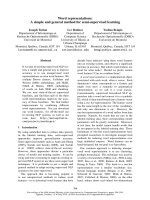
inner membrane (IM) [22]. As indicated in Fig. 1A, the
peptide conformation in buffer was predominantly dis-
ordered, whereas association of the peptide with lipid
vesicles induced a transition to a predominantly a-heli-
cal conformation. Complete binding of the peptide
to the lipid vesicles was manifested by the absence of
significant changes in the CD spectrum when the
lipid ⁄ peptide molar ratio was increased from 100 to 400.
The helical wheel diagram of Esc(1–18) in a perfect
a-helical conformation (Fig. 1B) shows amphipathicity
of the peptide, with hydrophobic and hydrophilic
residues segregating on opposite sides of the molecule.
Antibacterial activity
The activity of Esc(1–18) against E. coli ATCC 25922
was first evaluated by the microdilution broth assay to
determine the minimal inhibitory concentration (MIC),
using both a standard inoculum of 1 · 10
6
colony-
forming units (CFUs)ÆmL
)1
and 4 · 10
7
CFUÆmL
)1
,as
most of the experiments described below needed this
higher number of bacterial cells. As shown in Table 1,
where the frog skin membrane-active peptide tempo-
rin-1Tl [23] is included as a reference, the MIC of
Esc(1–18) in culture medium (Mueller–Hinton broth)
was found to be directly correlated with the number of
microbes present in the inoculum. Afterwards, to
examine the killing activity of Esc(1–18) against E. coli
and to determine whether this process was affected by
the ionic strength of the incubation medium, we
assayed the peptide’s bactericidal effect, as defined in
Experimental procedures, after 1.5 h of incubation
with bacteria, either in Mueller–Hinton broth, sodium
phosphate buffer (pH 7.4), or NaCl ⁄ P
i
(Table 1).
Interestingly, in all cases, a reduction in the number of
viable cells of ‡ 3 log
10
CFUÆmL
)1
(99.9% mortality)
was achieved at twice the MIC (16 lm) when a stan-
dard inoculum was used. In contrast, with the higher
number of bacteria (4 · 10
7
CFUÆmL
)1
), Esc(1–18)
displayed a bactericidal effect at 32 lm, a concentra-
tion equal to the MIC, under these conditions
(Table 1). Furthermore, to estimate the peptide’s abil-
ity to retain such activity under experimental condi-
tions closer to those encountered in vivo, antimicrobial
assays were performed in the presence of human
serum. It is noteworthy that, unlike temporin-1Tl
(Table 1) and other natural antimicrobial peptides,
such as human b-defensin 2 and dermaseptin S, which
lost their bacteriostatic effect in the presence of
20–30% serum (MIC ‡ 200 lm) [24,25], Esc(1–18) was
able to partially preserve its antibacterial activity at a
higher serum percentage (70%), with MIC and bacteri-
cidal concentration values of 32 and 64 lm, respec-
tively (Table 1), using a standard inoculum. As the
peptide’s degradation by serum enzymes was prevented
by heating serum at 56 °C (see Experimental proce-
dures), our findings suggest that serum components do
not strongly bind to Esc(1–18) and therefore do not
significantly affect the availability of active peptide
molecules.
λ (nm)
[Θ] (mdeg·cm
2
·dmol
–1
)
A
B
Fig. 1. Secondary structure of Esc(1–18). (A) CD spectra of the
peptide in sodium phosphate buffer (pH 7.4) (solid line) and after
association with PE ⁄ PG vesicles (dotted line, peptide 10 l
M, lipid
1m
M; broken line, peptide 5 lM, lipid 2 mM). (B) Helical wheel plot
of Esc(1–18): hydrophilic, hydrophobic and potentially positively
charged residues are represented as circles, diamonds and penta-
gons, respectively. The peptide is amidated at its C-terminus.
L. Marcellini et al. Esc(1–18) and E. coli membrane permeation/proteome
FEBS Journal 276 (2009) 5647–5664 ª 2009 The Authors Journal compilation ª 2009 FEBS 5649
The killing kinetics occurred on a quite fast time
scale, causing more than 90% microbial deaths within
10 min, at the MIC (Fig. 2). The latter result indicates
a substantial difference from those antimicrobial pep-
tides that preferentially act on intracellular targets and
over a longer time scale, and do not manifest any
lethal activity at their MICs [11,26].
Mode of action studies
It is well known that the mode of action of antimicro-
bial peptides depends on the mode(s) of their inter-
action with the cell membrane. However, before
reaching it, the peptide needs to bind and traverse
the cell wall, which, in Gram-negative bacteria, is sur-
rounded by an outer membrane (OM), composed
mainly of the anionic LPS (or endotoxin), which forms
a barrier to protect bacteria from many hydrophilic
and hydrophobic molecules, including some antimicro-
bial peptides [27]. Therefore, we first investigated the
ability of Esc(1–18) to bind LPS and penetrate the
E. coli OM.
LPS binding properties
LPS films have been used as suitable model systems to
mimic the outer layer of the Gram-negative OM
[28,29]. To investigate the binding of Esc(1–18) to LPS,
we monitored the changes in surface pressure of mono-
layers of commercially available LPS from E. coli
O111:B4 upon a peptide’s insertion, using the method
described in Experimental procedures. Esc(1–18) effi-
ciently penetrated E. coli LPS monolayers, as mani-
fested by the increase in film surface pressure (Fig. 3).
Under experimental conditions, monolayer penetration
reached a substantial stability around 1.0 lm Esc(1–18)
(Fig. 3A), which was then selected as the peptide con-
centration for subsequent experiments. When data
from similar measurements were analyzed in terms of
change in surface pressure (Dp) versus initial surface
pressure (p
0
), the critical surface pressure correspond-
ing to the LPS lateral packing density preventing the
intercalation of Esc(1–18) into E. coli LPS films could
be derived by extrapolating the Dp)p
0
slope to
Dp = 0, yielding a value of 47 mNÆm
)1
(Fig. 3B).
The kinetics of the insertion of the peptide into the
LPS monolayer were characterized by a rapid and
marked enhancement of surface pressure that followed
soon after injection of the peptide into the subphase,
the lag phase for this process being too short to be
measurable with our instrumentation (Fig. 3C). In a
typical experiment, within the first 60 s after peptide
injection, p attained a value that was slightly over 85%
1 x 10
7
1 x 10
6
CFU
1 x 10
5
Time (min)
1 x 10
4
0 5 10 15 20 25 30
Fig. 2. Time-kill curves for E. coli ATCC 25922 and Esc(1–18). Bac-
teria (4 · 10
7
CFUsÆmL
)1
) were grown in Mueller–Hinton broth at
37 °C and diluted in sodium phosphate buffer (pH 7.4). About
4 · 10
6
CFUs in 100 lL were incubated with Esc(1–18) at the MIC
(32 l
M; ) and at a sublethal dose (0.25 lM; ). The control (r)
consisted of bacteria incubated in the absence of peptide. Aliquots
were withdrawn, diluted in Mueller-Hinton broth and plated on agar
plates for CFUs counting. Data are the means ± standard devia-
tions of three independent experiments. Similar results were
obtained when bacteria were suspended in Mueller–Hinton broth or
NaCl ⁄ P
i
, and therefore are not shown.
Table 1. Antibacterial activity of Esc(1–18) and temporin-1Tl on E. coli ATCC 25922. The bactericidal activity is defined as the concentration
of peptide that is sufficient to reduce the number of viable bacteria by ‡ 3 log
10
CFUsÆmL
)1
after 1.5 h of incubation. The values found for
temporin-1Tl are in parentheses.
CFUÆmL
)1
MIC (lM) Bactericidal activity (lM)
Incubation medium Incubation medium
Mueller–Hinton
broth 70% serum
Mueller–Hinton
broth
Sodium phosphate
buffer (pH 7.4) NaCl ⁄ P
i
70% serum
1 · 10
6
8 (8) 32 (> 128) 16 (32) 16 (4) 16 (8) 64 (> 128)
4 · 10
7
32 128 32 32 32 128
Esc(1–18) and E. coli membrane permeation/proteome L. Marcellini et al.
5650 FEBS Journal 276 (2009) 5647–5664 ª 2009 The Authors Journal compilation ª 2009 FEBS
of the value recorded at the end of measurement
(Fig. 3C). This initial surge was then followed by a
slower increase in p for approximately the next 19 min,
when a plateau was reached, and no more significant
variation in p was observed for at least the next
16 min. This general kinetics pattern was apparently
independent of the initial surface pressure and from
peptide concentration, and was similar to that recorded
for temporin-1Tl interacting with a monolayer made of
the same type of LPS [30].
OM permeability
The permeabilization of the OM was determined by
investigating the periplasmic b-lactamase activity
against its specific substrate CENTA [31]. A plot of
enzyme release, as a function of peptide concentration,
is shown in Fig. 4A. Interestingly, there was a dose-
dependent perturbation of the OM, and the greatest
perturbation was obtained at the MIC of the peptide
(32 lm with 4 · 10
7
CFUÆmL
)1
). The rate of CENTA
hydrolysis, upon addition of 1 · MIC of Esc(1–18) to
the cells, was also monitored for 20 min, and the
amount hydrolyzed was found to be 70% of the
total within the first 5 min (Fig. 4B).
IM permeability
Next, the effect of the peptide on the E. coli IM was
analyzed by measuring the intracellular influx of
SYTOX Green [32]. This cationic dye, which is
excluded by intact membranes, but not from those with
lesions large enough to allow its entrance, dramatically
increases its fluorescence when bound to intracellular
nucleic acids (Fig. 5). The data revealed that Esc(1–18)
augmented the permeability of the IM, with kinetics
superimposable on those of the OM permeation
(although with a slightly longer lag time), reaching a
final value in about 15–20 min and in a concentration-
dependent fashion. However, membrane permeation
caused by Esc(1–18) was not maximal at levels up to
twice the MIC. This was manifested by a further
enhancement of fluorescence, following the addition of
a detergent for the complete solubilization of phospho-
lipid bilayers (Fig. 5, arrow at 20 min). Then, to inves-
tigate the size of membrane lesions induced by the
peptide, we assessed the release of intracellular com-
pounds, such as the cytoplasmic b-galactosidase, whose
Stokes radius is equal to 69 A
˚
[33]. As reported in
Fig. 6, the enzyme release was almost 40% of maxi-
mum when the peptide concentration was equal to the
MIC. These results underscore a disturbance of the IM,
although to a smaller extent than that of the OM, and
30
25
20
15
Dp (mN·m
–1
)
10
5
0
0.4 0.8 1.2
1.6 2
Peptide concentration (µ
M)
40
35
30
25
20
Dp (mN·m
–1
)
p
0
(mN·m
–1
)
15
10
5
0
48
44
4036322824201612840
p (mN·m
–1
)
50
45
40
35
30
25
20
15
10
240020001600
1200
8004000
5
0
Time (s)
A
B
C
Fig. 3. Insertion of Esc(1–18) into E. coli O111:B4 LPS monolay-
ers. (A) Increments of surface pressure of E. coli LPS monolayers
due to the addition of Esc(1–18) to the subphase are illustrated as
a function of peptide concentration at an initial surface pressure
varying between 19.2 and 21.0 mNÆm
)1
, or (B) an initial surface
pressure, with 1.0 l
M peptide. (C) Typical kinetics of surface pres-
sure increase related to Esc(1–18) penetration into E. coli LPS
monolayers (p
0
= 14.2, with 1.0 lM peptide; an arrow indicates
peptide injection into the subphase). Each data point represents
the mean of triplicate measurements. The standard deviation
varied between 0.1 and 0.9 mNÆm
)1
and, for the sake of clarity, is
not shown.
L. Marcellini et al. Esc(1–18) and E. coli membrane permeation/proteome
FEBS Journal 276 (2009) 5647–5664 ª 2009 The Authors Journal compilation ª 2009 FEBS 5651
indicate the existence of a direct correlation between
the peptide dose and the extent of both microbial death
and membrane disturbance.
Synergistic activities with conventional
antibiotics
Checkerboard titrations were carried out using Esc(1–
18) in combination with different classes of clinically
available antibiotics. As illustrated in Table 2, a clear
synergism was noted when the peptide was mixed with
cephalosporin C, erythromycin, nalidixic acid, netilmi-
4000
5000
2000
3000
4000
0
1000
0
5
10
15
20 25 30
Time (min)
Fluorescence (arbitrary units)
Fig. 5. Effect of Esc(1–18) on the permeation of the E. coli
ATCC 25922 IM. Cells (4 · 10
7
CFUsÆmL
)1
) were incubated with
1 l
M SYTOX Green in NaCl ⁄ P
i
. When basal fluorescence reached a
constant value, the peptide was added (first arrow, t = 0), and
changes in fluorescence were monitored (k
excitation
= 485 nm, k
emis-
sion
= 535 nm) and plotted as arbitrary units. SDS (0.1% in chloro-
form) was added for the maximal membrane permeation (second
arrow, t = 20 min). Data points represent the mean of triplicate
samples with standard deviation values not exceeding 2.5% from a
single experiment, representative of three different experiments.
The peptide concentrations used were as follows: 2 l
M (s); 4 lM
(*); 8 lM (e); 16 lM (d); 32 lM ( ); and 64 lM ( ). Fluorescence
values of control (bacteria without peptide) were subtracted from
each sample.
100
60
80
0
20
40
% of lactamase release
0
5101520
Time (min)
0.05
0.06
0.07
A
B
Absorbance
0.02
0.03
0.04
0
0.01
Control 4 8 16 32
After
detergent
lysis
Peptide concentration (µM)
Fig. 4. Permeation of E. coli OM by Esc(1–18). (A) Effects of differ-
ent concentrations of Esc(1–18) on permeation of the OM of E. coli
ATCC 25922 (4 · 10
7
CFUÆmL
)1
), were followed spectrophotometri-
cally by measuring the activity of periplasmic b-lactamase. The cells
were resuspended in sodium phosphate buffer (pH 7.4) + 100 m
M
NaCl, and incubated with different concentrations of peptide at
37 °C for 20 min. The enzyme activity was measured in the culture
filtrate by following the hydrolysis of 80 l
M CENTA at 405 nm. The
absorbances of all peptide-treated samples, bacteria without peptide
(control) and bacteria after lysis with 0.1% SDS in chloroform are
reported on the y-axis. The values are the means of three inde-
pendent measurements ± standard deviations. (B) Kinetics of OM
permeabilization caused by 1 · MIC of Esc(1–18) (32 l
M). Bacteria
(4 · 10
7
CFUÆmL
)1
) were incubated with the peptide at different
time intervals, and b-lactamase activity was detected as described
above and expressed as percentage of the total obtained after cell
lysis. Data are the means ± standard deviations of three indepen-
dent experiments.
60
70
40
50
60
10
20
30
% of total
0
4 8 16 32
Peptide concentration (µ
M)
Fig. 6. Bacterial viability and b-galactosidase activity of E. coli
ATCC 25922 culture after treatment with Esc(1–18). Bacterial cells
(4 · 10
7
CFUsÆmL
)1
) were grown in Mueller–Hinton broth at 37 °C,
diluted in sodium phosphate buffer (pH 7.4), and incubated with
the peptide at different concentrations for 20 min at 37 °C. The
number of surviving cells (
) is given as the percentage of the
total. b-Galactosidase activity was measured in the culture filtrate
by following the hydrolysis of 2 m
M Gal-ONp at 420 nm. Enzymatic
activity detected in the control (bacteria without peptide) was sub-
tracted from all values, which are expressed as percentage of the
total (e). Complete enzyme activity was determined by treating
bacteria with 0.1% SDS in chloroform. The values are the means
of three independent measurements ± standard deviations.
Esc(1–18) and E. coli membrane permeation/proteome L. Marcellini et al.
5652 FEBS Journal 276 (2009) 5647–5664 ª 2009 The Authors Journal compilation ª 2009 FEBS
cin, and rifampicin [a fractional inhibitory concentra-
tion (FIC) £ 0.5 indicates synergy; see Experimental
procedures]. To obtain insights into the mode of action
underlying the synergistic activity, we investigated the
bactericidal action of the combination of Esc(1–18)
and erythromycin, the antibiotic that gives the best
synergy with the peptide, as indicated by the lowest
FIC (Table 2). Erythromycin is a hydrophobic mole-
cule that inhibits protein synthesis by blocking either
the peptidyltransferase reaction or the translocation
step, but cannot easily traverse the OM of Gram-
negative bacteria [34].
As expected, erythromycin displayed a weak bacte-
ricidal effect, causing about 35% microbial death at a
very high concentration (256 lgÆmL
)1
) and within 3 h
of incubation (Fig. 7). Interestingly, when sublethal
concentrations of Esc(1–18) and erythromycin were
combined, 8% and 90% killing were detected after
20 min and 3 h, respectively (Fig. 7). These results
provide additional support for the membrane-permea-
bilizing properties of Esc(1–18). Indeed, as no reduc-
tion in the number of viable cells was observed
within the first 20 min [killing kinetics of Esc(1–18)],
but a reduction was observed after a longer time
(2–3 h) (Fig. 7), corresponding to the time-kill kinetics
of erythromycin, we can assume that the synergistic
activity between the two compounds is the result of
increased access of erythromycin to its intracellular
target, because of increased peptide-induced perme-
ability of the cytoplasmic membrane and ⁄ or the LPS
layer.
Permeabilization of large unilamellar vesicles
(LUVs)
The peptide’s ability to alter the structure of the
plasma membrane of E. coli cells by a nonstereo-
specific process was also confirmed by employing
calcein-loaded liposomes made of PE ⁄ PG (7 : 3, w:w).
Different concentrations of peptide were added to
LUV suspensions, and membrane permeability was
measured by following fluorescence recovery due to
calcein leakage from the liposomes [35]. Calcein leakage
occurred immediately after peptide addition, and
reached a plateau within the first 15 min (Fig. 8A).
Figure 8B shows the dose–response curve of peptide-
induced calcein release from PE ⁄ PG vesicles. The data
clearly show a membrane-perturbing activity of Esc(1–
18). Note that this activity increased in a dose-depen-
dent manner and reached its maximum ( 65% calcein
leakage) at a peptide ⁄ lipid molar ratio of 1.5. These
results are comparable with those found for other
membrane-active antimicrobial peptides, such as cathe-
licidin LL-37 [36]. However, at a peptide⁄ lipid molar
ratio as low as 0.04, Esc(1–18) was more active than
cathelicidin LL-37 [36]. As illustrated in Fig. 8B,
Esc(1–18) did not fully permeabilize the lipid vesicles,
and the calcein leakage diminished when the peptide⁄
lipid molar ratio exceeded 1.5, probably because of the
peptide’s aggregation at high concentrations. Taken
together, these observations are in line with those
made above using intact cells (Figs 5 and 6), and are
consistent with the suggestion that Esc(1–18) binds
and destabilizes the bacterial membrane, but to a lesser
Table 2. Interaction of Esc(1–18) with conventional antibiotics
against E. coli ATCC 25922. The ranges of concentrations tested
were as follows: 0.25–64 mgÆL
)1
for Esc(1–18) and 0.25–
256 mgÆL
)1
for the other antimicrobial agents. FIC indices were
interpreted as follows: FIC £ 0.5, synergy; 0.5 < FIC <1, additivity;
1 £ FIC < 4, indifference; and FIC ‡ 4, antagonism.
Compound MIC (lgÆmL
)1
) FIC
Ampicillin 4 1.06
Carbenicillin 64 0.73
Cephalosporin C 128 0.46
Ceftazidime 0.5 0.90
Chloramphenicol 8 0.83
Erythromycin 32 0.36
Imipenem 0.5 1.16
Nalidixic acid 8 0.40
Netilmicin 8 0.43
Oxacillin 8 1.11
Rifampicin 8 0.40
Streptomycin 4 1.10
Tazocin 4 0.73
Tetracycline 2 0.98
80
100
120
CFU (%)
40
20
60
80
0
20
60 120 180
Incubation time (min)
Fig. 7. Synergistic effect in the bactericidal activity of erythromycin
and Esc(1–18). E. coli cells (1 · 10
6
CFUsÆmL
)1
) were incubated in
Mueller–Hinton broth (diluted 1 : 2 with distilled water) in the pres-
ence of 256 lgÆmL
)1
erythromycin (white bars), a sublethal concen-
tration of erythromycin (8 lgÆmL
)1
, gray bars), or Esc(1–18)
(1 lgÆmL
)1
, squared bars), and with the combination of erythromy-
cin and Esc(1–18) at their sublethal doses (black bars). Aliquots
were withdrawn at the time intervals indicated, and plated for
counting. The percentages of viable cells with respect to the con-
trol (bacteria not treated) are reported on the y-axis. Data are the
means ± standard deviations of three independent experiments.
L. Marcellini et al. Esc(1–18) and E. coli membrane permeation/proteome
FEBS Journal 276 (2009) 5647–5664 ª 2009 The Authors Journal compilation ª 2009 FEBS 5653
extent than temporin-1Tl [37]. According to what has
been stated for other antimicrobial peptides [38], such
a discrepancy between the two frog skin peptides
might be related to a higher fraction of membrane-
bound active temporin-1Tl than of Esc(1–18).
The ability of Esc(1–18) to induce the leakage of
liposome-encapsulated markers of different sizes was
also monitored. PE ⁄ PG LUVs were preloaded with
fluorescein isothiocyanate–dextrans (FITC-Ds) of 4,
10, 40 or 70 kDa average molecular mass (FITC-D 4,
FITC-D 10, FITC-D 40, and FITC-D 70), and then
incubated with the peptide. The data shown in Fig. 9
reveal that Esc(1–18) is able to cause the release of the
four dextrans used in a dose-dependent manner, and
with a dependence on the size of the liposome-
entrapped probe. This indicates that Esc(1–18) does
not have a detergent-like effect on the membrane [39],
and that membrane lesions produced by this peptide
are larger than 58 A
˚
(Stokes radius of FITC-D 70
[40]), which is in agreement with its ability to promote
the release of b-galactosidase from E. coli cells.
Scanning electron microscopy (SEM)
The effect(s) of Esc(1–18) on E. coli morphology were
visualized by SEM (Fig. 10). The exposure of
4 · 10
7
cellsÆmL
)1
at the corresponding MIC of
Esc(1–18) resulted in an irregular rod form with a deep
wrinkling of the cell surface (within 5 min). However,
all of these changes became more pronounced after a
longer incubation time (20 min). With reference to
untreated cells, bacteria appeared flat, with a collapsed
cell structure and surface corrugation similar to that
induced by temporin-1Tl [41], but in a milder form.
Transmission electron microscopy (TEM)
TEM was then used to directly examine the damage to
bacteria induced by the peptide. A local disturbance to
the membrane was noted after the first 5 min of pep-
tide treatment, and this was followed by more damage
and loss of cellular integrity, with a partial discharge
of the cellular contents, within 20 min (Fig. 11). These
results correlate with the killing kinetics of the peptide,
and show that the antibacterial activity of Esc(1–18) is
concomitant with its membrane-perturbing activity.
A
50
60
70
B
10
20
30
40
Calcein leakage(%)
0
0.0001 0.001 0.01 0.1 1 10
(Peptide) : (Lipid)
[Peptide]:[Lipid]
50
60
3
1.5
0.4
0.08
0.01
0.0012
20
30
40
Calcein leakage (%)
0
10
510150
Time (min)
Fig. 8. Calcein leakage from PE ⁄ PG LUVs after Esc(1–18) treat-
ment. (A) Time course of calcein release from PE ⁄ PG (7 : 3, w ⁄ w)
calcein-loaded LUVs (final lipid concentration 200 l
M) after addition
of Esc(1–18) (arrow at time zero) at different concentrations. Control
(broken line) consisted of liposomes not treated with the peptide.
Calcein release was detected fluorimetrically (k
excitation
= 485 nm,
k
emission
= 535 nm). The percentage of leakage was calculated as
100(F
1
– F
0
) ⁄ (F
t
– F
0
), where F
1
and F
t
denote the fluorescence
before and after the addition of detergent (0.1% Triton X-100),
respectively, and F
0
represents the fluorescence of intact vesicles.
Data points are means with standard deviations not exceeding 4%
from a single experiment, representative of three independent mea-
surements. (B) Esc(1–18) was added to PE ⁄ PG LUVs (7 : 3, w ⁄ w)
at concentrations of 0.125–660 l
M. Calcein release was detected as
described above, after 15 min of peptide treatment. Data points are
means with standard deviations not exceeding 3% from a single
experiment, representative of four independent measurements.
30
40
50
60
Dextran release (%)
0
10
20
0 0.1 0.2 0.3 0.4 0.5 0.6 0.7 0.8 0.9 1
(Peptide) : (Lipid)
Fig. 9. Effect of Esc(1–18) on the release of FITC-D from PE ⁄ PG
liposomes. Liposomes containing FITC-D 4 (r), FITC-D 10 (
),
FITC-D 40 (
) or FITC-D 70 (s) were prepared as described in
Experimental procedures, and incubated in the presence of differ-
ent concentrations of the peptide for 15 min at 37 °C. Dextran
release was detected fluorimetrically (k
excitation
= 470 nm; k
emission
=
520 nm). Leakage was calculated as 100(F
1
– F
0
) ⁄ (F
t
– F
0
), where
F
0
represents the fluorescence of intact vesicles, and F
1
and F
t
denote the intensities of the fluorescence achieved by peptide and
Triton X-100 treatment, respectively. Values are means of three
independent measurements ± standard deviations.
Esc(1–18) and E. coli membrane permeation/proteome L. Marcellini et al.
5654 FEBS Journal 276 (2009) 5647–5664 ª 2009 The Authors Journal compilation ª 2009 FEBS
Proteomic analysis
To determine whether Esc(1–18) could evoke a cellular
reaction by modifying, within 20 min, the expression
levels of proteins under conditions where the peptide
did not affect the viability of E. coli or reduced it by
40% (2 and 16 lm peptide, respectively; data not
shown), the bacterial proteome was analyzed by means
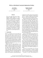
of 2D-PAGE and MS. This analysis revealed a similar
pattern of responses to both sublethal and lethal pep-
tide doses, consisting of only a few significant varia-
tions in protein expression (11 protein spots) as
compared with untreated cells. The majority of these
spots (Fig. 12) were identified by peptide mass finger-
printing, reported in Table 3. In particular, reductions
in the expression levels of a number of OM proteins
(OMPs), such as OMPc, nmpC, and OMP F, all of
which form passive diffusion pores allowing the pas-
sage of small molecular weight hydrophilic materials
[42], were detected in peptide-treated bacteria
(Table 3), with stronger reductions being seen at 16 lm
Esc(1–18). Otherwise, a slight increase in OMP W
expression was found at 16 lm. Note that the function
of this protein is not completely understood; however,
recent data have suggested that it may be involved in
the protection of bacteria against various forms of
environmental stresses [43]. Overexpression of trigger
factor (TF) was also caused by both peptide concen-
trations. TF in E. coli is a ribosome-associated chaper-
one that initiates folding of newly synthesized proteins
[44]. The enhanced production of TF might contribute
to more streamlined de novo protein folding, by shield-
ing nascent polypeptides on the ribosome, and thereby
shortening degradation or aggregation processes [45].
In addition, as shown in Table 3, exposure of bacteria
to Esc(1–18) gave rise to a drop in the level of the fol-
lowing enzymes: (a) glucosamine-fructose-6-phosphate
aminotransferase, which catalyzes the formation of
glucosamine 6-phosphate, a precursor of cell wall
peptidoglycan synthesis [46]; and (b) the dihydro-
lipoyllysine-residue succinyltransferase component of
2-oxoglutarate dehydrogenase complex, which catalyzes
the conversion of a-ketoglutarate into succinyl-CoA as
part of the tricarboxylic acid cycle [47].
Discussion
The repertoire of antimicrobial peptides has dramati-
cally increased during the past two decades, and
> 800 antimicrobial peptides have been isolated from
Control
MIC, 5 min
MIC, 20 min
Fig. 10. Scanning electron micrographs of
Esc(1–18)-treated E. coli ATCC 25922 cells
(4 · 10
7
CFUsÆmL
)1
). Upper panels: control
bacteria. Middle panels: bacteria after 5 min
of treatment with Esc(1–18) at the MIC
(32 l
M). Lower panels: bacteria after 20 min
of treatment with Esc(1–18) at the MIC.
See Results for other experimental details
and descriptions of the images. Each image
has been magnified · 10 000 or · 20 000.
L. Marcellini et al. Esc(1–18) and E. coli membrane permeation/proteome
FEBS Journal 276 (2009) 5647–5664 ª 2009 The Authors Journal compilation ª 2009 FEBS 5655
different plant and animal sources, with more than 400
isoforms being obtained from amphibian species. This
article discusses the antibacterial activity and mode of
action of the N-terminal region of esculentin-1b, an
antimicrobial peptide from the skin of P. lessonae ⁄ ridi-
bundus. As no activity against microorganisms had
been previously observed with the 19–46 fragment of
this peptide, possibly because of its low positive charge
at neutral pH (+1 versus +5 for the whole molecule)
[48], we analyzed the antibacterial activity of the
1–18 N-terminal portion of esculentin-1b. Surprisingly,
this activity was found to be similar to that of the full-
length natural peptide [48,49], whereas complementary
insecticidal properties were ascribed to the 19–46
fragment [50]. Recent experiments have underscored
the fact that Esc(1–18) possesses a wide spectrum of
antimicrobial activity against several species of Gram-
positive bacteria, Gram-negative bacteria, Candida and
multidrug-resistant nosocomial pathogens, without
being hemolytic [20,48].
Regardless of the precise mode of action, the
effect(s) of antimicrobial peptides in general depends
upon their interaction with the microbial membrane
[51,52]. In particular, the first step in this process is the
electrostatic attraction between the cationic peptide
and the negatively charged components of the cell
envelope, such as the phosphate groups within the
LPS molecules of the OM in Gram-negative bacteria
or the lipoteichoic acids on the surface of Gram-posi-
tive bacteria. In the case of Gram-negative bacteria,
antimicrobial peptides initially cross the LPS layer, in
a self-promoted uptake process driven by hydrophobic
interactions, and subsequently reach the IM [51]. Nev-
ertheless, studies performed with intact bacteria have
shown that antimicrobial peptides, e.g. pleurocidin
derivatives and buforin 2, do not disturb the
membrane of E. coli at their minimal antimicrobial
concentrations, but rather traverse it, accumulate
intracellularly, and damage a variety of essential vital
processes to mediate the lethal event, which occurs
only at multiples of the MICs [7,11,26].
In this study, we have shown that Esc(1–18) dis-
plays rapid bactericidal activity, at the MIC, against
E. coli (Fig. 2), concomitant with alteration of its
inner and outer membranes (Figs 4–6). As shown by
the biophysical and biochemical assays, this peptide
strongly bound LPS and completely permeated the
LPS OM (Figs 3 and 4). In addition, the intracellular
influx of SYTOX Green (Fig. 5), the extracellular
leakage of b-galactosidase (Fig. 6), calcein and dex-
tran release from liposomes mimicking the E. coli IM
(Figs 8 and 9) and electron microscopy images
(Figs 10 and 11) suggest that Esc(1–18) is a mem-
brane-active peptide which kills bacteria by, primarily,
injuring their membranes. This interpretation is fur-
ther supported by the small changes in the proteomic
1 µm
A
1 µm
B
1 µm
C
Fig. 11. Transmission electron micrographs of Esc(1–18)-treated
E. coli ATCC 25922 cells (4 · 10
7
CFUsÆmL
)1
). (A) Representative
control. (B) Representative bacterium after 5 min of peptide treat-
ment at the MIC (32 l
M). (C) Representative bacterium after
20 min of peptide treatment at the MIC. See Results for other
experimental details and descriptions of the images.
Esc(1–18) and E. coli membrane permeation/proteome L. Marcellini et al.
5656 FEBS Journal 276 (2009) 5647–5664 ª 2009 The Authors Journal compilation ª 2009 FEBS
profiling of bacteria upon treatment with either suble-
thal or lethal peptide doses.
Unlike DNA microarray analysis, which has proven
to be a successful tool for the monitoring of whole
genome expression profiles at the mRNA level [53],
proteomic analysis has been found to be very useful
for comparing changes in the expression levels of many
proteins, under antibiotic treatment or other environ-
mental conditions [54]. Importantly, this approach
represents the most powerful method for providing
a better understanding of complex biological processes,
as well as post-translational modifications of proteins,
which cannot be obtained from mRNA expression
profiles [55]. In peptide-treated bacteria, a decrease in
the levels of OMPc, OMP F, and nmp proteins, which
allow the passive diffusion of hydrophilic molecules
across the OM, would represent a cellular reaction that
compensates for the stress provoked upon contact with
a membrane-active antimicrobial peptide. In line with
this explanation is the greater production of TF and
OMP W, at the highest peptide concentration used, to
guarantee bacteria a more protected environment,
which would be particularly important for increasing
their viability. Furthermore, the exposure of bacteria
to Esc(1–18) would cause a slowdown of metabolic
activities, which is in agreement with the lower levels of
glucosamine-fructose-6-phosphate and dihydrolipoyl-
lysine-residue succinyltransferase component of 2-oxo-
glutarate dehydrogenase complex.
Esc(1–18) did not cause bacteria to disintegrate and
did not form blebs on their surface but, rather, emptied
the cells, causing the loss of cellular material through
CAB
+
pH 3–10
–
6703
6703
6703
6707
6707
6707
2702
2702
CB
A
0407
0407
0407
2702
2702
2702
5603
5603
5603
5103
5103
5103
Fig. 12. Two-dimensional maps of the E. coli proteome. Representative 2D gels of total protein extracts from E. coli ATCC 25922 cells. (A)
Control. (B, C). After Esc(1–18) treatment at 2 l
M and 16 lM, respectively. The region of the gels containing differentially expressed protein
spots is magnified in the lower panels. Protein spots that were identified by PMF are labeled with circles and numbers.
L. Marcellini et al. Esc(1–18) and E. coli membrane permeation/proteome
FEBS Journal 276 (2009) 5647–5664 ª 2009 The Authors Journal compilation ª 2009 FEBS 5657
the peptide-induced membrane breakages, and substan-
tial roughening of their surface. The peptide might bind
to the membrane surface in a carpet-like arrangement,
inserting into the polar phospholipid headgroups. This
would generate an unfavorable tension, resulting in the
formation of transient breakages with a size larger than
58 A
˚
, leading to bacterial death [9,56].
In addition, as suggested by the synergistic bacteri-
cidal activity of Esc(1–18) when combined with ery-
thromycin, an increased peptide-induced membrane
permeability, at subinhibitory peptide concentrations,
would make it easier for hydrophobic drugs to enter
the cells and to induce their toxic effect.
This work provides four interesting findings. The
first is the ability of Esc(1–18) to display fast bacterici-
dal activity, at the MIC, under both standard and
physiological conditions. The second is its ability to
simultaneously kill E. coli and permeate, in a dose-
dependent manner, its outer and inner membranes, but
without causing cell lysis. The third is the ability to
modify, within 20 min, the expression levels of a lim-
ited number of bacterial proteins, at either lethal or
sublethal concentrations. These findings rule out the
possibility that variations in the production of these
proteins account for the killing process of Esc(1–18).
Note that only a few studies on the effect(s) of antimi-
crobial peptides on the proteomes of microorganisms
have been performed to date. Interestingly, proteomic
and transcriptomic analysis of the yeast Saccharo-
myces cerevisiae, following exposure to a similar anti-
microbial peptide [esculentin-1a(1–21)], had shown
downregulation of enzymes of the lower glycolytic
pathway as well as a decrease in actin level, resulting
in dramatic changes in cell physiology [57]. It is wor-
thy of remark that both fragments of esculentin pep-
tide were found to affect the integrity of the microbial
plasma membrane and the synthesis of the microbial
cell wall. To the best of our knowledge, this study rep-
resents the first example of the effects of an antimicro-
bial peptide from frog skin on the proteome of
bacteria, and demonstrates that the bacterial mem-
branes are the major targets of its mechanism of
action. Fourth, Esc(1–18) synergizes with conventional
antibiotics in the inhibition of microbial growth. All of
these properties, together with potent activity against a
broad spectrum of multidrug-resistant clinical isolates
[20] and a lack of lytic effects on human erythrocytes
[48], lymphocytes, and keratinocytes (data not shown),
make Esc(1–18) a very attractive membrane-active
antimicrobial peptide for in-depth analysis of biological
properties. More specifically, it can be considered to
be a promising template for: (a) the production of less
toxic anti-infective preparations with new modes of
action and with the ability to elicit few changes in the
Table 3. Protein spots identified by PMF.
Spot
no.
Fold change
Protein name
UniProt
accession
no.
Theoretical
pI ⁄ M
r
Score
a
No. of
matching
peptides
Sequence
coverage
(%)
Peptide concentration (l
M)
216
0407
b
Outer membrane protein
C precursor
Q8CVW1 4.59 ⁄ 41.22 172 17 70 )1.66 )2.50
0407 Outer membrane porin
protein nmpC precursor
P21420 4.64 ⁄ 40 72 12 37
0407 Outer membrane protein F P02931 4.76 ⁄ 39.33 135 13 57
6703 Glucosamine-fructose-6-phosphate
aminotransferase
Q8XEG2
c
5.56 ⁄ 67.136 123 17 33 )1.50 )2.00
Q8FBT4
P17169
6707 Glucosamine-fructose-6-phosphate
aminotransferase
Q8XEG2
c
5.56 ⁄ 67.136 125 17 33 )1.60 )2.22
Q8FBT4
P17169
2702 Trigger factor Q0TKK5 4.81 ⁄ 48.25 72 11 29 +3.08 +2.51
5103 Outer membrane protein W P0A915 6.03 ⁄ 22.9 79 5 50 )1.50 +2.07
5603 Dihydrolipoyllysine residue
succinyltransferase component of
2-oxoglutarate dehydrogenase
complex
P0AFG6 5.58 ⁄ 43.88 60 10 30 )2.08 )1.92
a
The MASCOT score represents the probability that the observed match is a random event. Protein scores greater than 61 are significant
(P < 0.05).
b
This spot contains three different OMPs.
c
The three indicated UniProt accession numbers correspond to glucosamine-fructose-
6-phosphate aminotransferase from different E. coli strains. This protein was found in spot 6703 and spot 6707.
Esc(1–18) and E. coli membrane permeation/proteome L. Marcellini et al.
5658 FEBS Journal 276 (2009) 5647–5664 ª 2009 The Authors Journal compilation ª 2009 FEBS
proteome of the target microorganism and no microbial
resistance; and (b) the design of potential coadjuvants
of those antimicrobial agents that are already available.
Experimental procedures
Materials
Synthetic Esc(1–18) was purchased from GENEPEP
(Prades le Lez, France). The purity of the peptide, its
sequence and its concentration were determined as previ-
ously described [48]. Culture media, antibiotics, 2-nitrophe-
nyl b-d-galactoside (Gal-ONp) calcein and LPS from
E. coli serotype O111:B4 were all purchased from Sigma (St
Louis, MO, USA). SYTOX Green was from Molecular
Probes (Invitrogen, Carlsbad, CA, USA). Egg yolk PG and
PE were purchased from Avanti Polar Lipids (Alabaster,
AL, USA). FITC-Ds were purchased from Sigma. All other
chemicals were reagent grade. For antimicrobial assays,
the commercially available quality control strain E. coli
ATCC 25922 was used.
Penetration into LPS monolayers
Insertion of Esc(1–18) into LPS monolayers spread at an
air ⁄ buffer (5 mm Hepes, pH 7) interface was monitored by
measuring surface pressure (p) with a Wilhelmy wire
attached to a microbalance (DeltaPi, Kibron Inc., Helsinki,
Finland) connected to a PC and by using circular glass
wells (subphase volume 0.5 mL). After evaporation of
solvent and stabilization of monolayers at different initial
surface pressures (p
0
), the peptide (0.1–2 lm) was injected
into the subphase, and the increment in surface pressure of
the LPS film upon intercalation of the peptide dissolved in
the subphase was monitored for the next 37 min. The
difference between the initial surface pressure and the value
observed after the penetration of Esc(1–18) into the film
was taken as Dp.
Antibacterial activity
Susceptibility testing was performed by the microbroth dilu-
tion method according to the procedures outlined by the
National Committee for Clinical Laboratory Standards
(2001), using sterile 96-well plates. Stock solutions of
Esc(1–18) were prepared in serial two-fold dilutions in 20%
ethanol; 4 lL was then added to 46 lL of Mueller–Hinton
broth, previously placed in the wells of the microtiter plate.
Aliquots (50 lL) of bacteria in mid-log phase, at a concen-
tration of 1 · 10
6
or 4 · 10
7
CFUÆmL
)1
, were then added
to each well.
The range of peptide dilutions used was 1–128 lm. Inhi-
bition of growth was determined by measuring the absor-
bance at 595 nm with a 450-Bio-Rad Microplate Reader
after incubation for 18–20 h at 37 °C. Antibacterial activity
was expressed as MIC, the concentration of peptide at
which 100% inhibition of growth was observed.
Bactericidal activity
The bactericidal activity of Esc(1–18) against E. coli
ATCC 25922 was evaluated by a liquid microdilution assay
as described previously [41], in four different incubation
media: sodium phosphate buffer (pH 7.4); Mueller–Hinton
broth; NaCl ⁄ P
i
; and 70% human serum (inactivated by
heating at 56 °C for 30 min). Briefly, exponentially growing
bacteria were incubated at 37 °C for 1.5 h in the presence of
different concentrations of peptide (serial two-fold dilutions
ranging from 1 to 128 lm) dissolved in 100 lL of medium.
Following incubation, the samples were plated onto LB agar
plates. The number of surviving bacteria, expressed as
CFUs, was determined after overnight incubation at 37 °C.
Bactericidal activity was defined as the peptide concentra-
tion necessary to cause a reduction in the number of viable
bacteria of ‡ 3 log
10
CFUÆmL
)1
[24]. Controls were run
without peptide and in the presence of peptide solvent (20%
ethanol) at a final concentration of 0.6%.
Time-kill investigation
About 4 · 10
6
CFUs in 100 lL of sodium phosphate buffer
(pH 7.4) were incubated at 37 °C with Esc(1–18) at the
MIC (32 lm) and a subinhibitory concentration (0.25 lm).
Aliquots of 10 lL were withdrawn at different intervals,
diluted in Mueller–Hinton broth, and spread onto LB agar
plates. After overnight incubation at 37 °C, the number of
CFUs was counted. Controls were run without peptide and
in the presence of peptide solvent (20% ethanol) at a final
concentration of 0.6%.
Peptide effect in combination with conventional
antibiotics
Combinations of Esc(1–18) and antibiotics with different
chemical characteristics, in two-fold serial dilutions in
water, were tested for their synergistic effect by a checker-
board titration method. The ranges of drug dilutions used
were 0.25–64 lgÆmL
)1
for Esc(1–18) and 0.25–256 lgÆmL
)1
for the conventional antibiotics. The mean FIC index for
combinations of two peptides was calculated according to
the equation
X
ðFIC
A
þ FIC
B
Þ=n ¼
X
ðA=MIC
A
þ B=MIC
B
Þ=n
where A and B are the MICs of drug A and drug B in the
combination, MIC
A
and MIC
B
are the MICs of drug A
and drug B alone, FIC
A
and FIC
B
are the FICs of drug A
and drug B and n is the number of wells per plate used to
L. Marcellini et al. Esc(1–18) and E. coli membrane permeation/proteome
FEBS Journal 276 (2009) 5647–5664 ª 2009 The Authors Journal compilation ª 2009 FEBS 5659
calculate the FIC. The FIC indices were interpreted as fol-
lows: FIC £ 0.5, synergy; 0.5 < FIC <1, additivity;
1 £ FIC < 4, indifference; and FIC ‡ 4, antagonism [58].
The synergistic effect in the bactericidal activity of the com-
bination Esc(1–18) + erythromycin was also determined.
E. coli cells (1 · 10
6
CFUsÆmL
)1
) were incubated in Muel-
ler–Hinton broth (diluted 1 : 2 with distilled water) at
37 °C in the presence of 256 lgÆmL
)1
erythromycin, a sub-
lethal concentration of erythromycin (8 lgÆmL
)1
) or Esc(1–
18) (1 lgÆmL
)1
), and with the combination erythromycin +
Esc(1–18) at their sublethal doses. Aliquots were withdrawn
at specific time intervals (20, 60, 120 and 180 min), and
plated for counting.
Permeation of the bacterial OM
OM permeability was assessed by measuring the activity of
the periplasmic b-lactamase. Briefly, E. coli ATCC 25922
cells were grown at 37 °C in Mueller–Hinton broth to
a D
590 nm
of 0.8, and then washed and resuspended in
sodium phosphate buffer (pH 7.4) + 100 mm NaCl. About
4 · 10
6
cells (100 lL of bacterial suspension at a concentra-
tion of 4 · 10
7
CFUsÆmL
)1
) were incubated with different
concentrations of peptide (ranging from 4 to 32 lm) for
20 min at 37 °C. The bacterial culture was then passed
through a 0.2 lm filter, and a b-lactamase substrate
(CENTA, a synthesized chromogenic cephalosporin, with a
highly reactive b-lactam ring [31]) was added to a final con-
centration of 80 lm. Hydrolysis of the b-lactam ring, which
causes a color change from light yellow (k
max
: 340 nm) to
chrome yellow (k
max
: 405 nm), was recorded at 405 nm,
using a spectrophotometer (UV-1700 Pharma Spec Shima-
dzu, Tokyo, Japan). An increase in absorbance results in
an increase in OM permeability [31]. The same amount of
bacteria without Esc(1–18) was used as a control, whereas
the maximal membrane perturbation was obtained after
lysing bacteria with 0.1% SDS in chloroform (three drops
to 1 mL of bacterial suspension).
Permeation of the bacterial IM
To assess the ability of Esc(1–18) to alter the permeability
of the IM of E. coli,4· 10
6
cells were mixed with 1 lm
SYTOX Green in NaCl ⁄ P
i
for 5 min in the dark. After
addition of peptide, the increase in fluorescence, owing to
the binding of the dye to intracellular DNA, was measured
at 37 °C in a microplate counter (Wallac 1420 Victor
3Ô
;
Perkin Elmer, Foster City, CA, USA), using 485 and
535 nm filters for excitation and emission wavelengths,
respectively. The peptide concentrations used ranged from
2to64lm. Controls were cells without peptide. The ability
of Esc(1–18) to cause more pronounced damage to the
cytoplasmic membrane was determined by measuring the
release of b -galactosidase into the culture medium, using
Gal-ONp as a substrate [41]. As described above, E. coli
cells were grown at 37 °C in Mueller–Hinton broth supple-
mented with 1 mm isopropyl thio-b-d-galactoside to a
D
590 nm
of 0.8, and then washed and resuspended in
sodium phosphate buffer (pH 7.4). About 4 · 10
6
cells were
incubated with different concentrations of Esc(1–18) for
20 min at 37 °C. Controls were bacteria without peptide,
whereas the maximal membrane perturbation was obtained
after lysing bacteria with 0.1% SDS in chloroform (three
drops to 1 mL of bacterial suspension), as described above.
At the end of the incubation time, 2 lL aliquots were with-
drawn, diluted 1 : 100 in Mueller–Hinton broth, and spread
onto LB plates for counting. The bacterial culture was then
passed through a 0.2 lm filter, and the hydrolysis of Gal-
ONp was recorded in the culture filtrate at 420 nm using a
spectrophotometer (UV-1700 Pharma Spec Shimadzu).
Calcein-loaded and dextran-loaded LUV and
leakage assay
Lipid films of PE and PG were prepared by dissolving dry
lipids (2 mg of PE ⁄ PG mixture, 7 : 3, w ⁄ w) in chloro-
form ⁄ methanol (2 : 1, v ⁄ v) and evaporating the solvents
under a nitrogen stream. The lipid film was then hydrated
with 10 mm Tris and 150 mm NaCl (pH 7.4) containing
60 mm calcein solution. The liposome suspension was
extruded 10 times through a polycarbonate filter (pore size,
0.1 lm), and free calcein was removed by gel filtration,
using a Sephadex G-25 column (1.5 · 10 cm; Pharmacia
Biotech AB) at room temperature. Calcein entrapped in the
vesicles is highly concentrated, and the fluorescence is self-
quenched. Calcein release due to membrane permeation
induced by the peptide was monitored at 37 °C by the fluo-
rescence increase (k
excitation
= 485 nm; k
emission
= 535 nm).
Complete dye release was obtained using 0.1% Triton
X-100, which causes total destruction of lipid vesicles [59].
The apparent percentage of leakage value was calculated
according to the following equation [60]: leak-
age (%) = 100(F
1
– F
0
) ⁄ (F
t
– F
0
), where F
0
represents the
fluorescence of intact vesicles, and F
1
and F
t
denote the
intensities of the fluorescence achieved by peptide and
Triton X-100 treatment, respectively.
FITC-D was dissolved in Hepes buffer (10 mm Hepes,
150 mm NaCl, 0.1 mm EDTA, pH 7.4) at a concentration
of 4 mm [61].
The lipid film of PE ⁄ PG was resuspended in FITC-D
buffer, subjected to several cycles of freezing and thawing,
and then extruded as described above. FITC-D LUVs were
separated from nonencapsulated dextrans, using a Sepha-
dex G-50 (for FITC-D 4) or Sephadex G-200 (for FITC-
D 10, FITC-D 40, and FITC-D 70) gel filtration column
[62]. The self-quenching properties of entrapped FITC-D
were used in this series of measurements, and peptide-
induced dextran leakage was detected after 15 min of
peptide treatment at 37 °C, by increases in fluorescence
(k
excitation
= 470 nm; k
emission
= 520 nm). Complete leakage
Esc(1–18) and E. coli membrane permeation/proteome L. Marcellini et al.
5660 FEBS Journal 276 (2009) 5647–5664 ª 2009 The Authors Journal compilation ª 2009 FEBS
was achieved by treating vesicles with 0.1% Triton X-100.
The percentage of dye release was evaluated with the equa-
tion given above.
CD analysis
CD experiments were performed using a JASCO J-600
spectropolarimeter with a 1 mm path length cell. The CD
spectra of the peptide were recorded at 25 °C at 0.2 nm
intervals from 195 to 250 nm, at a concentration of 5 lm ,
in sodium phosphate buffer (pH 7.4) or in a suspension of
lipid vesicles composed of PE ⁄ PG (7 : 3, w ⁄ w), to mimic
the E. coli inner membrane [22], extruded to a diameter of
50 nm. For each spectrum, CD data from eight scans were
averaged and expressed as per residue molar ellipticity (h).
SEM
E. coli ATCC 25922 cells were grown in Mueller–Hinton
broth to a logarithmic phase, harvested by centrifugation at
1000 g for 10 min, washed twice with 10 mm sodium phos-
phate buffer (pH 7.4), and then resuspended in the same
buffer. About 4 · 10
6
cells were incubated at 37 °C for up
to 20 min with 32 lm Esc(1–18). Controls were run in the
presence of the peptide solvent (20% ethanol), at a final
concentration of 0.6%. The volume was adjusted to
200 lL, and each sample was spread on a poly(l-lysine)-
coated 12 · 12 mm glass slide to immobilize bacterial cells.
Glass slides were incubated at 37 °C for 90 min. Slide-
immobilized cells were fixed with 2.5% glutaraldehyde in
0.1 m potassium phosphate buffer, extensively washed with
the same buffer, and dehydrated with a graded ethanol ser-
ies. After critical point drying and gold coating, the samples
were observed with a Philips XL 30 CP instrument.
TEM
Samples containing E. coli ATCC 25922 cells (4 · 10
6
cells)
in sodium phosphate buffer (pH 7.4) were incubated with
32 m Esc(1–18) for 5 and 20 min, and then centrifuged at
300 g for 20 min. Controls were performed in the presence
of peptide solvent (20% ethanol), at a final concentration
of 0.6%. The pellets were resuspended in sodium phosphate
buffer (pH 7.4); a drop containing the bacteria was depos-
ited onto a carbon-coated grid and negatively stained with
phosphotungstic acid solution (2%, w ⁄ v) (pH 6.8). The
grids were examined using a Philips CM 100 electron
microscope.
Preparation of E. coli protein extract for 2D-PAGE
E. coli cells (4 · 10
8
mL
)1
) were treated with 2 and 16 lm
Esc(1–18) in sodium phosphate buffer (pH 7.4) and incu-
bated at 37 ° C for 20 min. Treated and untreated cells were
harvested by centrifugation at 10 000 g for 15 min at 4 °C,
and total protein extract was obtained using the Proteo-
Extract Complete Bacterial Proteome Extraction kit (Cal-
biochem cat. 539770), according to the manufacturer’s
instructions. The protein concentration was determined by
the Bradford assay.
2D-PAGE
Samples from three independent experiments were analyzed
in triplicate. IEF was performed on an Ettan IPG-Phor sys-
tem (Amersham Biosciences, Uppsala, Sweden), at 16 °C
and under a current limit of 50 lA per strip. Sixty micro-
grams of protein in a final volume of 350 lL of a solution
containing 8 m urea, 4% Chaps, 65 mm dithioerythritol
(DTE), 0.5% (v ⁄ v) ampholine pH 3–10 NL and a trace of
bromophenol blue were loaded onto 18 cm pH 3–10 NL
Immobiline DryStrip (IPG strip; Amersham Biosciences).
The strip rehydration step was performed at 16 °Cata
constant voltage of 30 V for 4 h and for an additional 5 h
at 50 V. Damp electrode pads were positioned under the
rehydrated strip over the electrodes. The IEF step was
performed using the following parameters: 400 V for 2 h,
800 V for 1 h, 1200 V for 2 h, 3000 V for 3 h, and 8000 V
for 6–8 h, until the total voltage reached 70 kVh. Immedi-
ately after the IEF run, IPG strips were equilibrated for
12 min in 6 m urea, 30% (v ⁄ v) glycerol, 2% (w ⁄ v) SDS,
50 mm Tris ⁄ HCl (pH 6.8), and 2% (w ⁄ v) DTE, and for
5 min in a similar solution, with a trace of bromophenol
blue, in which 2% DTE was replaced with 2.5% (w ⁄ v)
iodoacetamide. The second dimension of electrophoresis
was run on 9–16% linear gradient polyacrylamide gels
(18 cm · 20 cm · 1.5 mm) at 40 mA per gel constant current
at 10 °C for 5 h, until the dye front reached the bottom
of the gel. Gels were stained with colloidal Coomassie blue;
images were acquired on a BioRad GS-800 Calibrated
Imaging Densitometer (Bio-Rad, Veenendaal, The Nether-
lands), and analyzed with the Bio-Rad pdquest software,
version 7.1.0. For each spot, an average quantity derived
from each replicate group and a coefficient of variation
(CV) were calculated. Note that only those spots with a
CV £ 5% were considered to be ‘valid’ spots for the perfor-
mance of differential analysis. Spots that were at least two-
fold upregulated or downregulated and with a t-test P-value
less than 0.05 were considered to be proteins with signifi-
cantly altered expression, and were thus selected for identi-
fication by peptide mass fingerprinting (PMF).
Protein identification by peptide mass
fingerprinting (MALDI-TOF MS)
Protein spots were manually excised from the electrophore-
sis gel, washed with high-purity water and with 50% aceto-
nitrile ⁄ water, and then dehydrated with 100% acetonitrile.
L. Marcellini et al. Esc(1–18) and E. coli membrane permeation/proteome
FEBS Journal 276 (2009) 5647–5664 ª 2009 The Authors Journal compilation ª 2009 FEBS 5661
The gel slices were swollen at room temperature in 20 lL
of 40 mm NH
4
HCO
3
⁄ 10% acetonitrile containing
25 ngÆlL
)1
trypsin (Trypsin Gold, MS grade; Promega,
Madison, WI, USA).
After 1 h, 50 lLof40mm NH
4
HCO
3
⁄ 10% acetonitrile
was added, and digestion proceeded overnight at 37 °C.
The generated peptides were extracted with 50% acetoni-
trile ⁄ 5% trifluoroacetic acid (TFA) (two steps, 20 min at
room temperature each), dried by vacuum centrifugation,
suspended in 0.1% TFA, passed through microZipTip C18
pipette tips (Milllipore, Bedford, MA, USA), and directly
eluted with the MS matrix solution (10 mgÆmL
)1
a-cyano-
4-hydroxycinnamic acid in 50% acetonitrile ⁄ 1% TFA).
Mass spectra of the tryptic peptides were obtained using a
Voyager-DE MALDI-TOF mass spectrometer (Applied
Biosystems). PMF database searching was performed using
the mascot search engine ()
in the ncbinr ⁄ swiss-prot databases. Parameters were set to
allow one missed cleavage per peptide, a mass tolerance of
0.5 Da, and for carbamido-methylation of cysteines to be
considered as a fixed modification and oxidation of methio-
nines as a variable modification. The criteria used to accept
identifications included the extent of sequence coverage, the
number of matched peptides, and probabilistic score, as
detailed in Table 3.
Acknowledgements
We thank M. Simmaco for use of the facilities and
platforms available in the DiMA Unit of the Sant’An-
drea Hospital. This work was funded in part by Italian
Ministero dell’Universita
`
e Ricerca (PRIN 2005 proto-
col no. 2005062410) and by grants from the Universita
`
di Roma La Sapienza and Istituto di Biologia e Pato-
logia Molecolari of the National Research Council.
References
1 Zasloff M (2002) Antimicrobial peptides of multicellular
organisms. Nature 415, 389–395.
2 Brown KL & Hancock RE (2006) Cationic host defense
(antimicrobial) peptides. Curr Opin Immunol 18,
24–30.
3 Hancock RE & Diamond G (2000) The role of cationic
antimicrobial peptides in innate host defenses. Trends
Microbiol 8, 402–410.
4 Peschel A & Sahl HG (2006) The co-evolution of host
cationic antimicrobial peptides and microbial resistance.
Nat Rev Microbiol 4, 529–536.
5 Hancock RE & Sahl HG (2006) Antimicrobial and
host-defense peptides as new anti-infective therapeutic
strategies. Nat Biotechnol 24, 1551–1557.
6 Wenzel RP & Edmond MB (2000) Managing antibiotic
resistance. N Engl J Med 343, 1961–1963.
7 Jenssen H, Hamill P & Hancock RE (2006) Peptide
antimicrobial agents. Clin Microbiol Rev 19, 491–511.
8 Rossi LM, Rangasamy P, Zhang J, Qiu XQ & Wu GY
(2008) Research advances in the development of peptide
antibiotics. J Pharm Sci 97, 1060–1070.
9 Shai Y (2002) Mode of action of membrane active anti-
microbial peptides. Biopolymers 66, 236–248.
10 Boman HG, Agerberth B & Boman A (1993) Mecha-
nisms of action on Escherichia coli of cecropin P1 and
PR-39, two antibacterial peptides from pig intestine.
Infect Immun 61, 2978–2984.
11 Patrzykat A, Friedrich CL, Zhang L, Mendoza V &
Hancock RE (2002) Sublethal concentrations of pleu-
rocidin-derived antimicrobial peptides inhibit macromo-
lecular synthesis in Escherichia coli. Antimicrob Agents
Chemother 46, 605–614.
12 Kragol G, Lovas S, Varadi G, Condie BA, Hoffmann
R & Otvos L Jr (2001) The antibacterial peptide pyr-
rhocoricin inhibits the ATPase actions of DnaK and
prevents chaperone-assisted protein folding. Biochemis-
try 40, 3016–3026.
13 Podda E, Benincasa M, Pacor S, Micali F, Mattiuzzo
M, Gennaro R & Scocchi M (2006) Dual mode of
action of Bac7, a proline-rich antibacterial peptide.
Biochim Biophys Acta 1760, 1732–1740.
14 Simmaco M, Mignogna G & Barra D (1998) Antimi-
crobial peptides from amphibian skin: what do they tell
us? Biopolymers 47, 435–450.
15 Mangoni ML, Miele R, Renda TG, Barra D &
Simmaco M (2001) The synthesis of antimicrobial
peptides in the skin of Rana esculenta is stimulated by
microorganisms. FASEB J 15, 1431–1432.
16 Barra D, Simmaco M & Boman HG (1998) Gene-
encoded peptide antibiotics and innate immunity. Do
‘animalcules’ have defense budgets? FEBS Lett 430,
130–134.
17 Boland MP & Separovic F (2006) Membrane interac-
tions of antimicrobial peptides from Australian tree
frogs. Biochim Biophys Acta 1758, 1178–1183.
18 Mangoni ML, Papo N, Saugar JM, Barra D, Shai Y,
Simmaco M & Rivas L (2006) Effect of natural
L- to D-amino acid conversion on the organization,
membrane binding, and biological function of the
antimicrobial peptides bombinins H. Biochemistry 45,
4266–4276.
19 Mangoni ML (2006) Temporins, anti-infective peptides
with expanding properties. Cell Mol Life Sci 63, 1060–
1069.
20 Mangoni ML, Maisetta G, Di Luca M, Gaddi LM,
Esin S, Florio W, Brancatisano FL, Barra D, Campa
M & Batoni G (2008) Comparative analysis of the
bactericidal activities of amphibian peptide analogues
against multidrug-resistant nosocomial bacterial strains.
Antimicrob Agents Chemother 52, 85–91.
Esc(1–18) and E. coli membrane permeation/proteome L. Marcellini et al.
5662 FEBS Journal 276 (2009) 5647–5664 ª 2009 The Authors Journal compilation ª 2009 FEBS
21 Conlon JM (2008) Reflections on a systematic nomen-
clature for antimicrobial peptides from the skins of
frogs of the family Ranidae. Peptides 29, 1815–1819.
22 Sal-Man N, Oren Z & Shai Y (2002) Preassembly of
membrane-active peptides is an important factor in their
selectivity toward target cells. Biochemistry 41, 11921–
11930.
23 Mangoni ML & Shai Y (2009) Temporins and their
synergism against Gram-negative bacteria and in lipo-
polysaccharide detoxification. Biochim Biophys Acta
1788, 1610–1619.
24 Maisetta G, Batoni G, Esin S, Florio W, Bottai D, Fav-
illi F & Campa M (2006) In vitro bactericidal activity
of human beta-defensin 3 against multidrug-resistant
nosocomial strains. Antimicrob Agents Chemother 50,
806–809.
25 Oren Z, Hong J & Shai Y (1997) A repertoire of novel
antibacterial diastereomeric peptides with selective cyto-
lytic activity. J Biol Chem 272, 14643–14649.
26 Park CB, Kim HS & Kim SC (1998) Mechanism of
action of the antimicrobial peptide buforin II: bufo-
rin II kills microorganisms by penetrating the cell mem-
brane and inhibiting cellular functions. Biochem Biophys
Res Commun 244, 253–257.
27 Hancock RE & Bell A (1988) Antibiotic uptake into
gram-negative bacteria. Eur J Clin Microbiol Infect Dis
7, 713–720.
28 Gutsmann T, Hagge SO, Larrick JW, Seydel U &
Wiese A (2001) Interaction of CAP18-derived peptides
with membranes made from endotoxins or phospho-
lipids. Biophys J 80, 2935–2945.
29 Clausell A, Garcia-Subirats M, Pujol M, Busquets MA,
Rabanal F & Cajal Y (2007) Gram-negative outer and
inner membrane models: insertion of cyclic cationic
lipopeptides. J Phys Chem B 111, 551–563.
30 Giacometti A, Cirioni O, Ghiselli R, Mocchegiani F,
Orlando F, Silvestri C, Bozzi A, Di Giulio A, Luzi C,
Mangoni ML et al. (2006) Interaction of antimicrobial
peptide temporin L with lipopolysaccharide in vitro and
in experimental rat models of septic shock caused by
gram-negative bacteria. Antimicrob Agents Chemother
50, 2478–2486.
31 Bebrone C, Moali C, Mahy F, Rival S, Docquier JD,
Rossolini GM, Fastrez J, Pratt RF, Frere JM & Galleni
M (2001) CENTA as a chromogenic substrate for
studying beta-lactamases. Antimicrob Agents Chemother
45, 1868–1871.
32 Rosenfeld Y, Barra D, Simmaco M, Shai Y & Mangoni
ML (2006) A synergism between temporins toward
Gram-negative bacteria overcomes resistance imposed
by the lipopolysaccharide protective layer. J Biol Chem
281, 28565–28574.
33 Johansson E, Majka J & Burgers PM (2001) Structure
of DNA polymerase delta from Saccharomyces cerevisi-
ae. J Biol Chem 276, 43824–43828.
34 Scudamore RA, Beveridge TJ & Goldner M (1979)
Outer-membrane penetration barriers as components of
intrinsic resistance to beta-lactam and other antibiotics
in Escherichia coli K-12. Antimicrob Agents Chemother
15, 182–189.
35 Breukink E, van Kraaij C, Demel RA, Siezen RJ,
Kuipers OP & de Kruijff B (1997) The C-terminal
region of nisin is responsible for the initial interaction
of nisin with the target membrane. Biochemistry 36,
6968–6976.
36 Steffen H, Rieg S, Wiedemann I, Kalbacher H, Deeg
M, Sahl HG, Peschel A, Gotz F, Garbe C & Schittek B
(2006) Naturally processed dermcidin-derived peptides
do not permeabilize bacterial membranes and kill
microorganisms irrespective of their charge. Antimicrob
Agents Chemother 50, 2608–2620.
37 Rinaldi AC, Mangoni ML, Rufo A, Luzi C, Barra
D, Zhao H, Kinnunen PK, Bozzi A, Di Giulio A &
Simmaco M (2002) Temporin L: antimicrobial,
haemolytic and cytotoxic activities, and effects on
membrane permeabilization in lipid vesicles. Biochem
J 368, 91–100.
38 Stella L, Mazzuca C, Venanzi M, Palleschi A, Didone
M, Formaggio F, Toniolo C & Pispisa B (2004) Aggre-
gation and water-membrane partition as major determi-
nants of the activity of the antibiotic peptide trichogin
GA IV. Biophys J 86, 936–945.
39 Ladokhin AS, Selsted ME & White SH (1997) Sizing
membrane pores in lipid vesicles by leakage of
co-encapsulated markers: pore formation by melittin.
Biophys J 72, 1762–1766.
40 Lieber MR & Steck TL (1982) A description of the
holes in human erythrocyte membrane ghost J Biol
Chem 257, 11651–11659.
41 Mangoni ML, Papo N, Barra D, Simmaco M, Bozzi A,
Di Giulio A & Rinaldi AC (2004) Effects of the antimi-
crobial peptide temporin L on cell morphology, mem-
brane permeability and viability of Escherichia coli.
Biochem J 380, 859–865.
42 Bolter B & Soll J (2001) Ion channels in the outer mem-
branes of chloroplasts and mitochondria: open doors or
regulated gates? EMBO J 20, 935–940.
43 Hong H, Patel DR, Tamm LK & van den Berg B
(2006) The outer membrane protein OmpW forms an
eight-stranded beta-barrel with a hydrophobic channel.
J Biol Chem 281, 7568–7577.
44 Merz F, Boehringer D, Schaffitzel C, Preissler S,
Hoffmann A, Maier T, Rutkowska A, Lozza J, Ban N,
Bukau B et al. (2008) Molecular mechanism and
structure of trigger factor bound to the translating
ribosome. EMBO J 27, 1622–1632.
45 Hoffmann A, Merz F, Rutkowska A, Zachmann-Brand
B, Deuerling E & Bukau B (2006) Trigger factor forms
a protective shield for nascent polypeptides at the ribo-
some. J Biol Chem 281, 6539–6545.
L. Marcellini et al. Esc(1–18) and E. coli membrane permeation/proteome
FEBS Journal 276 (2009) 5647–5664 ª 2009 The Authors Journal compilation ª 2009 FEBS 5663
46 Vollmer W (2008) Structural variation in the glycan
strands of bacterial peptidoglycan. FEMS Microbiol
Rev 32, 287–306.
47 Jones DD & Perham RN (2008) The role of loop and
beta-turn residues as structural and functional determi-
nants for the lipoyl domain from the Escherichia coli
2-oxoglutarate dehydrogenase complex. Biochem J 409,
357–366.
48 Mangoni ML, Fiocco D, Mignogna G, Barra D &
Simmaco M (2003) Functional characterisation of the
1–18 fragment of esculentin-1b, an antimicrobial
peptide from Rana esculenta. Peptides 24, 1771–1777.
49 Simmaco M, Mignogna G, Barra D & Bossa F (1994)
Antimicrobial peptides from skin secretions of
Rana esculenta. Molecular cloning of cDNAs encoding
esculentin and brevinins and isolation of new active
peptides. J Biol Chem 269, 11956–11961.
50 Ponti D, Mangoni ML, Mignogna G, Simmaco M &
Barra D (2003) An amphibian antimicrobial peptide
variant expressed in Nicotiana tabacum confers resis-
tance to phytopathogens. Biochem J 370, 121–127.
51 Hancock RE & Rozek A (2002) Role of membranes in
the activities of antimicrobial cationic peptides. FEMS
Microbiol Lett 206, 143–149.
52 Giuliani A, Pirri G, Bozzi A, Di Giulio A, Aschi M &
Rinaldi AC (2008) Antimicrobial peptides: natural
templates for synthetic membrane-active compounds.
Cell Mol Life Sci 65, 2450–2460.
53 Lockhart DJ & Winzeler EA (2000) Genomics, gene
expression and DNA arrays. Nature 405, 827–836.
54 Bandow JE, Brotz H, Leichert LI, Labischinski H & Hec-
ker M (2003) Proteomic approach to understanding anti-
biotic action. Antimicrob Agents Chemother 47, 948–955.
55 Han MJ & Lee SY (2006) The Escherichia coli prote-
ome: past, present, and future prospects. Microbiol Mol
Biol Rev 70, 362–439.
56 Chen FY, Lee MT & Huang HW (2002) Sigmoidal
concentration dependence of antimicrobial peptide
activities: a case study on alamethicin. Biophys J 82,
908–914.
57 Gamberi T, Cavalieri D, Magherini F, Mangoni ML,
De Filippo C, Borro M, Gentile G, Simmaco M &
Modesti A (2007) An integrated analysis of the effects
of esculentin 1–21 on Saccharomyces cerevisiae. Biochim
Biophys Acta 1774, 688–700.
58 Lewis RE, Diekema DJ, Messer SA, Pfaller MA &
Klepser ME (2002) Comparison of Etest, chequerboard
dilution and time-kill studies for the detection of syn-
ergy or antagonism between antifungal agents tested
against Candida species. J Antimicrob Chemother 49 ,
345–351.
59 Makovitzki A, Avrahami D & Shai Y (2006) Ultrashort
antibacterial and antifungal lipopeptides. Proc Natl
Acad Sci USA 103, 15997–16002.
60 Matsuzaki K (1999) Why and how are peptide–lipid
interactions utilized for self-defense? Magainins and
tachyplesins as archetypes. Biochim Biophys Acta 1462,
1–10.
61 Choi MJ, Kang SH, Kim S, Chang JS, Kim SS, Cho H
& Lee KH (2004) The interaction of an antimicrobial
decapeptide with phospholipid vesicles. Peptides 25,
675–683.
62 Shangguan T, Alford D & Bentz J (1996) Influenza-
virus–liposome lipid mixing is leaky and largely insensi-
tive to the material properties of the target membrane.
Biochemistry 35, 4956–4965.
Esc(1–18) and E. coli membrane permeation/proteome L. Marcellini et al.
5664 FEBS Journal 276 (2009) 5647–5664 ª 2009 The Authors Journal compilation ª 2009 FEBS