Báo cáo khoa học: ATP-binding domain of heat shock protein 70 is essential for its effects on the inhibition of the release of the second mitochondria-derived activator of caspase and apoptosis in C2C12 cells potx
Bạn đang xem bản rút gọn của tài liệu. Xem và tải ngay bản đầy đủ của tài liệu tại đây (509.96 KB, 10 trang )
ATP-binding domain of heat shock protein 70 is essential
for its effects on the inhibition of the release of the
second mitochondria-derived activator of caspase and
apoptosis in C2C12 cells
Bimei Jiang
1
, Kangkai Wang
1
, Pengfei Liang
2
, Weimin Xiao
1
, Haiyun Wang
1
and Xianzhong Xiao
1
1 Department of Pathophysiology, Xiangya School of Medicine, Central South University, Changsha, Hunan, China
2 Department of Burns and plastic surgery, Xiangya Hospital, Central South University, Changsha, Hunan, China
Apoptosis is characterized by specific morphological
and biochemical hallmarks, including cell shrinkage,
membrane blebbing, nuclear breakdown and DNA
fragmentation. As a form of programmed cell death, it
is indispensable for many normal cellular functions,
such as embryo development, tissue homeostasis and
regulation of the immune system [1]. Malfunctions of
apoptosis have been implicated in human diseases,
including myocardial infarction, neurodegenerative dis-
eases, cancer and ischemic stroke [2–4]. Several factors,
including ATP depletion, calcium fluxes and reactive
oxygen species, have been proposed to cause apoptosis
and ⁄ or cytochrome c release in myocytes [5,6].
Caspases, a family of cysteine proteases, are key
components in mammalian apoptosis. They are present
in cells as inactive precursors and are activated by
proteolytic cleavage [7]. In mammals, mitochondrial
damage induced by diverse extracellular stress causes
the release of cytochrome c from the mitochondria
into the cytoplasm [8]. In the cytosol, cytochrome c
associates with apoptosis protease-activating factor-1
(Apaf-1) and then binds to and activates caspase-9 in
the presence of dATP ⁄ ATP [9]. This leads to proteo-
lytic activation of a common set of downstream prote-
ases, including caspases-3 and -7, and subsequent cell
death. It has recently been shown that a novel
Keywords
apoptosis; heat shock protein 70; hydrogen
peroxide; mitochondria; Smac
Correspondence
X. Xiao, Department of Pathophysiology,
Xiangya School of Medicine, Central South
University, Changsha, Hunan 410008, China
Fax ⁄ Tel: +86 731 2355019
E-mail:
(Received 8 December 2008, revised 14
February 2009, accepted 2 March 2009)
doi:10.1111/j.1742-4658.2009.06989.x
Hydrogen peroxide (H
2
O
2
) is a well known oxidative stress inducer causing
apoptosis of many cells. Previously, we have shown that heat shock pre-
treatment blocked the release of the second mitochondria-derived activator
of caspase (Smac) to the cytosol and inhibited apoptosis of C2C12 myo-
blast cells in response to H
2
O
2
. The present study aimed to elucidate the
underlying mechanism by over-expressing a major stress-inducible protein,
heat shock protein (HSP) 70, and characterizing the resulting cellular
changes. We demonstrate that HSP70 over-expression markedly inhibited
the release of Smac and prevented the activation of caspases-9 and -3 and
apoptosis in C2C12 cells under H
2
O
2
treatment. However, no direct inter-
action between HSP70 and Smac was observed by co-immunoprecipitation.
Mutational analysis demonstrated that the ATP-binding domain of HSP70,
rather than the peptide-binding domain, was essential for these observed
HSP functions. Taken together, our results provide evidence supporting the
role of HSP70 in the protection of C2C12 cells from H
2
O
2
-induced and
Smac-promoted apoptosis by preventing the release of Smac from mito-
chondria, thereby inhibiting activation of caspases-9 and -3. This mecha-
nism of HSP70 action is dependent on its ATP-binding domain but
independent of its interaction with Smac protein.
Abbreviations
AIF, apoptosis-inducing factor; Apaf-1, apoptotic protease activating factor-1; FITC, fluorescein isothiocyanate; HSP, heat shock protein; IAP,
inhibitor of apoptosis protein; JNK, Jun kinase; PI, pyridine iodination; Smac, second mitochondria-derived activator of caspase.
FEBS Journal 276 (2009) 2615–2624 ª 2009 The Authors Journal compilation ª 2009 FEBS 2615
mitochondrial protein, second mitochondria-derived
activator of caspase (Smac, also known as DIABLO),
is released into the cytosol in response to apoptotic
stimuli, such as UVB irradiation, etoposide and gluco-
corticoids [10,11]. Smac promotes caspase activation
by eliminating inhibition of caspases by inhibitor of
apoptosis protein (IAP) and is known to be a new and
important regulator of apoptosis in a variety of cancer
cells. The evidence obtained in our previous study also
revealed a vital role for Smac in the apoptosis of myo-
cytes induced by oxidative stress [12,13].
As a major stress-inducible heat shock protein, heat
shock protein (HSP) 70 has been shown to protect
cells from a number of apoptotic stimuli, including
heat shock, tumor necrosis factor, growth factor with-
drawal, oxidative stress and radiation [14,15]. Over-
expression of HSP70, which is known to comprise a
major self-preservation protein in the heart, has been
reported to enhance myocardial tolerance to ischemia–
reperfusion injury in transgenic animals [16].
Furthermore, HSP70 has been shown to exert its anti-
apoptotic function downstream of cytochrome c release
but upstream of caspase-3 activation along the stress-
induced apoptosis pathway [17]. It prevents caspase-3
and stress-activated protein kinase ⁄ Jun kinase (JNK)
activation [18] and mitochondrial depolarization [19],
blocks apoptosome formation and activation of
caspase-9 [20], and inhibits the release of apoptosis-
inducing factor (AIF) from mitochondria [21].
In our previous study using mouse myogenic C2C12
cells, heat shock pretreatment also prevented apoptosis
induced by oxidative stress [13]. However, whether the
protective effects of HSP70 are mediated by a mecha-
nism involving the release of Smac from mitochondria
remains to be elucidated. To this end, in the present
study, we over-expressed HSP70 and characterized the
subsequent cellular changes using C2C12 as an in vitro
system.
Results
Over-expression of HSP70 inhibits oxidative
stress-induced release of Smac from
mitochondria in C2C12 myogenic cells
To explore the effect of the change in HSP70 protein
expression on hydrogen peroxide (H
2
O
2
)-induced
apoptosis, C2C12 myogenic cells were transfected with
an expression vector with cDNA encoding the full-
length HSP70 protein or the empty vector. After
selection with G418, stably-transfected C2C12 cell
lines that constitutively expressed human HSP70 were
isolated. Two clones, termed HSP70-1 and HSP70-2,
showing different levels of HSP70 proteins by
immunoblot analysis were selected for further
study (Fig. 1A). The levels of HSP70 expression in
both C2C12 lines were similar or even below the
elevated endogenous HSP70 expression induced by
heat stress (Fig. 1A).
The levels of Smac in the soluble cytoplasm and
mitochondria were analyzed by western blot before
and after exposure to 0.5 mm H
2
O
2
for 2 h. In the
nontransfected control cells before heat shock, Smac
was detected in the motichondrial fraction but not in
the cytosolic fraction, consistent with its known subcel-
lular location. After exposure of cells to H
2
O
2
for 2 h,
Smac accumulated in the cytosol and the protein level
dramatically increased by 30-fold compared to the
control, as estimated by densitometry (Fig. 1B), indi-
cating the release of Smac from mitochondria into the
cytoplasm. Concordantly, the protein level in the mito-
chondria was significantly decreased. In the transfected
cells, HSP70 over-expression inhibited the release of
Smac from mitochondria into the cytosol in a dose-
dependent manner. Under the same conditions, the
absence of another mitochondrial marker cytochrome
oxidase subunit II in the cytosolic fractions indicated
that mitochondrial integrity was preserved and translo-
cation of Smac from mitochondria to the cytosol was
not due to mitochondrial breakdown.
Over-expression of HSP70 inhibits oxidative
stress-induced apoptosis in C2C12 myogenic cells
We next examined the effects of HSP70 over-expres-
sion on oxidative stress-induced apoptosis in C2C12
myogenic cells. As shown in Fig. 2, after treatment
with H
2
O
2
(0.5 mm) for different times, the vector-
transfected control cells underwent apoptosis, as indi-
cated by an apoptotic cell population in the flow
cytometry analysis. The percentages of apoptotic cells
were decreased in both of the HSP70 over-expressed
lines, indicating that HSP70 over-expression protected
cells from H
2
O
2
-induced cytotoxicity. The protective
effects of HSP70 were correlated with the level of
HSP70 expression because the clone with higher
HSP70 expression demonstrated a more significant
reduction of the apoptotic cell population (Fig. 2B).
Furthermore, over-expression of HSP70 displayed an
inhibitory effect on the activation of caspases-9 and -3
induced by H
2
O
2
, and such inhibition was also corre-
lated with the level of HSP70 expression (Fig. 2A).
The protective effect of HSP70 against H
2
O
2
-induced
apoptosis was further verified by the decrease in DNA
laddering in HSP70 over-expressed cells after H
2
O
2
treatment (Fig. 2C).
ATP-binding domain of HSP70 inhibits Smac release B. Jiang et al.
2616 FEBS Journal 276 (2009) 2615–2624 ª 2009 The Authors Journal compilation ª 2009 FEBS
No direct interaction between HSP70 and Smac
Because HSP70 inhibited the release of Smac and
apoptosis induced by H
2
O
2
in C2C12 myogenic cells,
we tested whether HSP70 inhibited the release of Smac
through direct interaction. As shown in Fig. 3, no
direct interaction between HSP70 and Smac was
detected in cell-free extracts prepared either from
untreated control cells or H
2
O
2
-treated (0.5 mm for
2 h) cells, indicating that interaction with Smac is not
required with respect to the role of HSP70 in the inhi-
bition of the release of Smac and apoptosis.
The role of the ATP-binding domain of HSP70
in the prevention of the release of Smac and
apoptosis after exposure to H
2
O
2
To determine which region of HSP70 is responsible for
its anti-apoptotic effects, C2C12 myogenic cells were
transiently transfected with expressing plasmids
pcDNA3.1-HSP70
WT
, and pcDNA3.1-HSP70
DATP-BD
or pcDNA3.1-HSP70
DPBD
. First, correct protein
expression from all cell lysates was confirmed by
western blot analysis with HSP70 antibody, showing
immunoreactive bands of the expected sizes (Fig. 4B).
Next, whether the protective potency of HSP70 would
be annulled by deletion of the ATP-binding domain or
the peptide-binding domain was investigated. As
shown in Fig. 5, over-expression of both mutant
HSP70
DPDB
and full-length HSP70
WT
similarly inhib-
ited the release of Smac from mitochondria, but
mutant HSP70
DATP-BD
lost its ability to inhibit the
release of Smac. These results suggest that the ATP-
binding domain is required for prevention of the
release of Smac from mitochondria.
Similarly, over-expression of HSP70
DPDB
behaved
similarly to full-length HSP70 (HSP70
WT
) in other
functional assays, including the inhibition of the acti-
vation of caspases-9 and -3 (Fig. 6A) after exposure to
H
2
O
2
for 8 h, as well as the inhibition of H
2
O
2
-
induced apoptosis as assessed by the percentage of
apoptotic cells (P < 0.05) (Fig. 6B) and cell viability
(Fig. 6C). By contrast, in these experiments conducted
under the same treatment conditions, HSP70
DATP-BD
over-expression abolished the function of full-length
HSP70 (P < 0.05). No toxic effects were observed
after transfection with the vectors described above.
Discussion
Our previous study demonstrated that heat shock pre-
treatment led to the up-regulation of HSP70 expression
and the inhibition of H
2
O
2
-mediated Smac release and
pcDNA3.1
A
B
HSP70-1 HSP70-2 HS
HSP70
GAPDH
*
##
HSP70-1
HSP70-2
pcDNA3.1
pcDNA3.1
0
pcDNA3.1 HSP70-1 HSP70-2 HS
Ratio of HSP70 to GAPDH
2
4
6
8
10
12
14
H
2
O
2
Smac
COXII
Loading control
*
#
#
Cyto
60
pcDNA3.1
pcDNA3.1 + H
2
O
2
HSP70-1 + H
2
O
2
HSP70-2 + H
2
O
2
50
40
30
20
10
0
C
y
to Mit
Ratio of Smac to loading
control
Mit Cyto Mit Cyto Mit Cyto Mit
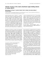
Fig. 1. Over-expression of HSP70 inhibited H
2
O
2
-induced Smac
release in C2C12 cells. (A) Cell lysates from C2C12 clones
over-expressing HSP70 or vector control plasmid (pcDNA3.1) were
immunoblotted with monoclonal anti-HSP70 serum. Immunoblot
analysis of b-actin was used as the loading control. A representative
experiment is shown. Hybridization signals were quantified and nor-
malized to GAPDH signals and are presented as the fold increase
over the respective controls. HS, Heat stress. (B) Vector control
(pcDNA3.1) and HSP70-over-expressing (HSP70-1, HSP70-2) C2C12
cells were either kept untreated or treated with 0.5 m
M of H
2
O
2
for
2 h, then harvested, lysed under conditions that kept mitochondria
intact, and centrifuged to obtain a supernatant (Cyto) and a pellet
fraction (Mit) as described in the Experimental procedures. The
presence of Smac in the different fractions was determined by
immunoblot analysis. Mitochondrial protein cytochrome oxidase
subunit II was used as a marker of mitochondrial protein and
Ponceau S staining was used as the loading control. Hybridization
signals were quantified and normalized to GAPDH signals and are
presented as the fold increase over the respective controls. *Signifi-
cant difference (P < 0.05) compared to the pcDNA3.1 control group.
B. Jiang et al. ATP-binding domain of HSP70 inhibits Smac release
FEBS Journal 276 (2009) 2615–2624 ª 2009 The Authors Journal compilation ª 2009 FEBS 2617
apoptosis in C2C12 myogenic cells [12], although the
correlation between the two events remains unknown.
In the present follow-up study, we engineered two
C2C12 cell lines with constitutive HSP70 expression at
a level similar to that of the endogenous proteins
induced by heat shock. This system mimics the anti-
apoptotic effects of heat shock and is very instrumen-
tal with respect to our investigation of the role of
HSP70. The results demonstrate that H
2
O
2
treatment
induced C2C12 cell apoptosis; however, HSP70 over-
expression significantly prevented such stress-induced
apoptosis. Because these effects were similar to those
of our previous observations for the same cells under-
going heat-shock, HSP70 is most likely to be the key
0
Caspase-3
pcDNA3.1
PI
pcDNA3.1 + H
2
O
2
10
1
10
2
10
3
10
4
10
1
10
2
10
3
10
4
10
1
10
0
45
Annexin V-FITC
40
35
30
25
20
15
10
5
0
*
#
#
pcDNA3.1
HSP70-1
HSP70-2
pcDNA3.1 + H
2
O
2
HSP70-1 + H
2
O
2
HSP70-2 + H
2
O
2
% Apoptotic cells
10
2
10
3
10
4
10
1
10
0
10
2
10
3
10
4
10
1
10
0
10
2
10
3
10
4
Q1
Q2
Q4
Q3
Q1
Q2
Q4
Q3
Q1
Q2
Q4
Q3
Q1
Q2
Q4
Q3
Q1
Q2
Q4
Q3
Q1
Q2
Q4
Q3
HSP70-1
HSP70-1 + H
2
O
2
HSP70-2
HSP70-2 + H
2
O
2
Caspase-9
pcDNA3.1
MpcDNA3.1 1 2 pcDNA3.1
HSP70 HSP70
0.5m
M H
2
O
2
1 2
pcDNA3.1 + H
2
O
2
HSP70-1 + H
2
O
2
HSP70-2 + H
2
O
2
500 bp
300 bp
100 bp
HSP70-1
HSP70-2
Caspases activity (folds)
0.5
1
1.5
2
2.5
3
3.5
A
B
C
*
*
#
#
#
#
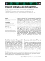
Fig. 2. Over-expression of HSP70 inhibited H
2
O
2
-induced apoptosis in C2C12 cells. (A) Cells over-expressing HSP70 and its deletion
mutants were treated with or without 0.5 m
M of H
2
O
2
for 8 h. Cells were harvested and cell lysates were assayed for protease activity of
caspases-9 or -3 using caspase fluorescent assay kits, and apoptotic cells were identified by elevated activation of caspases-9 and -3. The
experiment was repeated three times, with similar results being obtained in each case. Data are the mean ± SEM of triplicate samples. (B)
Cells were exposed to 0.5 m
M H
2
O
2
for 24 h. Cells were then processed for annexin V-FITC and pyridine iodination (PI) co-staining and ana-
lyzed by flow cytometry. Q3 cells were regarded as control cells, whereas Q4 cells were considered as a measure of early apoptosis, Q2
cells were considered as cells at late apoptosis and Q1 cells were considered as being under necrosis. Next, quantitation of apoptotic cells
was determined. Results are representative of three independent experiments. Data are the mean ± SEM of triplicate samples. *Significant
difference (P < 0.05) compared to the pcDNA3.1 control group; #Significant difference (P < 0.05) compared to the group (*) that was signifi-
cantly different from the pcDNA3.1 control group. (C) Cytosolic DNA was extracted from control and H
2
O
2
-exposed (24 h) C2C12 cells. DNA
samples (4 lg) were electrophoresed on agarose gels to visualize DNA laddering. M, DNA marker.
ATP-binding domain of HSP70 inhibits Smac release B. Jiang et al.
2618 FEBS Journal 276 (2009) 2615–2624 ª 2009 The Authors Journal compilation ª 2009 FEBS
player mediating the anti-apoptotic effects, which is
consistent with the general functional role of the chap-
erone protein. Our previous studies demonstrated that
H
2
O
2
at 0.5 mmolÆL
)1
induced apoptosis significantly,
but only affected a minimal number of cells (approxi-
mately 10%). In the present study, we demonstrated
that the levels of HSP70 protein expression in C2C12
myogenic cells stably transfected with the gene for
HSP70 were as high as those in cells pretreated with
heat shock, and that the ectopic expression of wild-
type HSP70 inhibited not only H
2
O
2
-mediated Smac
release, but also H
2
O
2
-induced apoptosis in transfected
C2C12 cells. Furthermore, there was no direct interac-
tion between HSP70 and Smac proteins, and the
ATP-binding domain of HSP70, rather than the pep-
tide-binding domain, was essential for this specific
function of the protein. Recent studies have revealed
that HSP70-mediated protection is essential for cells
aiming to combat stress and avoid cell death [14,22].
As three key modulators responsible for apoptosis,
cytochrome c, AIF and Smac are released into the
cytosol during stress, where they activate the caspase
cascade and subsequently cause cell death. HSP70 can
inhibit the release of cytochrome c and AIF from
mitochondria and prevent subsequent cell death
[21,23]. In the present study, we demonstrated that
HSP70 inhibited Smac release and the activation of
caspases-9 and -3, thereby preventing DNA fragmenta-
tion and apoptosis in cells under H
2
O
2
-induced oxida-
tive stress. This is similar to the protective effects of
another heat-shock protein, HSP27, against apoptosis,
as previously reported [24].
The molecular chaperone HSP70 has been shown
to inhibit stress-induced apoptosis by interacting
with apoptotic-associated factors. For example,
HSP70 directly interacts with JNK, resulting in the
suppression of JNK-mediated apoptosis [25]. HSP70
physically interacts with Apaf-1, blocking Apaf-1 ⁄
cytochrome c-mediated caspase activation [20]. HSP70
also binds to and antagonizes AIF, thereby inhibiting
HSP70
H
S
P7
0
+ H
2
O
2
IB: HSP70
IB: Smac
IgG Serum Lysate HSP70 Smac HSP70 Smac
IP IP IP
Fig. 3. No interaction was found between HSP70 and Smac. Vec-
tor control (C2C12-C) and HSP70-over-expressing (C2C12-HSP70)
cells were either kept untreated or treated with 0.5 m
M of H
2
O
2
for
2 h. Cells were harvested and lysed. Next, whole-cell lysates were
immunoprecipitated with polyclonal anti-HSP70 or polyclonal anti-
Smac sera. Immunoprecipitations were further analyzed by immu-
noblots probed with Smac antibody or polyclonal HSP70 antibody,
respectively.
HSP70
WT
A
B
N
ATP-BD
ATP-BD
EEVD
C
C
C
EEVD
EEVD
1 383 542 646
N
N
HSP70
ΔAPBD
HSP70
ΔATP-BD
HSP70
WT
70 kDa
IB: Hsp70
52 kDa
28 kDa
IB: Actin
HSP70
’ΔPBD
HSP70
’ΔATP-BD
PBD
PBD
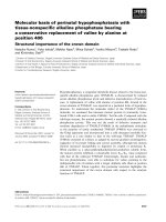
Fig. 4. Deletion mutants of HSP70 were constructed and transf-
ected. A schematic drawing is shown of the HSP70 deletion
mutants employed in the present study. (A) Deleted amino acids
are indicated by the dotted lines. ATP-BD, 1-383AA, 42 kDa; PBD,
384-542AA, 18 kDa. (B) Western blot analysis demonstrated the
levels of expression of the HSP70 proteins after deletion mutants
of HSP70 were transfected.
Cyto Mit Cyto Mit Cyto Mit Cyto Mit
pcDNA3.1 HSP70
WT
HSP70
ΔPBD
HSP70
ΔATP-BD
H
2
O
2
2 h
Smac
COX II
Loading control
Fig. 5. The ATP-binding domain of HSP70 is the essential region
for inhibition of Smac release. Cells over-expressing HSP70 or its
deletion mutants were treated with 0.5 m
M of H
2
O
2
for 2 h, har-
vested, lysed under conditions that kept mitochondria intact, and
then centrifuged to obtain a supernatant (Cyto) and a pellet fraction
(Mit) as described in the Experimental procedures. Protein protein
contents were determined by the Bradford assay (Bio-Rad,
Hercules, CA, USA), and equal amounts of proteins (10–20 lg)
were loaded in each lane and separated by SDS-PAGE. Levels of
Smac in the different fractions were determined by immunoblot
analysis. Cytochrome oxidase subunit II (COX II) was used as a
marker of mitochondrial protein and Ponceau S staining was used
to visulize equal protein loadings.
B. Jiang et al. ATP-binding domain of HSP70 inhibits Smac release
FEBS Journal 276 (2009) 2615–2624 ª 2009 The Authors Journal compilation ª 2009 FEBS 2619
caspase-independent apoptosis [23]. However, the
results obtained in the present study suggest that the
inhibitory effect of HSP70 on the release of Smac and
H
2
O
2
-mediated and Smac-promoted apoptosis is not
attributable to a direct physical interaction between
HSP70 and Smac.
HSP70 contains three functional regions: the ATP-
binding domain, the peptide-binding domain, and the
EEVD motif. Although the EEVD motif is considered
to be involved in the chaperone function of HSP70,
and was assumed to mediate cytoprotection by restor-
ing damaged or unfolded proteins under stress, the
roles of other domains of HSP70 in anti-apoptosis
remain highly controversial. Some studies have pro-
posed that the ATP-binding domain of human HSP70
is not required in HSP70-mediated JNK suppression,
inhibition of cytochrome c release and caspase activa-
tion, and protection of cells from injury [26]. By con-
trast, other studies have shown that the ATP-binding
domain of HSP70 is essential for its anti-apoptotic role.
For example, deletional analysis demonstrated that
the ATP-binding domain is essential for inhibiting
the release of cytochrome c from mitochondria [27].
3
A
*
*
#
#
#
#
2
2.5
1
1.5
0.5
Caspases activity (folds)
0
Caspase-3
70
60
50
40
*
*
#
#
#
#
pcDNA3.1
pcDNA3.1 + H
2
O
2
HSP70 + H
2
O
2
HSP70
ΔATP-BD
+ H
2
O
2
HSP70
ΔPBD
+ H
2
O
2
30
20
10
0
12 h 24 h
Time (h)
% Apoptotic cells
B
a
cd e
b
pcDNA3.1
HSP70 + H
2
O
2
HSP70
ΔATP-BD
+ H
2
O
2
HSP70
ΔPBD
+ H
2
O
2
pcDNA3.1 + H
2
O
2
Caspase-9
pcDNA3.1
pcDNA3.1 + H
2
O
2
HSP70 + H
2
O
2
HSP70
ΔATP-BD
+ H
2
O
2
C
1.2
*
#
#
1
0.8
0.6
0.4
0.2
0
pcDNA3.1
pcDNA3.1 + H
2
O
2
Hsp70 + H
2
O
2
Hsp70
ΔATP-BD
+ H
2
O
2
Hsp70
Δ
PBD
+ H
2
O
2
Cell viability
HSP70
ΔPBD
+ H
2
O
2
Fig. 6. ATP-binding domain of HSP70 is essential for the inhibition
of H
2
O
2
-induced activation of caspases-9 and -3 and apoptosis. (A)
The effects of HSP70 and its deletion mutant proteins on the acti-
vation of caspases-9 and -3 were analyzed. Cells over-expressing
HSP70 and its deletion mutants were treated with or without
0.5 m
M of H
2
O
2
for 8 h. Cells were harvested and cell lysates were
assayed for protease activity of caspases-9 or -3 using caspase
fluorescent assay kits. Data of caspase fluorescent assay were
obtained from four independent experiments. *Significant differ-
ence (P < 0.05) compared to the pcDNA3.1 control group; #Signifi-
cant difference (P < 0.05) compared to the group (*) that was
significantly different from the pcDNA3.1 control group (n = 8). (B)
Measurement of percentages of apoptotic cells. Twenty-four hours
after transfer, cells were treated with 0.5 m
M H
2
O
2
for 12 or 24 h,
and then stained with Hoechst 33258. Under a fluorescence micro-
scope, apoptotic cells, which contained condensed chromatin frag-
ments, were scored and expressed as a percentage of the total
cell number counted. Data are the mean ± SEM. *Significant differ-
ence (P < 0.05) compared to the pcDNA3.1 control group; #Signifi-
cant difference (P < 0.05) compared to the group (*) that was
significantly different from the pcDNA3.1 control group (n = 5).
(a–f) Cells incubated with H
2
O
2
for 24 h. (C) Determination of cell
viability. Approximately 2000 cells were plated in each well of
96-well plates. After 24 h of incubation, 0.5 m
M of H
2
O
2
was added
and cell viability was measured by an 3-(4,5-dimethylthiazol-2-yl)-
2,5-diphenyl-tetrazolium bromide assay after exposure to H
2
O
2
for
24 h. The experiment was repeated three times, with essentially
the same results being obtained in each case. Data are the
mean ± SEM of triplicate samples. *Significant difference
(P < 0.05) compared to the pcDNA3.1 control group; #Significant
difference (P < 0.05) compared to the group (*) that was signifi-
cantly different from the pcDNA3.1 control group (n = 5).
ATP-binding domain of HSP70 inhibits Smac release B. Jiang et al.
2620 FEBS Journal 276 (2009) 2615–2624 ª 2009 The Authors Journal compilation ª 2009 FEBS
The ATP-binding domain of HSP70 is important
for the interaction of HSP70 with apoptosis signal-
regulating kinase 1 (ASK1) and the inhibition of
ASK1-induced apoptosis in vitro [28]. Furthermore, the
ATP-binding domain of HSP70 is critical for sequester-
ing AIF in the cytosol [29]. In the present study, we
demonstrated that the ATP-binding domain of HSP70
was indispensable for inhibition of Smac release from
mitochondria as well as apoptotic events in C2C12
myogenic cells.
The molecular mechanism by which HSP70 and
HSP70
DPBD
interfere with Smac release and apoptosis
induced by oxidative-stress is still not fully understood.
The mitochondrial pathway of cell death is controlled
by Bcl-2 family proteins, a group of anti-apoptotic and
pro-apoptotic proteins that regulate the passage of
small molecules such as cytochrome c, Smac ⁄ DIABLO
and apoptosis-inducing factor (which activate caspase
cascades) through the mitochondrial transition pore
[30]. Bcl-2 is the prototype of the bcl-2 family of
proteins and is distributed in the mitochondria,
endoplasmic reticulum and nuclear envelope. With a
well-established role with respect to protecting cells
against a variety of apoptotic stimuli, it mainly acts at
the mitochondrial level [31]. A previous study [32]
demonstrated that HSP70 inhibits heat-induced apop-
tosis by preventing Bax translocation. Furthermore,
over-expression of HSP70 was associated with reduced
apoptotic cell death and an increased expression of the
anti-apoptotic protein, Bcl-2 [33]. On the basis of the
available evidence, HSP70 and HSP70
DPBD
may also
suppress Smac release and apoptosis by regulating the
expression of these pro-apoptotic or anti-apoptotic
bcl-2 family proteins.
In summary, using the H
2
O
2
-induced oxidative stress
model, the present study has revealed an important
anti-apoptotic role of HSP70, which comprises a
mechanism that involves the inhibition of Smac release
from mitochondria, and the suppression of caspase
activation. Such a mechanism is independent of the
interaction of HSP70 with Smac but requires the
ATP-binding domain of the protein. However, it
remains to be determined how these findings are
connected with the known functions of many other
cellular molecules.
Experimental procedures
Cell culture and treatment
C2C12 myogenic cells were cultured in DMEM supple-
mented with 10% heat-inactivated fetal bovine serum at
37 °C in the presence of 5% CO
2
under a humidified atmo-
sphere. H
2
O
2
diluted in NaCl ⁄ P
i
(137 mm NaCl, 2.68 mm
KCl, 10 mm Na
2
HPO
4
, 1.76 mm KH
2
PO
4
, pH = 7.4) was
used in the medium at a final concentration of 0.5 mm.
Heat shock treatment
Subconfluent cultured cells in 50-mm dishes were subjected
to hyperthermia of 42 ± 0.3 °C for 1 h in a water bath
before being allowed to recover for 12 h at 37 °Cina
humidified atmosphere containing 5% CO
2
. As a control,
cells were cultured under normal conditions without hyper-
thermia.
Construction of HSP70 and its truncated mutants
Full-length human HSP70 cDNA was obtained as a gener-
ous gift from I. Benjemin (University of Utah Health
Sciences Center, Salt Lake City, UT, USA) It was direc-
tionally cloned between KpnI and BamHI sites into the
mammalian expression vector pcDNA3.1(-)-His-myc. At
the same time, this cDNA was used as the template for
PCR amplification of two HSP70 truncated mutants with
deletion of the ATP-binding domain (HSP70
DATP-BD
)or
the peptide-binding domain (HSP70
DPBD
) using primer
pairs (Table 1). All DNA digested fragments were purified
using a gel purification kit (Invitrogen, Carlsbad, CA,
USA), and subsequently ligated into pcDNA3.1(-)-His-myc
vector overnight at 4 °C with T4 DNA polymerase (Pro-
mega, Madison, WI, USA). The correct insets were verified
by sequencing and digestion. The final constructs were
named pcDNA3.1-HSP70
WT
, pcDNA3.1-HSP70
DATP-BD
or
pcDNA3.1-HSP70
DPBD
(Fig. 4A).
Table 1. Sequences of primers used to construct pcDNA3.1-HSP70WT, pcDNA3.1-HSP70
DATP-BD
or pcDNA3.1-HSP70
DPBD
plasmids.
Primers Sequence (5¢-to3¢)
Sense of pcDNA3.1-HSP70
WT
AAAAGGATCCAAATGGCCAAAGCCGCGGCG
Antisense of pcDNA3.1-HSP70
WT
TCGGGTACCGGATCTACCTCCTCAATGGTG
Sense of pcDNA3.1-HSP70
DPBD
CTGATGGGGGACTCCTACGCCTTCAACATGAAGAGC
Antisense of pcDNA3.1-HSP70
DPBD
GAAGGCGTAGGAGTCCCCCATCAGGATGGCCGCCTG
Sense of pcDNA3.1-HSP70
DATP-BD
AAAAGGATCCAAAGTCCGAGAACTGGCAGGAC
Antisense of pcDNA3.1-HSP70
DATP-BD
TCGGGTACCGGATCTACCTCCTCAATGGTG
B. Jiang et al. ATP-binding domain of HSP70 inhibits Smac release
FEBS Journal 276 (2009) 2615–2624 ª 2009 The Authors Journal compilation ª 2009 FEBS 2621
Lipofectamine-mediated gene transfection
C2C12 myogenic cells were cultured to sub-confluence and
transfected with each of the expression plasmids manufac-
tured as described in the above steps, or the empty vector
without the cDNA (control) with a Lipofectamine-mediated
method (Lipofectamine 2000, Invitrogen), as described
previously [13].
Preparation of mitochondrial and cytosolic
fractions
The subcellular fractions of C2C12 myogenic cells treated
with or without H
2
O
2
were isolated as described previously
[13].
Western blot analysis
Western blotting with anti-HSP70 and anti-Smac sera was
performed as described previously [34].
Caspase activity assay
Caspase activation was determined according to the method
described previously [13].
Flow cytometric analysis
Both adherent and floating cells were collected after treat-
ment, washed with ice-cold NaCl ⁄ P
i
, and stained with
fluorescein isothiocyanate (FITC)-conjugated annexin V
(BD Biosciences, Franklin Lakes, NJ, USA) and pyridine
iodination (PI) for 20 min at room temperature in the dark.
The stained cells were then analyzed by a flow cytometer
(Beckman Coulter, Fullerton, CA, USA). FITC-conjugated
annexin V binds to phosphatidylserine molecules present
only at the surface of apoptotic cells but not non-apoptotic
cells due to the loss of plasma membrane asymmetry early
in apoptosis. Cells were simultaneously stained with PI to
discriminate membrane-permeable necrotic cells from FITC-
labeled apoptotic cells. Apoptotic cells were identified as
those with positive staining only to annexin V-FITC and
not to PI, and the results were expressed as the proportion
of these cells among the total number of cells analyzed.
Hoechst 33258 staining
Hoechst 33258 staining was performed as described previ-
ously [12,13].
Detection of DNA fragmentation
Floating and adherent cells (5 · 10
7
) were combined and
pelleted by centrifugation at 400 g for 5 min, and washed
twice with NaCl ⁄ P
i
. Cell pellets were resuspended in 200 lL
of lysis buffer [10 mm Tris–HCl (pH 8.0), 10 mm EDTA,
0.5% Triton X-100 and 0.1 mgÆmL
)1
RNase A] and incu-
bated at 37 °C for 1 h. Cell lysates were then treated with
protease K (0.2 mgÆmL
)1
)at54°C for 30 min. The genomic
DNA was isolated by two with two rounds of phenol–chlo-
roform extraction followed by an additional chloroform
extraction. DNA pellet was then washed in 70% ethanol
and resuspended in 1 mm EDTA and 10 mm Tris–HCl
(pH 8.0) at a final concentration of 20 lgÆmL
)1
. Aliquots
were electrophoresed on a 1.5% agarose gel containing ethi-
dium bromide, and photographed under UV illumination. A
GeneRuler 100 bp DNA ladder (MBI Fermentas, Hanover,
MD, USA) was utilized as DNA size marker.
Co-immunoprecipitation assay
For co-immunoprecipitation, transiently transfected C2C12
cells were lyzed with pre-cold RIPA buffer (150 mmolÆL
)1
NaCl, 1% NP40, 0.5% deoxycholic acid sodium salt, 0.1%
SDS, 50 mmolÆL
)1
Tris pH 8.0, 1 mm phenylmethanesulfo-
nyl fluoride and complete protease inhibitor tablet) at 4 °C
for 5 min. To reduce nonspecific combination, lysates con-
taining 500 lg of total protein were pre-immunized with
25 lL of a slurry of protein A ⁄ G coupled to agarose beads
(Invitrogen) overnight at 4 °C on a rotating wheel. Aliquots
of the pre-cleared supernatants were then each incubated
with 2 lg of appropriate mouse monoclonal anti-HSP70
serum, polyclonal rabbit anti-Smac serum (R&D Systems,
Minneapolis, MN, USA), normal mouse immunoglobu-
lin G (control for anti-HSP70) or normal rabbit serum
(control for anti-Smac) added into 25 lL of protein A ⁄ G
slurry coupled to agarose beads (Invitrogen) for 5 h at 4 °C
on a rotating wheel. Protein A ⁄ G beads were collected by
centrifugation at 4 °C followed by a total of four additional
washes lysis buffer containing 200 mm NaCl. Immune com-
plexes were eluted by twice by sample buffer (2% SDS, 2 m
2-mercaptoethanol) after boiling at 100 ° C for 10 min.
Proteins were separated by electrophoresis on SDS-PAGE
followed by immunoblotting with polyclonal anti-HSP70
and anti-Smac sera, as described previously [24]. As the
controls of total antigens in the lysates before co-immuno-
precipitation, portions of lysates (1 : 20) were also resolved
on SDS-PAGE and immunoblotted with anti-HSP70 or
anti-Smac sera.
Cell viability assay
To determine cell viability, 3-(4,5-dimethylthiazol-2-yl)-2,5-
diphenyl-tetrazolium bromide (0.5 mg) was added to
1 mL of cell suspension (1 · 10
6
cellsÆmL
)1
in 24-well
plates). After 4 h of incubation, cells were washed three
times with NaCl ⁄ P
i
(pH 7.4). The insoluble formazan
product was dissolved in dimethylsulfoxide and D
490
of
each culture well was then measured using a microplate
reader (Titertek Multiskan Plus, Flow Laboratories,
ATP-binding domain of HSP70 inhibits Smac release B. Jiang et al.
2622 FEBS Journal 276 (2009) 2615–2624 ª 2009 The Authors Journal compilation ª 2009 FEBS
McClean, VA, USA). The attenuance of formazan
formed in control cells was considered as 100% viability.
Statistical analysis
Data are expressed as the mean ± SEM of the indicated
number of separate experiments. Differences between two
groups were analyzed using an unpaired Student’s t-test.
Differences among three or more groups were analyzed by
one-way analysis of variance followed by the Student–New-
man–Keuls post-hoc test. P < 0.05 was considered statisti-
cally significant.
Acknowledgements
This study was supported by the grants from the
National Basic Research Program of China
(2007CB512007), the National Natural Science Foun-
dation of China (30700290) and Special Funds for
PhD Training from the Ministry of Education of
China (20060533009).
References
1 Steller H (1995) Mechanisms and genes of cellular
suicide. Science 267, 1445–1449.
2 Shishido T, Woo CH, Ding B, McClain C, Molina CA,
Yan C, Yang J & Abe J (2008) Effects of
MEK5 ⁄ ERK5 association on small ubiquitin-related
modification of ERK5: implications for diabetic ventric-
ular dysfunction after myocardial infarction. Circ Res
102, 1416–1425.
3 Burke RE (2008) Programmed cell death and new dis-
coveries in the genetics of parkinsonism. J Neurochem
104, 875–890.
4 Engel T, Kannenberg F, Fobker M, Nofer JR, Bode
G, Lueken A, Assmann G & Seedorf U (2007)
Expression of ATP binding cassette-transporter
ABCG1 prevents cell death by transporting
cytotoxic 7beta-hydroxycholesterol. FEBS Lett 581,
1673–1680.
5 Yaglom JA, Ekhterae D, Gabai VL & Sherman MY
(2003) Regulation of necrosis of H9c2 myogenic cells
upon transient energy deprivation. Rapid deenergization
of mitochondria precedes necrosis and is controlled by
reactive oxygen species, stress kinase JNK, HSP72 and
ARC. J Biol Chem 278, 50483–50496.
6 Dougherty CJ, Kubasiak LA, Frazier DP, Li H, Xiong
WC, Bishopric NH & Webster KA (2004) Mitochon-
drial signals initiate the activation of c-Jun N-terminal
kinase (JNK) by hypoxia-reoxygenation. FASEB J 18,
1060–1070.
7 Thornberry NA & Lazebnik Y (1998) Caspases:
enemies within. Science 281, 1312–1316.
8 Sun XM, MacFarlane M, Zhuang J, Wolf BB, Green
DR & Cohen GM (1999) Distinct caspase cascades are
initiated in receptor-mediated and chemical-induced
apoptosis. J Biol Chem 274, 5053–5060.
9 Li P, Nijhawan D, Budihardjo I, Srinivasula SM,
Ahmad M, Alnemri ES & Wang X (1997) Cytochrome
c and dATP-dependent formation of Apaf-1 ⁄ caspase-9
complex initiates an apoptotic protease cascade. Cell
91, 479–489.
10 Du C, Fang M, Li Y, Li L & Wang X (2000) Smac, a
mitochondrial protein that promotes cytochrome
c-dependent caspase activation by eliminating IAP
inhibition. Cell 102, 33–42.
11 Verhagen AM, Ekert PG, Pakusch M, Silke J, Connolly
LM, Reid GE, Moritz RL, Simpson RJ & Vaux DL
(2000) Identification of DIABLO, a mammalian protein
that promotes apoptosis by binding to and antagonizing
IAP proteins. Cell 102, 43–53.
12 Jiang B, Xiao W, Shi Y, Liu M & Xiao X (2005) Role
of Smac ⁄ DIABLO in hydrogen peroxide-induced apop-
tosis in C2C12 myogenic cells. Free Radic Biol Med 39,
658–667.
13 Jiang B, Xiao W, Shi Y, Liu M & Xiao X (2005) Heat
shock pretreatment inhibited the release of Smac ⁄ DIA-
BLO from mitochondria and apoptosis induced by
hydrogen peroxide in cardiomyocytes and C2C12
myogenic cells. Cell Stress Chaperones 10, 252–262.
14 Lui JC & Kong SK (2007) Heat shock protein 70 inhib-
its the nuclear import of apoptosis-inducing factor to
avoid DNA fragmentation in TF-1 cells during erythro-
poiesis. FEBS Lett 581, 109–117.
15 Zhao Y, Wang W & Qian L (2007) Hsp70 may protect
cardiomyocytes from stress-induced injury by inhibiting
Fas-mediated apoptosis. Cell Stress Chaperones 12, 83–
95.
16 Jayakumar J, Suzuki K, Khan M, Smolenski RT, Farrell
A, Latif N, Raisky O, Abunasra H, Sammut IA, Mur-
tuza B et al. (2000) Gene therapy for myocardial protec-
tion: transfection of donor hearts with heat shock protein
70 gene protects cardiac function against ischemia-reper-
fusion injury. Circulation 102, III302–III306.
17 Li CY, Lee JS, Ko YG, Kim JI & Seo JS (2000) Heat
shock protein 70 inhibits apoptosis downstream of cyto-
chrome c release and upstream of caspase-3 activation.
J Biol Chem 275, 25665–25671.
18 Lee JS, Lee JJ & Seo JS (2005) HSP70 deficiency results
in activation of c-Jun N-terminal kinase, extracellular
signal-regulated kinase, and caspase-3 in
hyperosmolarity-induced apoptosis. J Biol Chem 280,
6634–6641.
19 Creagh EM, Carmody RJ & Cotter TG (2000) Heat
shock protein 70 inhibits caspase-dependent and -inde-
pendent apoptosis in Jurkat T cells. Exp Cell Res 257,
58–66.
B. Jiang et al. ATP-binding domain of HSP70 inhibits Smac release
FEBS Journal 276 (2009) 2615–2624 ª 2009 The Authors Journal compilation ª 2009 FEBS 2623
20 Saleh A, Srinivasula SM, Balkir L, Robbins PD &
Alnemri ES (2000) Negative regulation of the Apaf-1
apoptosome by Hsp70. Nat Cell Biol 2, 476–483.
21 Ravagnan L, Gurbuxani S, Susin SA, Maisse C, Daugas
E, Zamzami N, Mak T, Ja
¨
a
¨
ttela
¨
M, Penninger JM, Garri-
do C et al. (2001) Heat-shock protein 70 antagonizes
apoptosis-inducing factor. Nat Cell Biol 3, 839–843.
22 Feng X, Bonni S & Riabowol K (2006) HSP70 induc-
tion by ING proteins sensitizes cells to tumor necrosis
factor alpha receptor-mediated apoptosis. Mol Cell Biol
26, 9244–9255.
23 Didelot C, Schmitt E, Brunet M, Maingret L, Parcellier
A & Garrido C (2006) Heat shock proteins: endogenous
modulators of apoptotic cell death. Handb. Exp. Phar-
macol. 26, 171–198.
24 Chauhan D, Li G, Hideshima T, Podar K, Mitsiades C,
Mitsiades N, Catley L, Tai YT, Hayashi T, Shringarpure
R et al. (2003) Hsp27 inhibits release of mitochondrial
protein Smac in multiple myeloma cells and confers dexa-
methasone resistance. Blood 102, 3379–3386.
25 Park HS, Lee JS, Huh SH, Seo JS & Choi EJ (2001)
Hsp72 functions as a natural inhibitory protein of c-Jun
N-terminal kinase. EMBO J 20, 446–456.
26 Mayer MP & Bukau B (2005) Hsp70 chaperones: cellu-
lar functions and molecular mechanism. Cell Mol Life
Sci 62, 670–684.
27 Mosser DD, Caron AW, Bourget L, Meriin AB, Sher-
man MY, Morimoto RI & Massie B (2000) The chaper-
one function of hsp70 is required for protection against
stress-induced apoptosis. Mol Cell Biol 20, 7146–7159.
28 Park HS, Cho SG, Kim CK, Hwang HS, Noh KT,
Kim MS, Huh SH, Kim MJ, Ryoo K, Kim EK et al.
(2002) Heat shock protein hsp72 is a negative regulator
of apoptosis signal-regulating kinase 1. Mol Cell Biol
22, 7721–7730.
29 Ruchalski K, Mao H, Li Z, Wang Z, Gillers S, Wang
Y, Mosser DD, Gabai V, Schwartz JH & Borkan SC
(2006) Distinct hsp70 domains mediate apoptosis-induc-
ing factor release and nuclear accumulation. J Biol
Chem 281, 7873–7880.
30 Kim R (2005) Recent advances in understanding the cell
death pathways activated by anticancer therapy. Cancer
103, 1551–1560.
31 Scorrano L & Korsmeyer SJ (2003) Mechanisms of
cytochrome c release by proapoptotic BCL-2 family
members. Biochem Biophys Res Commun 304, 437–
444.
32 Stankiewicz AR, Lachapelle G, Foo CP, Radicioni SM
& Mosser DD (2005) Hsp70 inhibits heat-induced apop-
tosis upstream of mitochondria by preventing Bax
translocation. J Biol Chem 280, 38729–38739.
33 Yenari MA, Liu J, Zheng Z, Vexler ZS, Lee JE & Giff-
ard RG (2005) Antiapoptotic and anti-inflammatory
mechanisms of heat-shock protein protection. Ann N Y
Acad Sci 1053, 74–83.
34 Liang P, Jiang B, Yang X, Xiao X, Huang X, Long J,
Zhang P, Zhang M, Xiao M, Xie T et al. (2008)
The role of peroxisome proliferator-activated receptor-
beta ⁄ delta in epidermal growth factor-induced HaCaT
cell proliferation. Exp Cell Res 314, 3142–3151.
ATP-binding domain of HSP70 inhibits Smac release B. Jiang et al.
2624 FEBS Journal 276 (2009) 2615–2624 ª 2009 The Authors Journal compilation ª 2009 FEBS