Báo cáo khoa học: High levels of structural disorder in scaffold proteins as exemplified by a novel neuronal protein, CASK-interactive protein1 pot
Bạn đang xem bản rút gọn của tài liệu. Xem và tải ngay bản đầy đủ của tài liệu tại đây (1.65 MB, 13 trang )
High levels of structural disorder in scaffold proteins as
exemplified by a novel neuronal protein, CASK-interactive
protein1
Annama
´
ria Bala
´
zs
1,
*, Veronika Csizmok
2,
*, La
´
szlo
´
Buday
1,2
, Marianna Raka
´
cs
2
, Robert Kiss
3
,
Mo
´
nika Bokor
4
, Roopesh Udupa
2
,Ka
´
lma
´
n Tompa
4
and Peter Tompa
2
1 Department of Medical Chemistry, Semmelweis University Medical School, Budapest, Hungary
2 Biological Research Center, Institute of Enzymology, Hungarian Academy of Sciences, Budapest, Hungary
3 Laboratory of Structural Chemistry and Biology, Institute of Chemistry, Eo
¨
tvo
¨
s Lora
´
nd University, Budapest, Hungary
4 Research Institute for Solid State Physics and Optics, Hungarian Academy of Sciences, Budapest, Hungary
Keywords
anchor; docking; post-synaptic density;
scaffold; unstructured
Correspondence
P. Tompa, Institute of Enzymology,
Biological Research Center, Hungarian
Academy of Sciences, Karolina ut 29, 1113
Budapest, Hungary
Fax: +36 1 466 5465
Tel: +36 1 279 3143
E-mail:
*These authors contributed equally to this
work
(Received 26 February 2009, revised 15
April 2009, accepted 12 May 2009)
doi:10.1111/j.1742-4658.2009.07090.x
CASK-interactive protein1 is a newly recognized post-synaptic density
protein in mammalian neurons. Although its N-terminal region contains
several well-known functional domains, its entire C-terminal proline-rich
region of 800 amino acids lacks detectable sequence homology to any
previously characterized protein. We used multiple techniques for the struc-
tural characterization of this region and its three fragments. By bioinfor-
matics predictions, CD spectroscopy, wide-line and
1
H-NMR spectroscopy,
limited proteolysis and gel filtration chromatography, we provided evidence
that the entire proline-rich region of CASK-interactive protein1 is intrinsi-
cally disordered. We also showed that the proline-rich region is biochemi-
cally functional, as it interacts with the adaptor protein Abl-interactor-2.
To extend the finding of a high level of disorder in this scaffold protein, we
collected 74 scaffold proteins (also including proteins denoted as anchor
and docking), and predicted their disorder by three different algorithms.
We found that a very high fraction (53.6% on average) of the residues fall
into local disorder and their ordered domains are connected by linker
regions which are mostly disordered (64.5% on average). Because of this
high frequency of disorder, the usual design of scaffold proteins of short
globular domains (86 amino acids on average) connected by longer linker
regions (140 amino acids on average) and the noted binding functions of
these regions in both CASK-interactive protein1 and the other proteins
studied, we suggest that structurally disordered regions prevail and play
key recognition roles in scaffold proteins.
Structured digital abstract
l
MINT-7034649: Caskin1 (uniprotkb:Q8VHK2) physically interacts (MI:0915) with L1CAM
(uniprotkb:
P32004)bytwo hybrid (MI:0018)
l
MINT-7034677: Caskin1 (uniprotkb:Q8VHK2) physically interacts (MI:0915) with NCK1
(uniprotkb:
P16333)bytwo hybrid (MI:0018)
l
MINT-7034706: Caskin1 (uniprotkb:Q8VHK2) physically interacts (MI:0915) with Stathmin-3
(uniprotkb:
Q9NZ72)bytwo hybrid (MI:0018)
Abbreviations
Abi2, Abl-interactor-2; Caskin1, CASK-interactive protein1; CBP, CREB-binding protein; GFP, green fluorescent protein; GST, glutathione
transferase; IDP, intrinsically disordered protein; IUP, intrinsically unstructured protein; MAPK, mitogen-activated protein kinase; PRD,
proline-rich region; PSD, post-synaptic density; SAM, sterile a motif; Ste5, Sterile 5.
3744 FEBS Journal 276 (2009) 3744–3756 ª 2009 The Authors Journal compilation ª 2009 FEBS
Introduction
Recently, novel scaffold proteins have been discovered
in the brain, particularly in neuronal cells, and are
referred to as the CASK-interactive protein (Caskin)
family [1]. Caskin1 and its isoform Caskin2, which are
present in the post-synaptic density (PSD), are multi-
domain proteins possessing six ankyrin repeats, two
sterile a motifs (SAM domains) and a single SH3
domain in the N-terminal part (cf. Fig. 1). In contrast,
there are no recognizable domains in the C-terminal
part, which is dominated by a long proline-rich region
[1], designated as the proline-rich domain (PRD) in
this work. Caskin1 can bind the Cask adaptor protein
[1], Abl-interactor-2 (Abi2), and another nine proteins
shown in this work, and is presumably involved
in the signal pathway related to the Abl tyrosine
kinases (A. Balazs, V. Csizmok, P. Tompa, R. Udupa
& L. Buday, unpublished results). The molecular
mechanism of the function of Caskins is not known at
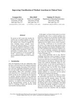
Fig. 1. The diagram at the bottom shows a schematic representation of the domain structure of Caskin1. The N-terminal half contains six
ankyrin repeats, one SH3 domain and the two SAM domains, whereas the C-terminal half contains no recognizable domain, and has been
designated as a proline-rich region ⁄ domain (PRD). The proline-rich region was cut into three parts (PRD1, PRD2 and PRD3), cloned and char-
acterized individually in this work. Above the scheme is the prediction by the IUPred algorithm, which shows that the entire PRD region is
probably intrinsically disordered (the score is above 0.5).
l
MINT-7034579: Caskin1 (uniprotkb:Q8VHK2) physically interacts (MI:0915) with ABI2 (uni-
protkb:
Q9NYB9)bytwo hybrid (MI:0018)
l
MINT-7034720: Caskin1 (uniprotkb:Q8VHK2) physically interacts (MI:0915) with Synapto-
tagmin (uniprotkb:
P21579)bytwo hybrid (MI:0018)
l
MINT-7034691: Caskin1 (uniprotkb:Q8VHK2) physically interacts (MI:0915) with Neurexin-2
(uniprotkb:
Q9P2S2)bytwo hybrid (MI:0018)
l
MINT-7034617: Caskin1 (uniprotkb:Q8VHK2) physically interacts (MI:0915) with CASK
(uniprotkb:
P07498)bytwo hybrid (MI:0018)
l
MINT-7034748: Caskin1 (uniprotkb:Q8VHK2) physically interacts (MI:0915) with SIAH1
(uniprotkb:
Q8IUQ4)bytwo hybrid (MI:0018)
l
MINT-7034663: Caskin1 (uniprotkb:Q8VHK2) physically interacts (MI:0915) with Myosin-Ib
(uniprotkb:
O43795)bytwo hybrid (MI:0018)
l
MINT-7034734: Caskin1 (uniprotkb:Q8VHK2) physically interacts (MI:0915) with Septin-4
(uniprotkb:
O43236)bytwo hybrid (MI:0018)
l
MINT-7034634: Caskin1 (uniprotkb:Q8VHK2) physically interacts (MI:0915) with EPHA2
(uniprotkb:
P29317)bytwo hybrid (MI:0018)
l
MINT-7034765, MINT-7034783: Caskin1 (uniprotkb:Q8VHK2) physically interacts
(
MI:0915) with ABI2 (uniprotkb:Q9NYB9)bypull down (MI:0096)
V. Csizmok et al. High levels of disorder in scaffold proteins
FEBS Journal 276 (2009) 3744–3756 ª 2009 The Authors Journal compilation ª 2009 FEBS 3745
present. As a result of their large size, the capacity to
bind multiple partners and the lack of catalytic
domains, they probably fall into the class of scaffold
proteins, which bind components of a signal trans-
duction pathway simultaneously and ensure the speci-
ficity and efficiency of signal propagation [1]. As
the long regions of these proteins often lack any
sequence similarity to other proteins and appear to
lack folded structural domains, we anticipated that
structural disorder may be a general feature of scaffold
proteins. In a recent review, structural disorder in
several scaffold proteins and in other proteins of
multiple binding partners (without adherence to the
accepted definition of scaffolds) has been suggested
and analysed [2].
As a result of the rapid advance of knowledge
on intrinsically disordered ⁄ unstructured proteins
(IDPs ⁄ IUPs), the concept of protein disorder has
gained general recognition recently [3–6]. Physical
evidence exists for the disorder of about 500 proteins
[7], and bioinformatics predictions suggest that disor-
der is prevalent in the proteome of eukaryotes, with
more than 10% of their proteins being fully disordered
[8–10]. Disorder is most often implicated in signalling
and regulatory functions, and its functional benefits
often manifest themselves in protein–protein recogni-
tion [5,11]. One advantage often referred to is that
their extended structure enables IDPs to have a large
interaction capacity with small protein size [12], which
might be directly related to the involvement of disor-
der in scaffold proteins. In fact, there is an elevated
level of disorder in hub proteins, i.e. proteins involved
in multiple interactions [13–16], and disorder increases
with the number of proteins in multiprotein complexes
[17]. The functional role of structural disorder has
been noted in a few scaffold proteins, such as Sterile 5
(Ste5) [18], BRCA1 [19], CREB-binding protein (CBP)
[4] and Mypt1 [20], and it has been suggested that
flexibility provided by disorder is instrumental in
overcoming steric hindrance in the assembly of large
multiprotein complexes [21].
Motivated by the apparent relationship between pro-
tein disorder and scaffold function, in this article, we
provide evidence that the proline-rich region of the
newly recognized Caskin1 is intrinsically disordered.
To extend this finding, we also collected 74 scaffold
proteins (also including proteins denoted as anchor
and docking) and examined them by three different
disorder predictor algorithms, i.e. IUPred [22,23],
VSL2 [24,25], and FoldIndex [26]. We found that, in
these proteins, the frequency of disorder is very high
(49.7%, 63.36% and 47.82% predicted by IUPred,
VSL2 and FoldIndex, respectively), which is similar to
that in the most disordered functional class, RNA
chaperones [27]. The implications of these findings
with respect to the function of Caskin1, and of scaf-
fold proteins in general, are discussed.
Results
Structural characterization of Caskin1 fragments
As described in the introductory paragraphs, the
N-terminal half of Caskin1 contains a number of
well-known domains involved in protein–protein inter-
action, such as the ankyrin repeats, SH3 and SAM
domains (Fig. 1). The three-dimensional structures of
these domains have been well characterized [28–30].
However, the C-terminal part of Caskin1 does not
contain any domain, but possesses several proline-rich
stretches. Because proline is incompatible with repeti-
tive secondary structural elements [31] and is known
to be enriched in IDPs [5], we assumed that the
C-terminus of Caskin1 might be intrinsically disor-
dered. This expectation was first confirmed by bioin-
formatics predictions by the IUPred algorithm
(Fig. 1). High IUPred scores indicate that the entire
proline-rich region of Caskin1 (amino acids 603–1430)
is disordered.
To confirm this prediction, a variety of experimental
approaches were also applied, as earlier it has been
suggested [5] that, as a result of the limitations of most
techniques, a multitude of approaches need to be
applied for the conclusive demonstration of disorder.
The full-length proline-rich region of Caskin1 with a
histidine tag on its C-terminus (PRD-His) was cloned
and expressed in bacteria. However, the expression of
this construct was rather difficult because of the high
proteolytic sensitivity of the protein, characteristic of
IDPs. Therefore, only CD, gel filtration and limited
proteolysis experiments could be performed, which do
not require large amounts of protein. For detailed
studies, the full-length proline-rich region was cut into
three parts, selected for splitting at sites of high local
disorder in the IUPred prediction (PRD1-His, Lys603–
Lys804; PRD2-His, Val805–Ala1199; PRD3-His,
Glu1200–Glu1430), cloned into PQE2 and pET20b
vectors with a C-terminal His tag and expressed in
Escherichia coli.
One important feature of IDPs is their heat stability.
Therefore, purification of the full-length proline-rich
region and its fragments from the bacterial extracts
was started by boiling the proteins at 100 °C for 5 min
and loading the supernatants on an Ni–agarose affinity
chromatograph. The heat stability of the fragments
and of full-length PRD-His during purification
High levels of disorder in scaffold proteins V. Csizmok et al.
3746 FEBS Journal 276 (2009) 3744–3756 ª 2009 The Authors Journal compilation ª 2009 FEBS
provides the first line of experimental evidence for
disorder.
The CD spectrum of PRD-His shows a minimum at
202 nm (Fig. 2A), which is characteristic of a protein
in a largely disordered conformation. The CD spectra
of the separate PRDs also show characteristic minima
around 200 nm (Fig. 3A), which underscores the
unstructured nature of these regions. In the case of
PRD2-His, and a little less in the case of PRD3-His, a
small shoulder at around 220 nm appears, which indi-
cates secondary structural elements in this region of
the protein. In addition, the sum of the spectra of the
three fragments almost completely reproduces the spec-
trum of full-length PRD-His (Fig. 3B), which confirms
the overall random structure of the proline-rich region,
i.e. the lack of discernible long-range interactions in
this region of Caskin1.
Another characteristic feature of IDPs is their
extreme sensitivity to proteolysis [5]. At typical prote-
ase concentrations at which globular proteins are
hardly affected, these proteins are degraded rapidly
and completely. In accordance with this, PRD-His
shows a greater sensitivity to proteolysis with a prote-
ase of wide substrate specificity, subtilisin, than does
the globular control protein BSA (Fig. 2B); this pro-
vides an indication of its disordered conformation.
Gel filtration data also verify the disordered nature
of the proline-rich region, as the apparent molecular
mass (m
app
) of PRD-His (334.5 kDa) is 3.9 times
higher than the real value (85.9 kDa) (Fig. 2C). The
three fragments also show a high apparent molecular
mass: 4.5 (PRD1-His, 95.5 kDa), 2.2 (PRD2-His,
91.9 kDa) and 5.4 (PRD3-His, 125.4 kDa) times
higher than the real molecular mass (21, 41.7 and
23.2 kDa, respectively) (Fig. 3D). Because the column
was calibrated with globular proteins, these ratios sug-
gest a largely unfolded conformational state, as values
of m
app
⁄ m = 4–5 are typical of fully disordered
proteins [20].
We have demonstrated previously that the high
hydration potential of IDPs can be detected by wide-
line
1
H-NMR measurements [32,33]. This technique is
suitable for the measurement of the amount of bound
water after freezing out bulk water. We compared the
temperature dependence of the mobile water fractions
of the three fragments PRD1-His, PRD2-His and
PRD3-His (Fig. 3C). The amount of water in the
hydrate layer far exceeds that of BSA and approaches
that of ERD10, an IDP characterized previously [33],
which provides further evidence for the open and
largely solvent-exposed nature. It is of note that the
mobile water fraction of PRD2-His shows some devia-
tion from that of the other two fragments, i.e. the level
of hydration of this fragment is lower than that of the
other two, which indicates some local preference for
ordering within this region.
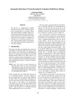
Fig. 2. Structural characterization of the proline-rich region of
Caskin1 (PRD-His). (A) CD spectrum of PRD-His; the large minimum
at 202 nm is typical of IDPs. (B) Limited proteolysis experiment with
a broad substrate specificity enzyme, subtilisin, at 1 : 2000 enzyme
to substrate ratio. Aliquots were withdrawn at times 0 s, 10 s, 30 s
and 1 min, and run on SDS-PAGE. Caskin1 is much more sensitive to
the enzyme than is the control globular protein BSA. (C) Gel filtration
chromatography of control globular proteins (
, see Materials and
methods) and PRD-His of Caskin1 (h). PRD is an extended, random
coil-like protein, with an m
app
value 3.9 times that of its real m value.
V. Csizmok et al. High levels of disorder in scaffold proteins
FEBS Journal 276 (2009) 3744–3756 ª 2009 The Authors Journal compilation ª 2009 FEBS 3747
The one-dimensional
1
H-NMR spectra of the PRD-
His fragments (Fig. 4) also underscores a largely disor-
dered conformational state. Chemical shifts show a
poor dispersion, i.e. amide proton signals are clustered
within a half-p.p.m. range centred at 8 p.p.m., whereas
the methyl group protons are clustered at around
1 p.p.m. Such a limited dispersion and signal overlap
in
1
H chemical shifts are typical of IDPs [34].
The proline-rich regions of Caskin1 interact
with Abi2
To demonstrate that the proline-rich regions character-
ized above are biochemically functional, we studied the
interaction of Caskin1 fragments with Abi2, which is
an adaptor protein identified originally by its inter-
action with Abl tyrosine kinase [35]. Caskin1 was cut
into five regions and expressed as glutathione transfer-
ase (GST) fusion proteins. These protein regions repre-
sent the ankyrin repeats and the SH3 domain together
(ANK ⁄ SH3-GST), the two SAM domains (SAM-GST)
and the three proline-rich regions (PRD1–3-GST) of
the C-terminal PRD. The full-length PRD of Caskin1
was also expressed (PRD-GST). Green fluorescent pro-
tein (GFP)-tagged Abi2 was expressed in COS7 cells,
extracts of which were used for the GST pull-down
assay. As shown in Fig. 5, the first and second proline-
rich regions of Caskin1 (PRD1-GST and PRD2-GST)
A B
C D
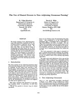
Fig. 3. Structural characterization of fragments of PRD. (A) Far-UV CD spectra of PRD1-His (blue), PRD2-His (green) and PRD3-His (red). All
spectra show a characteristic minimum at around 200 nm, which underscores the unstructured nature of the proline-rich region. (B) Compar-
ison of the far-UV CD spectrum of the full-length PRD-His (full line) and the sum of the spectra of PRD1-His, PRD2-His and PRD3-His (bro-
ken line). The sum of the spectra of the three fragments reproduces the spectrum of PRD-His, which confirms the overall random structure
of the full-length proline-rich region and the lack of appreciable long-range structural organization within this region of the protein. (C) The
temperature dependence of the mobile water fraction of PRD1-His (blue), PRD2-His (green) and PRD3-His (red), compared with that of the
globular control BSA (cyan) and the disordered control ERD10 (black). The large amount of water in the hydrate layer of PRDs suggests their
open, solvent-exposed conformations. (D) Gel filtration chromatography of the fragments PRD1-His, PRD2-His and PRD3-His shows that all
three fragments have an extended conformation with m
app
values 4.5, 2.2 and 5.4 times higher than the real m values, respectively.
High levels of disorder in scaffold proteins V. Csizmok et al.
3748 FEBS Journal 276 (2009) 3744–3756 ª 2009 The Authors Journal compilation ª 2009 FEBS
were able to interact with the GFP-Abi2 protein,
whereas ANK ⁄ SH3-GST, SAM-GST and PRD3-GST
did not show an association. It is worth noting that
the second proline-rich region showed significantly
increased interaction compared with the first, suggest-
ing that PRD2-GST contains the major binding site
for Abi2 (Fig. 5). This is supported by the finding that
the full-length PRD-GST has binding characteristics
similar to that of PRD2-GST. These in vitro data
suggest that the proline-rich fragments of C-terminal
Caskin1 are functional and may interact with SH3
domain-containing proteins, such as Abi2. [We have
also found an in vivo association and colocalization of
Abi2 with Caskin1 (A. Balazs, V. Csizmok, P. Tompa,
R. Udupa & L. Buday, unpublished results).]
Caskin1 is a scaffold protein
Although the exact function of Caskin1 is uncertain,
several observations suggest that it probably belongs
to the family of scaffold proteins. Scaffold proteins are
signalling proteins that typically have multiple binding
domains for simultaneous interaction with a variety of
partners. They have no catalytic activity, but tether
several signalling proteins to organize them into path-
ways, thus providing directionality and specificity in
signalling. For example, the Shank proteins serve as
important scaffold molecules modulating signalling
pathways at the post-synaptic sites of brain excitatory
synapses [36]. Ste5 serves in the yeast mating pathway,
ensuring that components of the mitogen-activated
protein kinase (MAPK) cascade, also involved in
osmoresponse and filamentation pathways, act specifi-
cally [18]. In our case, Caskin1 has been found in a
yeast two-hybrid screen to bind about 10 other part-
ners besides Abi2 (Table 1), and several points suggest
that it is a bona fide scaffold protein: (a) Caskin1 has
a modular structure with several of its domains and
non-domain regions involved in protein–protein inter-
actions; (b) none of its domains shows catalytic func-
tion; (c) it has 11 different partners all involved in
signal transduction; (d) it is preferentially located in
the PSD, known to harbour many proteins of signal-
ling and scaffold function (e.g. PSD95, Shank, Homer,
etc. [37]); (e) it has long uncharacterized regions which
lack sequence similarity to other proteins, and has
been shown here to be intrinsically disordered. The
appearance and functional role of structural disorder
have been explicitly noted in other scaffold proteins,
such as Ste5 [18], BRCA1 [19], CBP [4] and Mypt1
[20]. Thus, we decided to study this feature in detail to
gain further insight into the possible importance of
disorder in Caskin1 function and the class of scaffolds
in general.
The collection of scaffold proteins for bioinformatics
study, however, is hampered by the lack of consensus
on the definition of these proteins. In this article, we
focus on three classes of complex-forming proteins of
related function, also including anchor and docking
proteins. The prototype for anchor proteins is the
A
B
Fig. 4. (A,B) One-dimensional
1
H-NMR spectrum of PRD3-His
shows a narrow p.p.m. range and limited dispersion, typical for an
unfolded polypeptide. (B is the enlarged part of A between 6.3 and
8.8 p.p.m.).
Fig. 5. Proline-rich regions of Caskin1 interact with Abi2. Lysates
of COS7 cells expressing GFP-Abi2 were subjected to affinity purifi-
cation with the following Caskin1 GST fusion proteins (20 lgÆ
point
)1
) immobilized on glutathione–agarose beads: the ankyrin
repeats and the SH3 domain (ANK ⁄ SH3-GST), the SAM domains
(SAM-GST) and the three proline-rich regions (PRD1–3-GST). The
full-length PRD-GST was also used. Bound proteins were eluted by
SDS sample buffer, subjected to 7.5% SDS-PAGE, transferred to
nitrocellulose and immunoblotted with monoclonal anti-GFP IgG.
Lysates of COS7 cells immunoblotted with anti-GFP IgG are also
shown (bottom panel).
V. Csizmok et al. High levels of disorder in scaffold proteins
FEBS Journal 276 (2009) 3744–3756 ª 2009 The Authors Journal compilation ª 2009 FEBS 3749
A-kinase anchoring protein, which localizes protein
kinase A to different subcellular compartments [38].
Docking proteins, in general, have an N-terminal
membrane targeting element, typically a Pleckstrin
homology domain, a myristoylation site or a short
transmembrane domain. After direct or indirect inter-
actions with a tyrosine kinase, the docking protein
becomes tyrosine phosphorylated on multiple sites that
can interact with signalling proteins containing SH2
domains. Insulin receptor substrate 1, for example, con-
tains an N-terminal Pleckstrin homology domain and a
phosphotyrosine-binding domain, and nearly 20 poten-
tial tyrosine phosphorylation sites at the C-terminus
[39]. As suggested above, scaffold proteins are able to
interact with many different proteins at the same time,
but they are typically not subject to phosphorylation,
which creates novel binding sites. The lack of consensus
on these definitions is also indicated by the sole study
addressing the structural disorder in scaffold proteins
[2], in which several proteins clearly not of scaffold
function (e.g. p53, a transcription factor and voltage-
activated potassium channel, a binding partner of the
scaffold protein PSD95 [40]) were involved. Our study
encompasses proteins involved in the formation of mul-
tiprotein complexes, which have modular organization.
We collected 74 such proteins by literature search and
analysed their disorder by three different algorithms.
Prediction of disorder in scaffold proteins
The structural disorder of the 74 scaffold, docking and
anchor proteins was predicted by three different algo-
rithms, i.e. IUPred, VSL2 and FoldIndex (Table S1,
see Supporting information). We found that the ratio
of residues in local disorder was very high (49.7%,
63.36% and 47.82% predicted by IUPred, VSL2 and
FoldIndex, respectively) in these proteins, which is
comparable with the ratio found in the most disor-
dered protein families i.e. proteins involved in tran-
scription or signal transduction [41] and in RNA
chaperones [27]. This high level of disorder suggests
functional importance in scaffolds.
Further, we asked whether disorder can be ascribed
to regions intervening between the noted functional
domains in these proteins. To this end, their sequences
were analysed to localize their structured PFAM
domains. Some, described in detail in the literature,
are shown in Fig. 6. The analysis of regions connecting
the domains gave a very high disorder ratio: 61.13%,
77.53% and 54.84% predicted by IUPred, VSL2 and
FoldIndex, respectively; in certain proteins, such as
GRB2-associated proteins, it exceeded 90% (Table S1).
To demonstrate that these intervening regions are not
merely there to connect ordered functional domains,
we characterized their length distribution in the exam-
ined proteins (Fig. 7). Although globular domains tend
to be short and show a rather normal distribution,
with an average length of 86 amino acids, the distribu-
tion of linker regions is wide, with an average length
of 140 amino acids, and a maximal length as long as
1579 amino acids (in BRCA1).
Discussion
Our knowledge of the structure of scaffold proteins is
largely limited to those regions for which three-dimen-
sional structure has been established. However, if we
consider that the binding of numerous proteins in tight
proximity is rather difficult in the case of a rigid, glob-
ular structure, it is reasonable to assume that these
proteins contain long, disordered regions. Nevertheless,
the occurrence of disorder and its functional conse-
quences in scaffold proteins have never been examined
systematically. The present study provides evidence for
the extensive disorder of Caskin1 and also for the class
of scaffold proteins in general. Overall, the level of dis-
order exceeds that of the functional class so far consid-
ered to be the most disordered: RNA chaperones [27].
It is known that, in proteins associated with signal
transduction, transcription and RNA chaperone activi-
ties, the ratio of amino acids in locally disordered
regions is very high, on the order of 50–60%. These high
levels are thought to result from the functional advanta-
ges provided by disorder, which enables functions that
cannot be carried out by globular proteins. One advan-
tage of the extended, disordered conformation is an
Table 1. Results from the two-hybrid screen using a fragment of
Caskin1 (amino acids 280–963) as bait and a human fetal cDNA
library. The numbers in parentheses represent the number of identi-
cal clones obtained.
Clone Protein Function
1 (12) Abl-interactor-2 (Abi2) Adaptor protein
2 (2) CASK Scaffold protein
3 (1) EphA2 Receptor tyrosine kinase
4 (1) L1CAM Cell adhesion molecule
5 (2) Myosin IB Class I myosin
6 (1) Nck1 Adaptor protein
7 (1) Neurexin 2 Neuronal cell adhesion
molecule
8 (1) Stathmin-like
3 protein
Stathmin family protein
9 (1) Synaptotagmin Mediator of Ca
2+
-regulated
vesicle fusion
10 (7) Septin 4 Cell cycle regulator in yeast
11 (12) Siah1 Ubiquitin ligase
High levels of disorder in scaffold proteins V. Csizmok et al.
3750 FEBS Journal 276 (2009) 3744–3756 ª 2009 The Authors Journal compilation ª 2009 FEBS
enhanced interaction capacity of the protein [12], which
is also manifested in the elevated level of disorder of
hub proteins [13–16] and the increase in disorder with
complex size [17]. As disordered regions are often
directly involved in protein–protein interactions [42],
these points help us to interpret the possible role of
disorder in Caskin1 and in other scaffold proteins. To
obtain a balanced view of structural disorder, it should
also be taken into consideration that it may also pose a
danger to the cell, such as the occurrence of oncogenic
fusion proteins in cancer and amyloid aggregates in
neurodegenerative diseases [4]. It is probably a result of
these adverse effects that the cellular level of IDPs is
tightly regulated by several mechanisms [43].
Caskin1 is present in the PSD of neuronal cells.
Within its N-terminal half, it contains some well-char-
acterized domains, which are involved in the interac-
tion with Cask [1], but the C-terminal, proline-rich
region has never been examined. According to our
structural studies, this entire region is intrinsically dis-
ordered, and proline-rich regions are known to interact
with SH3, WW and other domains of cognate proteins
[31,44]. Indeed, PRD of Caskin1 contains several
consensus SH3 binding sites, and we postulate that it
is involved in multiple interactions with other PSD
proteins. In this study, we have shown that the
proline-rich regions interact with the Abi2 protein,
which have SH3 domains (we have also found the
in vivo association of Abi2 with Caskin1; A. Balazs,
V. Csizmok, P. Tompa, R. Udupa & L. Buday,
unpublished results). In this sense, PRD of Caskin1
might function in a manner similar to the long, central,
disordered region of BRCA1, which harbours binding
motifs for multiple partners in DNA repair [19]. A fur-
ther point on the function of PRD of Caskin1 is that
all of our studies point to a local tendency of ordering
in the middle PRD2 segment (amino acids 805–1199).
The level of hydration of this fragment is lower than
that of the other two and the results of CD analysis
also show some deviation from a fully disordered,
random coil-like state. By gel filtration chromatogra-
phy, this region also shows less extended conformation
than the rest of PRD. As a local tendency for ordering
is a sign of sites poised for interactions [45,46], it
Fig. 6. Schematic representation of the domain structure of selected scaffold proteins. The scheme shows the domain architecture of 20
selected scaffold proteins representing 20 families described in detail in the literature established by PFAM. Long grey lines connecting the
domains are regions with no recognizable similarity to known proteins.
Fig. 7. Length distribution of domains and linker regions in scaffold
proteins. The numbers of occurrences of domains (light grey) and
linker regions (dark grey) with their indicated lengths in the 74 scaf-
fold proteins (Table S1) are given. The occurrence was calculated
for 50 amino acid length bins, always including the upper limit. At
the end of the scale, linkers above 800 amino acids in length are
grouped (their maximum length extends to 1579 amino acids).
V. Csizmok et al. High levels of disorder in scaffold proteins
FEBS Journal 276 (2009) 3744–3756 ª 2009 The Authors Journal compilation ª 2009 FEBS 3751
is conceivable that this middle segment of PRD in
Caskin1 is a primary site of interaction with multiple
partners in PSD, especially as PRD2 is the major
binding site for Abi2. All of these inferences on the
function of Caskin1 are perfectly in line with the
organization of PSD. PSD is a dynamic multiprotein
complex attached to the post-synaptic membrane,
composed of several hundred proteins, including recep-
tors and channels, cell adhesion proteins, cytoskeletal
proteins, G-proteins and their modulators, and signal-
ling molecules including kinases and phosphatases [47].
A variety of scaffold proteins, such as members of the
MAGUK, Shank and Homer families, serve to orga-
nize PSD. As a result of its modular character and
ability to form multiprotein interactions, we suggest
that Caskin1 is a novel scaffold protein in PSD.
Previous scattered observations with other scaffold
proteins [4,18,19], our novel data on Caskin1 and the
noted functional advantages of disorder related to
molecular recognition [12,42,48] point towards the gen-
eral role of disorder in scaffold proteins. This inference
was underscored by the prediction of disorder for a
collection of 74 scaffold proteins: on average, 53.6%
of their amino acids were in locally disordered regions.
Disorder, however, is not evenly distributed in the
sequences, as shown by the consideration of only the
regions connecting PFAM domains. The predicted
average disorder for these regions is 64.5%, which sug-
gests that scaffold proteins are constructed as beads on
a string from globular domains connected by occasion-
ally very long linker regions. Because these linkers
cover 65.8% of the total length of scaffold proteins on
average, and their average length far exceeds that of
the globular domains, there is no doubt that disorder
in these proteins fulfils very important functions, prob-
ably commensurable in importance with that of
ordered domains.
Actual data on some scaffold proteins provide
evidence that these regions are much more than mere
passive linkers of functional globular domains. For
example, BRCA1 contains an approximately 1500-
amino-acid-long central region between the N-terminal
RING domain and C-terminal BRCT domain [19].
Although it lacks stable structural elements or recog-
nizable domains, this region is implicated in binding
not only DNA, but numerous proteins involved in
DNA damage response and repair [49,50]. Another
scaffold protein, Mypt1, also contains a long disor-
dered segment in its N-terminal region, and this
segment is involved in binding to the type 1 protein
phosphatase [51]. CBP has also been amply character-
ized in this respect. This protein contains seven globu-
lar domains and intervening disordered regions. At
least two regions of specific partner-binding function,
the nuclear receptor interaction domain and the
nuclear receptor co-activator-binding domain, reside in
the disordered regions of the protein [4]. In the case of
Ste5, the scaffold protein that binds several kinases of
the MAPK pathway, binding of Fus3 has been shown
to fall into a locally disordered region [18].
These data on scaffold proteins suggest that their
long disordered regions present binding sites for their
partners. As a result of their extended conformation,
they have a large potential binding capacity, being able
to anchor multiple partners next to each other. Inter-
action sites in disordered regions, termed preformed
structural elements [45], linear motifs [48], primary
contact sites [52] or molecular recognition features
[46], usually only constitute a few residues, and thus
enable a very economical and high-capacity binding of
partners. Furthermore, these regions are often the sites
of post-
translational modifications [48,53], and may themselves
affect the activity of the bound partner [18], which sug-
gests a rather elaborate and complex binding ⁄ organiz-
ing role in the function of scaffold proteins. We hope
that this suggestion provides novel insight into the
function of scaffold proteins, and will instigate the
design of novel experimental approaches aimed at
resolving the structure and function of these important
proteins.
Materials and methods
DNA constructs
The full-length rat Caskin1 cDNA was kindly provided by
Thomas Su
¨
dhof (University of Texas Southwestern Medical
Center, Dallas, TX, USA), and the full-length Abi2 cDNA
was donated by Ann Marie Pendergast (Duke University
Medical Center, Durham, NC, USA). Caskin1 cDNA was
amplified by a high-fidelity DNA polymerase and subcloned
into the pcDNA 3.1 ⁄ V5-His TOPO vector (Invitrogen, San
Diego, CA, USA). The full-length Abi2 was amplified by
PCR and subcloned into the BamHI site of the pEGFP-C1
vector (BD Biosciences Clontech, San Jose, CA, USA).
cDNAs corresponding to the ankyrin repeats and SH3
domain (ANK ⁄ SH3-GST, amino acids 1–346), SAM
domains (SAM-GST, amino acids 347–610), proline-rich
region 1 (PRD1-GST, amino acids 603–804), proline-rich
region 2 (PRD2-GST, amino acids 804–1199), proline-rich
region 3 (PRD3-GST, amino acids 1200–1430) and the
full-length PRD of Caskin1 (PRD-GST, amino acids
603–1430) were amplified by PCR and subcloned into the
EcoRI ⁄ SalI sites of the pGEX-4T1 vector (Amersham
Biosciences, Fairfield, CT, USA) as GST fusion proteins.
High levels of disorder in scaffold proteins V. Csizmok et al.
3752 FEBS Journal 276 (2009) 3744–3756 ª 2009 The Authors Journal compilation ª 2009 FEBS
For pull-down experiments, GST fusion proteins were puri-
fied by binding to glutathione–agarose (Sigma, St Louis,
MO, USA) without elution. Protein purification was moni-
tored on Coomassie blue-stained SDS–PAGE gels: the
majority of the GST proteins gave single bands.
The full-length proline-rich region of Caskin1 (PRD-His)
and its fragments (PRD1-His, PRD2-His, PRD3-His) were
also subcloned into the BamHI ⁄ XhoI sites of the expression
vector pQE2 (Qiagen, Venlo, the Netherlands) with a
C-terminal His tag. PRD2, because of poor expression of
the protein, was further subcloned into the NdeI ⁄ XhoI sites
of the expression vector pET20b (Novagen, San Diego,
CA, USA) with a C-terminal His tag. In all cases, the con-
structs were verified by DNA sequencing (MWG-Biotech,
Ebersburg, Germany).
Protein purification
For structural characterization, the full-length PRD of
Caskin1 and its fragments were expressed in the E. coli strain
BL21 Star. The expression of the proteins was induced by
0.5 mm isopropyl thio-b-d-galactoside at 30 °C for 3 h. The
proteins were purified to homogeneity from cellular extracts
by heat treatment of the supernatants (5 min · 100 ° C), fol-
lowed by nickel nitrilotriacetic acid affinity chromatography
(Qiagen). For further purification, the dialysed proteins were
loaded onto an SP-Sepharose ion exchange chromatograph
(Amersham) in a buffer of 20 mm Tris, 1 mm EDTA, pH 7.5,
and then eluted by a linear salt gradient (50–500 mm NaCl).
Fractions with the highest level of protein were pooled, dialy-
sed into 20 mm Tris, 150 mm NaCl, 1 mm EDTA, pH 7.5
and stored frozen at )20 °C in aliquots. The purity of the
constructs was demonstrated by SDS-PAGE (Fig. S1, see
Supporting information).
CD measurements
CD spectra were recorded at a protein concentration of
0.1 mgÆmL
)1
in 10 mm Na
2
HPO
4
, 150 mm NaCl, pH 7.5 in
a cuvette (path length, 1 mm) on a Jasco J-720 spectropola-
rimeter (Jasco, Oklahoma City, OK, USA) in a continuous
mode with a bandwidth of 1 nm, response time of 8 s and
scan speed of 20 nmÆmin
)1
. All spectra shown were
obtained by subtracting the buffer spectrum and averaging
10 separate scans.
Gel filtration chromatography
The unfolded nature of PRD and its fragments was also
characterized by gel filtration chromatography. The
proteins (200 lL) were run on an Amersham Biosciences
Superdex 200 (1 · 30 cm) column at 0.5 mLÆmin
)1
in a
buffer of 50 mm Na
2
HPO
4
, 150 mm NaCl, pH 7.0 on an
Amersham Biosciences FPLC system. The proteins were
detected at 280 nm. The column was calibrated using the
following globular proteins (m in parentheses): ribonuclease
A (13.7 kDa), chymotrypsinogen A (25.0 kDa), ovalbumin
(43.0 kDa), BSA (67 kDa) and alcohol dehydrogenase
(146.8 kDa). The m values of the proteins were determined
from the calibration curve constructed by plotting log m
values of calibration proteins vs. the elution volume. The
hydrodynamic dimension was characterized by the ratio of
m
app
, determined by gel filtration chromatography, and the
absolute value of m, calculated from the amino acid
sequence of the protein.
NMR spectroscopy
1
H-NMR spectra of PRD1, PRD2 and PRD3 were
recorded at 500 MHz on a Bruker DRX instrument
(Bruker, Billerica, MA, USA); 16 000 complex data points
were acquired in the direct dimension at 300 K using a
spectral width of 12 p.p.m. Data were zero-filled and
processed with a shifted quadratic sinbell plus exponential
window function. For water suppression, the 3-9-19 pulse
sequence with gradients was used [54].
Wide-line NMR spectrometry
The mobile proton (water) fraction was measured directly
by two
1
H-NMR methods: by measuring the free induction
decay signal or recording Carr–Purcell–Meiboom–Gill echo
trains. The determination of the mobile water fraction is
based on the comparison of the signal intensity or echo
amplitude extrapolated to t = 0 with the corresponding
values measured at a temperature at which the whole sam-
ple is in the liquid state. Details of the applied method have
been described elsewhere [32,33,55].
The effect of freezing on protein solutions was controlled
by the comparison of NMR parameters obtained before
and after a freeze–thaw cycle at temperatures above 0 °C.
We found that the freeze–thaw cycle caused no observable
changes for the studied samples as far as the measured
NMR parameters were concerned. The temperature was
controlled by an open-cycle Oxford cryostat with a stability
of ± 0.1 °C; the uncertainty of the temperature scale was
±1°C.
1
H-NMR measurements and data acquisition were
accomplished using a Bruker SXP 4-100 NMR pulse spec-
trometer at x
0
⁄ 2p = 82.55 MHz with a stability of better
than ± 10
)6
. The data points in the figures are based on
spectra recorded by averaging signals to reach a signal to
noise ratio of 50. The number of averaged NMR signals
was varied to achieve the desired signal quantity for each
sample and for unfrozen water quantities. The sensitivity of
the NMR spectroscope on sample change was controlled
by measuring the length of the p ⁄ 2 pulse to obtain reliable
M
0
values [55]. The extrapolation to zero time was
performed by fitting a stretched exponential.
V. Csizmok et al. High levels of disorder in scaffold proteins
FEBS Journal 276 (2009) 3744–3756 ª 2009 The Authors Journal compilation ª 2009 FEBS 3753
Antibodies and cell lines
Monoclonal antibody raised against GFP was supplied by
the Cancer Research UK Hybridoma Development Unit,
London, UK. COS7 cells were grown in Dulbecco’s modi-
fied Eagle’s medium (DMEM) supplemented with 10%
fetal bovine serum, penicillin (100 unitsÆmL
)1
) and strepto-
mycin (50 lgÆmL
)1
).
Transient transfection
Lipofectamine was obtained from Invitrogen and used for
the transfection of COS7 cells according to the manufac-
turer’s instructions. Briefly, 1 · 10
6
cells were plated on to
10 cm Petri dishes 24 h prior to transfection; 7 lg of the
various plasmid constructs and 50 lL of lipofectamine were
added to each well in 5 mL OptiMEM (Gibco, North
Andover, MA, USA). After 5 h, the cells were washed once
with DMEM and cultured in their regular medium.
Protein precipitation and western blotting
COS7 cells were washed with ice-cold NaCl ⁄ P
i
and lysed in
2 mL of ice-cold 50 mm Hepes buffer, pH 7.4, containing
100 mm NaCl, 1% Triton X-100, 20 mm NaF, 1 mm
EGTA, 1 mm Na
3
VO
4
,1mm p-nitrophenylphosphate,
10 mm benzamidine, 1 mm phenylmethylsulphonyl fluoride
and 25 lg ÆmL
)1
each of leupeptin, soybean trypsin inhibitor
and aprotinin. The lysates were clarified by centrifugation
at 15 000 g for 10 min at 4 °C. The lysates were then pre-
cipitated with 20 lg of the indicated GST-fusion protein
immobilized on glutathione–agarose (Sigma) for 1 h at
4 °C. Protein precipitates were washed three times with ice-
cold NaCl ⁄ P
i
, pH 7.4, containing 0.4% Triton X-100 and
eluted with SDS sample buffer. Bound proteins were sepa-
rated by SDS-PAGE and, because of the small amount of
proteins, transferred to nitrocellulose membrane and immu-
noblotted with the indicated antibodies. Blots were devel-
oped by the enhanced chemiluminescence (ECL; Amersham
Biosciences) system.
Collection of scaffold proteins and bioinformatics
predictions
We collected a number of anchor, docking and scaffold pro-
teins (denoted collectively as scaffold proteins) from the litera-
ture and by screening the UniProt knowledgebase (Table S1).
For the prediction of disorder, three different algorithms,
i.e. IUPred [13,23], VSL2 [24,25] and FoldIndex [26], were
used (, />disprot/predictorVSL2.php, />fldbin/findex).
The domain prediction was performed by the PFAM
algorithm ( for all the scaffold
proteins.
Acknowledgements
This research was supported by grants OTKA K60694
and K61555 from the Hungarian Scientific Research
Fund, ETT 245 ⁄ 2006 from the Hungarian Ministry of
Health, Miha
´
ly Pola
´
nyi Program (Agency for Research
Fund Management and Research Exploitation, KPI)
and the International Senior Research Fellowship
ISRF 067595 from the Wellcome Trust. R.U. acknowl-
edges support by the Marie Curie RTN ‘ENDOCYTE’
from the European Union FP6 program. Peter Ba
´
nki
is acknowledged for technical assistance with wide-line
NMR spectrometry.
References
1 Tabuchi K, Biederer T, Butz S & Sudhof TC (2002)
CASK participates in alternative tripartite complexes
in which Mint 1 competes for binding with caskin 1, a
novel CASK-binding protein. J Neurosci 22, 4264–
4273.
2 Cortese MS, Uversky VN & Keith Dunker A (2008)
Intrinsic disorder in scaffold proteins: getting more
from less. Prog Biophys Mol Biol 98 , 85–106.
3 Tompa P & Fuxreiter M (2008) Fuzzy complexes: poly-
morphism and structural disorder in protein–protein
interactions. Trends Biochem Sci 33, 2–8.
4 Dyson HJ & Wright PE (2005) Intrinsically unstruc-
tured proteins and their functions. Nat Rev Mol Cell
Biol 6, 197–208.
5 Tompa P (2002) Intrinsically unstructured proteins.
Trends Biochem Sci 27, 527–533.
6 Uversky VN, Oldfield CJ & Dunker AK (2005) Show-
ing your ID: intrinsic disorder as an ID for recognition,
regulation and cell signaling. J Mol Recognit 18, 343–
384.
7 Sickmeier M, Hamilton JA, LeGall T, Vacic V, Cortese
MS, Tantos A, Szabo B, Tompa P, Chen J, Uversky
VN et al. (2007) DisProt: the Database of Disordered
Proteins. Nucleic Acids Res 35, D786–D793.
8 Dunker AK, Obradovic Z, Romero P, Garner EC &
Brown CJ (2000) Intrinsic protein disorder in complete
genomes. Genome Inform Ser Workshop Genome Inform
11, 161–171.
9 Ward JJ, Sodhi JS, McGuffin LJ, Buxton BF & Jones
DT (2004) Prediction and functional analysis of native
disorder in proteins from the three kingdoms of life.
J Mol Biol 337, 635–645.
10 Tompa P, Dosztanyi Z & Simon I (2006) Prevalent
structural disorder in E. coli and S. cerevisiae proteo-
mes. J Proteome Res 5, 1996–2000.
11 Tompa P (2005) The interplay between structure and
function in intrinsically unstructured proteins. FEBS
Lett 579, 3346–3354.
High levels of disorder in scaffold proteins V. Csizmok et al.
3754 FEBS Journal 276 (2009) 3744–3756 ª 2009 The Authors Journal compilation ª 2009 FEBS
12 Gunasekaran K, Tsai CJ, Kumar S, Zanuy D & Nussi-
nov R (2003) Extended disordered proteins: targeting
function with less scaffold. Trends Biochem Sci 28,
81–85.
13 Dosztanyi Z, Chen J, Dunker AK, Simon I & Tompa P
(2006) Disorder and sequence repeats in hub proteins
and their implications for network evolution. J Prote-
ome Res 5, 2985–2895.
14 Ekman D, Light S, Bjorklund AK & Elofsson A (2006)
What properties characterize the hub proteins of the
protein–protein interaction network of Saccharomy-
ces cerevisiae? Genome Biol 7, R45.
15 Haynes C, Oldfield CJ, Ji F, Klitgord N, Cusick ME,
Radivojac P, Uversky VN, Vidal M & Iakoucheva LM
(2006) Intrinsic disorder is a common feature of hub
proteins from four eukaryotic interactomes. PLoS Com-
put Biol 2, e100.
16 Patil A & Nakamura H (2006) Disordered domains and
high surface charge confer hubs with the ability to inter-
act with multiple proteins in interaction networks.
FEBS Lett 580, 2041–2045.
17 Hegyi H, Schad E & Tompa P (2007) Structural disor-
der promotes assembly of protein complexes. BMC
Struct Biol 7, 65.
18 Bhattacharyya RP, Remenyi AGood MC, Bashor CJ,
Falick AM & Lim WA (2006) The Ste5 scaffold allos-
terically modulates signaling output of the yeast mating
pathway. Science 311, 822–826.
19 Mark WY, Liao JC, Lu Y, Ayed A, Laister R, Szymc-
zyna B, Chakrabartty A & Arrowsmith CH (2005)
Characterization of segments from the central region of
BRCA1: an intrinsically disordered scaffold for multiple
protein–protein and protein–DNA interactions? J Mol
Biol 345, 275–287.
20 Csizmok V, Szollosi E, Friedrich P & Tompa P (2006)
A novel two-dimensional electrophoresis technique for
the identification of intrinsically unstructured proteins.
Mol Cell Proteomics 5, 265–273.
21 Namba K (2001) Roles of partly unfolded conforma-
tions in macromolecular self-assembly. Genes Cells 6,
1–12.
22 Dosztanyi Z, Csizmok V, Tompa P & Simon I (2005)
IUPred: web server for the prediction of intrinsically
unstructured regions of proteins based on estimated
energy content. Bioinformatics 21, 3433–3434.
23 Dosztanyi Z, Csizmok V, Tompa P & Simon I (2005)
The pairwise energy content estimated from amino acid
composition discriminates between folded and
intrinsically unstructured proteins. J Mol Biol 347, 827–
839.
24 Peng K, Radivojac P, Vucetic S, Dunker AK &
Obradovic Z (2006) Length-dependent prediction
of protein intrinsic disorder. BMC Bioinformatics 7,
208.
25 Obradovic Z, Peng K, Vucetic S, Radivojac P &
Dunker AK (2005) Exploiting heterogeneous sequence
properties improves prediction of protein disorder. Pro-
teins 61(Suppl 7), 176–182.
26 Prilusky J, Felder CE, Zeev-Ben-Mordehai T, Rydberg
EH, Man O, Beckmann JS, Silman I & Sussman JL
(2005) FoldIndex: a simple tool to predict whether a
given protein sequence is intrinsically unfolded. Bioin-
formatics 21, 3435–3438.
27 Tompa P & Csermely P (2004) The role of structural
disorder in the function of RNA and protein chaper-
ones. FASEB J 18, 1169–1175.
28 Li SS (2005) Specificity and versatility of SH3 and other
proline-recognition domains: structural basis and impli-
cations for cellular signal transduction. Biochem J 390,
641–653.
29 Mayer BJ (2001) SH3 domains: complexity in modera-
tion.
J Cell Sci 114, 1253–1263.
30 Kim CA & Bowie JU (2003) SAM domains: uniform
structure, diversity of function. Trends Biochem Sci 28,
625–628.
31 Williamson MP (1994) The structure and function of
proline-rich regions in proteins. Biochem J 297, 249–
260.
32 Bokor M, Csizmok V, Kovacs D, Banki P, Friedrich P,
Tompa P & Tompa K (2005) NMR relaxation studies
on the hydrate layer of intrinsically unstructured pro-
teins. Biophys J 88, 2030–2037.
33 Tompa P, Banki P, Bokor M, Kamasa P, Kovacs D,
Lasanda G & Tompa K (2006) Protein–water and
protein–buffer interactions in the aqueous solution of
an intrinsically unstructured plant dehydrin: NMR
intensity and DSC aspects. Biophys J 91, 2243–2249.
34 Dyson HJ & Wright PE (2004) Unfolded proteins and
protein folding studied by NMR. Chem Rev 104, 3607–
3622.
35 Dai Z & Pendergast AM (1995) Abi-2, a novel SH3-
containing protein interacts with the c-Abl tyrosine
kinase and modulates c-Abl transforming activity.
Genes Dev 9, 2569–2582.
36 Sheng M & Kim E (2000) The Shank family of scaffold
proteins. J Cell Sci 113 (Pt 11), 1851–1856.
37 Feng W & Zhang M (2009) Organization and dynamics
of PDZ-domain-related supramodules in the postsynap-
tic density. Nat Rev Neurosci 10, 87–99.
38 Pawson T & Scott JD (1997) Signaling through scaf-
fold, anchoring, and adaptor proteins. Science 278,
2075–2080.
39 Thirone AC, Huang C & Klip A (2006) Tissue-specific
roles of IRS proteins in insulin signaling and glucose
transport. Trends Endocrinol Metab 17, 72–78.
40 Magidovich E, Orr I, Fass D, Abdu U & Yifrach O
(2007) Intrinsic disorder in the C-terminal domain of
the Shaker voltage-activated K
+
channel modulates its
V. Csizmok et al. High levels of disorder in scaffold proteins
FEBS Journal 276 (2009) 3744–3756 ª 2009 The Authors Journal compilation ª 2009 FEBS 3755
interaction with scaffold proteins. Proc Natl Acad Sci
USA 104, 13022–13027.
41 Iakoucheva LM, Brown CJ, Lawson JD, Obradovic Z &
Dunker AK (2002) Intrinsic disorder in cell-signaling and
cancer-associated proteins. J Mol Biol 323, 573–584.
42 Dyson HJ & Wright PE (2002) Coupling of folding and
binding for unstructured proteins. Curr Opin Struct Biol
12, 54–60.
43 Gsponer J, Futschik ME, Teichmann SA & Babu MM
(2008) Tight regulation of unstructured proteins: from
transcript synthesis to protein degradation. Science 322,
1365–1368.
44 Kay BK, Williamson MP & Sudol M (2000) The impor-
tance of being proline: the interaction of proline-rich
motifs in signaling proteins with their cognate domains.
FASEB J 14, 231–241.
45 Fuxreiter M, Simon I, Friedrich P & Tompa P (2004)
Preformed structural elements feature in partner recog-
nition by intrinsically unstructured proteins. J Mol Biol
338, 1015–1026.
46 Vacic V, Oldfield CJ, Mohan A, Radivojac P,
Cortese MS, Uversky VN & Dunker AK (2007)
Characterization of molecular recognition features,
MoRFs, and their binding partners. J Proteome
Res 6, 2351–2366.
47 Beresewicz M (2007) Scaffold proteins (MAGUK,
Shank and Homer) in postsynaptic density in the cen-
tral nervous system. Postepy Biochem 53, 188–197.
48 Fuxreiter M, Tompa P & Simon I (2007) Structural dis-
order imparts plasticity on linear motifs. Bioinformatics
23, 950–956.
49 Wang Q, Zhang H, Kajino K & Greene MI (1998)
BRCA1 binds c-Myc and inhibits its transcriptional and
transforming activity in cells. Oncogene 17, 1939–1948.
50 Zhang H, Somasundaram K, Peng Y, Tian H, Bi D,
Weber BL & El-Deiry WS (1998) BRCA1 physically
associates with p53 and stimulates its transcriptional
activity. Oncogene 16, 1713–1721.
51 Toth A, Kiss E, Herberg FW, Gergely P, Hartshorne
DJ & Erdodi F (2000) Study of the subunit interactions
in myosin phosphatase by surface plasmon resonance.
Eur J Biochem 267, 1687–1697.
52 Csizmok V, Bokor M, Banki P, Klement E
´
,
Medzihradszky KF, Friedrich P, Tompa K & Tompa P
(2005) Primary contact sites in intrinsically
unstructured proteins: the case of calpastatin and
microtubule-associated protein 2. Biochemistry 44,
3955–3964.
53 Iakoucheva LM, Radivojac P, Brown CJ, O’Connor
TR, Sikes JG, Obradovic Z & Dunker AK (2004) The
importance of intrinsic disorder for protein phosphory-
lation. Nucleic Acids Res 32, 1037–1049.
54 Piotto M, Saudek V & Sklenar V (1992) Gradient-
tailored excitation for single-quantum NMR
spectroscopy of aqueous solutions. J Biomol NMR 2,
661–665.
55 Tompa K, Banki P, Bokor M, Lasanda G & Vasaros L
(2003) Diffusible and residual hydrogen in amorphous
Ni(Cu)-Zr-H alloys, J. Alloys Comp. 350, 52–55.
Supporting information
The following supplementary material is available:
Fig. S1. SDS-PAGE with the various forms of Caskin1
with the different tags.
Table S1. Predicted disorder of scaffold proteins.
This supplementary material can be found in the
online version of this article.
Please note: As a service to our authors and readers,
this journal provides supporting information supplied
by the authors. Such materials are peer-reviewed and
may be re-organized for online delivery, but are not
copy-edited or typeset. Technical support issues arising
from supporting information (other than missing files)
should be addressed to the authors.
High levels of disorder in scaffold proteins V. Csizmok et al.
3756 FEBS Journal 276 (2009) 3744–3756 ª 2009 The Authors Journal compilation ª 2009 FEBS