Báo cáo khoa học: Weak oligomerization of low-molecular-weight protein tyrosine phosphatase is conserved from mammals to bacteria pot
Bạn đang xem bản rút gọn của tài liệu. Xem và tải ngay bản đầy đủ của tài liệu tại đây (745.14 KB, 12 trang )
Weak oligomerization of low-molecular-weight protein
tyrosine phosphatase is conserved from mammals to
bacteria
Jascha Blobel
1
, Pau Bernado
´
1
, Huimin Xu
2
, Changwen Jin
2
and Miquel Pons
1,3
1 Laboratory of Biomolecular NMR, Institute for Research in Biomedicine, Barcelona, Spain
2 Beijing Nuclear Magnetic Resonance Center, College of Life Sciences, College of Chemistry and Molecular Engineering, Peking University,
Beijing, China
3 Departament de Quı
´
mica Orga
`
nica, Universitat de Barcelona, Spain
Introduction
Low-molecular-weight protein tyrosine phosphatases
(lmwPTPs) constitute one of the four families of protein
tyrosine phosphatases [1]. In eukaryotic cells, lmwPTPs
participate in platelet-derived growth factor (PDGF)-
induced mitogenesis [2] and insulin-mediated mitotic
signaling [3]. Dephosphorylation of different substrates,
such as the ephrin [4] or fibroblast growth factor
receptor, resulting in cell proliferation [5], has also been
Keywords
low-molecular-weight protein tyrosine
phosphatase (lmwPTP); phosphatase
regulation; protein oligomerization; signaling
pathways; supramolecular proenzyme
Correspondence
M. Pons, Laboratory of Biomolecular NMR,
Institute for Research in Biomedicine,
Parc Cientı
´
fic de Barcelona, Baldiri Reixac,
10, 08028 Barcelona, Spain
Fax: +34934039976
Tel: +34934034683
E-mail:
(Received 13 April 2009, revised 5 June
2009, accepted 9 June 2009)
doi:10.1111/j.1742-4658.2009.07139.x
The well-characterized self-association of a mammalian low-molecular-
weight protein tyrosine phosphatase (lmwPTP) produces inactive oligomers
that are in equilibrium with active monomers. A role of the inactive oligo-
mers as supramolecular proenzymes has been suggested. The oligomeriza-
tion equilibrium of YwlE, a lmwPTP from Bacillus subtilis, was studied by
NMR. Chemical shift data and NMR relaxation confirm that dimerization
takes place through the enzyme’s active site, and is fully equivalent to the
dimerization previously characterized in a eukaryotic low-molecular-weight
phosphatase, with similarly large dissociation constants. The similarity
between the oligomerization of prokaryotic and eukaryotic phosphatases
extends beyond the dimer and involves higher order oligomers detected by
NMR relaxation analysis at high protein concentrations. The conservation
across different kingdoms of life suggests a physiological role for lmwPTP
oligomerization in spite of the weak association observed in vitro. Struc-
tural data suggest that substrate modulation of the oligomerization equilib-
rium could be a regulatory mechanism leading to the generation of
signaling pulses. The presence of a phenylalanine residue in the dimeriza-
tion site of YwlE, replacing a tyrosine residue conserved in all eukaryotic
lmwPTPs, demonstrates that lmwPTP regulation by oligomerization can be
independent from tyrosine phosphorylation.
Structured digital abstract
l
MINT-7148507: ywle (uniprotkb:P39155) and YwlE (uniprotkb:P39155) bind (MI:0407)by
nuclear magnetic resonance (
MI:0077)
Abbreviations
AIR, ambiguous interaction restraints; BPTP, Bos taurus protein tyrosine phosphatase; BR, best representative; EM, energy minimization;
HSQC, heteronuclear single-quantum correlation; lmwPTP, low-molecular-weight protein tyrosine phosphatase; PDGF, platelet-derived
growth factor; SAXS, small-angle X-ray scattering; YwlE, Bacillus subtilis protein tyrosine phosphatase.
4346 FEBS Journal 276 (2009) 4346–4357 ª 2009 The Authors Journal compilation ª 2009 FEBS
suggested. The regulation of lmwPTPs is not completely
understood and different mechanisms have been
described [6]. Reversible inactivation by oxidation of
the catalytic cysteine of phosphatases has been shown
to provide a regulation mechanism [7]. In the case of
lmwPTP, the presence of an additional cysteine results
in the reversible formation of a disulfide bond [8].
Phosphorylation of a tandem repeat of tyrosine residues
(YY-loop) lining the active site [9] has also been
suggested as a possible regulation mechanism leading to
either activation or inactivation, depending on the
substrate involved [9–12]. Dimerization of Bos taurus
lmwPTP (BPTP) has been observed in crystals [13] and
has also been shown to take place in solution [14], and
the protein evolves further into higher molecular weight
species which stand in fast equilibrium with the mono-
mer and dimer [15]. Dimerization involves the tyrosines
of the YY-loop and residues of the active site, leading
to an intrinsically inactive species. It has been suggested
that lmwPTP oligomerization could be an additional
regulation mechanism, although its physiological
relevance remains unproven [13]. A major objection is
the high dissociation constant of the lmwPTP dimer
in vitro, although crowding conditions inside the
cytoplasm may enhance oligomerization [16,17].
Prokaryotic lmwPTPs have recently been identified
and have been far less studied than eukaryotic
lmwPTPs [18]. Some prokaryotic lmwPTPs are viru-
lence factors that mimic eukaryotic phosphatases and
dephosphorylate eukaryotic proteins, thereby interfer-
ing with the host defense response. An example is a
phosphatase from Mycobacterium tuberculosis released
into the extracellular medium, from which it is proba-
bly translocated into macrophages, interfering with the
host signaling pathways [19].
Endogenous prokaryotic lmwPTPs participate in the
regulation of bacterial metabolism [20]. They can be
divided into two types on the basis of their sequence and
biological characteristics [21]. The first type binds and
dephosphorylates endogenous kinases (BY-kinases),
with which they often appear to be co-regulated in a
single operon [22–24]. They control the biosynthesis
and transport of virulence factors, such as exo- and
capsular polysaccharides [25–28]. One member is Wzb
from Escherichia coli, which acts with the kinase Wzc
in the regulation of colanic acid production [29–31].
The second type has been found in Gram-positive
bacteria [32]. Although little is known about their
function to date, one representative, YwlE from Bacil-
lus subtilis, has been identified as the cognate phosp-
hotyrosine-phosphatase of McsB, McsA and CtsR
[33]. It has been suggested that they are essential in
certain processes, such as bacterial stress resistance.
Comparison of lmwPTPs from phylogenetically dis-
tant species (endogenous prokaryotic and eukaryotic
forms) is expected to identify conserved features that
may shed light on the intrinsic regulation mechanisms
of this class of phosphatases.
Prokaryotic and eukaryotic lmwPTPs show low
sequence homology (less than 30% sequence identity)
[20], but a comparison of the presently available X-ray
[33–38] and NMR [20,39] structures shows a well-
conserved tertiary structure exhibiting a Rossman fold
[40].
It has been proposed that substrate specificity is
mainly determined by the residues lining the active site.
Eukaryotic and prokaryotic lmwPTPs acting on
eukaryotic substrates as virulence factors show high
similarity in these residues [34–37].
Endogenous prokaryotic lmwPTPs acting on pro-
karyotic substrates have different specificities and
correspondingly different residues lining the active site.
Wzb, the only representative of type one prokaryotic
lmwPTPs with an available experimental structure
[20], possesses mainly hydrophobic residues GAL-
VGKGA (38–45), compared with polar and aromatic
residues in the case of eukaryotes. The B. taurus and
human forms share the sequence SDWNVGRSP
(47–55), forming the so-called W-loop lining the active
site. The second type of endogenous lmwPTP, repre-
sented by YwlE [39], has a loop with the sequence
FASPNGKA(40–47), containing both polar and
hydrophobic residues. In both types of endogenous
lmwPTP, W49, suggested to be important for eukary-
otic substrate recognition, is missing [20].
The YY-loop is the second flexible loop that
appears over the active site in all lmwPTPs. The
name comes from the two consecutive tyrosine resi-
dues found in eukaryotic lmwPTPs. All known
eukaryotic and prokaryotic lmwPTPs bear at least
one tyrosine in this region, with the exception of
YwlE, which possesses a nonphosphorylatable phen-
ylalanine (F120). Thus, although phosphorylation
and dimerization could be related regulatory events
in most lmwPTPs, phosphorylation cannot be a regu-
latory mechanism of YwlE. However, dimerization,
as shown below, is conserved. The dimerization of
both eukaryotic and prokaryotic lmwPTPs involves
the active site, and the best docking model of the
prokaryotic dimer is very similar to the crystal struc-
ture of the eukaryotic dimer. Furthermore, both
phosphatases evolve to similar higher order oligo-
mers, as detected by NMR relaxation. The evolution-
ary conservation of the oligomerization process
strongly suggests a functional role in spite of the
large dissociation constants measured in vitro.
J. Blobel et al. Conserved weak protein interactions in lmwPTP
FEBS Journal 276 (2009) 4346–4357 ª 2009 The Authors Journal compilation ª 2009 FEBS 4347
Results
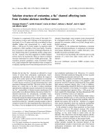
Concentration-dependent chemical shifts and
docking calculations
Figure 1A compares the
15
N–
1
H heteronuclear single-
quantum correlation (HSQC) spectra of YwlE
recorded at protein concentrations of 0.05 and 1.0 mm.
Although most of the peaks remain unperturbed, sev-
eral cross-peaks show unequivocal concentration-
dependent chemical shifts suggesting oligomerization
(Fig. 1A). The combined
1
H and
15
N chemical shift
changes are shown in Fig. 1B. The residues showing
the largest variations are located in the neighboring
region of the active site (Fig. 1C). Residues V39–A51,
forming what would be the W-loop in eukaryotic
lmwPTPs, as well as residue T84, facing the W-loop
on the opposite site of the active site crevice, are
among the most affected. Residues N10 and S14,
located in the crevice of the active site, also show
chemical shift changes. Furthermore, the aromatic resi-
dues Y127 and F120 around the YY-loop, just outside
the active site, are also affected. Only one perturbed
residue (V32) is placed in a remote position from the
A
B
D
C
Fig. 1. Concentration-dependent chemical shifts. (A) Overlay of expanded regions of
15
N–
1
H HSQC spectra of 0.05 mM (black) and 1.0 mM
(blue) YwlE. (B) Combined
15
N and
1
H chemical shift changes. (C) Chemical shift mapping. Residues showing changes above the threshold,
indicated by the black broken line in (B), are highlighted on the structure of YwlE (1zgg). The W- and YY-loops are shown in blue and red,
respectively. The active site residues are shown in yellow. (D) Sequence alignment of YwlE and BPTP (adapted from Lescop et al. [19]).
Residues showing the largest concentration-dependent chemical shift changes are highlighted in green. The active site of BPTP is
highlighted in yellow.
Conserved weak protein interactions in lmwPTP J. Blobel et al.
4348 FEBS Journal 276 (2009) 4346–4357 ª 2009 The Authors Journal compilation ª 2009 FEBS
active site. A similar set of residues was perturbed by
concentration changes of eukaryotic BPTP [14].
Figure 1D compares the chemical shift differences
associated with the same concentration changes
superimposed onto the aligned sequences of YwlE and
BPTP.
The oligomerization of BPTP has been characterized
extensively by NMR chemical shifts [14], NMR relaxa-
tion [15],
129
Xe-NMR [41], analytical ultracentrifuga-
tion [13], X-ray [13] and small-angle X-ray scattering
(SAXS) [42]. At low concentrations (< 0.25 mm), only
monomeric and dimeric forms are detectable. At
higher concentrations, NMR relaxation,
129
Xe-NMR
and SAXS experiments detect the formation of larger
oligomers. NMR relaxation data can be explained by
the presence of an additional species which is compati-
ble with a tetramer, although more complex oligomeri-
zation processes cannot be ruled out.
Chemical shift and ultracentrifugation data of BPTP
have been analyzed previously as a two-state mono-
mer–dimer equilibrium, and this was shown to be a
correct approximation up to a concentration of
0.5 mm. Assuming that the monomer–dimer equilib-
rium is also the dominant oligomerization process in
YwlE, we used the chemical shift restraints to model
the structure of the dimer using the molecular docking
program haddock 2.0 [43]. The NMR structure of
YwlE (1zgg) was used as the model for the monomeric
form. The docking protocol and the definition of
active and passive restraints are described in Materials
and methods.
The 200 best solutions clustered into five families.
The five families are related by the relative rotation of
the two lmwPTP monomers around an axis going
through their active sites. The RMSD values, buried
surface, energy components, haddock scoring of all
families (Table S1) and Ramachandran map analysis
of the best family (Table S2) are given as Supporting
information. The most populated family (containing
more than 50% of the solutions) includes the two
structures with the best haddock scoring terms
(E
haddock
= )107.8 and )101.0 kcalÆmol
)1
). Consi-
dering the average of the 10 best structures in each
family, the same family also shows the largest buried
surface area (1278 ± 135 A
˚
2
) and the largest desolva-
tion energy ()38.3 ± 5.6 kcalÆmol
)1
).
The best representative (BR) of the most populated
family, sharing an RMSD with all other family mem-
bers of 3.8 ± 1.1 A
˚
, shows very good agreement with
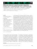
the crystallographic dimer of BPTP [13]. The resulting
superposition of both dimers is shown in Fig. 2A.
In the docking model of the YwlE dimer, the flex-
ible loops around the active site (W- and YY-loops)
are involved in the stabilization of the dimer, in full
agreement with the crystallographic dimer of BPTP.
Other structural similarities between the prokaryotic
model and the eukaryotic dimer are the aromatic
residue F120 and the catalytically active C7 of
YwlE, which occupy the positions of Y131 and C12
of BPTP. The second residue defining the active site
motif of all phosphatases, namely the arginine placed
six residues after the catalytic cysteine, is also
located in equivalent positions in both prokaryotic
and eukaryotic dimeric lmwPTPs (Fig. 2B). Residue
W49 of BPTP, after which the eukaryotic W-loop is
named, is replaced by F40 in the prokaryotic form
(Fig. 2B).
A
B
Fig. 2. (A) Best YwlE dimeric docking model (green) superimposed
onto the crystallographic BPTP dimer (gray). (B) Expansion of the
active site of YwlE (green) displaying functionally important side-
chains (colored by atom type) superimposed with the equivalent
side-chains of BPTP (gray). The labels show the structurally equiva-
lent YwlE ⁄ BPTP residues. C7 ⁄ C12 and R13 ⁄ R18 participate in the
catalytic activity, and F40 ⁄ W49 and F120 ⁄ Y131 are involved in
dimerization.
J. Blobel et al. Conserved weak protein interactions in lmwPTP
FEBS Journal 276 (2009) 4346–4357 ª 2009 The Authors Journal compilation ª 2009 FEBS 4349
Self-association monitored by NMR relaxation
The concentration-dependent chemical shift changes in
YwlE are smaller in magnitude than those observed
for BPTP.
15
N-NMR relaxation rates are very sensitive
to self-association equilibria as they strongly depend
on the rotational correlation time of all the species in
solution. Therefore, we used longitudinal (R
1
) and
transverse (R
2
)
15
N-NMR relaxation rates measured at
different protein concentrations to analyze the oligo-
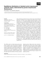
merization of lmwPTP. Figure 3 shows the average
R
2
⁄ R
1
values (<R
2
⁄ R
1
>) of all measured residues
at different YwlE concentrations. The increase in
<R
2
⁄ R
1
> at higher concentrations confirms the self-
association of YwlE.
When multiple species coexist in fast exchange in
solution, the measured
15
N-NMR relaxation rates are
the concentration-weighted averages of the individual
relaxation rates of each species. In NMR relaxation
experiments, all residues in the protein are sensing the
changes in the correlation time associated with the for-
mation of higher molecular weight species, whereas
only residues in the interface of the dimer show
changes in chemical shifts. The
15
N-NMR relaxation
approach makes use of a large number of independent
measurements (approximately the number of residues
times the number of concentrations), giving the possi-
bility to distinguish between different oligomerization
models. However, the structure of the monomer and
dimer are required to analyze the data.
The combined use of NMR spin relaxation and
hydrodynamic calculations has previously been shown
to be a powerful tool for the characterization of weak
protein–protein interactions in solution [15, 41, 44–48].
This approach assumes that the lifetime of the differ-
ent oligomeric species is significantly longer than their
rotational correlation times, but the exchange is still
fast on the chemical shift time-scale. Under these
conditions, the measured relaxation rates are inter-
preted as the weighted average of the relaxation rates
of the coexisting individual species [48].
A detailed analysis of the oligomerization process
was carried out by comparing the residue-specific
relaxation rates obtained experimentally with those
derived from hydrodynamic calculations of the experi-
mental monomer structure and the haddock model
for the dimer, as described below.
The theoretical relaxation data of the monomer and
the haddock model of the dimer were computed using
HydroNMR [49,50], and the dissociation constant of
the dimer (K
d
), giving the relative populations of
monomer and dimer at different concentrations, was
fitted to reproduce the experimental R
2
⁄ R
1
values of
the individual residues (see Materials and methods).
This approach gives a well-defined minimum (v
2
=
1.128) with a K
d
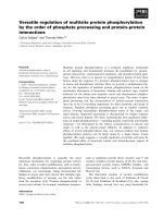
value of 5.20 ± 0.20 mm. The experi-
mental and fitted R
2
⁄ R
1
values for all four relaxation
datasets are shown in Fig. 4A–D. The residue-specific
residuals are given in Fig. S1.
Good agreement was observed at the lower protein
concentrations, but the calculated R
2
⁄ R
1
values at
higher lmwPTP concentrations were systematically
lower than the experimental values (Fig. 4A). A statis-
tically significant better fit was obtained by the inclu-
sion of an additional species (an isotropic tetramer) in
the equilibrium, as observed previously in the case of
BPTP [15,41,42].
As described previously for BPTP, we modeled the
higher oligomers of YwlE as a single isotropic tetra-
mer, i.e. with the same relaxation rates, R
1T
= 0.689
and R
2T
= 42.4 s
)1
, for all residues (see Materials
and methods). This procedure provided a much better
agreement with the experimental relaxation data
(v
2
= 0.921), as shown in Fig. 4F–I,, yielding thermo-
dynamic constants of K
d
= 7.59 ± 0.73 mm and
K
t
= 0.27 ± 0.10 mm. The improvement derives
mainly from the better reproduction of the high-
concentration data (cf. Fig. 4E, J). The reported uncer-
tainties were obtained from a Monte-Carlo analysis of
the model as described in Materials and methods. The
improvement in the figure of merit v
2
from 1.128 to
0.921 is statistically significant according to the F-test
(P <10
)10
), indicating that the YwlE oligomerization
equilibrium involves at least three species. An equiva-
lent situation has been demonstrated previously in the
case of BPTP [15]. The dependence of the dissociation
constants on the assumed values of R
1T
and R
2T
was
checked by testing values diverging by ± 20% from
the initial estimates. The variations in K
d
and K
t
were
0.23 mm and 0.11 mm, respectively, centered around
the initially estimated dissociation constants.
Fig. 3. Average R
2
⁄ R
1
(<R
2
⁄ R
1
>) values measured at different
YwlE concentrations.
Conserved weak protein interactions in lmwPTP J. Blobel et al.
4350 FEBS Journal 276 (2009) 4346–4357 ª 2009 The Authors Journal compilation ª 2009 FEBS
Figure 5 compares the concentration dependence of
the molar fractions of the different oligomers of YwlE
and BPTP calculated from the dissociation constants
determined by NMR.
Discussion
Evolutionary conservation of specific sequence or
structural features in homologous proteins is consid-
ered to be a relevant criterion for their physiological
significance. Intermolecular interactions are also vali-
dated by their conservation in phylogenetically distant
species. In this work, we show that similar oligomeri-
zation processes are conserved in eukaryotic and pro-
karyotic lmwPTPs.
Previous studies of lmwPTP from B. taurus have
demonstrated the formation of oligomers in solution
[13–15,41,42]. A dimer showing intermolecular contacts
involving the active site and tyrosine residues in the
YY-loop was observed in crystals [13] and was con-
firmed to exist in solution [14]. As the active site is
closed by the second molecule on dimer formation, this
species is intrinsically inactive. Higher order oligomers
were also detected by concentration-dependent NMR
relaxation rate measurements [15],
129
Xe-NMR [41]
and SAXS [42]. The oligomerization processes were
found to correspond to weak interactions in vitro, with
A
B
C
D
E
F
G
H
I
J
Fig. 4. R
2
⁄ R
1
values of individual residues measured at the four YwlE concentrations (mM) of 1.00 [(A) and (F) in blue], 0.50 [(B) and (G) in
green], 0.25 [(C) and (H) in dark yellow] and 0.10 [(D) and (I) in red]. Calculated values using the best-fitted parameters for the monomer–
dimer (A–D) and monomer–dimer–tetramer (F–I) models are shown in black. The contribution of the highest concentration experiments to
the fitting error of individual residues is shown in (E) for the monomer–dimer model and in (J) for the monomer–dimer–tetramer model. The
individual contributions from all the concentrations are given as Supporting information.
Fig. 5. Molar fractions of the different species present in the YwlE
(full lines) and BPTP (broken lines) oligomerization equilibria. Mono-
mers are shown in red, dimers in green and tetramers in blue.
Points and circles represent the experimental points with their
uncertainties.
J. Blobel et al. Conserved weak protein interactions in lmwPTP
FEBS Journal 276 (2009) 4346–4357 ª 2009 The Authors Journal compilation ª 2009 FEBS 4351
dissociation constants in the millimolar range, and, in
spite of the putative regulatory role that can be associ-
ated with an interaction that changes the phosphatase
activity reversibly, the large dissociation constants shed
some doubt on the physiological relevance of this
mechanism.
YwlE from B. subtilis is a prokaryotic lmwPTP
involved in the regulation of endogenous bacterial meta-
bolic processes [33], not influenced, like prokaryotic
virulence factors, by the interaction of bacteria with
eukaryotic hosts. As such, the comparison with BPTP is
expected to genuinely display the effects of evolution in
lmwPTPs of two phylogenetically distant organisms.
The very similar fold of YwlE and eukaryotic lmwPTP
monomers provides a strong case for a common evolu-
tionary origin, in spite of only a modest sequence homol-
ogy. Low sequence homology is expected between
homologous proteins from very distant species, and
stresses the relevance of the conservation of specific resi-
dues or residue classes in particular sites. Here, we have
analyzed the oligomerization of YwlE using NMR
chemical shift and
15
N-NMR relaxation measurements
at different protein concentrations in order to compare it
with the previously reported oligomerization of BPTP.
The structure of the YwlE dimer was modeled by
computational docking of two monomers using experi-
mental NMR chemical shifts as constraints. The BR of
the family with the highest average haddock score,
which is also the most populated, shows the highest
similarity to the crystal structure of dimeric BPTP. A
detailed comparison of the specific residues forming
the dimer interface confirmed that the dimers formed
by eukaryotic and prokaryotic lmwPTP are, indeed,
homologous structures. Two pairs of aromatic residues
seem to play equivalent roles in the formation of the
dimer in YwlE and BPTP: F120 ⁄ Y131 and F40 ⁄ W49
from the prokaryotic and eukaryotic lmwPTPs, respec-
tively. F120 and Y131 occupy similar positions in the
interaction surface, close to the active site cysteines C7
and C12 of YwlE and BPTP, respectively. Tyrosines
131 and 132 are highly conserved in all eukaryotic
lmwPTPs and are phosphorylation sites implicated in
lmwPTP regulation.
The other pair of aromatic residues involves the
highly conserved tryptophan, which is present in nearly
all eukaryotic lmwPTPs and gives the name to the
W-loop. This flexible loop shows maximal variations
between eukaryotic and endogenous prokaryotic
lmwPTPs, in agreement with the different characteris-
tics of their respective substrates. W49 is missing in
prokaryotic lmwPTP, whereas the role of its indole
side-chain in the stabilization of the dimer seems to be
taken on by the phenyl group of F40 of YwlE.
Residues N44 in YwlE and R53 in BPTP, both
located in the W-loop, seem to play equivalent roles in
the stabilization of the dimers by interacting with their
related residues N44 and R43 of the second molecule
of the dimer.
The physical characteristics of the residue pairs
F40 ⁄ W49, N44 ⁄ R53 and F120 ⁄ Y131 of YwlE and
BPTP are conserved in PtpB, a lmwPTP from the
Gram-negative bacterium Salmonella aureus (F36 and
N40) [51], and Etp from E. coli (H42 and K46).
Escherichia coli Wzb, a member of the first type of
endogenous lmwPTPs, does not have the aromatic
homolog to F40 ⁄ W49 in the substrate recognition
loop. However, concentration-dependent
15
N-NMR
relaxation data for this protein point to the formation
of higher molecular weight species [20]. The apparent
correlation time increases from 9.95 ns (0.1 mm)to
10.7 ns (0.3 mm) and, compared with the theoretical
value for the monomer (9.21 ns) predicted from its
NMR structure (2fek) using HydroNMR [49,50], sug-
gests a weak self-association mechanism, similar to
that observed for YwlE and BPTP.
The similarity of the lmwPTP dimers of the prokary-
otic and eukaryotic forms, involving structurally
related, although not identical, residues, strongly sug-
gests that the conserved feature is, indeed, the forma-
tion of a dimer, and rules out the alternative
explanation that dimer formation is a necessary side-
effect of the conservation of the active site and sur-
rounding substrate recognition and regulation loops.
Phosphorylation of the adjacent tyrosine residues
Y131 and Y132 in eukaryotic lmwPTP is considered to
be a regulation mechanism. Interestingly, the crystal
structure of the BPTP dimer, with a bound phosphate
and the tyrosine side-chain pointing to the active site
of the second molecule, is reminiscent of the expected
reaction product of a dephosphorylation reaction,
although dimerization does not require the previous
phosphorylation of the tyrosine. The observation of
dimeric YwlE having a phenylalanine residue in the
dimer interface in place of a tyrosine confirms that
lmwPTP dimerization is independent of phosphoryla-
tion. However, the structure of the YwlE dimer sug-
gests that the active site of each molecule is occupied
by the residues of the other molecule forming the
dimer, and therefore the dimer is an inactive species.
Modulation of the monomer–dimer equilibrium could
therefore provide a mechanism to modulate phospha-
tase activity.
The formation of higher oligomers also seems to
be conserved between eukaryotic and prokaryotic
lmwPTPs. Higher oligomers formed by the interac-
tion of dimers are also expected to be enzymatically
Conserved weak protein interactions in lmwPTP J. Blobel et al.
4352 FEBS Journal 276 (2009) 4346–4357 ª 2009 The Authors Journal compilation ª 2009 FEBS
inactive. The W49G BPTP mutant, in which dimeri-
zation is prevented, shows a much lower tendency to
oligomerize (results not shown). Larger oligomers are
known to be preferentially stabilized by crowding con-
ditions as found inside the cell [16,52]. Crowding is
expected to decrease the effective dissociation constant
of a lmwPTP tetramer (72 kDa) by a factor of
10
1
–10
2
. Larger aggregates or aggregates of higher
molecular weight can give reductions of 10
3
–10
5
in the
dissociation constants inside the cell relative to the
values that would be determined in vitro [53]. A
moderate decrease in the dissociation constants of
BPTP oligomers has been observed in vitro in the
presence of a low-molecular-weight crowder [41].
Oligomer formation, therefore, could be a requirement
to ensure that the effective dissociation constants
match the protein concentrations in vivo.
One can speculate on the hypothetical advantages
of a regulation mechanism based on the equilibrium
between active monomers and an inactive dimer, in
which the active site is also the dimerization site. Pro-
vided that the stability of the dimer is only moderate,
increasing the concentration of possible substrates
would cause an increase in the concentration of active
monomers, as substrate binding would compete with
dimerization. This suggests that oligomeric lmwPTP
would provide a latent reservoir of phosphatase
(equivalent to a proenzyme form) that could be acti-
vated by phosphorylated substrates, and would return
to the inactive form after the substrate has been
exhausted by the activity of the phosphatase. This
self-regulating mechanism would allow for the genera-
tion of ‘phosphorylation pulses’ following kinase
activity.
Materials and methods
Protein preparation and NMR experiments
Sample preparation and NMR measurements, including
HSQC and
15
N-NMR relaxation studies, have been
described elsewhere [39]. Briefly, reducing conditions were
ensured by the presence of 30 mm dithiothreitol in solution
made up of 50 mm Tris ⁄ HCl buffer at pH 7.5, 50 mm NaCl
and 10% D
2
O. No phosphate was present. The NMR
experiments were performed at 25 °C on a 600 MHz instru-
ment. Spectra were analyzed using NMRPipe [54].
15
N-NMR relaxation rates R
1
and R
2
were measured at 0.1,
0.25, 0.5 and 1.0 mm total protein concentrations. Changes
in chemical shift were monitored by comparing measure-
ments at 0.05 and 1.00 mm lmwPTP. The combined
changes in chemical shift (Dd) were calculated using the
relationship
Dd ¼½Dd
2
H
þðDd
N
=5Þ
2
1=2
ð1Þ
where Dd
H
and Dd
N
are the changes in chemical shift
observed in the
1
H and
15
N dimensions, respectively.
Generation of a dimeric model by computational
docking
Models of potential lmwPTP dimers were generated from
the first model of the NMR structure 1zgg using haddock
2.0 [46]. This approach is implemented in cns [55] (cns
Version 1.2 used here) and takes advantage of experimen-
tally measured data reporting on perturbations in the pro-
tein–protein interface. Here, concentration-dependent
chemical shifts, as detected by HSQC experiments, were
used. As suggested by the authors of haddock, residues
showing strong chemical shift changes in combination with
a solvent accessibility of greater than 50% were chosen as
‘active’ ambiguous interaction restraints (AIRs). These
included the three residues F40, N44 and F120. A second
group of residues serving as a less restrictive type of
restraint, generally referred to as ‘passive’ AIRs, was made
up of residues showing strong chemical shift changes and
also their direct neighbors, both of which had to exhibit a
surface accessibility of greater than 30%. The 13 residues
G9, N31, N33, S42, P43, G45, T49, H50, T84, H85, G121,
I126 and K128 passed the restriction criteria for passive
AIRs. The solvent accessibility of all residues was deter-
mined by the program naccess [56] from the monomer.
haddock docking was launched using the default settings
of the program, whereas python scripts derived from aria
[57] were used to analyze all results and automate the pro-
cess. The process consists of two docking stages: a rigid
body energy minimization (EM) and a semi-rigid simulated
annealing in torsion angle space. It should be noted that,
during the rigid body EM, a 180° rotated model was gener-
ated from each rigid-docked structure to amplify the diver-
sity amongst dimeric models. In the second stage, all
residues making intermolecular contacts within a 5 A
˚
cut-
off were considered as flexible. No symmetry restraints were
enforced. The full electrostatic binding energy was pre-
served throughout the protocol. In the scoring of the final
dimeric models, the contributions from van der Waals’ and
electrostatic interactions, AIR distance restraints and desol-
vation energies, as well as buried surface area terms,
were scaled and summed in the haddock scoring term
(E
haddock
) as suggested by the authors. haddock 2.0 gener-
ated 1000 dimers during hard docking. The best 200 dimers
were retained for the semi-flexible simulated annealing step.
The refined final 200 dimers were sorted into families using
the program cluster_struc, a part of the haddock program
package, using a maximum RMSD difference of 12 A
˚
amongst the family members in combination with a mini-
mum family size of five structures. The BR of a family is
J. Blobel et al. Conserved weak protein interactions in lmwPTP
FEBS Journal 276 (2009) 4346–4357 ª 2009 The Authors Journal compilation ª 2009 FEBS 4353
the structure that has the lowest average RMSD to all the
remaining members. The BR of the most populated family,
which has the highest average haddock score, as well as
the largest average buried surface area and the highest des-
olvation energy amongst the 10 best members (Fig. S1),
was chosen as the model of the YwlE dimer.
Hydrodynamic calculations
15
N-NMR relaxation rates for monomer and dimer were
calculated using the program HydroNMR [49]. Hydrogens
were added to the dimer using a program from the WHAT
IF server ( />The atomic element radius was assigned to be 3.3 A
˚
and
the atomic distance (N–H) 1.02 A
˚
[50]. The temperature
was set to 25 °C, the viscosity to 8.91 · 10
)3
P and the
NMR field strength to 14.09 T. For the monomer of
B. subtilis, a rotational correlation time of s
c
Mon
= 8.65 ns
(<R
2
⁄ R
1
> = 8.36) was calculated, exhibiting an anisot-
ropy of D
par
⁄ D
per
= 1.18. The dimer had a s
c
Dim
value of
20.00 ns (<R
2
⁄ R
1
> = 43.14) and D
par
⁄ D
per
= 1.58, being
clearly the most anisotropic species. For the tetramer, no
structure is available, but it is expected to be a compact
globular object. When calculating the theoretical relaxation
rates for the tetramer, solvent depletion effects arising from
protein–protein interfaces must be accounted for. This can
be achieved using the rotational correlation time of the
dimer (s
c
Dim
) for the calculation of the rotational correla-
tion time of a solvent-depleted monomer (s
c
DMon
) through
the relationship s
c
Dmon
= s
c
Dim
⁄ 2.78, giving s
c
DMon
=
7.19 ns [58,59]. The theoretical rotational correlation time
for the tetramer can then be calculated by s
c
Tet
= ns
c
DMon
,
with n = 4, resulting in s
c
Tet
= 28.78 ns [15]. From s
c
Tet
,
theoretical R
1T
and R
2T
values for a spherical body of
0.689 and 42.4 s
)1
(R
2
⁄ R
1
= 61.52) are calculated for a
magnetic field of 14.09 T. The two relaxation rates are used
for all residues of the tetramer in the fitting protocol of
experimental
15
N-NMR relaxation data.
The rotational correlation time of E. coli lmwPTP Wzb
was calculated from the best energy model of the NMR
structure 2fek [20] using HydroNMR [49,50] at 25 °C and
14.09 T.
15
N-NMR relaxation data analysis
Experimental
15
N-NMR relaxation data were filtered for
the fitting as described elsewhere [41]. Briefly, data from
individual residues were not used when any of the following
three situations were encountered: (a) heteronuclear Over-
hauser effect < 0.6, (b) large (> 25%) experimental errors
when compared with the relaxation rates R
2
⁄ R
1
and (c)
large disagreement (> 25%) between the experimental and
simulated R
2
⁄ R
1
values using the relevant model, filtering
for residues affected by chemical exchange. A total of 95
experimental R
2
⁄ R
1
values were extracted, leaving 313
R
2
⁄ R
1
values spanning the four protein concentrations of
0.10, 0.25, 0.50 and 1.00 mm lmwPTP in the case of the
monomer–dimer model and 309 R
2
⁄ R
1
values for the
monomer–dimer–tetramer equilibrium. The average relative
relaxation rates (<R
2
⁄ R
1
>) were calculated for each
protein concentration.
Experimental R
1
and R
2
values are the concentration-
weighted average of the relaxation rates of all participating
species. Equation (2) shows the calculation of the relaxation
rates for the monomer–dimer–tetramer model, with M
being the molar fraction of monomer, D the molar fraction
of dimer and T the molar fraction of tetramer.
R
n
¼ M Á R
nM
þ D Á R
nD
þ T Á R
nT
ð2Þ
The relaxation rate R
n
of each species is denoted with the suf-
fix corresponding to each species. n is equal to 1 and 2 in the
case of longitudinal and transverse relaxation, respectively.
The equilibrium parameters were determined by minimiz-
ing the error function defined in Eqn (3).
v
2
¼ 1=N ÁðR
i
R
j
½ðR
2
=R
1
Þ
exp
ij
ÀðR
2
=R
1
Þ
theo
ij
2
=½EðR
2
=R
1
Þ
exp
ij
2
Þ
ð3Þ
where i and j denote different residues (i) at varying protein
concentrations (j ), and E(R
2
⁄ R
1
)
exp
is the corresponding
experimental error. N is the number of experimental data
used.
For the monomer–dimer model, the fitting of the theoret-
ical relaxation rates of two species to the experimental
values adjusts a single parameter, namely the dissociation
constant K
d
connecting the relative concentrations of
monomer [M] and dimer [D] over all concentration ranges
through Eqn (4). The presence of a tetramer, being a
dimer of dimers, is accounted for by the dissociation
constant K
t
given by Eqn (5). [T] denotes the concentration
of tetramer.
K
d
¼½M
2
=½Dð4Þ
K
t
¼½D
2
=½Tð5Þ
The minimization protocol consists of a grid search for
each variable, followed by the minimization of all variables
together using the function fmincon as implemented in
Matlabª. Errors in the calculated parameters are deter-
mined using the Monte-Carlo method. Thus, a new set of
synthetic relaxation rates is calculated from the determined
dissociation constants, whereas a further term is added to
each relative relaxation rate R
2
⁄ R
1
calculated from the
experimental error multiplied by a random chosen value
obtained from a Gaussian distribution. The newly gener-
ated relaxation rate data set is fitted to the applied model.
The process is repeated 100 times. The final error in the dis-
sociation constants is equal to the standard deviation of the
100 obtained dissociation constants for each species. For
Conserved weak protein interactions in lmwPTP J. Blobel et al.
4354 FEBS Journal 276 (2009) 4346–4357 ª 2009 The Authors Journal compilation ª 2009 FEBS
the fitting of the experimental relaxation data to the mono-
mer–dimer–tetramer model, a further type of error estima-
tion is used to prove the validity of the results obtained
using the minimization protocol with Monte-Carlo error
estimation with respect to the relaxation rates of the tetra-
mer (R
1T
and R
2T
). Thus, 100 new sets of R
1T
and R
2T
were generated with a variation of 20% from their theoreti-
cally determined values (0.689 and 42.4 s
)1
) using a Gauss-
ian distribution. Each set was used for a new fitting to the
experimental relaxation data using the minimization proto-
col as described above.
Acknowledgements
The authors thank Dr Ewen Lescop (ICSN-CNRS) for
useful discussions. This work was partially supported
by funds from the Spanish Ministry of Education
(BIO2007-63458 to MP). J.B. is a recipient of a pre-
doctoral fellowship from the Spanish Ministerio de
Education y Ciencia. P.B. holds a Ramo
´
n y Cajal con-
tract that is partially financed by the Spanish Ministry
of Education and by funds provided to the IRB by the
Generalitat de Catalunya.
References
1 Fauman EB & Saper MA (1996) Structure and function
of the protein tyrosine phosphatases. Trends Biochem
Sci 21, 413–417.
2 Chiarugi P, Cirri P, Marra F, Raugei G, Fiaschi T,
Camici G, Manao G, Romanelli RG & Ramponi G
(1998) The Src and signal transducers and activators of
transcription pathways as specific targets for low molec-
ular weight phosphotyrosine-protein phosphatase in
platelet-derived growth factor signaling. J Biol Chem
273, 6776–6785.
3 Chiarugi P, Cirri P, Marra F, Raugei G, Camici G,
Manao G & Ramponi G (1997) LMW-PTP is a
negative regulator of insulin-mediated mitotic and
metabolic signaling. Biochem Biophys Res Commun 238,
676–682.
4 Stein E, Lane AA, Cerretti DP, Schoeklmann HO,
Schroff AD, van Etten RL & Daniel TO (1998) Eph
receptors discriminate specific ligand oligomers to
determine alternative signaling complexes, attachment,
and assembly responses. Genes Dev 12, 667–678.
5 Rigacci S, Rovida E, Bagnoli S, Dello Sbarba P & Berti
A (1999) Low M(r) phosphotyrosine protein phospha-
tase activity on fibroblast growth factor receptor is not
associated with enzyme translocation. FEBS Lett 459,
191–194.
6 Raugei G, Ramponi G & Chiarugi P (2002) Low molec-
ular weight protein tyrosine phosphatases: small, but
smart. Cell Mol Life Sci 59, 941–949.
7 Tonks NK (2005) Redox redux: revisiting PTPs and the
control of cell signaling. Cell 121, 667–670.
8 Chiarugi P, Fiaschi T, Taddei ML, Talini D, Giannoni
E, Raugei G & Ramponi G (2001) Two vicinal cysteines
confer a peculiar redox regulation to low molecular
weight protein tyrosine phosphatase in response to
platelet-derived growth factor receptor stimulation.
J Biol Chem 276, 33478–33487.
9 Rigacci S, Degl’Innocenti D, Bucciantini M, Cirri P,
Berti A & Ramponi G (1996) pp60v-src phosphorylates
and activates low molecular weight phosphotyrosine-
protein phosphatase. J Biol Chem 271, 1278–1281.
10 Tailor P, Gilman J, Williams S, Couture C & Mustelin
T (1997) Regulation of the low molecular weight
phosphotyrosine phosphatase by phosphorylation at
tyrosines 131 and 132. J Biol Chem 272, 5371–5374.
11 Bucciantini M, Chiarugi P, Cirri P, Taddei L, Stefani
M, Raugei G, Nordlund P & Ramponi G (1999) The
low Mw phosphotyrosine protein phosphatase behaves
differently when phosphorylated at Tyr131 or Tyr132
by Src kinase. FEBS Lett 456, 73–78.
12 Schwarzer D, Zhang Z, Zheng W & Cole PA (2006)
Negative regulation of a protein tyrosine phosphatase
by tyrosine phosphorylation. J Am Chem Soc 128,
4192–4193.
13 Tabernero L, Evans BN, Tishmack PA, van Etten RL
& Stauffacher CV (1999) The structure of the bovine
protein tyrosine phosphatase dimer reveals a potential
self-regulation mechanism. Biochemistry 38,
11651–11658.
14 A
˚
kerud T, Thulin E, van Etten RL & Akke M
(2002) Intramolecular dynamics of low molecular
weight protein tyrosine phosphatase in monomer–
dimer equilibrium studied by NMR: a model for
changes in dynamics upon target binding. J Mol Biol
322, 137–152.
15 Bernado
´
P, A
˚
kerud T, Garcı
´
a de la Torre J, Akke M &
Pons M (2003) Combined use of NMR relaxation
measurements and hydrodynamic calculations to study
protein association. Evidence for tetramers of low
molecular weight protein tyrosine phosphatase in
solution. J Am Chem Soc 125, 916–923.
16 Minton AP (1997) Influence of excluded volume upon
macromolecular structure and associations in ‘crowded’
media. Curr Opin Biotechnol 8, 65–69.
17 Ellis RJ (2001) Macromolecular crowding: obvious but
underappreciated.
Trends Biochem Sci 26, 597–604.
18 Li Y & Strohl WR (1996) Cloning, purification, and
properties of a phosphotyrosine protein phosphatase
from Streptomyces coelicolor A3(2). J Bacteriol 178,
136–142.
19 Koul A, Choidas A, Treder M, Tyagi AK, Drlica K,
Singh Y & Ullrich A (2000) Cloning and characteriza-
tion of secretory tyrosine phosphatases of Mycobacte-
rium tuberculosis . J Bacteriol 182, 5425–5432.
J. Blobel et al. Conserved weak protein interactions in lmwPTP
FEBS Journal 276 (2009) 4346–4357 ª 2009 The Authors Journal compilation ª 2009 FEBS 4355
20 Grangeasse C, Cozzone AJ, Deutscher J & Mijakovic I
(2007) Tyrosine phosphorylation: an emerging regula-
tory device of bacterial physiology. Trends Biochem Sci
32, 86–94.
21 Lescop E, Hu Y, Xu H, Hu W, Chen J, Xia B & Jin C
(2006) The solution structure of Escherichia coli Wzb
reveals a novel substrate recognition mechanism of pro-
karyotic low molecular weight protein-tyrosine phos-
phatases. J Biol Chem 281, 19570–19577.
22 Whitfield C & Roberts IS (1999) Structure, assembly
and regulation of expression of capsules in Escherichi-
a coli. Mol Microbiol 31, 1307–1319.
23 Grangeasse C, Doublet P, Vincent C, Vaganay E,
Riberty M, Duclos B & Cozzone AJ (1998) Functional
characterization of the low-molecular-mass phospho-
tyrosine-protein phosphatase of Acinetobacter johnsonii.
J Mol Biol 278, 339–347.
24 Preneta P, Jarraud S, Vincent C, Doublet P, Duclos B,
Etienne J & Cozzone AJ (2002) Isolation and character-
ization of a protein-tyrosine kinase and a phosphotyro-
sine-protein phosphatase from Klebsiella pneumoniae.
Comp Biochem Physiol 131, 103–112.
25 Vincent C, Doublet P, Grangeasse P, Vaganay E,
Cozzone AJ & Duclos B (1999) Cells of Escherichia coli
contain a protein-tyrosine kinase, Wzc, and a phosp-
hotyrosine-protein phosphatase, Wzb. J Bacteriol 181,
3472–3477.
26 Bugert P & Geider K (1997) Characterization of the
amsI gene product as a low molecular weight acid
phosphatase controlling exopolysaccharide synthesis of
Erwinia amylovora. FEBS Lett 400, 252–256.
27 Ilan O, Bloch Y, Frankel G, Ullrich H, Geider K &
Rosenshine I (1999) Protein tyrosine kinases in bacterial
pathogens are associated with virulence and production
of exopolysaccharide. EMBO J 18, 3241–3248.
28 Grangeasse C, Obadia P, Mijakovic I, Deutscher J,
Cozzone AJ & Doublet P (2003) Autophosphorylation
of the Escherichia coli protein kinase Wzc regulates
tyrosine phosphorylation of Ugd, a UDP-glucose
dehydrogenase. J Biol Chem 278, 39323–39329.
29 Cozzone AJ, Grangeasse C, Doublet P & Duclos P
(2004) Protein phosphorylation on tyrosine in bacteria.
Arch Microbiol 181, 171–181.
30 Stevenson G, Andrianapolous K, Hobbs M & Reeves
PR (1996) Organization of the Escherichia coli K-12
gene cluster responsible for production of the extracellu-
lar polysaccharide colanic acid. J Bacteriol 178,
4885–4893.
31 Vincent C, Duclos B, Grangeasse C, Vaganay E, Riber-
ty M, Cozzone A & Doublet P (2000) Relationship
between exopolysaccharide production and protein-tyro-
sine phosphorylation in gram-negative bacteria. J Mol
Biol 304, 311–321.
32 Mijakovic I, Musumeci L, Tautz L, Petranovic D,
Edwards RA, Jensen RP, Mustelin T, Deutscher J &
Bottini N (2005) In vitro characterization of the Bacil-
lus subtilis protein tyrosine phosphatase YwqE. J Bacte-
riol 187 , 3384–3390.
33 Kirstein J & Turgay K (2005) A new tyrosine phosphor-
ylation mechanism involved in signal transduction in
Bacillus subtilis. J Mol Microbiol Biotechnol 9
, 182–188.
34 Zhang M, van Etten RL & Stauffacher CV (1994) Crys-
tal structure of bovine heart phosphotyrosyl phospha-
tase at 2.2-A
˚
resolution. Biochemistry 33, 11097–11105.
35 Zhang M, Stauffacher CV, Lin D & van Etten RL
(1998) Crystal structure of a human low molecular
weight phosphotyrosyl phosphatase. Implications for
substrate specificity. J Biol Chem 273, 21714–21720.
36 Zabell APR, Schroff AD Jr, Bain BE, van Etten RL,
Wiest O & Stauffacher CV (2006) Crystal structure of
the human B-form low molecular weight phosphotyro-
syl phosphatase at 1.6-A
˚
resolution. J Biol Chem 281,
6520–6527.
37 Madhurantakam C, Rajakumara E, Mazumdar PA,
Saha B, Mitra D, Wiker HG, Sankaranarayanan R &
Das AK (2005) Crystal structure of low-molecular-
weight protein tyrosine phosphatase from Mycobacte-
rium tuberculosis at 1.9-A
˚
resolution. J Bacteriol 187,
2175–2181.
38 Wang S, Stauffacher CV & van Etten RL (2000)
Structural and mechanistic basis for the activation of a
low-molecular weight protein tyrosine phosphatase by
adenine. Biochemistry 39, 1234–1242.
39 Xu H, Xia B & Jin C (2006) Solution structure of a
low-molecular-weight protein tyrosine phosphatase from
Bacillus subtilis. J Bacteriol 188, 1509–1517.
40 Tabernero L, Aricescu AR, Jones EY & Szedlacsek SE
(2008) Protein tyrosine phosphatases: structure–function
relationships. FEBS J 275, 867–882.
41 Blobel J, Schmidl S, Vidal D, Nisius L, Bernado
´
P,
Millet O, Brunner E & Pons M (2007) Protein tyrosine
phosphatase oligomerization studied by a combination
of
15
N-NMR relaxation and
129
Xe-NMR. Effect of
buffer containing arginine and glutamic acid. JAm
Chem Soc 129, 5946–5953.
42 Blobel J, Bernado
´
P, Svergun DI, Tauler R & Pons M
(2009) Low-resolution structures of transient protein–
protein complexes using small angle x-ray scattering.
J Am Chem Soc 131, 4378–4386.
43 Dominguez C, Boelens R & Bonvin AM (2003) HAD-
DOCK: a protein–protein docking approach based on
biochemical or biophysical information. J Am Chem
Soc 125, 1731–1737.
44 Noguera V, Walker O, Rouhier N, Jacquot JP, Krimm
I & Lancelin JM (2005) NMR reveals a novel glutare-
doxin–glutaredoxin interaction interface. J Mol Biol
353, 629–641.
45 Garcı
´
a de la Torre J, Bernado
´
P & Pons M (2005)
Hydrodynamic models and computational methods for
NMR relaxation. Methods Enzymol 394, 419–430.
Conserved weak protein interactions in lmwPTP J. Blobel et al.
4356 FEBS Journal 276 (2009) 4346–4357 ª 2009 The Authors Journal compilation ª 2009 FEBS
46 van Dijk AD & Bonvin AM (2006) Solvated docking:
introducing water into the modeling of biomolecular
complexes. Bioinformatics 22, 2340–2347.
47 Jensen MR, Kristensen SM, Keeler C, Christensen HE,
Hodson ME & Led JJ (2008) Weak self-association of
human growth hormone investigated by nitrogen-15
NMR relaxation. Proteins 73, 161–172.
48 Fushman D, Cahill S & Cowburn D (1997) The main-
chain dynamics of the dynamin pleckstrin homology
(PH) domain in solution: analysis of
15
N relaxation
with monomer ⁄ dimer equilibration. J Mol Biol 266,
173–194.
49 Garcı
´
a de la Torre J, Huertas ML & Carrasco B (2000)
HYDRONMR: prediction of NMR relaxation of glob-
ular proteins from atomic-level structures and hydro-
dynamic calculations. J Magn Reson 147, 138–146.
50 Bernado
´
P, Garcia de la Torre J & Pons M (2002)
Interpretation of
15
N NMR relaxation data of globu-
lar proteins using hydrodynamic calculations with
HYDRONMR. J Biomol NMR 23, 139–150.
51 Soulat D, Vaganay E, Duclos B, Genestier AL, Etienne
J & Cozzone AJ (2002) Staphylococcus aureus contains
two low-molecular-mass phosphotyrosine protein phos-
phatases. J Bacteriol 184, 5194–5199.
52 Ralston GB (1990) Effects of crowding in protein solu-
tions. J Chem Educ 67, 857–860.
53 Zimmerman SB & Trach SO (1991) Estimation of mac-
romolecule concentrations and excluded volume effects
for the cytoplasm of Escherichia coli. J Mol Biol 222,
599–620.
54 Delaglio F, Grzesiek S, Vuister GW, Zhu G, Pfeifer J
& Bax A (1995) NMRPipe: a multidimensional spectral
processing system based on UNIX pipes. J Biomol
NMR 6, 277–293.
55 Bruenger AT, Adams PD, Clore GM, DeLano WL,
Gros P, Grosse-Kunstleve RW, Jiang JS, Kuszewski J,
Nilges M, Pannu NS et al. (1998) Crystallography &
NMR system: a new software suite for macromolecular
structure determination. Acta Crystallogr D Biol Crys-
tallogr 54, 905–921.
56 Hubbard SJ & Thornton JM (1993) ‘NACCESS’, Com-
puter Program, Department of Biochemistry and Molec-
ular Biology, University College London.
57 Linge JP, O’Donoghue SI & Nilges M (2001)
Automated assignment of ambiguous nuclear Over-
hauser effects with ARIA. Methods Enzymol 339,
71–90.
58 Garcı
´
a Bernal JM & Garcı
´
a de la Torre J (1981) Trans-
port properties of oligomeric subunit structures. Bio-
polymers 20, 129–139.
59 Carrasco B & Garcı
´
a de la Torre J (1999) Improved
hydrodynamic interaction in macromolecular bead
models. J Chem Phys 111, 4817–4826.
Supporting information
The following supplementary material is available:
Fig. S1. Individual residue contributions to the fitting
error of relaxation data to different oligomerization
models.
Table S1. haddock average parameters of the 10 best
structures of each family.
Table S2. Ramachandran analysis of the 10 best struc-
tures of family 1.
This supplementary material can be found in the
online article.
Please note: As a service to our authors and readers,
this journal provides supporting information supplied
by the authors. Such materials are peer-reviewed and
may be re-organized for online delivery, but are not
copy-edited or typeset. Technical support issues arising
from supporting information (other than missing files)
should be addressed to the authors.
J. Blobel et al. Conserved weak protein interactions in lmwPTP
FEBS Journal 276 (2009) 4346–4357 ª 2009 The Authors Journal compilation ª 2009 FEBS 4357