Báo cáo khoa học: Distinguishing between calpain heterodimerization and homodimerization doc
Bạn đang xem bản rút gọn của tài liệu. Xem và tải ngay bản đầy đủ của tài liệu tại đây (411.56 KB, 10 trang )
Distinguishing between calpain heterodimerization
and homodimerization
Ravikiran Ravulapalli
1
, Robert L. Campbell
1
, Sherry Y. Gauthier
1
, Sirano Dhe-Paganon
2
and Peter
L. Davies
1
1 Department of Biochemistry, Queen’s University, Kingston, Canada
2 Structural Genomics Consortium and the Department of Physiology, University of Toronto, Canada
Calpains are a family of intracellular cysteine prote-
ases. They are Ca
2+
-dependent and function by modu-
lating the biological activities of target proteins
through selective cleavage [1]. Genome sequencing
projects have revealed numerous calpain isoforms in
vertebrates, invertebrates, plants, microorganisms and,
recently, in kinetoplastid parasites [1–7]. In the human
genome, 14 different calpain isoforms have been identi-
fied to date. Several calpain isoforms are ubiquitously
expressed, whereas many demonstrate tissue-specific
expression patterns [8]. Although their precise func-
tions are poorly understood, calpains are implicated in
many intracellular processes linked to calcium signal-
ing, such as cell motility, apoptosis, and cell cycle reg-
ulation, as well as cell-type-specific functions, such as
cell fusion in myoblasts and long-term potentiation in
neurons [9–12]. Several pathologic conditions (ischemic
injury, Alzheimer’s disease, limb-girdle muscular
dystrophy 2A, type II diabetes mellitus, gastric cancer,
etc.) have been associated with disturbances of the
Keywords
calcium; calpain; dimerization; EF-hand
protease
Correspondence
P. L. Davies, Department of Biochemistry,
Queen’s University, Kingston, ON K7L 3N6,
Canada
Fax: +1 613 533 2497
Tel: +1 613 533 2983
E-mail:
(Received 28 August 2008, revised 13
November 2008, accepted 4 December
2008)
doi:10.1111/j.1742-4658.2008.06833.x
The two main mammalian calpains, 1 and 2, are heterodimers of a large
80 kDa and a small 28 kDa subunit that together bind multiple calcium
ions during enzyme activation. The main contact between the two subunits
of these intracellular cysteine proteases is through a pairing of the fifth
EF-hand of their C-terminal penta-EF-hand (PEF) domains. From model-
ing studies and observation of crystal structures, it is not obvious why
these calpains form heterodimers with the small subunit rather than
homodimers of the large subunit, as suggested for calpain 3 (p94). There-
fore, we have used a differential tagging system to determine which of the
other PEF domain-containing calpains form heterodimers and which form
homodimers. His6-tagged PEF domains of calpains 1, 3, 9 and 13 were
coexpressed with the PEF domain of the small subunit that had been
tagged with an antifreeze protein. As predicted, the PEF domain of cal-
pain 1 heterodimerized and that of calpain 3 formed a homodimer. The
PEF domain of digestive tract-specific calpain 9 heterodimerized with the
small subunit, and that of calpain 13, prevalent in lung and testis, was
mainly found as a homodimer with a small amount of heterodimer. These
results indicate whether recombinant production of a particular calpain
requires coexpression of the small subunit, and whether this calpain is
likely to be active in a small subunit knockout mouse. Furthermore, as the
endogenous inhibitor calpastatin binds to PEF domains on the large and
small subunit, it is less likely that the homodimeric calpains 3 and 13 with
two active sites will bind or be silenced by calpastatin.
Abbreviations
AFP, antifreeze protein; PEF, penta-EF-hand.
FEBS Journal 276 (2009) 973–982 ª 2008 The Authors Journal compilation ª 2008 FEBS 973
calpain system [13–18]. Therefore, elucidating the spe-
cific role of calpains in these pathologies may facilitate
treatment of these diseases.
The ubiquitous and well-characterized members of
the family, calpains 1 and 2 (l-isoform and m-isoform,
respectively), are heterodimers, containing a large
80 kDa subunit (domains I–IV) and a small 28 kDa
subunit (domains V and VI) [19–21]. Both enzymes
share a papain-like protease core (domains I and II)
characterized by the presence of the catalytic triad resi-
dues cysteine, histidine and asparagine. Domains III
and IV are the C2-like and penta-EF-hand (PEF)
domains, respectively. The PEF domain (IV) of the
large subunit pairs with the homologous PEF
domain VI of the small subunit through EF-hand 5,
thus forming a heterodimer. In the absence of Ca
2+
,
both isoforms are catalytically inactive, and upon
binding Ca
2+
, the heterodimer undergoes multiple
structural changes to form the active calpain enzyme.
Structural events, such as autoproteolysis, subunit dis-
sociation, intradomain ⁄ interdomain rearrangement and
phospholipid binding, are suggested to be involved in
this complex regulation of activation [22–25].
Five of the human calpains (calpains 5, 6, 7, 10 and
15) have significantly different domain compositions
from those of the conventional calpain large subunit,
suggestive of distinct functions [25–29]. In particular,
they lack a PEF domain with which to dimerize, and
are presumed to be monomers. The other members of
the calpain family (calpains 3, 8, 9, 11, 12 and 13) do
have a PEF domain (domain IV). Considering their
similarity in domain arrangement to the classic cal-
pains 1 and 2, these isoforms have the potential to
form heterodimers with the small subunit. However,
recent biophysical studies on the recombinant PEF
domain of calpain 3 showed that it forms a very stable
homodimer [30]. Molecular modeling demonstrated
that this interaction could be the basis for homodimer-
ization of the whole enzyme. A 180 kDa protein was
formed by recombinant expression of inactive cal-
pain 3 in the absence of the small subunit, which is
consistent with homodimerization [31]. The situation
with native calpain 3 (p94) is unclear, because the
enzyme is unstable and rapidly autoproteolysed during
purification, but the small subunit does not seem to
copurify with the 94 kDa large subunit. Thus, it can-
not be assumed that the presence of a C-terminal PEF
domain in other calpain isoforms will lead to heterodi-
merization with the small subunit. One of the reasons
why it is important to establish which calpains form
heterodimers is that calpastatin, the natural inhibitor
of calpains 1 and 2 [32], binds to sites on the PEF
domains of both the large and small subunits [33,34].
In the presence of Ca
2+
, subdomains A and C of
calpastatin tightly associate with PEF domain IV of
the large subunit and domain VI of the small subunit,
respectively. This binding ensures a high local concen-
tration of subdomain B that binds and blocks the
active site cleft of the enzyme. In the absence of
the small subunit, calpastatin would lose one of its
binding sites and might not associate tightly enough
with the large subunit to inhibit it. More to the
point, a homodimer of the large subunit would have
two active sites at opposite ends of the molecule,
and these certainly could not both be inhibited by
one calpastatin inhibitory domain. In this context,
we sought to examine all known PEF domains from
human calpain isoforms, including calpain 3, to
establish whether they exist as heterodimers or
homodimers.
In order to screen these PEF domains, a coexpres-
sion system with differential tags on the recombinant
proteins was established. The human small subunit
lacking the glycine-rich domain (21 kDa) was tagged
with type III antifreeze protein (AFP) (7 kDa) [35] in
its place on the N-terminus, whereas the recombinant
domain IVs of other calpain isoforms had a His6-tag
on the N-terminus (Fig. 1). This approach gave us
the opportunity to exploit two distinct purification
methods, ice affinity purification [36] and Ni
2+
–nitri-
lotriacetate–agarose chromatography, to characterize
these recombinant proteins.
Results
Multiple constructs representing the domain IV region
of human calpain isoforms 1, 3, 8, 9, 11, 12 and 13
were designed in an effort to improve the likelihood of
expressing these recombinant isoforms. Recombinant
calpains 1, 3, 9 and 13 domain IV constructs produced
high yields when expressed alone or when coexpressed
with human small subunit (Table 1). Constructs of cal-
pains 8, 11 and 12 failed to express. Further trials to
stabilize their expression by coexpression with human
small subunit did not influence the yield.
Establishing the validity of the screening method
using calpain 1 domain IV
To test the functionality of the N-terminally AFP-
tagged small subunit in forming a natural heterodimer
[20,21], we coexpressed it with inactive rat calpain 2
(C105S-m-80 kDa) large subunit and with human cal-
pain 1 domain IV. The rat large subunit was chosen
for this purpose because the human ortholog is poorly
expressed in Escherichia coli and the residues involved
Calpain heterodimerization and homodimerization R. Ravulapalli et al.
974 FEBS Journal 276 (2009) 973–982 ª 2008 The Authors Journal compilation ª 2008 FEBS
in heterodimer formation are highly conserved in the
two mammals. As expected, both calpain 2 large sub-
unit and the isolated domain, calpain 1 domain IV
(21 kDa), formed heterodimers with recombinant
type III AFP-tagged human small subunit (28 kDa).
This was established by Ni
2+
–nitrilotriacetate–agarose
column purification, where both the coexpressed con-
structs were detected in the imidazole-eluted fractions
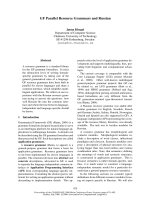
(Fig. 2A, lane 4; Fig. 2C, lane 3). In Fig. 2A lane 4,
the relative staining of large (80 kDa) and small
(21 kDa) subunits is consistent with their 1 : 1 stoichi-
ometry. When an immunoblot of the gel, shown in
Fig. 2A, was probed with antibody against AFP, the
only protein band detected was 28 kDa AFP-tagged
small subunit (Fig. 2B, lane 1). Similarly, when a
duplicate immunoblot was probed with antibody
against His-tag, the only protein band detected was
80 kDa His-tagged large subunit (Fig. 2B, lane 2).
One immediate advantage of the type III AFP-
tagged small subunit construct is the increase in its
molecular mass from 21 to 28 kDa, which readily
distinguishes it from domain IV constructs. Thus, in
lane 3 of Fig. 2C, the upper 28 kDa band of the small
subunit is well separated from the lower, more abun-
dant His6-tagged calpain 1 domain IV. Although the
presence of AFP-tagged small subunit in the affinity-
purified His6-tagged calpain 1 domain IV shows that
the two different PEF domains form heterodimers, the
relative staining of these two bands suggests that
calpain 1 domain IV is present in excess.
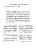
ABC
Fig. 1. Three possible scenarios derived from coexpression of recombinant PEF fusion proteins. (A) Homodimer model of His6-tagged PEF
domain. (B) Homodimer model of type III AFP-tagged PEF domain (calpain small subunit domain VI). (C) Heterodimer model of fusion protein
containing type III AFP-tagged (blue) small subunit (cyan) forming a dimer with His6-tagged (light brown) PEF domain (orange). Rat small
subunit (1AJ5) was used for modeling. All structures were drawn with
PYMOL [51].
Table 1. Screening results of domain IV constructs from calpains 1, 3, 8, 9, 11, 12, and 13. Column 1: calpains used for screening. Col-
umn 2: number of constructs designed and cloned. Column 3: number of constructs expressed. Column 4: yields of constructs when
expressed alone in the absence of human small subunit. Column 5: results from coexpression of the domain IV construct with human small
subunit. Column 6: results obtained by biophysical analysis of these constructs. NA, data not available; +++, very high expression; ++, high
expression; +, low expression.
Construct Cloned Expressed Yield Coexpression yield Dimerization
Calpain 1 domain IV 1 1 +++ +++ Heterodimer
Calpain 3 domain IV 5 5 +++
++
+++ Homodimer
Calpain 8 domain IV 5 Nil No expression No expression NA
Calpain 9 domain IV 14 11 +++ +++ Heterodimer
Calpain 11 domain IV 11 Nil No expression No expression NA
Calpain 12 domain IV 6 Nil No expression No expression NA
Calpain 13 domain IV 12 12 +++
++
+++ Homodimer
a
a
Predominant form found as homodimer.
R. Ravulapalli et al. Calpain heterodimerization and homodimerization
FEBS Journal 276 (2009) 973–982 ª 2008 The Authors Journal compilation ª 2008 FEBS 975
Calpain 3 domain IV is a homodimer
Calpain 3 domain IV is suggested to favor homodi-
merization, even though small subunit-containing
calpains are produced in muscle cells. Earlier studies
showed that recombinant calpain 3 domain IV, when
expressed in isolation, formed a homodimer [30]. In
further support of this argument, we show below that
His6-tagged recombinant calpain 3 domain IV coex-
pressed with type III AFP-tagged human small subunit
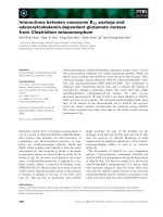
(28 kDa) exclusively forms a homodimer. Upon purifi-
cation by Ni
2+
–nitrilotriacetate–agarose chromato-
graphy, the 28 kDa subunit was not detected in the
imidazole-eluted fraction along with calpain 3 domain
IV (Fig. 3A, lane 4). The 28 kDa subunit was present
in the fractions that did not bind to the Ni
2+
–nitrilo-
triacetate–agarose column (Fig. 3A, lane 2). Indeed, it
was the most abundant protein in the flow-through
fraction from that column.
Calpain 9 domain IV forms a heterodimer with
the small subunit
The recombinant calpain 9 domain IV construct has
200 amino acids, including its His6 N-terminal tag. It
has a theoretical pI of 5.71 and a calculated molecular
mass of 23 130 Da. The amino acid sequence is 43%
identical with domain IV of calpain 1, and 40% identi-
cal with the small subunit (28 kDa). When calpain 9
domain IV was coexpressed with the 28 kDa small
subunit fusion protein, it formed a heterodimer. Both
subunits were detected in the imidazole-eluted fraction
(Fig. 3B, lane 3). Their stoichiometry was close to
1 : 1. To confirm the identity of the two subunits, the
gel was immunoblotted and probed with the two anti-
bodies used in Fig. 2B. The antibody against AFP
detected the upper band as a 28 kDa AFP-tagged
small subunit (Fig. 3C, lane 1). Similarly, the antibody
against His-tag reacted with the N-terminally His6-
tagged calpain 9 domain IV (Fig. 3C, lane 2).
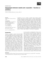
In the converse approach using ice affinity purifica-
tion, His6-tagged calpain 9 domain IV was included in
the ice because of its heterodimerization with the AFP-
tagged small subunit (Fig. 4, lane 2). Here, the amount
of the His6-tagged calpain 9 domain IV in the ice frac-
tion was slightly lower than would be predicted from
the expected 1 : 1 stoichiometry with the small subunit
as seen in the liquid fraction (Fig. 4, lane 3). This
seems to be due to a small amount of subunit dissocia-
tion that occurs as the ice grows over and pushes past
the adsorbed AFP-tagged subunit. The shearing forces
of the ice are apparently sufficient to disrupt quater-
A B C
Fig. 2. SDS ⁄ PAGE and immunoblot analysis of differentially tagged calpain 1 and 2 heterodimers. (A) Lane 1: molecular mass standards indi-
cated at the side of the gel. Lanes 2, 3 and 4: flow-through, wash and eluate samples, respectively, from the Ni
2+
–nitrilotriacetate–agarose
column chromatography of 80 kDa subunit (C105S-m-80 kDa) (triangle) coexpressed with 28 kDa AFP-tagged small subunit (dot). (B) Lanes 1
and 2: immunoblots of lane 4 from (A) probed with antibody against AFP and antibody against His-tag, respectively. (C) Lanes 1, 2 and 3:
flow-through, wash and eluate samples from the Ni
2+
–nitrilotriacetate–agarosecolumn chromatography of calpain 1 domain IV (C1DIV)
(square) coexpressed with 28 kDa AFP-tagged small subunit (dot). Both coexpressed constructs are predominantly detected in fractions
eluted with imidazole.
Calpain heterodimerization and homodimerization R. Ravulapalli et al.
976 FEBS Journal 276 (2009) 973–982 ª 2008 The Authors Journal compilation ª 2008 FEBS
nary structure in a portion of the dimers, but do not
break covalent bonds between the AFP moiety and a
fusion partner [36]. A similar partial dissociation of
subunits was seen during ice affinity purification of full
length l-calpain heterodimerized to the AFP-tagged
subunit (results not shown). The control experiment in
this series showed that His6-tagged calpain 9 domain
IV, when expressed alone, was not included in the ice
but remained in the liquid fraction (Fig. 4, lanes 4 and
5, respectively).
Calpain 13 domain IV
The recombinant calpain 13 domain IV construct con-
tains 174 amino acids, including the His6 N-terminal
tag. It has a theoretical pI of 6.75 and a calculated
molecular mass of 19 901 Da. Unlike other calpain
PEF domains, it has low sequence identity with domai-
n IV of calpain 1 (28%) and the small subunit (29%).
When the recombinant calpain 13 domain IV construct
was coexpressed with type III AFP-tagged human
small subunit (28 kDa), calpain 13 domain IV was pre-
dominantly seen in the eluant. The 28 kDa small sub-
unit was mainly observed in the flow-through,
although a faint band was seen in the wash and eluant
(Fig. 5). On the basis of these SDS ⁄ PAGE results, a
small amount of heterodimer is produced but cal-
pain 13 domain IV is predominantly a homodimer.
Discussion
The PEF domain was first described in calpain [37–
39], and has since been found in other proteins such as
ALG-2, grancalcin, sorcin and peflin [40,41]. It is char-
acterized by having a fifth EF-hand available to pair
with that of another PEF domain to form heterodi-
mers or homodimers. More than half of the human
calpain isoforms (1, 2, 3, 8, 9, 11, 12 and 13) have a
PEF domain. Of these, the ubiquitous well-studied cal-
pains 1 and 2 are known to form heterodimers with
the small subunit PEF domains. However, previous
investigations on calpain 3 suggest that PEF domain-
containing calpain isoforms need not necessarily form
a heterodimer, like calpains 1 and 2. In this study, we
set out to determine what kind of dimers the different
calpain isoforms make.
Modeling studies using shape complementarity as a
tool to measure the likelihood of forming a hetero-
dimer or homodimer were performed using calpain 2,
the previously generated model of calpain 3 domain IV
A
B
C
Fig. 3. SDS ⁄ PAGE and immunoblot analysis of calpain 3 domain IV (C3DIV) and calpain 9 domain IV (C9DIV) samples coexpressed with
small subunit. (A) Lane 1: molecular mass standards indicated at the side of the gel. Lanes 2, 3 and 4: flow-through, wash and eluate sam-
ples, respectively, from the Ni
2+
–nitrilotriacetate–agarose column chromatography of His-tagged (C3DIV) (triangle) coexpressed with 28 kDa
AFP-tagged small subunit (dot). Only the C3DIV domain is detected in the eluant. (B) Lanes 1–3: flow-through, eluate and wash samples
from the Ni
2+
–nitrilotriacetate–agarose column of His-tagged C9DIV (square) coexpressed with 28 kDa AFP-tagged small subunit (dot). Both
the human small subunit and C9DIV are present in the eluant. (C) Lanes 1 and 2: immunoblots of lane 3 from (B) probed with antibody
against AFP and antibody against His-tag, respectively.
R. Ravulapalli et al. Calpain heterodimerization and homodimerization
FEBS Journal 276 (2009) 973–982 ª 2008 The Authors Journal compilation ª 2008 FEBS 977
[30], and the small subunit structures as a guide. In
addition, models were generated for artificial structures
of the calpain 3 domain IV–small subunit heterodimer
and of the calpain 2 domain IV homodimer. Shape
complementarity values differed only slightly between
the different dimers. In order of best to worst, the
complementarity values were calpain 3 domain IV
homodimer (0.751), small subunit homodimer (0.751),
calpain 3 domain IV–small subunit heterodimer
(0.734), calpain 2 domain IV–small subunit hetero-
dimer (0.734) and calpain 2 domain IV homodimer
(0.715). These values are not significantly different
from each other, and therefore do not appear to pro-
vide a method for distinguishing correct from incorrect
dimers. Comparison of the buried surface areas for the
various complexes also shows little variation, with the
calpain 2 domain IV homodimer displaying the small-
est surface area (average value of 1182 A
˚
2
) as com-
pared to the others (average values ranging from 1311
to 1391 A
˚
2
). As tight packing of residues involved in
the dimerization interfaces might not be the only
factor influencing dimer formation, we used experimen-
tation to distinguish which isoforms form heterodimers
or homodimers.
The recombinant small subunit domain VI has a
molecular mass of 21 264 Da, and forms a homo-
dimer when expressed alone [42]. Its molecular mass
is close in value to those of isolated calpain PEF
domains (domain IV), making it hard to distinguish
whether they formed homodimers or heterodimers
when coexpressed. In order to overcome this uncer-
tainty, we devised a differential tag approach whereby
all the calpain PEF domains contain a His6 N-termi-
nal tag and the small subunit has an N-terminal
type III AFP tag (7 kDa), allowing us to distinguish
these two domains by size. Like the rat small subunit,
the recombinant 28 kDa human small subunit fusion
protein formed a homodimer when expressed alone
(results not shown).
Fig. 4. Ice affinity purification of type III AFP-tagged small subunit
and calpain 9 domain IV (C9DIV). Lane 1: molecular mass standards
indicated at the side of the gel. Lanes 2 and 3: equal volumes of
the ice and liquid fractions obtained from the distribution of coex-
pressed 28 kDa AFP-tagged small subunit (dot) with His-tagged
C9DIV (square). Lanes 4 and 5: equal volumes of the ice and liquid
fractions obtained from the distribution of His-tagged C9DIV
(square) in the absence of 28 kDa AFP-tagged small subunit.
Fig. 5. SDS ⁄ PAGE analysis of calpain 13 domain IV (C13DIV) sam-
ples from the Ni
2+
–nitrilotriacetate–agarose column. Lane 1: molec-
ular mass standards indicated at the side of the gel. Lanes 2, 3 and
4: flow-through, wash and eluate fractions from the column,
respectively. The 28 kDa subunit and C13DIV proteins are indicated
by dot and square symbols, respectively.
Calpain heterodimerization and homodimerization R. Ravulapalli et al.
978 FEBS Journal 276 (2009) 973–982 ª 2008 The Authors Journal compilation ª 2008 FEBS
Calpain 1, 3, 9 and 13 PEF domains were success-
fully cloned and coexpressed as soluble recombinant
products. However, numerous attempts to express cal-
pain 8, 11 and 12 PEF domain constructs in E. coli
were unsuccessful, and thus the dimerization potential
of these PEF domains could not be analyzed. As the
wild-type calpains 1 and 2 are both known to form
heterodimers, we used calpain 2 large subunit and cal-
pain 1 domain IV as controls in our experiments. Even
in the absence of its adjacent domains, calpain 1
domain IV formed a heterodimer with the small sub-
unit, rather than a homodimer. It should be noted that
this construct lacks the N-terminal anchor peptide,
which, on the basis of the structure of calpain 2
[19,21], should make additional heterodimerization
contacts between the large and small subunits.
Recombinant calpain 3 domain IV was previously
shown to form a homodimer when expressed alone [30].
In this study, it was coexpressed with small subunit
(28 kDa) but still formed a homodimer, further support-
ing the argument that calpain 3 is a natural homodimer.
Calpain 9 has been previously suggested to form a hete-
rodimer when coexpressed with small subunit in the
baculovirus expression system [43]. Coexpression of
recombinant proteins calpain 9 domain IV and small
subunit fusion product (28 kDa) led these proteins to
associate as a heterodimer, in agreement with these pre-
vious studies. As with calpain 1, the absence of the other
domains in the large subunit did not alter the propensity
of calpain 9 domain IV to heterodimerize. When
expressed alone, calpain 9 domain IV formed an oligo-
mer, unlike other PEF domains (results not shown).
Calpain 13 is a tissue-specific calpain expressed predom-
inantly in testis and lung. Its physiological role is not
well understood, and its dimerization state is unknown
[8]. Calpain 13 domain IV appeared as a predominant
homodimer when coexpressed with small subunit fusion
protein (28 kDa), although there were small amounts of
heterodimer present in the eluate from the Ni
2+
–nitri-
lotriacetate–agarose column.
Most of the PEF domains in calpain isoforms share
a high degree of sequence identity; however, it is not
clear why they prefer one form of dimerization over
the other. Further analysis of these constructs by
determining their structure through crystallography
may help us to gain more insight into the preference
for homodimerization versus heterodimerization.
Meanwhile, on the basis of these results, we predict
that calpain 9 can be bound and silenced by calpasta-
tin. Silencing of calpains 3 and 13 would require the
simultaneous binding of two calpastatin inhibitory
domains. Although this is a theoretical possibility,
especially as calpastatin has four inhibitory domains
and is an intrinsically unstructured protein, the
absence of a small subunit in these two calpains would
deprive calpastatin of one of its three calpain-binding
sequences. The loss of this binding site would signifi-
cantly weaken the overall binding interaction.
Experimental procedures
High-throughput cloning
The cDNA fragments encoding the domain IV regions of cal-
pains 1, 9, 11, 12 and 13 were obtained by PCR amplification
of full-length cDNA templates of human calpains 1, 9, 11, 12
and 13 obtained from the Mammalian Gene Collection,
using Expand high-fidelity DNA polymerase (Roche, India-
napolis, IN, USA). Human calpain 8 domain IV was
obtained by PCR amplification of reverse transcripts
(RT-PCR) of total RNA from human stomach (Stratagene,
La Jolla, CA, USA), using an RT-Thermoscript kit (Invitro-
gen, Carlsbad, CA, USA) and Expand high-fidelity DNA
polymerase. Human calpain 3 domain IV was obtained as
previously described [30]. Multiple constructs were designed
for each of these domains. The amplified fragments encoding
domain IV regions of calpains 1, 8, 9, 11, 12 and 13 were
inserted using the infusion ligation independent cloning
system (BD Biosciences, Mountainview, CA, USA) into a
modified pET28-LIC expression vector (EMD-Novagen,
Gibbstown, NJ, USA) using a 96-well format high-through-
put approach [44], downstream of the nucleotide sequence
encoding MGSSHHHHHHSSGLVPRLGS. This 20 amino
acid sequence contains a hexahistidine tag (His6-tag) and a
thrombin cleavage site.
Type III AFP-tagged human small subunit
The cDNA fragment encoding domain VI of the human
small subunit was obtained by PCR amplification of reverse
transcripts (RT-PCR) of total RNA from human stomach
(Stratagene, La Jolla, CA, USA), using an RT-Thermo-
script kit (Invitrogen, Carlsbad, CA, USA) and Expand
high-fidelity DNA polymerase (Roche, Indianapolis, IN,
USA). The amplified product was cloned into the modified
pET vector (pAC-pET) as previously described [45]. The
type III AFP sequence was previously prepared by gene
synthesis [46]. It was cloned into the pAC-pET vector 5¢ of
the truncated 21 kDa subunit sequence. At the protein
level, the two domains are joined by a linker of three
alanine residues.
Protein expression and purification by
Ni
2+
–nitrilotriacetate–agarose
The pET28-LIC vectors encoding the domain IV regions
were transformed along with the pAC-pET plasmid
R. Ravulapalli et al. Calpain heterodimerization and homodimerization
FEBS Journal 276 (2009) 973–982 ª 2008 The Authors Journal compilation ª 2008 FEBS 979
containing the small subunit fusion construct into E. coli
BL21(DE3) cells (Novagen) by electroporation. The trans-
formed cells were grown in 1 L of LB medium under kana-
mycin and ampicillin selection. The cells were grown to a
D
600 nm
of 0.8–1.0 at 37 °C. Protein expression was induced
at 16 °C using 0.4 mm isopropyl thio-b-d-galactoside for
16 h. The cells were collected by centrifugation, resuspended
in lysis buffer [25 mm Tris ⁄ HCl, pH 7.6, 5 mm EDTA, 5%
(v ⁄ v) glycerol, 10 m m 2-mercaptoethanol, and 0.1 mm phen-
ylmethanesulfonyl fluoride], and lysed by sonication. The
resulting lysate was clarified by centrifugation at 27 000 g
for 45 min. The supernatant obtained was incubated with
5mL Ni
2+
–nitrilotriacetate–agarose resin (Qiagen, Chats-
worth, CA, USA) for 30 min at 4 °C with constant stirring.
The Ni
2+
–nitrilotriacetate–agarose resin was later trans-
ferred to a column and washed with N-buffer (50 mm
Tris ⁄ HCl, pH 7.6, 100 mm NaCl, 5 mm imidazole, and
0.01% sodium azide). His6-tagged proteins were eluted with
the lysis buffer containing 250 mm imidazole. The samples
collected were later analyzed by SDS ⁄ PAGE. The inactive
recombinant rat calpain 2 large subunit (C105S-m-80 kDa)
was also coexpressed with the AFP-tagged small subunit
and purified as described previously [45].
Ice affinity purification
Ice affinity purification [36] was explored as a way of isolat-
ing and identifying products containing the type III AFP
fusion. In this method, the AFP fusion protein was
adsorbed from solution (50 mL) into growing polycrystal-
line ice frozen onto a cooled brass cold finger. The growth
of the ice was controlled by circulating cold ethylene glycol
solution through the hollow cold finger. After a thin layer
of ice ( 1 mm) had initially formed on the cold finger, it
was immersed in the AFP-containing solution prechilled to
1 °C in an insulated beaker. The solution was gently mixed
using a stir bar, and the temperature of the cold finger was
gradually reduced at a linear rate ()0.5 to )2.5 °C over
36 h), using a temperature-programmable water bath (Ne-
slab), until approximately half to two-thirds of the volume
was incorporated into the ice hemisphere. The ice hemi-
sphere was then removed from the liquid and allowed to
melt for 10 min to remove any protein that was nonspe-
cifically bound to the surface of the ice. The ice hemisphere
was melted to release the AFP. Samples (2 and 5 lL) from
both melted ice (ice fraction) and leftover liquid (liquid
fraction) were analyzed by SDS ⁄ PAGE [47].
Modeling studies
Shape complementarity of various dimer structures and
models was calculated using the program sc from the ccp4
program suite [48]. Crystal structures of the rat small sub-
unit homodimer (Protein Data Bank code: 1dvi) and of the
human calpain 2 heterodimer (Protein Data Bank code:
1kfu) were used as references. Homology models of the cal-
pain 3 domain IV homodimer, the heterodimer of calpain 3
domain IV with the small subunit and of a calpain 2
domain IV homodimer were generated using the program
modeller 9v3 [49]. The best of 100 models were then used
in an energy minimization and molecular dynamics proto-
col using the program gromacs 3.3 [50]. The protein was
solvated, energy minimized using the steepest descents pro-
tocol, and subjected to position-restrained molecular
dynamics to relax the solvent. This was followed by a 2 ns
molecular dynamics simulation. Structures were extracted
from the trajectory every 20 ps, and the surface comple-
mentarity at the dimer interface was calculated with the
program sc from the ccp4 program suite [48]. The average
sc value from these 100 structures is reported. For compar-
ison, the same molecular dynamics protocol was used on
the crystal structures of the rat small subunit homodimer
(Protein Data Bank code: 1dvi) and of the human calpain 2
heterodimer (Protein Data Bank code: 1kfu).
Immunoblotting
Immunoblotting was performed using 10% Tris ⁄ Tricine
SDS ⁄ PAGE gels transferred onto poly(vinylidene difluoride)
membranes. Polyclonal antibodies against the His-tag and
against type III AFP were raised in rabbits. The secondary
antibody was anti-(rabbit IgG) conjugated to horseradish
peroxidase (Promega, Madison, WI, USA), which was
detected by ECL (Perkin-Elmer, Fremont, CA, USA).
Acknowledgements
This research was funded by a grant to P. L. Davies
from the Canadian Institutes for Health Research.
P. L. Davies holds a Canada Research Chair in Pro-
tein Engineering. The Structural Genomics Consortium
is a registered charity (number 1097737) that receives
funds from the Canadian Institutes for Health
Research, the Canadian Foundation for Innovation,
Genome Canada through the Ontario Genomics Insti-
tute, GlaxoSmithKline, Karolinska Institute, the Knut
and Alice Wallenberg Foundation, the Ontario Inno-
vation Trust, the Ontario Ministry for Research and
Innovation, Merck & Co., Inc., the Novartis Research
Foundation, the Swedish Agency for Innovation Sys-
tems, the Swedish Foundation for Strategic Research,
and the Wellcome Trust.
References
1 Goll DE, Thompson VF, Li H, Wei W & Cong J
(2003) The calpain system. Physiol Rev 83, 731–801.
2 Pinter M, Stierandova A & Friedrich P (1992) Purifica-
tion and characterization of a Ca(2+)-activated thiol
Calpain heterodimerization and homodimerization R. Ravulapalli et al.
980 FEBS Journal 276 (2009) 973–982 ª 2008 The Authors Journal compilation ª 2008 FEBS
protease from Drosophila melanogaster. Biochemistry 31,
8201–8206.
3 Barnes TM & Hodgkin J (1996) The tra-3 sex determi-
nation gene of Caenorhabditis elegans encodes a mem-
ber of the calpain regulatory protease family. EMBO J
15, 4477–4484.
4 Huang X, Czerwinski E & Mellgren RL (2003) Purifica-
tion and properties of the Dictyostelium calpain-like
protein, Cpl. Biochemistry 42, 1789–1795.
5 Margis R & Margis-Pinheiro M (2003) Phytocalpains:
orthologous calcium-dependent cysteine proteinases.
Trends Plant Sci 8, 58–62.
6 Dear N, Matena K, Vingron M & Boehm T (1997)
A new subfamily of vertebrate calpains lacking a
calmodulin-like domain: implications for calpain
regulation and evolution. Genomics 45, 175–184.
7 Ersfeld K, Barraclough H & Gull K (2005) Evolution-
ary relationships and protein domain architecture in an
expanded calpain superfamily in kinetoplastid parasites.
J Mol Evol 61, 742–757.
8 Farkas A, Tompa P & Friedrich P (2003) Revisiting
ubiquity and tissue specificity of human calpains. Biol
Chem 384, 945–949.
9 Glading A, Lauffenburger DA & Wells A (2002)
Cutting to the chase: calpain proteases in cell motility.
Trends Cell Biol 12, 46–54.
10 Neumar RW, Xu YA, Gada H, Guttmann RP & Siman
R (2003) Cross-talk between calpain and caspase prote-
olytic systems during neuronal apoptosis. J Biol Chem
278, 14162–14167.
11 Janossy J, Ubezio P, Apati A, Magocsi M, Tompa P &
Friedrich P (2004) Calpain as a multi-site regulator of
cell cycle. Biochem Pharmacol 67, 1513–1521.
12 Suzuki T, Okumura-Noji K, Ogura A, Tanaka R,
Nakamura K & Kudo Y (1992) Calpain may produce
a Ca(2+)-independent form of kinase C in long-term
potentiation. Biochem Biophys Res Commun 189, 1515–
1520.
13 Cox NJ, Hayes MG, Roe CA, Tsuchiya T & Bell GI
(2004) Linkage of calpain 10 to type 2 diabetes:
the biological rationale. Diabetes 53(Suppl. 1),
S19–S25.
14 Kuwako K, Nishimura I, Uetsuki T, Saido TC &
Yoshikawa K (2002) Activation of calpain in cultured
neurons overexpressing Alzheimer amyloid precursor
protein. Brain Res Mol Brain Res 107, 166–175.
15 Enns D, Karmazyn M, Mair J, Lercher A, Kountchev J
& Belcastro A (2002) Calpain, calpastatin activities and
ratios during myocardial ischemia–reperfusion. Mol Cell
Biochem 241, 29–35.
16 Richard I, Broux O, Allamand V, Fougerousse F,
Chiannilkulchai N, Bourg N, Brenguier L, Devaud C,
Pasturaud P, Roudaut C et al. (1995) Mutations in the
proteolytic enzyme calpain 3 cause limb-girdle muscular
dystrophy type 2A. Cell 81, 27–40.
17 Yoshikawa Y, Mukai H, Hino F, Asada K & Kato I
(2000) Isolation of two novel genes, down-regulated in
gastric cancer. Jpn J Cancer Res 91, 459–463.
18 Wang KK & Yuen PW (1994) Calpain inhibition: an
overview of its therapeutic potential. Trends Pharmacol
Sci 15, 412–419.
19 Hosfield CM, Elce JS, Davies PL & Jia Z (1999) Crystal
structure of calpain reveals the structural basis for
Ca(2+)-dependent protease activity and a novel mode
of enzyme activation. EMBO J 18, 6880–6889.
20 Hosfield CM, Ye Q, Arthur JS, Hegadorn C, Croall
DE, Elce JS & Jia Z (1999) Crystallization and X-ray
crystallographic analysis of m-calpain, a Ca2+-depen-
dent protease. Acta Crystallogr D Biol Crystallogr 55,
1484–1486.
21 Strobl S, Fernandez-Catalan C, Braun M, Huber R,
Masumoto H, Nakagawa K, Irie A, Sorimachi H,
Bourenkow G, Bartunik H et al. (2000) The crystal
structure of calcium-free human m-calpain suggests an
electrostatic switch mechanism for activation by
calcium. Proc Natl Acad Sci USA 97, 588–592.
22 Moldoveanu T, Hosfield CM, Lim D, Elce JS, Jia Z
& Davies PL (2002) A Ca(2+) switch aligns the active
site of calpain. Cell 108, 649–660.
23 Reverter D, Strobl S, Fernandez-Catalan C, Sorimachi
H, Suzuki K & Bode W (2001) Structural basis for
possible calcium-induced activation mechanisms of
calpains. Biol Chem 382, 753–766.
24 Tompa P, Emori Y, Sorimachi H, Suzuki K & Fried-
rich P (2001) Domain III of calpain is a Ca2+-regu-
lated phospholipid-binding domain. Biochem Biophys
Res Commun 280, 1333–1339.
25 Suzuki K, Hata S, Kawabata Y & Sorimachi H (2004)
Structure, activation, and biology of calpain. Diabetes
53(Suppl. 1), S12–S18.
26 Waghray A, Wang DS, McKinsey D, Hayes RL &
Wang KK (2004) Molecular cloning and characteriza-
tion of rat and human calpain-5. Biochem Biophys Res
Commun 324, 46–51.
27 Tonami K, Kurihara Y, Aburatani H, Uchijima Y,
Asano T & Kurihara H (2007) Calpain 6 is involved in
microtubule stabilization and cytoskeletal organization.
Mol Cell Biol 27, 2548–2561.
28 Yorikawa C, Takaya E, Osako Y, Tanaka R, Terasawa
Y, Hamakubo T, Mochizuki Y, Iwanari H, Kodama T,
Maeda T et al. (2008) Human calpain 7 ⁄ PalBH associ-
ates with a subset of ESCRT-III-related proteins in its
N-terminal region and partly localizes to endocytic
membrane compartments. J Biochem 143, 731–745.
29 Dong B & Liu R (2008) Characterization of endoge-
nous and recombinant human calpain-10. Biochimie 90,
1362–1371.
30 Ravulapalli R, Diaz BG, Campbell RL & Davies PL
(2005) Homodimerization of calpain 3 penta-EF-hand
domain. Biochem J 388, 585–591.
R. Ravulapalli et al. Calpain heterodimerization and homodimerization
FEBS Journal 276 (2009) 973–982 ª 2008 The Authors Journal compilation ª 2008 FEBS 981
31 Kinbara K, Ishiura S, Tomioka S, Sorimachi H, Jeong
SY, Amano S, Kawasaki H, Kolmerer B, Kimura S,
Labeit S et al. (1998) Purification of native p94, a mus-
cle-specific calpain, and characterization of its autolysis.
Biochem J 335, 589–596.
32 Wendt A, Thompson VF & Goll DE (2004) Interaction
of calpastatin with calpain: a review. Biol Chem 385,
465–472.
33 Todd B, Moore D, Deivanayagam CC, Lin GD,
Chattopadhyay D, Maki M, Wang KK & Narayana
SV (2003) A structural model for the inhibition of
calpain by calpastatin: crystal structures of the native
domain VI of calpain and its complexes with calpastatin
peptide and a small molecule inhibitor. J Mol Biol 328,
131–146.
34 Hanna RA, Campbell RL & Davies PL (2008) Cal-
cium-bound structure of calpain and its mechanism of
inhibition by calpastatin. Nature 456, 409–412.
35 Jia Z, DeLuca CI, Chao H & Davies PL (1996) Struc-
tural basis for the binding of a globular antifreeze
protein to ice. Nature 384, 285–288.
36 Kuiper MJ, Lankin C, Gauthier SY, Walker VK &
Davies PL (2003) Purification of antifreeze proteins by
adsorption to ice. Biochem Biophys Res Commun 300,
645–648.
37 Lin GD, Chattopadhyay D, Maki M, Wang KK,
Carson M, Jin L, Yuen PW, Takano E, Hatanaka M,
DeLucas LJ et al. (1997) Crystal structure of calcium
bound domain VI of calpain at 1.9 A resolution and its
role in enzyme assembly, regulation, and inhibitor bind-
ing. Nat Struct Biol 4, 539–547.
38 Blanchard H, Grochulski P, Li Y, Arthur JS, Davies
PL, Elce JS & Cygler M (1997) Structure of a calpain
Ca(2+)-binding domain reveals a novel EF-hand and
Ca(2+)-induced conformational changes. Nat Struct
Biol 4, 532–538.
39 Kretsinger RH (1997) EF-hands embrace. Nat Struct
Biol 4, 514–516.
40 Maki M, Kitaura Y, Satoh H, Ohkouchi S & Shibata
H (2002) Structures, functions and molecular evolution
of the penta-EF-hand Ca2+-binding proteins. Biochim
Biophys Acta 1600, 51–60.
41 Jia J, Han Q, Borregaard N, Lollike K & Cygler M
(2000) Crystal structure of human grancalcin, a member
of the penta-EF-hand protein family. J Mol Biol 300,
1271–1281.
42 Blanchard H, Li Y, Cygler M, Kay CM, Simon J,
Arthur C, Davies PL & Elce JS (1996) Ca(2+)-binding
domain VI of rat calpain is a homodimer in solution:
hydrodynamic, crystallization and preliminary X-ray
diffraction studies. Protein Sci 5, 535–537.
43 Lee HJ, Tomioka S, Kinbara K, Masumoto H, Jeong
SY, Sorimachi H, Ishiura S & Suzuki K (1999) Charac-
terization of a human digestive tract-specific calpain,
nCL-4, expressed in the baculovirus system. Arch
Biochem Biophys 362, 22–31.
44 Benoit RM, Wilhelm RN, Scherer-Becker D &
Ostermeier C (2006) An improved method for fast,
robust, and seamless integration of DNA fragments
into multiple plasmids. Protein Expr Purif 45, 66–71.
45 Elce JS, Hegadorn C, Gauthier S, Vince JW & Davies
PL (1995) Recombinant calpain II: improved expression
systems and production of a C105A active-site mutant
for crystallography. Protein Eng 8, 843–848.
46 Chao H, Davies PL, Sykes BD & Sonnichsen FD
(1993) Use of proline mutants to help solve the NMR
solution structure of type III antifreeze protein. Protein
Sci 2, 1411–1428.
47 Marshall CB, Tomczak MM, Gauthier SY, Kuiper MJ,
Lankin C, Walker VK & Davies PL (2004) Partitioning
of fish and insect antifreeze proteins into ice suggests
they bind with comparable affinity. Biochemistry 43,
148–154.
48 Collaborative Computational Project, Number 4 (1994)
The CCP4 suite: programs for protein crystallography.
Acta Crystallogr D Biol Crystallogr 50, 760–763.
49 Sali A & Blundell TL (1993) Comparative protein mod-
elling by satisfaction of spatial restraints. J Mol Biol
234, 779–815.
50 Van Der Spoel D, Lindahl E, Hess B, Groenhof G,
Mark AE & Berendsen HJ (2005) GROMACS: fast,
flexible, and free. J Comput Chem 26, 1701–1718.
51 DeLano WL (2003) The PyMOL Molecular Graphics
System. DeLano Scientific, San Carlos, CA.
Calpain heterodimerization and homodimerization R. Ravulapalli et al.
982 FEBS Journal 276 (2009) 973–982 ª 2008 The Authors Journal compilation ª 2008 FEBS