Báo cáo khoa học: E2A participates in a fine control of pre-mature B-cell apoptosis mediated by B-cell receptor signaling via transcriptional regulation of survivin, IAP2 and caspase-8 genes pot
Bạn đang xem bản rút gọn của tài liệu. Xem và tải ngay bản đầy đủ của tài liệu tại đây (798.85 KB, 11 trang )
E2A participates in a fine control of pre-mature B-cell
apoptosis mediated by B-cell receptor signaling via
transcriptional regulation of survivin, IAP2 and
caspase-8 genes
Kenji Toyonaga
1,2,
*, Hidehiko Kikuchi
1,3,
*, Koki Yamashita
1
, Masami Nakayama
1
, Kazuo Chijiiwa
2
and Tatsuo Nakayama
1,3
1 Section of Biochemistry and Molecular Biology, Department of Medical Sciences, Miyazaki Medical College, University of Miyazaki, Japan
2 Section of Surgical Oncology and Regulation of Organ Function, Department of Medical Science, Miyazaki Medical College, University of
Miyazaki, Japan
3 Department of Life Science, Frontier Science Research Center, University of Miyazaki, Japan
It is widely known that B lymphocytes are essential to
immune responses in health and disease, and maintain
homeostasis by balancing cell viability and cell death
[1]. The molecular mechanisms of B-lymphocyte devel-
opment have mostly been studied in mammalian bone
marrow and/or peripheral lymphoid tissue (e.g. spleen).
Their development requires not only controlled lineage-
and locus-specific immunoglobulin gene recombination,
establishing unique antigen specificity of the B lympho-
cytes, but also developmental stage-specific gene expres-
sion participating in lymphoid cell proliferation and
synthesis of immune mediators [1–3]. In addition, vari-
ous factors and/or signals control various aspects of
the normal development of B lymphocytes and func-
tion of the immune system. Such developmental activi-
ties require numerous transcription factors, i.e. the
Keywords
apoptosis; B cell; caspase; E2A; survivin
Correspondence
T. Nakayama, Department of Life Science,
Frontier Science Research Center,
University of Miyazaki, 5200 Kihara,
Kiyotake, Miyazaki 889-1692, Japan
Fax: +81 985 85 6503
Tel: +81 985 85 3127
E-mail:
*These authors contributed equally to this
work
(Received 8 May 2008, revised
16 December 2008, accepted 24
December 2008)
doi:10.1111/j.1742-4658.2009.06881.x
Antigen binding to the B-cell receptor (BCR) of pre-mature B lymphocytes
induces their apoptotic cell death, but binding to the BCR of mature
B lymphocytes triggers activation and proliferation. Binding to pre-mature
B lymphocytes is thought not only to function as a mechanism to exclude
B-cell clones that possess the ability to react with self-antigen, but also to
act as a defense mechanism in auto-immune diseases. Cross-linking of
BCR of pre-mature B-cell lines, including the chicken DT40 cell line, with
anti-immunoglobulin IgG induces apoptotic cell death. Treatment with
phorbol 12-myristate 13-acetate/ionomycin, which mimics BCR stimula-
tion, is used to study intracellular signal transduction of B lymphocytes.
Here, by analyzing the E2A-deficient DT40 cell line, E2A
)/)
, we show that
E2A deficiency prevents certain levels of apoptotic cell death mediated by
BCR signaling. In addition, E2A deficiency-linked BCR signaling controls
the mimicked pre-mature B-cell apoptosis by PMA/ionomycin through ele-
vated survivin plus inhibitor of apoptosis 2 levels, and reduced caspase-3
and caspase-8 activities, resulting in increased amounts of ICAD (inhibitor
of caspase-activated DNase), compared with those in the presence of E2A,
followed by reduction of DNA fragmentation. These findings will contrib-
ute to the resolution of molecular mechanisms of negative selection of
B cells and also auto-immune diseases.
Abbreviations
AIF, apoptosis-inducing factor; AKT, acutely transforming retrovirus AKT8 in rodent T cell lymphoma; BCR, B-cell receptor; CAD, caspase-
activated DNase; EBF, early B-cell factor; FACS, fluorescence-activated cell sorter; GAPDH, glyceraldehyde 3-phosphate dehydrogenase;
GATA-3, GATA binding protein-3; HAT, histone acetyltransferase; HDAC, histone deacetylase; IAP, inhibitor of apoptosis; PARP, poly(ADP-
ribose)polymerase; PCAF, p300/CBP-associated factor; PMA, phorbol 12-myristate 13-acetate.
1418 FEBS Journal 276 (2009) 1418–1428 ª 2009 The Authors Journal compilation ª 2009 FEBS
basic helix-loop-helix transcription factors E2A, early
B cell factor (EBF), GATA-binding protein-3 (GATA-
3), Pax5, PU.1, Ikaros and Aiolos, etc [2–4]. Their
importance in B-cell development has been established
by knockout experiments on mouse hematopoietic stem
cells, in which all these functions are associated with
cessation of early stages of B-cell differentiation [3,5].
However, the expression of these factors is sustained
throughout development of lymphocytes in normal
mice after the observed block in their knockout mice
[6]. Thus, their physiological functions beyond develop-
mental arrest remain poorly understood.
Among these factors, E2A has been directly impli-
cated in transcriptional regulation of several B lineage-
specific genes, and has been shown to be essential for
Ig H- and L-chain recombination [5]. Moreover, E2A
is required to initiate the expression of some B lineage-
specific genes such as EBF, mb-1 and B29, but not to
maintain expression of these genes [5]. In general, E2A
has also been shown to promote proliferation and sur-
vival of various cell types [5,7]. Recently, it was
revealed that E2A might be a key regulator of apopto-
sis versus proliferation in lymphoid cells, in addition
to its B lineage-determining function [8–11]. E2A-defi-
cient mice develop T cell-derived lymphoma, and
enforced expression of an inhibitor of differentiation
proteins that indirectly inhibit E2A function [12,13]. In
addition, ectopic expression of E47 or E12 induced
apoptotic cell death in thymic lymphoma cells derived
from E2A-deficient mice [14]. These results indicate
that E2A might act as a tumor suppressor.
It is also accepted that B lymphocytes are suscepti-
ble to receptor- and mitochondria-initiated cell death
at various stages of peripheral differentiation and dur-
ing immune responses [1,15,16]. Recent genetic
evidence has contributed to understanding of the
BCR-dependent survival mechanism of mature B cells
[17,18]. It is also known that E2A plays important
roles in the apoptosis of B lymphocytes [9–11]. In
addition, Id3 protein, an E protein antagonist, induces
growth arrest and apoptosis in B-lymphocyte progeni-
tor cells [19]. However, the impact of B cell-specific
factors, including E2A, on the BCR-mediated apop-
tosis of pre-mature B cells remains unclear, when
apoptosis is triggered by antigen stimulation.
In order to better understand the roles of histone
acetyltransferases (HATs), histone deacetylases
(HDACs) and B cell-specific transcription factors in
B-cell functions, we have systematically generated vari-
ous homozygous mutants, including HDAC2
)/)
,
GCN5
)/)
, Aiolos
)/)
and E2A
)/)
, using gene-targeting
techniques on the DT40 cell line, which was established
from chicken pre-mature B lymphocytes [20]. Our
results show that HDAC2 controls the amount of IgM
H-chain at two stages: transcription of its gene and
alternative processing of its pre-mRNA [21]. Recently,
we revealed not only that HDAC2 upregulates gene
expression of EBF1, Pax5, Aiolos, Ikaros and HDAC7,
and down-regulates those of E2A, p300/CBP-associated
factor (PCAF), HDAC4 and HDAC5, but also that
E2A upregulates expression of IgM H- and L-chain
genes, but downregulates Aiolos, but Aiolos, EBF1,
Pax5, and Ikaros downregulate expression of these two
genes [22]. These results, together with others, indicate
that HDAC2 indirectly controls the expression of IgM
H- and L-chain genes, through upregulated transcrip-
tional regulation of EBF1, Pax5, Aiolos and Ikaros,
and downregulated transcriptional regulation of E2A.
Moreover, our results indicated that GCN5 functions as
a supervisor in normal cell-cycle progression, with com-
prehensive control over the expression of several cell
cycle-related genes, as well as apoptosis-related genes,
probably through alterations in the chromatin structure,
indicated by the changing acetylation status of core
histones surrounding these widely distributed genes [23].
Recently, we showed that GCN5 and BCR signaling
collaborate to induce apoptotic cell death of the DT40
cell line, through depletion of ICAD [inhibitor of
caspase-activated DNase (CAD)] and inhibitor of apop-
tosis 2 (IAP2), and activation of caspase activities [24].
Based on these results, in this study, as a first step
in elucidating the participation of B cell-specific factors
in pre-mature B-cell apoptosis mediated by BCR stim-
ulation by analyzing the E2A-deficient DT40 mutant
E2A
)/)
, we clarified the impact of E2A on apoptotic
cell death of the DT40 cell line, and show that E2A is
involved in fine control of pre-mature B-cell apoptosis
mediated by BCR signaling, via transcriptional regula-
tion of survivin, IAP2 and caspase-8 genes.
Results
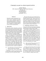
Insignificant influence of E2A deficiency on gene
expression of apoptosis-related factors
To assess the influence of E2A deficiency on gene
expression of apoptosis- and BCR signaling-related fac-
tors, and other factors, we performed semi-quantitative
RT-PCR on total RNAs prepared from DT40 and three
independent E2A
)/)
clones (Fig. 1). E2A deficiency did
not have a significant influence on transcription of most
of these genes, except for survivin (to approximately
200%), PKCa (to approximately 60%), PKCg (to
approximately 40%), PKCl (to approximately 40%)
and PKCf (to approximately 160%). In addition, we
performed immunoblot analyses to assess the influence
K. Toyonaga et al. Fine control of pre-mature B-cell apoptosis by E2A
FEBS Journal 276 (2009) 1418–1428 ª 2009 The Authors Journal compilation ª 2009 FEBS 1419
of E2A deficiency on the amounts of proteins whose
mRNA levels were altered as noted above. Consistent
with the results on mRNA levels, the protein levels for
survivin and PKCf were increased in E2A
)/)
and that
of PKCg was decreased (Fig. S1); PKCa and PKCl
could not be detected using the available antibodies. On
the other hand, transcription of various genes encoding
membrane-proximal factors, NF-jBs, transcription fac-
tors and B cell-related factors, amongst others, was not
altered in the E2A-deficient mutants (data not shown).
These insignificant effects of E2A deficiency on the
expression of numerous genes probably resulted in no
changes in the amount of apoptotic cells, as discussed
below (see Fig. 2).
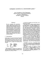
Moderate resistance to apoptosis is induced by
PMA/ionomycin in E2A
-/-
We examined the influences of phorbol 12-myristate
13-acetate (PMA)/ionomycin and etoposide on
cell-cycle progression and proliferation of DT40 and
E2A
)/)
cells. Cells cultured in the presence of PMA/
ionomycin for 24 h or etoposide for 6 h were analyzed
by fluorescence-activated cell sorter (FACS) after stain-
ing with propidium iodide (Fig. 2A). As expected, eto-
poside treatment caused apoptotic cell death for the
two cell lines at the same level because the drug inhib-
its the topoisomerase-2 activity that is essential for
DNA replication. Although PMA/ionomycin treatment
of DT40 cells resulted in cell-cycle distributions that
were quite different from those in the case of the eto-
poside treatment, it did induce apoptosis, and these
findings agree with those reported previously [24]. On
the other hand, although PMA/ionomycin treatment
of E2A
)/)
did not alter the cell-cycle distribution pat-
tern up to 24 h (cell growth was slightly delayed there-
after), the depletion of E2A slightly prevented
apoptotic cell death even in the presence of PMA/
ionomycin. To confirm these findings, we examined the
effects of PMA/ionomycin treatment on the viability
Fig. 1. Effect of E2A deficiency on gene expression of apoptosis-related factors, caspases, caspase-regulating factors, CAD/ICAD and PKCs.
Total RNAs were extracted from DT40 and three independent E2A
)/)
clones (1–3), and mRNA levels were determined by semi-quantitative
RT-PCR using appropriate primers. The chicken GAPDH gene was used as an internal control. The numbers under the panels indicate the
number of cycles used for PCR.
Fine control of pre-mature B-cell apoptosis by E2A K. Toyonaga et al.
1420 FEBS Journal 276 (2009) 1418–1428 ª 2009 The Authors Journal compilation ª 2009 FEBS
of DT40 and E2A
)/)
(Fig. 2B). As expected, the viabil-
ity of the two cell lines did not differ in the absence of
PMA/ionomycin. However, in the presence of PMA
and ionomycin, the viability of E2A
)/)
(approximately
50% at 48 h) was slightly higher than that of DT40,
which was dramatically reduced (approximately 20%
by 48 h). We undertook a comparative analysis of
changes in the morphological structure of nucleus as
an effect of PMA/ionomycin treatment in DT40 and
E2A
)/)
(Fig. 2C). Nuclear fragmentation, another
characteristic of apoptosis, was partially hindered in
E2A
)/)
, but it was clearly detected for DT40 in the
presence of PMA/ionomycin. These results show that
E2A deficiency leads to blockage of the induced apop-
totic cell death that is seen in the DT40 cell line when
treated with PMA/ionomycin.
Influence of depletion of B cell-specific transcrip-
tion factors, HDACs and HATs on resistance to
apoptosis induced by PMA/ionomycin treatment
Using gene-targeting techniques, we systematically gen-
erated several homozygous DT40 mutants that lacked
genes encoding B cell-specific transcription factors,
HDACs or HATs [20,22]. Our previous results
revealed that GCN5 and PMA/ionomycin treatment
AB
C
Fig. 2. Analyses of apoptosis in E2A
)/)
. (A) Effects of PMA/ionomycin and etoposide treatments on cell-cycle distributions of DT40 and
E2A
)/)
. DT40 and E2A
)/)
cells treated with etoposide (10 lgÆmL
)1
) for 6 h, or with PMA (10 ngÆmL
)1
) plus ionomycin (1 lM) for 24 h, were
processed for DNA content analysis by propidium iodide staining. Nuclei were analyzed by flow cytometry (FACSCalibur, Becton Dickinson
and Company, Franklin Lakes, NJ, USA), and data for DT40 and E2A
)/)
(clone 1) were plotted on linear histograms as relative cell number
(y axis) against red fluorescence intensity (x axis). The percentages of the various cell-cycle phases (sub-G
1
,G
1
, S and G
2
/M) for for DT40
and E2A
)/)
(clone 1), together with those of two other E2A
)/)
clones (2 and 3) are indicated in the table. (B) Sensitivity of DT40 (circles) and
E2A
)/)
(squares, triangles and diamonds) to PMA/ionomycin-mediated apoptotic cell death. Cells were resuspended in DMEM containing
10% v/v fetal bovine serum, and treated with (filled symbols) or without (open symbols) 10 ngÆmL
)1
PMA plus 1 lM ionomycin at 37 °C for
up to 48 h. Viable cells were counted by the trypan blue dye exclusion method. Data represent the mean of two separate experiments, and
error bars indicate the standard deviation. (C) Morphology of DT40 and E2A
)/)
(clone 1) cells treated with PMA/ionomycin. Cells were
cultured for 24 h without (no treatment) or with PMA/ionomycin (PMA/ionomycin), and their nucleus forms were analyzed by microscopy.
K. Toyonaga et al. Fine control of pre-mature B-cell apoptosis by E2A
FEBS Journal 276 (2009) 1418–1428 ª 2009 The Authors Journal compilation ª 2009 FEBS 1421
cooperatively induce apoptotic cell death in the DT40
cell line [24]. As a first step in elucidating the participa-
tion of E2A in apoptosis of DT40 cells, we examined
the effects of PMA/ionomycin treatment for these
homozygous DT40 mutants by FACS after staining
with propidium iodide and/or determination of cell
viability. Detailed information on the generation of
mutants lacking Helios, Pax5, MORF, MOZ and
MOZ/MORF will be shown elsewhere. As shown in
Table 1, depletion of HDAC2, as well as of E2A and
GCN5, prevented the apoptosis induced by PMA/ion-
omycin treatment, but depletion of Aiolos promoted
such apoptosis. No changes were detected in the
remaining single mutants lacking EBF, Helios, Pax5,
HDAC1, HDAC7, SIRT1, SIRT2, PCAF, HAT1,
MORF or MOZ, or in a double knockout mutant
lacking MOZ and MORF. These findings suggest that,
among the B cell-specific transcription factors, HDACs
and HATs examined, E2A, Aiolos, HDAC2 and
GCN5 preferentially participate in control of the apop-
totic cell death induced by PMA/ionomycin treatment
of the DT40 cell line.
Up- and downregulation of expression of the E2A
or Aiolos genes by PMA/ionomycin treatment
To determine whether or not expression of the
B cell-specific transcription factors, HDACs and HATs
mentioned above is influenced by PMA/ionomycin
treatment, DT40 and E2A
)/)
were cultured in the pres-
ence of PMA/ionomycin, and RT-PCR was performed
(Fig. 3 and Fig. S2). E2A deficiency dramatically
decreased the mRNA level of Aiolos (to approximately
30%), but did not have any effect on transcripts of
the remaining B cell-specific transcription factors, or
HDACs and HATs. On the other hand, PMA/ionomy-
cin treatment dramatically altered gene expression of
E2A in DT40 (to approximately 320% by 24 h) and
that of Aiolos in both DT40 and E2A
)/)
(to less than
10% by 24 h). However, in the two cell lines, the treat-
ment did not have significant effects on mRNA levels
of HDAC1, HDAC2, SIRT2, HAT1 and MORF, and
slightly distinct but almost similar effects on mRNA
levels of EBF, Pax5, HDAC7, SIRT1, GCN5, PCAF
and MOZ. These findings, together with those shown
in Table 1, indicate not only that HDAC2 and GCN5
are necessary for control of the apoptosis of the DT40
cell line mediated by PMA/ionomycin treatment, but
also that their own transcription is not influenced by
PMA/ionomycin. Therefore, the apoptotic cell death
of DT40 mediated by PMA/ionomycin treatment must
be under the control of the elevated or decreased
amounts of E2A or Aiolos.
Upregulation of survivin and IAP2 gene expres-
sion and no effect on caspase-8 gene expression
by PMA/ionomycin treatment in E2A
-/-
To further clarify the molecular mechanism linked to
the apoptotic induction of the DT40 cell line coopera-
tively mediated by E2A and BCR signaling, DT40 and
E2A
)/)
were cultured in the presence of PMA/ionomy-
cin, and RT-PCR was performed for various factors
(Fig. 3 and Fig. S2). E2A deficiency did not alter the
gene expression of the apoptosis-related factors bcl-2,
bcl-xL, bak, Apaf-1, cytochrome c, acutely transform-
ing retrovirus AKT8 in rodent T cell lymphoma
(AKT), apoptosis-inducing factor (AIF) and poly
(ADP-ribose)polymerase (PARP) (also shown in
Fig. 1). The PMA/ionomycin treatment did not have a
significant influence on the mRNA levels of bcl-2, bak,
Apaf-1, cytochrome c, AKT and AIF, and had slightly
distinct but almost similar effects on the mRNA levels
of bcl-xL and PARP in both DT40 and E2A
)/)
.
Similarly, depletion of E2A showed no effects on
gene expression of CAD and ICAD (also shown in
Fig. 1). PMA/ionomycin treatment showed similar
effects on the expression of CAD and ICAD genes in
both DT40 and E2A
)/)
, i.e. the CAD mRNA level
decreased by 3 h but thereafter increased to the con-
trol level by 24 h, and the ICAD mRNA level
Table 1. Influences of depletion of B cell-specific transcription fac-
tors, HDACs and HATs on resistance to apoptosis induced by
PMA/ionomycin treatment.
Mutants Reference
Resistance for
PMA/ionomycin
B cell-specific factors
E2A
)/)
[22] ›
Aiolos
)/)
[22] fl
EBF
)/)
[22] fi
Helios
)/)
Unpublished data fi
Pax5
)/)
[22] fi
HDACs
HDAC1
)/)
[21] fi
HDAC2
)/)
[21] ›
HDAC7
)/)
[23] fi
SIRT1
)/)
[31] fi
SIRT2
)/)
[31] fi
HATs
GCN5
)/)
[23] ›
PCAF
)/)
[23] fi
HAT1
)/)
[32] fi
MORF
)/)
Unpublished data fi
MOZ
)/)
Unpublished data fi
MOZ
)/)
/MORF
)/)
Unpublished data fi
Fine control of pre-mature B-cell apoptosis by E2A K. Toyonaga et al.
1422 FEBS Journal 276 (2009) 1418–1428 ª 2009 The Authors Journal compilation ª 2009 FEBS
decreased by 3 h and thereafter remained unchanged
at 24 h in the presence of PMA/ionomycin. These
findings indicate that E2A and BCR stimulation have
no effects on gene expression of CAD and ICAD, in
contrast to the effects of GCN5 and BCR stimula-
tion [24].
With regard to caspases, E2A depletion showed no
effects on expression of caspase-3, caspase-6, caspase-
8, caspase-9 and caspase-10 genes (also shown in
Fig. 1). On the other hand, in both DT40 and E2A
)/)
,
the caspase-6 mRNA level was decreased gradually
by PMA/ionomycin treatment by 24 h, and the cas-
pase-10 mRNA level was increased by 3 h and there-
after decreased dramatically by 24 h. Expression of
caspase-3 and caspase-9 remained unchanged in the
presence of PMA/ionomycin. Interestingly, in DT40,
PMA/ionomycin treatment increased the caspase-8
mRNA level by 3 h (to approximately 160%) and
this level remained unchanged at 24 h, but the treat-
ment showed no change in the transcript level of
caspase-8 in E2A
)/)
. These findings indicate that
expression of most caspase genes is not much influ-
enced by either E2A or BCR stimulation, except that
of caspase-8.
With regard to caspase-regulating factors, depletion
of E2A increased transcription of the survivin gene (to
approximately 220%), but did not have a significant
effect on expression of the FLIP, IAP1, IAP2 and
Smac genes. However, PMA/ionomycin treatment had
distinct effects on expression of these caspase inhibi-
tors. The IAP1 or Smac mRNA levels increased or
decreased slightly by 3 h and thereafter remained
unchanged in both DT40 and E2A
)/)
. The FLIP
mRNA level was slightly decreased at 3 h (to approxi-
mately 60%) and thereafter increased to the control
level in DT40, but remained unchanged in E2A
)/)
.
Fig. 3. Effects of PMA/ionomycin treatment on gene expression of B cell-specific factors, HDACs, HATs, apoptosis-related factors, CAD/
ICAD, caspases and caspase-regulating factors. Total RNAs were extracted from PMA/ionomycin-treated DT40 and E2A
)/)
(clone 1) at indi-
cated times up to 24 h, and then the mRNA levels of appropriate genes were determined by RT-PCR. The chicken GAPDH gene was used
as a control.
K. Toyonaga et al. Fine control of pre-mature B-cell apoptosis by E2A
FEBS Journal 276 (2009) 1418–1428 ª 2009 The Authors Journal compilation ª 2009 FEBS 1423
Interestingly, whereas the IAP2 mRNA level in DT40
was significantly decreased by PMA/ionomycin treat-
ment at 3 h (to approximately 30%) and thereafter
remained unchanged, the transcript level in E2A
)/)
was unchanged at 3 h, and thereafter decreased by
24 h (to approximately 20%). Furthermore, in E2A
)/)
,
the survivin mRNA level was maintained at a high
level (approximately 220%) when treated with PMA
and ionomycin for 3 h, and thereafter decreased to
slightly higher level than that in DT40; the mRNA
level was not influenced by the drug treatment in
DT40. These findings indicate not only that E2A
downregulates transcription of the survivin gene and
has no effects on that of FLIP, IAP1, IAP2 or Smac,
but also that BCR stimulation and E2A cooperatively
control expression of FLIP, IAP2 and survivin genes.
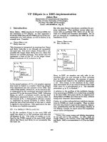
Resistance to PMA/ionomycin-mediated
apoptosis of E2A
-/-
is brought about by
increased amounts of survivin and IAP2, and
reduced activity of caspase-3
Next we examined the effect of PMA/ionomycin treat-
ment on cellular protein levels of survivin, IAP2 and
ICAD, which are proximal factors controlling CAD
activity for DNA fragmentation, by immunoblotting
using their specific antibodies (Fig. 4A). E2A deficiency
increased the protein levels of survivin (to approxi-
mately 190%), but had no effect on those of IAP2 and
ICAD. Consistent with previous results [24], PMA/ion-
omycin treatment in DT40 dramatically decreased the
protein levels of IAP2 and ICAD (to approximately
40% and less than 10%) by 24 h, but had a moderate
influence on that of survivin (approximately 60% at
24 h). Therefore, in DT40 cells treated with PMA/iono-
mycin, the time courses of alterations in the protein and
mRNA levels of survivin were virtually similar, and the
gradual reductions in protein levels of IAP2 plus ICAD
compared with acute decreases (unchanged thereafter)
of their mRNA levels agreed with previous results [24].
On the other hand, in E2A
)/)
, PMA/ionomycin
treatment did not influence the protein level of survivin
by 8 h and thereafter it decreased gradually. PMA/ion-
omycin treatment did not change the protein level of
IAP2. Further, the PMA/ionomycin treatment showed
a moderate reduction in the protein level of ICAD (to
approximately 40%) by 24 h in E2A
)/)
, i.e. the rate of
decrease in the amount of ICAD in the mutant was
slower than that in DT40. Thus, in E2A
)/)
, in the
presence of PMA/ionomycin, the time courses of alter-
ations in the mRNA and protein levels of survivin
were virtually similar up to 24 h, but the findings that
the protein levels of IAP2 or ICAD were increased or
slightly decreased by 24 h did not agree with the find-
ings that the mRNA levels of IAP2 and ICAD were
reduced slowly by 24 h or quickly by 3 h, respectively.
To resolve this discrepancy between the results
regarding the mRNA and protein levels of ICAD
(and also IAP2), we next examined the effects of PMA/
ionomycin treatment on caspase activities (Fig. 4B).
Interestingly, in E2A
)/)
, PMA/ionomycin treatment
showed slightly decreased activities of caspase-3 (to
approximately 50% by 16 h), caspase-8 (to approxi-
mately 70% by 16 h) and caspase-9 (to approximately
70% by 16 h), compared with those in DT40, probably
due to a balance of the amounts of each of the three
caspases and the inhibitors survivin and IAP2 (and also
FLIP and IAP1). The slightly decreased caspase-8
activity mediated by PMA/ionomycin treatment in
E2A
)/)
may have resulted from the balanced mRNA
(and probably protein) levels of caspase-8, which lead
to activation of pro-caspase-9, causing formation of the
active form of caspase-3, and of the inhibitors FLIP
(for caspase-8), survivin (for caspase-9) and IAP2 (for
caspase-3) [25]. In E2A
)/)
, the decreased activity of
caspase-3 mediated by PMA/ionomycin treatment must
depend on both the reduced amount of activated
caspase-3 itself as a result of decreased caspase-8 (and
probably caspase-9) activity, and the elevated protein
(and also mRNA) levels of the inhibitors survivin and
IAP2. As a result, the slow diminution of the protein
level of ICAD by PMA/ionomycin treatment in E2A
)/)
(Fig. 4A) must be due to its slight degradation medi-
ated by suppressed caspase-3 activity, although the
ICAD mRNA level was decreased when exposed for
3 h but thereafter remained unchanged, as did that in
DT40 (Fig. 3 and Fig. S2).
Because the alterations in the mRNA level of CAD
were the same in both DT40 and E2A
)/)
(Fig. 3 and
Fig. S2), and its protein could not be detected by the
available antibodies to assess CAD activity, we exam-
ined the effects of PMA/ionomycin treatment on DNA
fragmentation, a typical result of CAD activity
(Fig. 4C). The DNA fragmentation was found to be
more moderate for E2A
)/)
than that observed for
DT40, even in the presence of PMA/ionomycin up to
24 h. These results indicate that the CAD activity in
E2A
)/)
is suppressed by the moderately reduced
amount of its inhibitor ICAD compared with that in
DT40, resulted in decreased DNA fragmentation, a
characteristic of apoptosis.
Discussion
In recent years, numerous studies have been performed
to determine the physiological target genes of E2A.
Fine control of pre-mature B-cell apoptosis by E2A K. Toyonaga et al.
1424 FEBS Journal 276 (2009) 1418–1428 ª 2009 The Authors Journal compilation ª 2009 FEBS
A
B
C
Fig. 4. Analyses of mechanisms of the resistance to PMA/ionomycin-mediated apoptosis in E2A
)/)
. (A) Effects of PMA/ionomycin treatment
on protein levels of survivin, IAP2 and ICAD. Whole proteins were isolated from PMA/ionomycin-treated DT40 and E2A
)/)
at indicated times
up to 24 h, and subjected to SDS–PAGE followed by immunoblotting. Antibody binding was detected using secondary antibodies conjugated
to horseradish peroxidase, and then data analysis was performed using a luminescent image analyzer. Left panel: typical immunoblot pattern
(DT40 and E2A
)/)
clone 1). b-actin was used as a control. The apparent molecular masses of marker proteins are indicated. Right panel: time
courses of protein levels for survivin, IAP2 and ICAD after treatment with PMA/ionomycin in DT40 (circles) and three E2A
)/)
clones (1–3)
(squares, triangles and diamonds). Data are expressed as percentages of the control (DT40 at 0 h). (B) Effects of PMA/ionomycin treatment
on caspase activities. Cell lysates were prepared from PMA/ionomycin-treated DT40 (circles) and E2A
)/)
clones (1–3) (squares, triangles and
diamonds) at indicated times up to 16 h, and then caspase activity assays were performed using appropriate caspase assay kits. Absorbance
at 405 nm was measured to determine activities. Data represent the mean of two separate experiments, and error bars indicate standard
deviation. (C) Effects of PMA/ionomycin treatment on DNA fragmentation in DT40 and E2A
)/)
. DNA was isolated from DT40 and E2A
)/)
cells incubated for 0, 8, 16 and 24 h in the presence of PMA and ionomycin, and analyzed by 1.5% agarose gel electrophoresis. The sizes
of k-DNA digested with HindIII are indicated in kb. Left panel: typical electrophoregram of DNA extracted from PMA/ionomycin-treated DT40
and E2A
)/)
(clone 1). Right panel: electrophoregram of DNA extracted from PMA/ionomycin-treated DT40 and three E2A
)/)
clones (1–3)
at 16 h.
K. Toyonaga et al. Fine control of pre-mature B-cell apoptosis by E2A
FEBS Journal 276 (2009) 1418–1428 ª 2009 The Authors Journal compilation ª 2009 FEBS 1425
E2A directly activates the EBF gene [26] and regulates
expression of several genes, i.e. k5, Rag-1, Vj1 and jo,
that are involved in D-JH rearrangement, cell survival,
Igj rearrangement, etc [26,27]. E2A directly controls
IgH gene expression, and is involved in repressing the
Nfil3 and FGFR2 genes in pre-mature B lymphocytes
[28]. Thus, E2A is one of the most essential regulators
at multiple stages of B-cell development. In T lympho-
cytes, PLCc2, Cdk6, CD25, Tox, Gadd45a, Gadd45b,
Gfi1, Gfi1b, Socs1, Socs3, Id2, Eto2, Xbp1 etc have
been identified as novel E47 target genes using an E2A-
deficient lymphoma cell line [29]. Recently, we reported
that apoptosis of the chicken DT40 cell line, a pre-
mature B-cell line, is cooperatively controlled by GCN5
and BCR stimulation via complex transcriptional
regulation of a number of genes encoding BCR signal-
ing-related factors, B cell-specific factors, transcription
factors and apoptosis-related factors, indicating that
both are necessary for apoptosis of DT40 cells [24]. In
DT40 cells, BCR signaling is transduced from BCR
and membrane-proximal factors (Syk, BTK, BLNK
and PLCc2, etc), via mainly PKCd, PKCe and PKCf,
to NF-jBs (probably c-Rel and NFp50). This activated
signal mediated by BCR signaling is probably trans-
ducted separately into two apoptotic pathways, i.e.
direct transduction of the signal into the CAD/ICAD
system and transduction of the activated signal into the
caspase cascade pathway. However, understanding of
the participation of most B cell-specific factors in the
apoptotic process has remained elusive.
Lack of E2A partially prevents the apoptotic cell
death seen in DT40 cells treated with PMA/ionomycin,
which mimics BCR stimulation (Fig. 2); such apoptosis
is completely prevented by either GCN5 deficiency [24]
or HDAC2 deficiency, and is significantly accelerated
by Aiolos deficiency (Table 1). By analyzing E2A
)/)
,we
revealed that E2A upregulates the expression of PKCa,
PKCg and PKCl genes, and downregulates the expres-
sion of survivin and PKCf genes, among the numerous
factors examined (Fig. 1 and unpublished data). PMA/
ionomycin treatment increased expression of the E2A
gene and dramatically suppressed that of the Aiolos
gene in DT40, but in E2A
)/)
had no effects or similar
effects on the expression of other disrupted genes
(Fig. 3A) and genes encoding B cell-specific factors,
HDACs and HATs (our unpublished data). These
results suggest not only that, among the B cell-specific
factors tested, E2A or Aiolos participates preferentially
in suppression or acceleration of apoptosis of the DT40
cell line, but also that, among the HAT and HDAC
families tested, gene expression of GCN5 and HDAC2,
which are essential for apoptotic cell death, are not
influenced directly by PMA/ionomycin treatment. On
the other hand, in E2A
)/)
, the detected protein level of
Aiolos, which was already suppressed to a very low
level, is further reduced by the treatment, resulting in
suppression of apoptotic cell death compared with that
of DT40.
The alterations in expression of E2A and Aiolos
(and probably others) in DT40 when exposed to
PMA/ionomycin accompany altered expression of vari-
ous genes encoding apoptosis-related factors, caspases,
caspase-regulating factors and CAD/ICAD (Fig. 3 and
Fig. S2), and almost all of these results agreed with
those in a previous report [24]. In E2A
)/)
, the altered
expression of Aiolos (and probably others) in the
presence of PMA/ionomycin results in (and/or accom-
panies) slightly different effects on expression of the
genes mentioned above, i.e. the influences of PMA/ion-
omycin treatment on expression of the survivin and
IAP2 (and probably FLIP) genes were more moderate
in the mutant than in DT40, and no effect was
observed on expression of caspase-8. However, in
DT40, the PMA/ionomycin-induced alterations in gene
expression of various B cell-specific factors, HDACs,
HATs, apoptosis-related factors, caspases, caspase-reg-
ulating factors and CAD/ICAD led to changes in the
activities of caspase-3, caspase-8 and caspase-9, and in
the protein levels of IAP2 and ICAD (Fig. 4A,B), con-
sistent with previous results [24]. Interestingly, in
E2A
)/)
, the noticeable alterations in gene expression of
survivin and IAP2 accompanied by unchanged gene
expression of caspase-8 (and also various B cell-specific
factors, HDACs, HATs, apoptosis-related factors,
caspases, caspase-regulating factors and CAD/ICAD)
mediated by PMA/ionomycin results in suppression of
activities of caspase-3, caspase-8 and caspase-9, and
alterations in the protein levels of survivin, IAP2 and
ICAD (and probably FLIP) (Fig. 4A,B).
Finally, in E2A
)/)
, the slightly suppressed degrada-
tion of ICAD molecules as an effect of reduced cas-
pase-3 activity reduces CAD activity, leading to
moderate fragmentation of DNA molecules (Fig. 4C).
Thus, progress towards apoptotic cell death in E2A
)/)
is suppressed by collaboration of both BCR signaling
and E2A depletion, mainly via moderate changes in
amounts of the inhibitors survivin, IAP2 and ICAD
(and probably FLIP). Thus, E2A is involved in fine
control of pre-mature B-cell apoptosis mediated by
BCR signaling via transcriptional regulation of survi-
vin, IAP2, FLIP and caspase-8 genes.
The observations in this study regarding the par-
ticipation in apoptosis of Aiolos and HDAC2, which
are now being further studied by us, as well as that
of GCN5, amongst others, will be useful in elucidat-
ing not only the linkage between BCR signaling and
Fine control of pre-mature B-cell apoptosis by E2A K. Toyonaga et al.
1426 FEBS Journal 276 (2009) 1418–1428 ª 2009 The Authors Journal compilation ª 2009 FEBS
apoptosis cascades in pre-mature B-cell lines, but
also the molecular mechanism of negative selection
or development of B lymphocytes through cross-talk
among B-cell signaling, B cell-specific transcriptional
regulation and epigenetic chromatin topology altera-
tions. These results, combined with other findings
obtained in the future, may contribute to clinical
understanding of auto-immune diseases and B-cell
lymphomas.
Experimental procedures
Materials
PMA was purchased from Calbiochem (Darmstadt, Ger-
many) and ionomycin was purchased from Sigma (St Louis,
MO, USA). The antibodies used were anti-ICAD (Santa
Cruz Biotechnology Inc., Santa Cruz, CA, USA), anti-
cIAP-2 (Chemicon, Temecula, CA, USA), horseradish per-
oxidase-conjugated goat anti-rabbit immunoglobulin and
horseradish peroxidase-conjugated rabbit anti-mouse immu-
noglobulin (Dako Inc., Glostrup, Denmark).
Cell cultures and apoptosis induction
DT40 cells and all subclones were cultured essentially as
described previously [24]. Apoptosis was induced as follows:
cells (2 · 10
6
) in 10 mL of culture medium were incubated
with 10 ngÆmL
)1
PMA plus 1 lm ionomycin or with
10 lgÆmL
)1
etoposide at 37 °C. Viable cells were counted
by the trypan blue dye exclusion method. Flow cytometric
analyses, morphological analyses, the caspase activity assay
and the DNA fragmentation assay were performed as
described previously [24,30].
Semi-quantitative RT-PCR
Total RNAs were isolated from DT40 and its subclones.
Reverse transcription was performed using a first-strand
DNA synthesis kit (Toyobo, Osaka, Japan) at 42 °C for
20 min, followed by heating at 99 °C for 5 min. PCRs were
performed as described previously [24] using sense primers
and antisense primers synthesized according to the EST
data deposited in GenBank for the appropriate genes, and
listed in previous reports [22–24], except for SIRT1 (sense
primer 5¢-CTGTTTTTACCACCAAATCG-3¢ and antisense
primer 5¢-CAACTTGTTGCTTGTTGGAT-3¢) and SIRT2
(sense primer 5¢-ATGTCCCTCATGGGCTTCGG-3¢ and
antisense primer 5¢-TCACGGCTCTTTGTCGTCCC-3¢).
The chicken glyceraldehyde 3-phosphate dehydrogenase
(GAPDH) gene was used as an internal control. PCR prod-
ucts were subjected to 1.5% agarose gel electrophoresis,
and analyzed using an LAS-1000plus luminescent image
analyzer (Fujifilm, Tokyo, Japan).
Immunoblotting
Cells were treated with 10% trichroloacetic acid, collected
by centrifugation 20 000 g for 5 min at 4 °C, dissolved in
0.5 m Tris/HCl (pH 6.8) containing 2.5% SDS, 10% glyc-
erol and 5% 2-mercaptoethanol, and heated at 100 °C for
5 min. Immunoblotting was performed as described previ-
ously [24]. b-actin was used as a control.
Acknowledgements
We thank Y. Takami and H. Suzuki for technical sup-
port and H. K. Barman for editorial reading of the
manuscript. This work was supported in part by a
Grant-in-Aid for Scientific Research from the Ministry
of Education, Culture, Sports, Science and Technology
of Japan.
References
1 Cancro MP (2005) B cells and aging: gauging the inter-
play of generative, selective, and homeostatic events.
Immunol Rev 205, 48–59.
2 Singh H, Medina KL & Pongubala JMR (2005) Contin-
gent gene regulatory networks and B cell fate specifica-
tion. Proc Natl Acad Sci USA 102, 4949–4953.
3 Kee BL & Murre C (2001) Transcription factor regula-
tion of B lineage commitment. Curr Opin Immunol 13,
180–185.
4 Riley RL, Van der Put E, King AM, Fransca D &
Blomberg BB (2005) Deficient B lymphopoiesis in mur-
ine senescence: potential roles for dysregulation of E2A,
Pax-5, and STAT5. Semin Immunol 17, 330–336.
5 Greenbaum S & Zhuang Y (2002) Regulation of early
lymphocyte development by E2A family proteins. Semin
Immunol 14, 405–414.
6 Alinikula J, Lassila O & Nera KP (2006) DT40
mutants: models to study transcriptional regulation of
B cell development and function. In Reviews and
Protocols in DT40 Research, (Buerstedde JM &
Takeda S., eds) pp. 189–205. Springer-Verlag, Berlin,
Germany.
7 Riley RL, Blomberg BB & Frasca D (2005) B cells,
E2A, and aging. Immunol Rev 205, 30–47.
8 Ikawa T, Kawamoto H, Wright LYT & Murre C
(2004) Long-term cultured E2A-deficient hematopoietic
progenitor cells are pluripotent. Immunity 20, 349–360.
9 Lazorchak AS, Wojciechowski J, Dai M & Zhuang Y
(2006) E2A promotes the survival of precursor and
mature B lymphocytes. J Immunol 177, 2495–2504.
10 Lietz A, Janz M, Sigvardsson M, Jundt F, Dorken B &
Mathas S (2007) Loss of bHLH transcription factor
E2A activity in primary effusion lymphoma confers
resistance to apoptosis. Br J Haematol 137, 342–348.
K. Toyonaga et al. Fine control of pre-mature B-cell apoptosis by E2A
FEBS Journal 276 (2009) 1418–1428 ª 2009 The Authors Journal compilation ª 2009 FEBS 1427
11 Murre C (2005) Helix-loop-helix proteins and lympho-
cyte development. Nat Immunol 6, 1079–1086.
12 Rivera R & Murre C (2001) The regulation and func-
tion of the Id proteins in lymphocyte development.
Oncogene 20, 8308–8316.
13 Mikkers H, Allen J & Berns A (2002) Proviral activa-
tion of the tumor suppressor E2a contributes to T cell
lymphomagenesis in EmuMyc transgenic mice. Onco-
gene 21, 6559–6566.
14 Engel I & Murre C (1999) Ectopic expression of E47 or
E12 promotes the death of E2A-deficient lymphomas.
Proc Natl Acad Sci USA 96, 996–1001.
15 Carey GB, Donjerkovic D, Liu S, Hinshaw JA, Ton-
netti L, Davidson W & Scott DW (2000) B-cell receptor
and Fas-mediated signals for life and death. Immunol
Rev 176, 105–115.
16 Sen R (2006) Control of B lymphocyte apoptosis by
the transcription factor NF-jB. Immunity 25, 871–883.
17 Moscat J, Diaz-Meco MT & Rennert P (2003) NF-jB
activation by protein kinase C isoforms and B-cell func-
tion. EMBO Rep 4, 31–36.
18 Foreman AL, Van de Water J, Gougeon ML &
Gershwin ME (2007) B cells in autoimmune disease:
insights from analyses of immunoglobulin variable
(Ig V) gene usage. Autoimmun Rev 6, 387–401.
19 Kee BL, Rivera RR & Murre C (2001) Id3 inhibits
B lymphocyte progenitor growth and survival in
response to TGF-b. Nat Immunol 2, 242–247.
20 Kikuchi H, Barman HK, Nakayama M, Takami Y &
Nakayama T (2006) Participation of histones, histone
modifying enzymes and histone chaperones in verte-
brate cell functions. In Reviews and Protocols in DT40
Research, (Buerstedde JM & Takeda S., eds)
pp. 225–243. Springer-Verlag, Berlin, Germany.
21 Takami Y, Kikuchi H & Nakayama T (1999) Chicken
histone deacetylase-2 controls the amount of the IgM
H-chain at the steps of both transcription of its gene
and alternative processing of its pre-mRNA in the
DT40 cell line. J Biol Chem 274, 23977–23990.
22 Nakayama M, Suzuki H, Yamamoto-Nagamatsu N,
Barman HK, Kikuchi H, Takami Y, Toyonaga K,
Yamashita K & Nakayama T (2007) HDAC2 controls
IgM H- and L-chain gene expressions via EBF1, Pax5,
Ikaros, Aiolos and E2A gene expressions. Genes Cells
12, 359–373.
23 Kikuchi H, Takami Y & Nakayama T (2005) GCN5: a
supervisor in all-inclusive control of vertebrate cell cycle
progression through transcription regulation of various
cell cycle-related genes. Gene 347, 83–97.
24 Kikuchi H & Nakayama T (2008) GCN5 and BCR sig-
nalling collaborate to induce pre-mature B cell apopto-
sis through depletion of ICAD and IAP2 and activation
of caspase activities. Gene 419, 48–55.
25 Aggarwal BB, Bhardwaj U & Takada Y (2004) Regula-
tion of TRAIL-induced apoptosis by ectopic expression
of antiapoptotic factors. Vitam Horm 67, 453–483.
26 Kee BL, Quong MW & Murre C (2000) E2A proteins:
essential regulators at multiple stages of B-cell develop-
ment. Immunol Rev 175, 138–149.
27 Lazorchak A, Jones ME & Zhuang Y (2005) New
insights into E-protein function in lymphocyte develop-
ment. Trends Immunol 26, 334–338.
28 Greenbaum S, Lazorchak AS & Zhuang Y (2004) Dif-
ferential functions for the transcription factor E2A in
positive and negative gene regulation in pre-B lympho-
cyte. J Biol Chem 279, 45028–45035.
29 Schwartz R, Engel I, Fallahi-Sichani M, Petrie HT &
Murre C (2006) Gene expression patterns define novel
roles for E47 in cell cycle progression, cytokine-medi-
ated signaling, and T lineage development. Proc Natl
Acad Sci USA 103, 9976–9981.
30 Kikuchi H & Imajoh-Ohmi S (1995) Activation and
possible involvement of calpain, a calcium-activated
cysteine protease, in down-regulation of apoptosis of
human monoblast U937 cells. Cell Death Differ 2,
195–199.
31 Matsushita N, Takami Y, Kimura M, Tachiiri S, Ishiai
M, Nakayama T & Takata M (2005) Role of NAD-
dependent deacetylases SIRT1 and SIRT2 in radiation
and cisplatin-induced cell death in vertebrate cells.
Genes Cells 10, 321–332.
32 Barman HK, Takami Y, Ono T, Nishijima H,
Sanematsu F, Shibahara K & Nakayama T (2006)
Histone acetyltransferase 1 is dispensable for
replication-coupled chromatin assembly but contributes
to recover DNA damage created following replication
blockage in vertebrate cells. Biochem Biophys Res
Commun 345, 1547–1557.
Supporting information
The following supplementary material is available:
Fig. S1. Influences of E2A deficiency on protein levels
of survivin, PKCg and PKCf.
Fig. S2. Effects of the PMA/ionomycin treatment on
gene expression of B cell-specific factors, HDACs,
HATs, apoptosis-related factors, CAD/ICAD, caspases
and caspase-regulating factors in two E2A
)/)
clones.
This supplementary material can be found in the
online version of this article.
Please note: Wiley-Blackwell is not responsible for
the content or functionality of any supplementary
materials supplied by the authors. Any queries (other
than missing material) should be directed to the
corresponding author for the article.
Fine control of pre-mature B-cell apoptosis by E2A K. Toyonaga et al.
1428 FEBS Journal 276 (2009) 1418–1428 ª 2009 The Authors Journal compilation ª 2009 FEBS