Báo cáo khoa học: Solution structure of the bb¢ domains of human protein disulfide isomerase docx
Bạn đang xem bản rút gọn của tài liệu. Xem và tải ngay bản đầy đủ của tài liệu tại đây (806.4 KB, 10 trang )
Solution structure of the bb¢ domains of human protein
disulfide isomerase
Alexey Y. Denisov*, Pekka Ma
¨
a
¨
tta
¨
nen*, Christian Dabrowski, Guennadi Kozlov, David Y. Thomas
and Kalle Gehring
Department of Biochemistry, McGill University, and Groupe de Recherche Axe
´
sur la Structure des Prote
´
ines (GRASP), Montre
´
al, Canada
The endoplasmic reticulum (ER) is the cell compart-
ment where membrane and secretory proteins fold.
The rate-limiting step for the folding of many proteins
is the formation of disulfide bonds. As polypeptides
are synthesized, their cysteine thiols enter the oxidizing
environment of the ER and form covalent intramolec-
ular and intermolecular disulfide links. Although this
oxidative folding process occurs spontaneously [1],
non-native disulfide-bonded intermediates often occur,
acting as kinetic traps along the folding pathway [2,3].
To avoid these, the ER contains a large family of
enzymes called protein disulfide isomerases (PDIs) that
catalyze both disulfide bond formation and the rear-
rangement of incorrect disulfide bonds [4–7].
PDI family members are loosely defined by
homology to thioredoxin and ER localization. There
are at least 17 PDI family proteins in humans, 13 of
which contain CXXC active-site motifs, and 9 have
been shown to catalyze disulfide-exchange reactions
[4,5]. The best studied and most abundant member
of the family is PDI, a ubiquitous enzyme found at
very high concentrations in the ER. Its concentra-
tion has been estimated to be 10 lm in dog pancre-
atic microsomes [8], the highest of all ER resident
proteins. PDI has four thioredoxin-like domains,
a-b-b¢-a¢, where the two a domains contain catalytic
CGHC motifs, and the two b domains lack the
conserved cysteine residues and are noncatalytic. The
linkers between the domains are generally short.
The longest is a stretch of 19 amino acids between
the b¢ and a¢ domains, referred to as the x-linker
[9].
Keywords
chaperone; endoplasmic reticulum; NMR
solution structure; protein disulfide
isomerase family
Correspondence
K. Gehring, Department of Biochemistry,
McGill University, 3655 Promenade Sir
William Osler, Montreal, QC H3G 1Y6,
Canada
Fax: +1 (514) 398 7384
Tel: +1 (514) 398 7287
E-mail:
*These authors contributed equally to this
work
(Received 16 November 2008, revised 30
December 2008, accepted 30 December
2008)
doi:10.1111/j.1742-4658.2009.06884.x
Protein disulfide isomerase is the most abundant and best studied of the
disulfide isomerases that catalyze disulfide bond formation in the endoplas-
mic reticulum, yet the specifics of how it binds substrate have been elusive.
Protein disulfide isomerase is composed of four thioredoxin-like domains
(abb¢a¢). Cross-linking studies with radiolabeled peptides and unfolded pro-
teins have shown that it binds incompletely folded proteins primarily via
its third domain, b¢. Here, we determined the solution structure of the sec-
ond and third domains of human protein disulfide isomerase (b and b¢,
respectively) by triple-resonance NMR spectroscopy and molecular model-
ing. NMR titrations identified a large hydrophobic surface within the b¢
domain that binds unfolded ribonuclease A and the peptides mastoparan
and somatostatin. Protein disulfide isomerase-catalyzed refolding of
reduced ribonuclease A in vitro was inhibited by these peptides at concen-
trations equal to their affinity to the bb¢ fragment. Our findings provide a
structural basis for previous kinetic and cross-linking studies which
have shown that protein disulfide isomerase exhibits a saturable, substrate-
binding site.
Abbreviations
ER, endoplasmic reticulum; GST, glutathione S-transferase; HSQC, heteronuclear single-quantum correlation; PDI, protein disulfide
isomerase; RDC, residual dipolar coupling; RNase A, ribonuclease A.
1440 FEBS Journal 276 (2009) 1440–1449 ª 2009 The Authors Journal compilation ª 2009 FEBS
The structure of yeast PDI has been determined in
two crystal forms [10,11]. In both structures, the
protein adopts a U shape with the catalytic a and a¢
domains on the same side of the protein. Compari-
son of the two structures shows that considerable
flexibility exists in the interdomain linkers. The larg-
est difference is a twist of over 120° in the relative
orientations of the a and b domains. In one struc-
ture, the catalytic cysteines face each other; in the
other, the catalytic residues of the a domain face
away from the a¢ domain. The crystal structures also
revealed the presence of a hydrophobic pocket
(which faces inwards at the base of the U) in the b¢
domain. This b¢-domain pocket was postulated to be
the site for binding of incompletely folded proteins,
along with adjoining contiguous portions of the a, b
and a¢ domains [12]. Cross-linking studies with radio-
labelled model peptides identified a homologous,
hydrophobic binding site on the b¢ domain of human
PDI [13].
Sequence identity between human and yeast PDI for
the b and b¢ domains is < 10% (Fig. 1), making it dif-
ficult to compare the two proteins accurately. The
structures of individual a [14], b [15], a¢ (Protein Data
Bank entry code 1X5C) and b¢ [16] domains of human
PDI have been solved by NMR or X-ray crystallogra-
phy. The overall shape of full-length human PDI has
been investigated by small-angle X-ray scattering and
shown, at low resolution, to adopt a flat annular
arrangement [17].
Here, we reported the solution structure of the bb¢
fragment of human PDI and addressed the question of
how PDI recognizes unfolded proteins. Using NMR
titrations, we mapped the region of PDI that binds
unfolded proteins and we showed that peptides which
bind to this region inhibit the PDI-catalyzed refolding
of ribonuclease A (RNase A).
Results
Spectra of the human PDI-bb¢ domains
Dimerization of PDI fragments containing the hydro-
phobic b¢ domain has complicated structural studies for
more than a decade [11]. NMR spectra of the isolated
b¢ domain of human PDI showed broad lines and mul-
tiple peaks as a result of the presence of a mixture of
monomeric and dimeric forms (Fig. S1). These forms
can be separated by gel filtration but rapidly exchange
at 20–30 °C( 20% of dimers in 2 h and more than
50% of dimers in 12 h, starting with pure 0.5 mm b¢
monomer at 30 °C). The
1
H-
15
N heteronuclear single-
quantum correlation (HSQC) spectrum of PDI-b¢ was
significantly better upon the addition of hydrophobic
compounds, such as peptide ligands or detergents (e.g.
0.3% Triton X-100), that dissociate the dimer (Fig. S1).
Alternatively, the b domain moderates the tendency of
the b¢ domain to dimerize and significantly slows inter-
conversion. The monomeric form of the PDI-bb¢ frag-
ment converts into < 10% of dimers in 12 h and
< 25% of dimers in 3 days, starting with 0.5 mm of
monomers at 30 °C. Most of the
1
H-
15
N HSQC signals
of the dimeric form of PDI-bb¢ coincide with the signals
of the monomeric form or are weak as a result of the
high molecular weight of the dimer. The monomeric
form gives good-quality spectra required for structural
studies. The
1
H-
15
N spectrum of the 25 kDa PDI-bb¢
fragment shows signal dispersion typical for a well-
folded protein and allows determination of the back-
bone and side-chain NMR signal assignments [18].
Protein structure and comparison
The solution structure of the human PDI-bb¢ fragment
was calculated based on 2200 NMR-derived con-
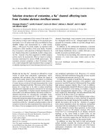
Fig. 1. Structure-based sequence alignment
of human and yeast PDI-bb¢ showing the
positions of the a-helices and the b-strands.
Color shading represents the size of the
amide chemical shift changes in human
PDI-bb¢ upon the binding of unfolded
RNase A (red, Dd > 0.10; yellow,
0.10 > Dd > 0.05 p.p.m.). Residues are
numbered from the initiator methionine
in the signal sequence.
A. Y. Denisov et al. Structure of the bb¢ domains from human PDI
FEBS Journal 276 (2009) 1440–1449 ª 2009 The Authors Journal compilation ª 2009 FEBS 1441
straints, including
1
H-
15
N residual dipolar couplings
(RDCs) (Fig. S2). The set of best structures is pre-
sented in Fig. 2A and the structural statistics are shown
in Table 1. The mean rmsd obtained from the average
structure was 0.7 A
˚
for backbone atoms. The greatest
uncertainties were in modeling helix a3 in the b domain
and the loop between b4¢ and b5¢ in the b¢ domain. A
ribbon representation of the PDI-bb¢ structure is pre-
sented in Fig. 2B. The structures of both b and b¢
domains corresponded to a babababba thioredoxin-
like fold, where the central five-stranded b-sheet is sur-
rounded by a-helices on both sides. Heteronuclear
15
N{
1
H} NOEs were in the range of 0.6–0.9 (Fig. S2),
indicating the absence of a flexible interdomain linker.
Contacts between the b and b¢ domains could be
observed as long-range NOEs between protons
H
a
(His231) ⁄ H
a
(Gly251), H
e1
(His231) ⁄ H
N
(Gly251),
Me(Val155) ⁄ H
N
(Leu234) and H
e1
(Phe209)⁄ H
N
(Leu236).
Analysis of RDCs for the two domains yielded the
same degree of alignment and rhombicity, which fur-
ther confirms the rigid structure of the bb¢ domain frag-
ment (Fig. S2). It is interesting to note that the protein
surface and electrostatic potential is quite different for
the b and b¢ domains (Fig. 2C).
The pairwise C
a
-atomic coordinate rmsd between
human PDI-bb¢ and the crystal structure of yeast PDI
[10] was 3.5 A
˚
for 198 structurally equivalent amino
acids (DALI Z-factor = 14.6). The principal differ-
ence between the protein fold of human and yeast PDI
bb¢ domains was an extra helix, a3, in the b domain of
the human protein and an extra a-helix in the b¢
domain from yeast (Figs 1 and 3B). In this sense, the
fold of human PDI-bb¢ is more similar to the fold of
human ERp57-bb¢. ERp57 is a disulfide isomerase that
has the same domain architecture as PDI but shares
very low sequence identity with PDI and is glycopro-
tein specific via interaction with calnexin or calreticu-
lin. The rmsd between human PDI-bb¢ and the crystal
structure of human ERp57-bb¢ [19] is 4.5 A
˚
for 209
amino acids (Z = 14.9). A comparison of the b
domain in our PDI-bb¢ structure with the reported
solution structure of the isolated b domain [15] gave
an rmsd of 1.6 A
˚
for 101 amino acids (Z = 16.1),
showing that the structure is not changed significantly
by interaction with the b¢ domain.
The pairwise C
a
-atomic coordinate comparison of
our solution structure of human PDI-bb¢ and the crys-
tal structure of the I289A mutant of the human PDI-
b¢x fragment [16] showed differences with an rmsd of
2.6 A
˚
for 116 amino acids (DALI Z-factor = 12.1).
Superimposition of these structures is shown in
Fig. 3C. The differences could result from (a) an effect
of the second b domain in our PDI-bb¢ structure, (b)
the I289A mutation, or (c) the presence of the x-linker
in the crystal structure. In the b¢x structure, the hydro-
phobic x-linker folds back and binds to the b¢ domain
in the region that we identified here as the hydropho-
bic peptide-binding pocket (vide infra).
Binding site for unfolded ligands
Analysis of the PDI-bb¢ electrostatic surface revealed a
highly hydrophobic region within the b¢ domain
(Fig. 2C). Previous work has demonstrated that the
amphipathic peptides mastoparan and D-somatostatin
can bind directly to PDI, and that this interaction is Tri-
A
B C
Fig. 2. The human PDI-bb¢ fold. Stereoview
of the backbone superposition for 10 low-
energy structures (A); ribbon representation
of the solution structure of PDI-bb¢ (B); and
color-coded surface of PDI-bb¢, with red
indicating negative electrostatic potential
and blue indicating positive potential (C).
Structure of the bb¢ domains from human PDI A. Y. Denisov et al.
1442 FEBS Journal 276 (2009) 1440–1449 ª 2009 The Authors Journal compilation ª 2009 FEBS
ton X-100 sensitive [13]. This finding was confirmed by
NMR titrations of PDI-bb¢ by mastoparan and somato-
statin peptides and unfolded RNase A protein. Compar-
ison of
1
H-
15
N HSQC spectra of PDI-bb¢ in the absence
or presence of unfolded ligands (Fig. S1) was indicative
of strong shifts of the NMR signals for residues in heli-
ces a1¢, a3¢ and all five b-strands of the b¢ domain. The
most strongly shifted HSQC signals were Thr241,
Ala245, Phe249, Gly250, His256, Asp297, Glu322,
Met324 when titrated with mastoparan (Fig. 4A),
Thr241, Gln243, Ile248, Gly250, Asp297, Arg300,
Ile318, Thr325 when titrated with somatostatin and
Thr241, Gly251, His256, Ile318, Thr319 and Glu321
when titrated with unfolded RNase A (Figs 1 and 4C).
The chemical shift changes were plotted throughout the
PDI-bb¢ sequence, and affected residues were mapped
onto the protein backbone trace (Fig. 4B,D). A close-up
view of the hydrophobic pocket in the b¢ domain is
shown in Fig. 3A. The binding pocket is large and could
accommodate multiple hydrophobic residues. The sig-
nals identified by NMR belong to hydrophobic residues
of the binding pocket or neighboring residues, which
could be influenced by steric contacts with the side
chains of the hydrophobic residues and small changes in
the conformation of the b¢ domain. From the NMR
titrations, the dissociation constant (K
d
) was 130 ±
30 lm for mastoparan and 35 ± 15 lm for both
somatostatin and unfolded RNase A (Fig. S2). The
higher affinity of somatostatin and unfolded RNase A
compared with mastoparan is probably a result of the
larger number of hydrophobic residues with aromatic
side chains. In control NMR titrations, folded RNase A
showed essentially no binding to PDI-bb¢ ( K
d
>2mm),
as hydrophobic patches of RNase A are not exposed to
solvent in the folded state.
The structure of the bb¢ domains of the glycopro-
tein-specific PDI homolog, ERp57, did not reveal a
similar hydrophobic-binding pocket [19]. ERp57
instead relies on substrate recruitment by the lectin-like
chaperones calnexin and calreticulin, which bind the
ERp57 b¢ domain on the surface opposite to the corre-
sponding hydrophobic surface in PDI [5]. Nonetheless,
many of the hydrophobic residues in the PDI-b¢ pocket
(shown in Fig. 3A) are similar in other PDI family
Table 1. Structural statistics for PDI-bb¢.
Restraints for structure calculations
Total restraints used 2191
Intraresidue NOEs 682
Sequential NOEs 531
Medium and long-range NOEs 294
Hydrogen bonds 92
/ and wbackbone angles 386
NH RDCs 206
Final energies (kcalÆmol
)1
)
Etotal 560 ± 35
Ebond 26 ± 7
Eangle 128 ± 26
Eimpr 20.8 ± 5.0
Erepel 217 ± 39
Enoe 12.6 ± 2.6
Ecdih 13.7 ± 3.3
Esani 36 ± 8
rmsd from idealized geometry
Bond (A
˚
) 0.0026 ± 0.0004
Bond angles (°) 0.35 ± 0.04
Improper torsions (°) 0.26 ± 0.04
rmsd for experimental restraints
Distances (A
˚
) 0.010 ± 0.001
Dihedral angles (°) 0.54 ± 0.07
RDCs
rmsd (Hz) 1.35 ± 0.05
Q-value 0.089 ± 0.004
Coordinate rmsd from the average structure (A
˚
)
a
Backbone atoms (N,C
a
,C¢) 0.72 ± 0.06
All heavy atoms 1.23 ± 0.05
Ramachandran analysis (%)
Residues in most favored regions 84.0 ± 2.2
Residues in additional allowed regions 12.8 ± 3.0
Residues in generously allowed regions 3.2 ± 1.2
a
For residues 137–350.
AB C
Fig. 3. (A) View of the peptide-binding hydrophobic pocket in the human PDI-b¢ domain with the residues displayed in stick representation.
(B) Superimposition of the solution structure of the human PDI-b¢ (blue) with the crystal structure of yeast PDI-b¢ (red, Protein Data Bank
entry code 2B5E). (C) Superimposition of the solution structure of human PDI-b¢ (blue) with the crystal structure of the human PDI-b¢x
I289A mutant (green, Protein Data Bank entry code 3BJ5). The x-linker tail of PDI-b¢x is shown in red.
A. Y. Denisov et al. Structure of the bb¢ domains from human PDI
FEBS Journal 276 (2009) 1440–1449 ª 2009 The Authors Journal compilation ª 2009 FEBS 1443
members (Fig. S3). It is likely that PDIp, PDILT,
ERp27 and ERp44 share features with PDI concerning
how they bind unfolded proteins [20–22].
Residue-specific interactions
The importance of individual amino acids in human
PDI for the binding of D-somatostatin was previously
investigated [23], but many of the reported mutations
were not in the region of the PDI-b¢ binding pocket.
In that study it was reported that the mutation of resi-
due Ile289 (numbered as Ile272 without the PDI signal
sequence), which is located at the bottom of the b¢
hydrophobic pocket (Fig. 3A), significantly reduced
cross-linking with D-somatostatin. To explore the role
of individual amino acids in the b¢ domain, we
prepared two mutants (I289A and F240E) in both the
bb¢ fragment and the full-length protein. Surprisingly,
NMR titration experiments of the binding of somato-
statin and mastoparan to the PDI-bb¢ I289A mutant
did not show a significant effect in comparison with
wild-type bb¢ domains (data not shown). We also
investigated the effect of the I289A mutant on the
PDI-catalyzed refolding of RNase A using a continu-
ous spectroscopic assay of 2¢3¢ cCMP hydrolysis
(Fig 5A). In agreement with the NMR titration, the
I289A mutant did not diminish the foldase activity of
the PDI. By contrast, the mutation of F240E strongly
decreased PDI-catalyzed refolding. This mutation
destablized the b¢ domain (most
1
H-
15
N HSQC signals
of the b¢-domain of the PDI-bb¢ F240E mutant were
shifted in comparison with wild-type protein and
strongly broadened) and prevented peptide binding in
the context of the bb¢ fragment.
To identify peptide residues involved in binding to
PDI, we carried out reciprocal NMR titrations by
observing changes in the signals for mastoparan
following the addition of PDI-bb¢ protein (Fig. 6). At
the lowest concentration of PDI-bb¢, at least half of
the 14 mastoparan signals were significantly shifted by
binding to the b¢ domain of PDI. No strong selectivity
in residue binding was found. At a protein ⁄
mastoparan ratio of 1 : 15, practically all of the mas-
toparan signals (except for those of the terminal amino
acids) were strongly broadened as a result of binding
to the PDI-bb¢ protein. Further work is necessary to
A
B
D
C
Fig. 4. Mapping residues involved in ligand binding. Magnitude of amide chemical shift changes in the primary sequence of PDI-bb¢ and
backbone trace of PDI-bb¢ colored according to the magnitude of the chemical shift changes upon binding mastoparan (A, B) and unfolded
RNase A (C, D).
Structure of the bb¢ domains from human PDI A. Y. Denisov et al.
1444 FEBS Journal 276 (2009) 1440–1449 ª 2009 The Authors Journal compilation ª 2009 FEBS
determine the precise roles of substrate residues and
residues in the b¢ domain, but our preliminary results
indicate that the binding reaction involves multiple
redundant interactions.
Inhibition of PDI in RNase A refolding
In order to understand better the contribution of the
b¢ binding site to PDI activity, the inhibitory influence
on the refolding of RNase A caused by the peptides
binding b¢ was examined. In the assay, incubation of
unfolded RNase with PDI led to RNase activity,
which was measured by the hydrolysis of tRNA. In
the absence of an inhibitor or in the presence of a
highly charged peptide, RNase A was rapidly refolded
in 10 min, leading to the disappearance of tRNA
(Fig. 5B). Addition of the hydrophobic peptides, mas-
toparan or D-somatostatin, inhibited RNase A refold-
ing at concentrations similar to their affinity to the bb¢
fragment. Mastoparan completely inhibited RNase A
A
B
Fig. 5. PDI-catalyzed RNase-refolding assays. (A) Mutagenesis of
the b¢ domain reduces the efficiency of PDI-catalyzed refolding of
RNase A in a simultaneous refolding and cCMP hydrolysis assay.
The refolding rate of the F240E mutant was 50% lower than that
of the wild-type PDI or the I289A mutant relative to spontaneous
refolding in the absence of PDI. A small increase in absorbance
was observed in the absence of RNase A. (B) Peptides that bind to
the b¢ domain inhibit PDI refolding of RNase A in a dose-dependent
manner. Folding reactions were carried out with the indicated con-
centrations of a control peptide (KEKEKVKQIPKAPK), mastoparan,
or D-somatostatin, and the activity of RNase A was measured in a
gel assay of tRNA hydrolysis. In the presence of the control pep-
tide, PDI rapidly refolded RNase A, leading to the complete degra-
dation of the substrate tRNA. Both mastoparan and D-somatostatin
blocked refolding.
Fig. 6. NMR titrations of 2 mM mastoparan by human PDI-bb¢
protein and a plot of the magnitude of changes in mastoparan
proton chemical shifts at a protein ⁄ mastoparan ratio of 1 : 15.
A. Y. Denisov et al. Structure of the bb¢ domains from human PDI
FEBS Journal 276 (2009) 1440–1449 ª 2009 The Authors Journal compilation ª 2009 FEBS 1445
refolding at 120 lm, whereas D-somatostatin blocked
refolding at concentrations between 30 and 60 lm.
These are similar to the K
I
of 80 lm reported for the
inhibition of PDI glutathione-insulin transhydrogenase
activity by the peptide somatostatin [24]. Control
experiments with di(o-aminobenzyl)-labeled oxidized
glutathione showed no inhibition of PDI oxido-reduc-
tase activity by D-somatostatin (data not shown).
A systematic study of the kinetics of PDI-mediated
RNase A refolding showed that the refolding rate of
RNase A is saturable with increasing concentrations of
unfolded RNase A [25]. The K
m
measured, 7 lm,is
close to the affinity measured by NMR for unfolded
RNase A binding to the isolated bb¢ domains. The sec-
ondary importance of other domains for the binding of
large protein substrates has been previously demon-
strated [13,26]. Mutational analysis of PDI revealed that
loss of the two cysteines in the C-terminal a¢ domain
increased the K
m
to 30 lm, and loss of an additional cys-
teine in the a domain resulted in an increase of the K
m
to
50 lm [25,27]. On the other hand, the role of the b
domain seems to be to act simply as a spacer to allow
room for the a and a¢ domains to interact with substrate
thiols. By NMR, we detected no interactions between
unfolded RNase A and the b domain.
Discussion
There are many examples of chaperone proteins that
bind unfolded protein segments via hydrophobic
patches. The best known is cytosolic Hsp70, which
binds and releases, through cycles of ATP binding and
hydrolysis, short stretches of hydrophobic polypeptides
that are in an extended conformation [28]. In Escheri-
chia coli, the ClpA–ClpP chaperones disaggregate and
unfold proteins in order to degrade them. ClpA binds
to substrates with low affinity, but broad specificity,
via a hydrophobic surface formed by two helices in its
N-terminal domain [29]. Multisubunit GroEL binds
in vivo to more than 10% of newly synthesized poly-
peptides [30] via a groove between two alpha helices
that is lined with hydrophobic residues [31]. Neverthe-
less, hydrophobic binding is not a universal mechanism
of chaperone function, and other chaperones use
charged and polar residues for interactions between
the chaperone and the substrate [31,32].
The relatively weak binding of PDI-bb¢ to peptides
and unfolded RNase A, and the large size of the
binding pocket, is consistent with a low degree of
specificity for hydrophobic ligands. High specificity is
not expected because PDI acts on many substrates
with different primary sequences. It is also important
that substrate proteins are released from PDI after
disulfide bond formation and protein folding. A
large, multivalent hydrophobic binding site is an
effective way to bind a variety of substrates when
unfolded and to release them once they acquire their
native conformation with fewer hydrophobic residues
exposed.
To conclude, structural analysis of the bb¢ fragment of
PDI has revealed a large hydrophobic surface that inter-
acts with peptides and unfolded RNase A. This site
appears to be responsible for the saturable kinetics
observed for RNase A folding by PDI, and blocking the
site strongly inhibits the activity of PDI. Structural anal-
ysis of the substrate-binding sites of other disulfide
isomerases should shed more light on their substrate
specificities and help to explain why such a large variety
of disulfide isomerases is found in the mammalian
ER [5].
Experimental procedures
Sample preparation
PDI was cloned from cDNA derived from human bronchial
epithelial cells. The bb¢ (residues P135–S357) and b¢
(residues L236–S357) fragments were subcloned into
pGEX-6P-1 (Amersham Pharmacia Biotech, Piscataway,
NJ, USA) and expressed in E. coli BL21 (DE3) as glutathi-
one S-transferase (GST) fusion proteins. To provide iso-
tope-labeled samples for NMR, cultures were grown at
37 °C on minimal M9 medium supplemented with
15
N
ammonium chloride and [
13
C]-glucose (Cambridge Isotopes
Laboratory, Andover, MA, USA) to produce uniformly
15
N- or
15
N,
13
C-labeled proteins. The protein was purified
by GST-affinity chromatography on a Glutathione Sepha-
rose 4B column (Amersham). PreScission protease (Amer-
sham) was used to cleave the fusion protein from GST. The
resulting proteins contained five extraneous N-terminal resi-
dues (GPLGS). Further purification was carried out using
gel-filtration chromatography on a Superdex-75 column.
Mass spectral analysis confirmed the sequence composition
of human PDI- bb ¢. The NMR samples contained 0.1-1 mm
protein in 90% H
2
O ⁄ 10% D
2
O, 25 mm sodium phosphate
buffer (pH 7.0), 70 mm NaCl, 0.5 mm EDTA and 5 mm
dithiothreitol.
Unlabeled 14 amino acid mastoparan INLKALAALAK
KIL, D-somatostatin AGSKNFFWKTFTSS and charged
KEKEKVKQIPKAPK peptides were chemically synthe-
sized at EZBiolab (Westfield, IN, USA) and additionally
purified by reverse-phase HPLC. Somatostatin AGCKN
FFWKTFTSC (‡ 97% pure by HPLC) was purchased
from Sigma (St Louis, MO, USA). Bovine pancreatic
RNase A from Sigma was unfolded and reduced for 20 min
at room temperature in 0.1 m Tris ⁄ HCl (pH 8.0) containing
6 m guanidine ⁄ HCl and 20 mm dithiothreitol [33]. Unfolded
Structure of the bb¢ domains from human PDI A. Y. Denisov et al.
1446 FEBS Journal 276 (2009) 1440–1449 ª 2009 The Authors Journal compilation ª 2009 FEBS
RNase A was desalted in 0.1% formic acid on a NAP-5
column (Amersham) and lyophilized. The maximum
solubility of the unfolded RNase A in the NMR phosphate
buffer was 0.2 mm.
Mutagenesis of PDI
Point mutants of full-length PDI and the bb¢ domains were
prepared in the vectors used for the expression of the wild-
type proteins using QuickchangeÔ site-directed mutagenesis
(Stratagene, La Jolla, CA, USA) with mismatched primers
and were verified by DNA sequencing.
NMR spectroscopy
NMR spectra were recorded at 30 °C on Bruker DRX
600 MHz and Varian Unity Inova 800 MHz spectrometers
equipped with triple-resonance cryoprobes and pulsed-field
gradients. Proton homonuclear NOEs were obtained from
15
N-edited and
13
C-edited NOESY spectra recorded at
800 MHz with a mixing time of 80 ms. Amide heteronuclear
15
N{
1
H} NOEs were measured to determine high-mobility
regions of protein [34].
1
H-
15
N RDCs with precision ± 1 Hz
were extracted from in-phase/anti-phase-HSQC experiments
[35] on an isotropic sample and on a sample containing
12 mgÆmL
)1
of Pf1 phage. NMR spectra were processed
using nmrpipe [36] and xwinnmr (Bruker Biospin, Milton,
Canada) software, and then analyzed using xeasy [37] and
nmrview [38]. Detailed analysis of ligand binding to PDI-bb¢
was carried out by comparison of chemical shifts for back-
bone amide signals in
1
H-
15
N HSQC spectra. HSQC spectra
were recorded at 1 : 2, 1 : 1, 2 : 1, 4 : 1 and 8 : 1 peptide to
protein ratios. The magnitude of amide chemical shift
changes was calculated as [(D
1
H shift)
2
+(D
15
N
shift · 0.2)
2
]
1 ⁄ 2
, in p.p.m. Values of dissociation constants
were obtained by monitoring the chemical shift changes as a
function on ligand concentration using a simple binding
model. A least-squares search was performed by varying the
values of K
d
and the chemical shift of fully saturated protein.
Standard deviations were derived for each K
d
value by com-
paring different cross-peaks in the HSQC spectra.
Assignments of the amide proton signals of mastoparan
were determined using 2D NOESY with a mixing time of
200 ms and TOCSY experiments on a 2 mm sample at 10 °C.
Structure calculations
Regions of a-helical or b-strand secondary structure were
determined based on C
a
-chemical shifts [39] and the NOE
patterns [40]. ARIA-assigned [41] and manually verified
NOEs were collected from
15
N- and
13
C-edited NOESY
spectra. Backbone angles were estimated from the chemical
shifts using the TALOS database [42]. The starting struc-
ture was generated with modeller [43] using the yeast PDI
crystal structure (Protein Data Bank entry code 2B5E) and
was in agreement with manually assigned NOEs. The pro-
tein structure was refined using the standard protocol in
CNS version 1.1 [44], and the structural statistics for the 10
best structures is shown in Table 1. The atomic coordinates
have been deposited as the Protein Data Bank entry 2K18.
The pairwise coordinate rmsd comparisons between differ-
ent proteins were obtained using dali [45]. module software
[46] was used for comparison of the RDCs with their back-
calculated values. Structural figures were generated using py-
mol [47] and molmol [48]. protskin (C. Deprez and
K. Gehring; soft-
ware was used for mapping chemical shift changes onto pro-
tein backbone traces. procheck-nmr software [49] was used
to check the protein stereochemical geometry (Table 1).
Refolding of bovine RNase A by PDI
PDI-catalyzed refolding of RNase A was measured in two
assays: a continuous spectroscopic assay of 2¢3¢ cCMP
hydrolysis; and a gel-based assay of RNA degradation.
PDI and unfolded RNase A were prepared as described
above. The first assay monitored the absorbance change at
296 nm and was carried out as previously described [50]
with the following modifications. Refolding was carried out
in 25 mm Hepes, pH 8.0, containing 0.5 mm oxidized gluta-
thione, 2 mm reduced glutathione, 0.75 mm CaCl
2
and
100 mm NaCl. The concentration of reduced RNase A in
the refolding reaction was 4.2 lm, and the concentration of
PDI was 0.6 lm. In the second assay, 0.18 lm RNase A
was refolded with 0.3 lm PDI in 25 mm Hepes, pH 8.0,
containing 0.5 mm oxidized glutathione, 2 mm reduced glu-
tathione, 0.75 m m CaCl
2
and 100 mm NaCl. Samples were
removed during folding and free thiols were blocked with
an equal volume of 0.5 m iodoacetamide. The RNase A
activity at each time-point was assayed by incubation with
10 lg of yeast tRNA (Sigma) for 15 min at 25 ° C followed
by electrophoresis in a 1% agarose gel containing ethidium
bromide for visualization. Gels were exposed to UV light
and photographed using an Alpha Innotech Alpha Imager.
Control experiments with di(o-aminobenzyl)-labeled oxi-
dized glutathione (a gift of Bulent Mutus) were carried out
as described previously [51].
Acknowledgements
The authors are grateful to Lloyd Ruddock for sharing
data and helpful discussions and to Tara Sprules for
assistance in running experiments at the Quebec-East-
ern Canada High Field NMR Facility. This work was
funded by operating grants to D. T. and K. G. from
the Canadian Institutes of Health Research (CIHR).
P. M. was supported by a CIHR Canada Graduate
Scholarships Doctoral Award.
A. Y. Denisov et al. Structure of the bb¢ domains from human PDI
FEBS Journal 276 (2009) 1440–1449 ª 2009 The Authors Journal compilation ª 2009 FEBS 1447
References
1 Anfinsen CB, Haber E, Sela M & White FH Jr (1961)
The kinetics of formation of native ribonuclease during
oxidation of the reduced polypeptide chain. Proc Natl
Acad Sci USA 47, 1309–1314.
2 Creighton TE (1997) Protein folding coupled to disul-
phide bond formation. Biol Chem 378, 731–744.
3 Weissman JS (1995) All roads lead to Rome. The multi-
ple pathways of protein folding. Chem Biol 2, 255–260.
4 Ellgaard L & Ruddock LW (2005) The human protein
disulphide isomerase family: substrate interactions and
functional properties. EMBO Rep 6, 28–32.
5 Maattanen P, Kozlov G, Gehring K & Thomas DY
(2006) ERp57 and PDI: multifunctional protein disul-
fide isomerases with similar domain architectures but
differing substrate-partner associations. Biochem Cell
Biol 84, 881–889.
6 Hatahet F & Ruddock LW (2007) Substrate recognition
by the protein disulfide isomerases. FEBS J 274, 5223–
5234.
7 Appenzeller-Herzog C & Ellgaard L (2007) The human
PDI family: versatility packed into a single fold. Bio-
chim Biophys Acta 1783, 535–548.
8 Bies C, Guth S, Janoschek K, Nastainczyk W, Volkmer
J & Zimmermann R (1999) A Scj1p homolog and fold-
ing catalysts present in dog pancreas microsomes. Biol
Chem 380, 1175–1182.
9 Freedman RB, Gane PJ, Hawkins HC, Hlodan R,
McLaughlin SH & Parry JW (1998) Experimental and
theoretical analyses of the domain architecture of mam-
malian protein disulphide-isomerase. Biol Chem 379,
321–328.
10 Tian G, Xiang S, Noiva R, Lennarz WJ & Schindelin
H (2006) The crystal structure of yeast protein disulfide
isomerase suggests cooperativity between its active sites.
Cell 124, 61–73.
11 Tian G, Kober FX, Lewandrowski U, Sickmann A,
Lennarz WJ & Schindelin H (2008) The catalytic activ-
ity of protein disulfide isomerase requires a conforma-
tionally flexible molecule. J Biol Chem 283, 33630–
33640.
12 Gruber CW, Cemazar M, Heras B, Martin JL & Craik
DJ (2006) Protein disulfide isomerase: the structure of
oxidative folding. Trends Biochem Sci 31, 455–464.
13 Klappa P, Ruddock LW, Darby NJ & Freedman RB
(1998) The b¢ domain provides the principal peptide-
binding site of protein disulfide isomerase but all
domains contribute to binding of misfolded proteins.
EMBO J 17, 927–935.
14 Kemmink J, Darby NJ, Dijkstra K, Nilges M & Creigh-
ton TE (1996) Structure determination of the N-terminal
thioredoxin-like domain of protein disulfide isomerase
using multidimensional heteronuclear
13
C ⁄
15
N NMR
spectroscopy. Biochemistry 35, 7684–7691.
15 Kemmink J, Dijkstra K, Mariani M, Scheek RM,
Penka E, Nilges M & Darby NJ (1999) The structure in
solution of the b domain of protein disulfide
isomerase. J Biomol NMR 13, 357–368.
16 Nguyen VD, Wallis K, Howard MJ, Haapalainen AM,
Salo KEH, Saaranen MJ, Sidhu A, Wierenga RK, Freed-
man RB, Ruddock LW et al. (2008) Alternative confor-
mations of the x region of human protein disulfide-
isomerase modulate exposure of the substrate-binding b¢
domain. J Mol Biol 383, 1144–1155.
17 Li SJ, Hong XG, Shi YY, Li H & Wang CC (2006) Annual
arrangement and collaborative actions of four domains
of protein-disulfide isomerase: a small angles X-ray scat-
tering study in solution. J Biol Chem 281, 6581–6588.
18 Denisov AY, Maattanen P, Sprules T, Thomas DY &
Gehring K (2007)
1
H,
13
C and
15
N resonance aassign-
ment of the bb¢ domains of human protein disulfide
isomerase. Biomol NMR Assign 1, 129–130.
19 Kozlov G, Maattanen P, Schrag JD, Pollock S, Cygler
M, Nagar B, Thomas DY & Gehring K (2006) Crystal
structure of the bb¢ domains of the protein disulfide
isomerase ERp57. Structure 14, 1331–1339.
20 Anelli T, Alessio M, Bachi A, Bergamelli L, Bertoli G,
Camerini S, Mezghrani A, Ruffato E, Simmen T &
Sitia R (2003) Thiol-mediated protein retention in the
endoplasmic reticulum: the role of ERp44. EMBO J 22,
5015–5022.
21 van Lith M, Hartigan N, Hatch J & Benham AM
(2005) PDILT, a divergent testis-specific protein disul-
fide isomerase with a non-classical SXXC motif that
engages in disulfide-dependent interactions in the endo-
plasmic reticulum. J Biol Chem 280 , 1376–1383.
22 Wang L, Wang L, Vavassori S, Li S, Ke H, Anelli T,
Degano M, Ronzoni R, Sitia R, Sun F et al. (2008)
Crystal structure of human ERp44 shows a dynamic
functional modulation by its carboxy-terminal tail.
EMBO Rep 9 , 642–647.
23 Pirneskoski A, Klappa P, Lobell M, Williamson RA,
Byrne L, Alanen HI, Salo KEH, Kivirikko KI, Freed-
man RB & Ruddock LW (2004) Molecular character-
ization of the principal substrate binding site of the
ubiquitous folding catalyst protein disulfide isomerase.
J Biol Chem 2004, 10374–10381.
24 Morjana NA & Gilbert HF (1991) Effect of protein and
peptide inhibitors on the activity of protein disulfide
isomerase. Biochemistry 30, 4985–4990.
25 Lyles MM & Gilbert HF (1994) Mutations in the thior-
edoxin sites of protein disulfide isomerase reveal func-
tional nonequivalence of the N- and C-terminal
domains. J Biol Chem 269, 30946–30952.
26 Darby NJ, Penka E & Vincentelli R (1998) The multi-
domain structure of protein disulfide isomerase is essen-
tial for high catalytic efficiency. J Mol Biol 276, 239–247.
27 Walker KW, Lyles MM & Gilbert HF (1996) Catalysis
of oxidative protein folding by mutants of protein
Structure of the bb¢ domains from human PDI A. Y. Denisov et al.
1448 FEBS Journal 276 (2009) 1440–1449 ª 2009 The Authors Journal compilation ª 2009 FEBS
disulfide isomerase with a single active-site cysteine.
Biochemistry 35, 1972–1980.
28 Young JC, Agashe VR, Siegers K & Hartl FU (2004)
Pathways of chaperone-mediated protein folding in the
cytosol. Nat Rev Mol Cell Biol 5, 781–791.
29 Xia D, Esser L, Singh SK, Guo F & Maurizi MR
(2004) Crystallographic investigation of peptide binding
sites in the N-domain of the ClpA chaperone. J Struct
Biol 146, 166–179.
30 Houry WA, Frishman D, Eckerskorn C, Lottspeich F
& Hartl FU (1999) Identification of in vivo substrates
of the chaperonin GroEL. Nature 402, 147–154.
31 Gomez-Puertas P, Martin-Benito J, Carrascosa JL,
Willison KR & Valpuesta JM (2004) The substrate
recognition mechanisms in chaperonins. J Mol Recognit
17, 85–94.
32 Hubbard TJP & Sander C (1991) The role of heat-
shock and chaperone proteins in protein folding: possi-
ble molecular mechanisms. Protein Eng 4, 711–717.
33 Hillson DA, Lambert N & Freedman RB (1984) Forma-
tion and isomerization of disulfide bonds in proteins: pro-
tein disulfide-isomerase. Meth Enzymol 107, 281–294.
34 Peng JW & Wagner G (1994) Investigation of protein
motions via relaxation measurements. Meth Enzymol
239, 563–596.
35 Ottiger M, Delaglio F & Bax A (1998) Measurement of
J and dipolar couplings from simplified two-dimensional
NMR spectra. J Magn Reson 131, 373–378.
36 Delaglio F, Grzesiek S, Vuister GW, Zhu G, Pfeifer J
& Bax A (1995) NMRpipe: a multidimensional spectral
processing system based on UNIX pipes. J Biomol
NMR 6, 277–293.
37 Bartels C, Xia TH, Billeter M, Guntert P & Wu
¨
thrich
K (1995) The program XEASY for computer-supported
NMR spectral analysis of biological macromolecules.
J Biomol NMR 6, 1–10.
38 Johnson BA & Blevins RA (1994) NMR View: a com-
puter program for the visualization and analysis of
NMR data. J Biomol NMR 4, 603–614.
39 Wishart DS & Sykes BD (1994) Chemical shifts as a tool
for structure determination. Meth Enzymol 239, 363–392.
40 Wu
¨
thrich K (1986) NMR of Proteins and Nucleic Acids.
John Wiley & Sons, New York.
41 Nilges M, Macias MJ, O’Donoghe SI & Oschkinat H
(1997) Automated NOESY interpretation with
ambiguous distance restraints: the refined NMR
solution structure of the pleckstrin homology domain
from beta-spectrin. J Mol Biol 269, 408–422.
42 Cornilescu G, Delaglio F & Bax A (1999) Protein
backbone angle restraints from searching a database for
chemical shift and sequence homology. J Biomol NMR
13, 289–302.
43 Sali A & Blundell TL (1993) Comparative protein
modelling by satisfaction of spatial restraints. J Mol
Biol 234, 779–815.
44 Bru
¨
nger AT, Adams PD, Clore GM, DeLano WL,
Gros P, Grosse-Kuntsleve RW, Jiang JS, Kuszewski J,
Nilges M, Pannu NS, et al. (1998) Crystallography and
NMR system: a new software suite for macromolecular
structure determination. Acta Crystallogr D (Biol
Crystallogr) 54, 905–921.
45 Holm L & Sander C (1995) Dali: a network tool for
protein structure comparison. Trends Biochem Sci 20,
478–480.
46 Dosset P, Hus JC, Marion D & Blackledge M (2001)
A novel interactive tool for rigid-body modeling of
multi-domain macromolecules using residual dipolar
couplings. J Biomol NMR 20, 223–231.
47 DeLano WL & Hus JC (2002) The PyMOL Molecular
Graphics System. DeLano Scientific, San Carlos, CA.
48 Koradi R, Billeter M & Wu
¨
thrich K (1996) MOLMOL:
a progam for display and analysis of macromolecular
structures. J Mol Graph 14, 51–55.
49 Laskowski RA, Rullmann JA, MacArthur MW,
Kaptein R & Thornton JM (1996) AQUA and PRO-
CHECK-NMR: programs for checking the quality
of protein structures solved by NMR. J Biomol NMR 8,
477–486.
50 Lyles MM & Gilbert HF (1991) Catalysis of the
oxidative folding of ribonuclease A by protein disulfide
isomerase: dependence of the rate on the composition
of the redox buffer. Biochemistry 30, 613–619.
51 Raturi A, Vacratsis PO, Seslija D, Lee L & Mutus B
(2005) A direct, continuous, sensitive assay for protein
disulphide-isomerase based on fluorescence self-quench-
ing. Biochem J 391, 351–357.
Supporting information
The following supplementary material is available:
Fig. S1. Comparison of
1
H-
15
N HSQC spectra of
human PDI-b¢ (A) and PDI-bb¢ (B) in the absence
(black) and presence (red) of mastoparan (at 8 : 1 pep-
tide-protein ratio).
Fig. S2. Values of the
15
N{
1
H} heteronuclear NOE for
backbone amides in human PDI-bb¢, the correlation
between the observed and back-calculated RDCs for
solution structure of human PDI-bb¢, and changes of
chemical shifts in PDI-bb¢ Asp297 versus peptide
concentrations.
Fig. S3. Multiple sequence alignment of the b¢ domains
for human PDI family.
This supplementary material can be found in the
online version of this article.
Please note: Wiley-Blackwell is not responsible for
the content or functionality of any supplementary
materials supplied by the authors. Any queries (other
than missing material) should be directed to the corre-
sponding author for the article.
A. Y. Denisov et al. Structure of the bb¢ domains from human PDI
FEBS Journal 276 (2009) 1440–1449 ª 2009 The Authors Journal compilation ª 2009 FEBS 1449