Báo cáo khoa học: Post-translational modifications in the active site region of methyl-coenzyme M reductase from methanogenic and methanotrophic archaea potx
Bạn đang xem bản rút gọn của tài liệu. Xem và tải ngay bản đầy đủ của tài liệu tại đây (1020.92 KB, 9 trang )
Post-translational modifications in the active site region
of methyl-coenzyme M reductase from methanogenic
and methanotrophic archaea
Jo
¨
rg Kahnt
1
,Ba
¨
rbel Buchenau
1
, Felix Mahlert
1
, Martin Kru
¨
ger
2
, Seigo Shima
1
and Rudolf K. Thauer
1
1 Max Planck Institute for Terrestrial Microbiology, Marburg, Germany
2 Bundesanstalt fu
¨
r Geowissenschaften und Rohstoffe, Hannover, Germany
Methane is formed in methanogenic archaea from
methyl-coenzyme M by reduction with coenzyme B.
This reaction is catalyzed by methyl-coenzyme M
reductase (MCR). The 300 kDa enzyme is composed
of three different subunits in an a
2
b
2
c
2
arrangement
and contains 2 mol of the nickel tetrapyrrole coen-
zyme F
430
, tightly bound. The prosthetic group has to
be in the Ni(I) oxidation state for the enzyme to be
active. Some methanogenic archaea contain two MCR
isoenzymes, designated MCR I and MCR II, the syn-
thesis of which is differentially regulated [1]. There is
circumstantial evidence that MCR is also involved in
the anaerobic oxidation of methane with sulfate by
methanotrophic archaea of the ANME-1, ANME-2 or
ANME-3 clusters [2–4].
The crystal structure of MCR I from Methanother-
mobacter marburgensis has been resolved to 1.16 A
˚
[5–8]. The structure revealed two identical F
430
-binding
sites, roughly 50 A
˚
apart. Each F
430
is buried deeply
within the protein complex and is accessible from the
protein surface only via a 50 A
˚
long channel, which at
its narrowest part is only 6 A
˚
in diameter. The channel
and the coenzyme-binding sites are formed mainly by
hydrophobic residues of subunits a, a¢, b and c , and
a¢, a, b¢ and c¢, respectively (the prime superscript indi-
cates the second identical subunit). Surprisingly, in
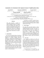
the active site region, five modified amino acids were
found: thioglycine a445, forming a thioxo peptide
(thioamide) bond with tyrosine a446, S-methylcyste-
ine a452, 2-(S)-methylglutamine a400, 1-N-methylhisti-
dine a257 (3-methylhistidine according to IUPAC
nomenclature) and 5-(S)-methylarginine a271 (Fig. 1).
The modifications are introduced after translation, as
the DNA sequence of the encoding mcrA gene shows
Keywords
methanogenic archaea; methanotrophic
archaea; methylated amino acids; methyl-
coenzyme M reductase; thioxo peptides
Correspondence
R. Thauer, Max-Planck-Institute fu
¨
r
terrestrische Mikrobiologie, Karl-von-Frisch-
Strasse, D-35043 Marburg, Germany
Fax: +49 6421 178109
Tel: +49 6421 178101
E-mail:
Website: />(Received 11 June 2007, revised 23 July
2007, accepted 26 July 2007)
doi:10.1111/j.1742-4658.2007.06016.x
Methyl-coenzyme M reductase (MCR) catalyzes the methane-forming step
in methanogenic archaea. Isoenzyme I from Methanothermobacter marbur-
gensis
2
was shown to contain a thioxo peptide bond and four methylated
amino acids in the active site region. We report here that MCRs from all
methanogens investigated contain the thioxo peptide bond, but that the
enzymes differ in their post-translational methylations. The MS analysis
included MCR I and MCR II from Methanothermobacter marburgensis,
MCR I from Methanocaldococcus jannaschii and Methanoculleus thermophi-
lus, and MCR from Methanococcus voltae, Methanopyrus kandleri and
Methanosarcina barkeri. Two MCRs isolated from Black Sea mats contain-
ing mainly methanotrophic archaea of the ANME-1 cluster were also ana-
lyzed.
Abbreviation
MCR, methyl-coenzyme M reductase.
FEBS Journal 274 (2007) 4913–4921 ª 2007 The Authors Journal compilation ª 2007 FEBS 4913
no unusual codons or unusual codon usages at the
positions in which the five modified amino acids were
found. Via in vivo labeling experiments with l-(methyl-
D
3
)-methionine, it was shown that the methyl groups
in the four methylated amino acids are introduced
cotranslationally or post-translationally by specific
S-adenosylmethionine-dependent protein methylases
[9]. How the sulfur is transferred into the carboxamide
group of glycine in the peptide chain remains to be
shown.
Neither the functions of the five modifications nor
whether the modifications are present in MCRs from
all methanogens
3
are known. Comparison of primary
structures deduced from the DNA sequences reveals
that the five amino acid positions are conserved in
MCRs from all methanogenic archaea [9]. However, in
the gene for the a-subunit of MCR from methano-
trophic archaea of the ANME-1 cluster, there is a
codon for a valine, whereas in mcrA from methano-
genic archaea, there is a codon for a glutamine [2].
Methanogenic archaea and methanotrophic archaea
all belong to the kingdom of Euryarchaeota. They are
classified on the basis of their 16S RNA sequence in
five orders, Methanobacteriales, Methanopyrales, Met-
hanococcales, Methanomicrobiales and Methanosarci-
nales [10]. The phylogenetic distance between the
archaeal orders is as large as between, for example,
proteobacteria and Gram-positive bacteria. The phy-
logeny is reflected in the primary structure of the
MCR a-subunit, which can therefore be used to clas-
sify methanogens [11].
In the work reported here, we have analyzed by MS
the MCR from at least one representative of each of
the five orders of methanogenic archaea and from
two methanotrophic archaea of the ANME-1 cluster.
We have included in the analysis both MCR I
and MCR II from Methanothermobacter marburgensis
(growth temperature optimum 65 °C) and MCR I
from a mesophilic (37 °C) and a hyperthermophilic
(85 °C) Methanococcus species. The analysis revealed
that the thioxo peptide bond is conserved in all MCRs
investigated, but that there are differences in the post-
translational methylations. Specifically, Cys a452 is not
methylated in the enzyme from the hyperthermophilic
Methanocaldococcus jannaschii and Methanopyrus
kandleri, and Gln a400 is not methylated in Methano-
sarcina barkeri.
Results
Up to now, the crystal structures of three methyl-coen-
zyme M reductases have been resolved, MCR isoen-
zyme I from Methanothermobacter marburgensis , MCR
from Methanosarcina barkeri,andMCRfromMethano-
pyrus kandleri [7]. In case of the enzyme from Methano-
thermobacter marburgensis and Methanosarcina barkeri,
the resolution was high enough to identify the
post-translational modifications; in the case of the
Methanopyrus kandleri enzyme, it was not. Attempts to
obtain diffracting crystals of MCR II from Methano-
thermobacter marburgensis and of the MCR from the
other methanogens mentioned below were not success-
ful. We therefore searched for post-translational
modifications in the active site region by subjecting the
a-subunit of MCRs to tryptic digestion, followed by
separation and sequencing of the peptides of interest.
Either before or after the tryptic digestion, any oxidized
cysteine residues were reduced with dithiothreitol and
subsequently alkylated with 4-vinylpyridine.
Either of two methods for sequencing the alkylated
tryptic peptides were employed: (a) the tryptic peptide
was subjected to partial hydrolysis with aminopepti-
dase M and the partial hydrolysate was then analyzed
O
-
O
NH
3
+
NH
+
N
H
3
C
H
3
C
S
O
-
O
NH
3
+
H
2
NN
O
-
NH O
NH
3
+
CH
3
H
H
2
N
O
-
OO
NH
3
+
H
3
C
N
S
H
N
1
-Methylhistidine
S-Methylcysteine
5-(S)-Methylarginine
2-(S)-Methylglutamine
Thiopeptide bound thioglycine
Fig. 1. Post-translationally modified amino acids in the a-subunit of
methyl-coenzyme M reductase isoenzyme I from Methanothermo-
bacter marburgensis. Two of these modified amino acids, 2-(S)-
methylgutamine and 5-(S)-methylarginine, have until now not been
found in any other protein. 1-N-Methylhistidine ¼ 3-methylhistidine
according to IUPAC nomenclature.
1
Methanogenic archaea methyl-coenzyme M reductase J. Kahnt et al.
4914 FEBS Journal 274 (2007) 4913–4921 ª 2007 The Authors Journal compilation ª 2007 FEBS
by MALDI-TOF MS (Fig. 2); or (b) the tryptic pep-
tide was subjected to MALDI-TOF MS ⁄ MS (Fig. 3).
As a control, the five modifications within the active
site region of MCR I from Methanothermobacter mar-
burgensis were found using both methods. Where indi-
cated, proteases other than trypsin were used for
peptide generation.
Tryptic peptide containing thioglycine and
S-methylcysteine or cysteine
In the tryptic digest of the MCR a-subunit from all
methanogens investigated, a peptide containing a thi-
oxo peptide was found (Table 1). The sequence
LGFY-thioxoglycine-YDLQDQC is highly conserved
in the nine tryptic peptides analyzed. Very few varia-
tions were found, namely a phenylalanine instead of a
tyrosine directly before the thioglycine (Methanoculleus
thermophilus and Methanosarcina barkeri) or directly
after the thioglycine (Methanosarcina barkeri), and an
alanine instead of an aspartate two positions from the
thioglycine (Methanopyrus kandleri). The cysteine fol-
lowing the conserved glutamine (Q) is methylated in
the two isoenzymes from Methanothermobacter mar-
burgensis and in the enzymes from Methanococcus vol-
tae, Methanoculleus thermophilus and Methanosarcina
barkeri, but is not methylated in the enzyme from Met-
hanopyrus kandleri and Methanocaldococcus jannaschii.
The thiocarbonyl group in thiopeptides can undergo
a slow reaction with water, in which the 32-sulfur is
replaced by a 16-oxygen. This explains why, in some
spectra, a parallel sequence shifted by 16 Da to smaller
masses was seen (Fig. 2).
In two MCRs from methanotrophic archaea of the
ANME-1 cluster [11,12] a tryptic peptide with the
N-terminal sequence LGFYGYDL QDQCTAC
5
was
found (amino acids modified in other MCR are in
bold type), but the glycine was not thioxylated and the
cysteine was not methylated (Table 1). As the MCR
was isolated from microbial mats from the Black Sea
and could not be tested for activity, the possibility can-
not be excluded that the enzyme once had a thioxo
group but lost it by spontaneous hydrolysis.
Peptides containing N-methylhistidine, 5-methyl-
arginine or 2-methylglutamine
The methylated histidine (H257) was found in the a-sub-
unit of MCRs from all methanogenic archaea and of the
two MCRs from the methanotrophic archaea of the
Intensity (%)
1325.5
1453.61
1566.74
1637.8
1800.92
2037.05
D Q L
A
Y Y F
Thio
Gly
G L
1873.94
*
*
*
*
*
Mass (m/z)
0
100
2184.15
2241.19
2353.28
1210.42
Fig. 2. MALDI-TOF MS of the peptide mixture generated via aminopeptidase M from the thioglycine-containing tryptic peptide of the MCR
a-subunit from Methanopyrus kandleri. The purified enzyme was denatured in SDS, digested with trypsin, and then alkylated with 4-vinylpyri-
dine. Subsequently, the tryptic peptides were separated by HPLC and analyzed by MALDI-TOF MS. The peptide with a mass of 2353.28 Da
(predicted to contain the thioglycine) was partially hydrolyzed by aminopeptidase M, and the partial hydrolysate was analyzed by MALDI-TOF
MS. From the peptide ladder, the N-terminal amino acid sequence was found to be LGFY-thioglycine-YALQD. The mass peaks labeled with
an asterisk have a mass that is 16 Da smaller than those of the respective thioglycine-containing peptides. The thiocarbonyl group in thiopep-
tides can undergo a slow reaction with water in which the 32-sulfur is replaced by an 16-oxygen. ThioGly, thioglycine.
J. Kahnt et al.
1
Methanogenic archaea methyl-coenzyme M reductase
FEBS Journal 274 (2007) 4913–4921 ª 2007 The Authors Journal compilation ª 2007 FEBS 4915
ANME-1 cluster (Table 2). The sequence around the
methylated histidine is shown in Fig. 4.
The methylated arginine (R271) is also highly con-
served in the a-subunit of MCRs from methanogenic
archaea. However, in the MCR from the methano-
trophic archaea of the ANME-1 cluster, the arginine
in the respective tryptic peptide was not methylated
(Table 2).
The glutamine (Q400) is not methylated in the
a-subunit of MCR from Methanosarcina barkeri.In
Table 1.
10;1110;11
Amino acid sequence of the MCR a-subunit tryptic peptide containing the thioglycine. G*, thioglycine; C, S-methylcysteine. Amino
acids modified in most MCR are indicated by bold type.
MCR from
Sequence of the thioglycine-containing
tryptic peptide
Methanothermobacter
marburgensis (MCR I)
LGFYG*YDLQDQC*GASNVFSIR
a,b
Methanothermobacter
marburgensis (MCR II)
LGFYG*YDLQDQC*GASNSLSIR
a
Methanopyrus
kandleri
LGFYG*YALQDQCGAANSLSVR
a
Methanocaldococcus
jannaschii (MCR I)
LGFYG*YDLQDQCGAANSLSFR
b
Methanococcus
voltae
LGFYG*YDLQDQC*GASNSLAIR
a,c
Methanoculleus
thermophilus (MCR I)
LGFFG*YDLQDQC*GSANSLSIR
c
Methanosarcina
barkeri
LGFFG*FDLQDQC*GATNVLSYQGDEGLPDELR
c
Methanotrophic archaea
of the ANME-1b cluster
LGFYGYDLQDQCTACGSYSYQSDEGMPFEMR
c,d
a
Sequence determined via aminopeptidase M and MALDI-TOF MS.
b
Sequence taken from Selmer et al. [9].
c
Sequence determined via
MALDI-TOF MS ⁄ MS.
d
Sequence of peptide obtained from two different MCRs isolated from the Black Sea mats.
Intensity (%)
Mass (
m/z
)
0
1257.15
R GF S A
Pyridyl-
ethyl
Cys
L N A QS D Q L D Y
321.43
608.27
521.31
408.34
722.27
793.26
864.25
921.23
1964.0
1891.63
1728.36
1613.4
1500.38
1372.26
1129.15
174.59
Thio
Gly
2127.99
Y F G L
2446.36
100
Fig. 3. MALDI-TOF MS ⁄ MS of the thioglycine-containing tryptic peptide of the MCR a-subunit from Methanocaldococcus jannaschii. The
purified enzyme was denatured in SDS, alkylated with 4-vinylpyridine, and then subjected to SDS ⁄ PAGE. After in-gel digestion of the a-sub-
unit with trypsin, the generated peptides were analyzed by MALDI-TOF MS. The peptide with a mass of 2446.36 Da (predicted to contain
the thioglycine) was then analyzed by MALDI-TOF MS ⁄ MS. From the fragment pattern, the following amino acid sequence is deduced:
LGFY-thioglycine-YDLQDQ-pyridylethylCys-GAANSLSFR. ThioGly, thioglycine.
1
Methanogenic archaea methyl-coenzyme M reductase J. Kahnt et al.
4916 FEBS Journal 274 (2007) 4913–4921 ª 2007 The Authors Journal compilation ª 2007 FEBS
the two MCRs from the ANME-1 cluster, there is a
valine instead of a glutamine (Table 2).
Discussion
Methanogenic archaea are dependent on methane
formation for growth, and thus on a functional methyl-
coenzyme M reductase [1,13]. Therefore, a genetic
analysis of methanogenic archaea with respect to the
function of the post-transcriptional modifications of
MCR is presently not possible [14,15]. A reversed
genetic approach is also not in sight, because the genes
required for the biosynthesis of the nickel cofactor F
430
[16] and for the post-translational modifications [9] are
not yet known [17,18]. Also, the in vitro reconstitution
of active enzyme from its subunits and cofactor has
proven very difficult; very little activity is recovered
[19]. Therefore, the function of the post-translational
modifications can at present only be approached by
comparison of MCRs from archaea differing in phylo-
genetic relationship and ⁄ or growth conditions.
An early hypothesis was that the post-translational
or cotranslational modifications modulate the kinetic
properties (K
m
and V
max
) of MCR, which is why we
started the analysis with isoenzyme II from Methano-
thermobacter marburgensis, which differs considerably
in K
m
and V
max
from isoenzyme I [20]. However, this
hypothesis had to be abandoned when we found that
the two isoenzymes were identical with respect to their
post-translational modifications (Table 2).
His a257 is methylated in all analyzed MCRs, indi-
cating an essential function (Table 2). The residue
1-N-methylhistidine is part of the coenzyme B-binding
site. Its unmethylated nitrogen atom Ne2 donates the
shortest of the three hydrogen bonds of the protein to
the phosphate group of coenzyme B. The distance of
2.65 A
˚
indicates the presence of a strong hydrogen
bond with fully overlapping orbitals. Owing to the
positive inductive effect, the pKa of the methylated
amino acid is expected to increase slightly (imidazole
and N-methylimidazole differ in their pKa by 0.1), and
this will affect the strength of coenzyme B binding. A
more important function of the methyl group may
involved improved orientation of the histidine inside
the coenzyme B-binding site. With the methylation, the
histidine residue is prevented from forming two alter-
nate nitrogen positions by rotating around the angle
v
2
, due to occlusion by a peptide oxygen from the pre-
ceding residue [21].
The functions of the three other methylations are
less obvious, in part because they are not conserved in
all MCR enzymes (Table 2). The reduction of methyl-
coenzyme M with coenzyme B takes place in a hydro-
phobic pocket in the complete absence of water. It
involves radical intermediates that are very reactive
[5,18]. This is probably why the active site of this
enzyme is lined up primarily with unreactive aromatic
and aliphatic amino acid residues. The methyl groups
of S-methylcysteine, 5-methylarginine and 2-methylglu-
tamine are oriented towards the active site chamber
Table 2. Amino acid modifications found in the a-subunit of MCRs from methanogenic archaea by MS analysis of tryptic peptides. Where
indicated, the a-subunit was subjected to proteolysis by chymotrypsin, endoproteinase AspN or BrCN rather than by trypsin. The data for
MCR I from Methanothermobacter marburgensis were taken from Selmer et al. [9]. NF, respective peptide not found.
MCR from Thio-Gly S-Methyl-Cys N-Methyl-His 5-Methyl-Arg 2-Methyl-Gln
Methanothermobacter
marburgensis (MCR I)
++ + + +
Methanothermobacter
marburgensis) (MCR II)
++ + +
a
+
Methanopyrus
kandleri
+ Cys + +
b
+
Methanocaldococcus
jannaschii (MCR I)
+ Cys + +
c
+
a
Methanococcus
voltae
++ + NF +
Methanoculleus
thermophilus (MCR I)
+ + NF +
a
+
Methanosarcina
barkeri
++ + +
a
Gln
Methanotrophic archaea
of the ANME-1 cluster
(two MCRs)
Gly Cys + Arg Val
a
a-Subunit cleaved with chymotrypsin.
b
a-Subunit cleaved with BrCN.
c
a-Subunit digested with AspN.
J. Kahnt et al.
1
Methanogenic archaea methyl-coenzyme M reductase
FEBS Journal 274 (2007) 4913–4921 ª 2007 The Authors Journal compilation ª 2007 FEBS 4917
and have no contact with solvent. The function of the
methylations could thus be to protect the respective
amino acid residues from reaction with the radical
intermediates. As the methylations provide the amino
acids with additional hydrophobic interactions,
another function could be to restrict the conforma-
tional flexibility of the residues, as has been discussed
for post-transcriptional tRNA modifications [22–24].
The observed differences in methylation are difficult
to explain, because they may be compensated for by
differences in positioning or properties of nearby resi-
dues, which are not directly evident.
A function for the thioxo peptide bond (for proper-
ties see Pfeifer et al. [25] and Artis & Lipton [26]) was
proposed in the catalytic cycle of MCR. In the final
step of the cycle, a disulfide anion radical of coen-
zyme M and coenzyme B is formed, which is oxidized
to the heterodisulfide by reducing the active site’s
nickel back to the Ni(I) oxidation state [5,18]. The thi-
oxo group is positioned such that it could be involved
in electron transport from the disulfide anion radical
to the nickel. The redox potential of the thioxo pep-
tide ⁄ radical couple is estimated to lie between that of
the disulfide ⁄ disulfide anion radical couple () 1.7 V)
[27] and the F
430
[Ni(II)] ⁄ F
430
[Ni(I)] couple () 0.64 V)
[18]. It has also been suggested that upon a one-elec-
tron reduction of the thioxo peptide bond, a change
from the trans to the cis conformation (similar to the
light-induced switch of the thioxo peptide bond from
trans to cis [28,29]) could occur, and that this confor-
mational change could be involved in coupling the two
active sites [4,30,31]. The finding of the thioxo peptide
Fig. 4. Partial amino acid sequence of the a-subunit of MCRs from six methanogenic archaea of five different orders and from two methano-
trophic archaea of the ANME-1 cluster. The numbering is that for the a-subunit of MCR I from Methanothermobacter marburgensis. The
amino acids, which in MCR from Methanothermobacter marburgensis are post-translationally modified, are highlighted in bold. The ANME-
1a sequence (2216) was provided by A. Meyerdierks, Max Planck Institute for Marine Microbiology in Bremen, and the ANME-1b sequence
was taken from Lyoyd et al. [40].
1
Methanogenic archaea methyl-coenzyme M reductase J. Kahnt et al.
4918 FEBS Journal 274 (2007) 4913–4921 ª 2007 The Authors Journal compilation ª 2007 FEBS
bond in MCRs from all methanogenic archaea sup-
ports this hypothesis, whereas the finding that the two
MCRs isolated from the Black Sea microbial mats
lacks this bond does not. As the thioxo peptide bond
can lose its sulfur by hydrolysis, and as the MCR
extracted from the microbial mats from the Black Sea
was inactive, the function of the thioxo peptide bond
as proposed above cannot yet be dismissed. This ques-
tion will remain open until methanotrophic archaea
can be grown in the laboratory and until active MCRs
can be isolated from them for analysis of the presence
of the thioxo peptide bond.
Methylated amino acids are found in many proteins,
although the presence of 5-methylarginine and 2-meth-
ylglutamine has, until now, only been reported for
MCR [9]. A thioxo peptide bond in a protein has not
been encountered before. However, such a bond has
been found, for example, in methanobactin from meth-
ane-oxidizing bacteria [32] and thioviridamide from
Streptomyces olivoviridis [33]. How these nonriboso-
mally synthesized polypeptides are thioxylated is not
known. A mechanism similar to that described for the
sulfuration of the C-terminal glycine in ThiS and
MoaD, involved in thiamine biosynthesis and molyb-
dopterin biosynthesis, respectively, and for the sulfura-
tion of uridine in tRNA to thiouridine can be
envisaged [34–36]. This mechanism probably also
applies for the synthesis of pyridine-2,6-bis(thiocarb-
oxylate) from dipicolinic acid in Pseudomonas stutzeri
[37–39].
Experimental procedures
Methanothermobacter marburgensis (DSMZ 2133) (growth
temperature optimum 65 °C), Methanopyrus kandleri
(DSMZ 6324) (98 °C), Methanococcus voltae (DSMZ 1537)
(37 °C), Methanocaldococcus jannaschii (DSMZ 2661)
(85 °C), Methanoculleus thermophilus (DSMZ 3915) (57 °C)
and Methanosarcina barkeri (strain Fusaro) (DSMZ 804)
(38 °C) were obtained from the Deutsche Sammlung von
Mikroorganismen und Zellkulturen (Braunschweig, Ger-
many). The first three methanogenic archaea were grown
on H
2
and CO
2
, Methanoculleus thermophilus on isopropa-
nol, H
2
and CO
2
, and Methanosarcina barkeri on methanol.
The microbial mats containing methanotrophic archaea of
the ANME-1 cluster were from the Black Sea [2]. MCR
was purified from cells by published procedures [1].
Of the methanogenic archaea mentioned above, Methano-
thermobacter marburgensis, Methanocaldococcus jannaschii
and Methanoculleus thermophilus contain two MCR iso-
enzymes, whereas Methanococcus voltae, Methanopyrus
kandleri and Methanosarcina barkeri do not have MCR
isoenzymes.
For determination of the amino acid modifications in the
a-subunit of MCR, the a-subunit was subjected to proteo-
lysis by trypsin, and then the tryptic peptides, after separa-
tion, were analyzed by MALDI-TOF MS. Where indicted,
the a-subunit was subjected to proteolysis by chymotrypsin
(sequencing grade; Roche, Mannheim, Germany), endopro-
teinase AspN (sequencing grade; Roche) or BrCN rather
than by trypsin.
MALDI-TOF MS analysis of tryptic peptides after
partial digestion with aminopeptidase M
Purified MCR (2 mg of protein in 25 lL) was supplemented
with SDS and dithiothreitol to final concentrations of 1.5%
and 200 mm, respectively, and then incubated for 10 min at
95 °C to fully denature the protein. Subsequently, the solu-
tion was diluted 10-fold with 100 mm NH
4
HCO
3
(pH 8.3),
supplemented with 0.2 mg of trypsin (diphenyl carbamyl
chloride
6
-treated, 11392 U mg
)1
; Fluka, Buchs, Switzerland),
and incubated for 12 h at 37 °C. Then, excess 4-vinylpyri-
dine (60 mm) was added to alkylate the cysteine-derived
thiol groups. After incubation for 30 min at 70 °C, the non-
reacted vinylpyridine was removed by evacuation to dryness
in a Savant-SpeedVac SC110; (Life Sciences International,
Frankfurt, Germany)
7
and the dried material containing the
tryptic peptides was redissolved in 400 lLofH
2
O. After
removal of SDS using a Detergent-Out column (Genotech,
St Louis, MO, USA), the peptides were separated by RP-
HPLC and analyzed by MALDI-TOF MS. The peptide
whose molecular mass matched with that predicted for the
modified amino acid containing tryptic peptide was partially
hydrolyzed with 10 mU of aminopeptidase M (Roche) at
pH 7.1 for 30 min at room temperature, and the partial
hydrolysate was then analyzed via MALDI-TOF MS
(Voyager DE-RP; ABI, Darmstadt, Germany; 337 nm N
2
laser). From the peptide ladder, the N-terminal amino acid
sequence of the peptides was obtained as shown in Fig. 2
for the thioglycine-containing tryptic peptide of the MCR
a-subunit from Methanopyrus kandleri.
MALDI-TOF MS ⁄ MS analysis of tryptic peptides
Purified MCR (0.15 mg) was denatured in SDS (0.15%)
containing dithiothreitol (6 mm), alkylated with excess 4-vi-
nylpyridine (30 mm) as described above, and then subjected
to SDS ⁄ PAGE followed by staining with Coomassie Blue.
The band corresponding to the a-subunit was cut out, di-
stained and dried under vacuum, and the dried material
was suspended in 30 lLof10mm NH
4
HCO
3
(pH 8.3) con-
taining 0.6 lg of trypsin (sequence grade, modified; Pro-
mega, Madison, WI, USA) and 10% acetonitrile. After
incubation at room temperature for 12 h, the trypsin digest
was extracted, and the extract was applied to a C18 Pep-
Map 100 nano LC column
8
(Dionex, Idstein, Germany) for
J. Kahnt et al.
1
Methanogenic archaea methyl-coenzyme M reductase
FEBS Journal 274 (2007) 4913–4921 ª 2007 The Authors Journal compilation ª 2007 FEBS 4919
the separation of the peptides, which were subsequently
analyzed by MALDI-TOF MS. The peptide whose mass
matched that predicted for the modified amino acid con-
taining tryptic peptide was analyzed by MALDI-TOF
MS ⁄ MS (Ultraflex; Bruker, Bremen, Germany). From the
fragment pattern, the amino acid sequence was obtained as
exemplified in Fig. 3 for the thioglycine-containing tryptic
peptide of the MCR a-subunit from Methanocaldococcus
jannaschii.
Acknowledgements
This work was supported by the Max Planck Society
and the Fonds der Chemischen Industrie.
References
1 Thauer RK (1998) Biochemistry of methanogenesis: a
tribute to Marjory Stephenson. Microbiology 144, 2377–
2406.
2 Kru
¨
ger M, Meyerdierks A, Glockner FO, Amann R,
Widdel F, Kube M, Reinhardt R, Kahnt J, Bocher R,
Thauer RK et al. (2003) A conspicuous nickel protein
in microbial mats that oxidize methane anaerobically.
Nature 426, 878–881.
3 Shima S & Thauer RK (2005) Methyl-coenzyme M
reductase (MCR) and the anaerobic oxidation of meth-
ane (AOM) in methanotrophic archaea. Curr Opin
Microbiol 8, 643–648.
4 Thauer RK & Shima S (2007) Methyl-coenzyme M
reductase in methanogens and methanotrophs. In
Archaea, Evolution, Physiology and Molecuar Biology
(Garrett R & Klenk H-P, eds), pp. 275–283. Blackwell
Publishing, Malden, MA.
5 Ermler U, Grabarse W, Shima S, Goubeaud M &
Thauer RK (1997) Crystal structure of methyl-coenzyme
M reductase: the key enzyme of biological methane for-
mation. Science 278, 1457–1462.
6 Grabarse W, Mahlert F, Duin EC, Goubeaud M,
Shima S, Thauer RK, Lamzin V & Ermler U (2001a)
On the mechanism of biological methane formation:
structural evidence for conformational changes in
methyl-coenzyme M reductase upon substrate binding.
J Mol Biol 309, 315–330.
7 Grabarse W, Mahlert F, Shima S, Thauer RK & Ermler
U (2000) Comparison of three methyl-coenzyme M
reductases from phylogenetically distant organisms:
unusual amino acid modification, conservation and
adaptation. J Mol Biol 303, 329–344.
8 Grabarse W, Shima S, Mahlert F, Duin EC, Thauer RK
& Ermler U (2001b) Methyl-coenzyme M reductase. In
Handbook of Metalloproteins (Messerschmidt A, Huber
R, Poulos T & Wieghardt K, eds), pp. 897–914. John
Wiley & Sons, Chichester.
9 Selmer T, Kahnt J, Goubeaud M, Shima S, Grabarse W,
Ermler U & Thauer RK (2000) The biosynthesis of
methylated amino acids in the active site region of
methyl-coenzyme M reductase. J Biol Chem 275, 3755–
3760.
10 Boone DR, Whitman WB & Rouvie
`
re P (1993) Diver-
sity and taxonomy of methanogens. In Methanogenesis
(Ferry JG, ed.), pp. 35–80. Chapman & Hall, New
York, NY.
11 Friedrich MW (2005) Methyl-coenzyme M reductase
genes: unique functional markers for methanogenic and
anaerobic methane-oxidizing Archaea. Methods Enzymol
397, 428–442.
12 Hallam SJ, Putnam N, Preston CM, Detter JC, Rokh-
sar D, Richardson PM & DeLong EF (2004) Reverse
methanogenesis: testing the hypothesis with environmen-
tal genomics. Science 305, 1457–1462.
13 Rother M, Boccazzi P, Bose A, Pritchett MA &
Metcalf WW (2005) Methanol-dependent gene expression
demonstrates that methyl-coenzyme M reductase is essen-
tial in Methanosarcina acetivorans C2A and allows isola-
tion of mutants with defects in regulation of the methanol
utilization pathway. J Bacteriol 187, 5552–5559.
14 Metcalf WW, Zhang JK, Apolinario E, Sowers KR &
Wolfe RS (1997) A genetic system for Archaea of the
genus Methanosarcina: liposome-mediated transforma-
tion and construction of shuttle vectors. Proc Natl Acad
Sci USA 94, 2626–2631.
15 Rother M & Metcalf WW (2005) Genetic technologies
for Archaea. Curr Opin Microbiol 8, 745–751.
16 Thauer RK & Bonacker LG (1994) Biosynthesis of
coenzyme F
430
, a nickel porphinoid involved in metha-
nogenesis. In The Biosynthesis of the Tetrapyrrole Pig-
ments. Ciba Foundation Symposium 180 (Chadwick DJ
& Ackrill K, eds), pp. 210–227. John Wiley & Sons,
Chichester.
17 Fricke WF, Seedorf H, Henne A, Kru
¨
er M, Liesegang H,
Hedderich R, Gottschalk G & Thauer RK (2006) The
genome sequence of Methanosphaera stadtmanae reveals
why this human intestinal archaeon is restricted to metha-
nol and H
2
for methane formation and ATP synthesis.
J Bacteriol 188, 642–658.
18 Jaun B & Thauer RK (2007) Methyl-coenzyme M
reductase and its nickel corphin coenzyme F430 in
methanogenic archaea. In Nickel and its Surprising
Impact in Nature (Sigel A, Sigel H & Sigel RKO, eds),
pp. 323–356. John Wiley & Sons, Chichester.
19 Hartzell PL & Wolfe RS (1986) Requirement of the
nickel tetrapyrrole F
430
for in vitro methanogenesis:
reconstitution of methylreductase component C from its
dissociated subunits. Proc Natl Acad Sci USA 83, 6726–
6730.
20 Bonacker LG, Baudner S, Mo
¨
rschel E, Bo
¨
cher R &
Thauer RK (1993) Properties of the two isoenzymes of
1
Methanogenic archaea methyl-coenzyme M reductase J. Kahnt et al.
4920 FEBS Journal 274 (2007) 4913–4921 ª 2007 The Authors Journal compilation ª 2007 FEBS
methyl-coenzyme M reductase in Methanobacterium
thermoautotrophicum. Eur J Biochem 217, 587–595.
21 Grabarse W (1999) Crystal structures of methyl-coen-
zyme M reductase from three methanogenic archaea.
PhD Thesis, Johann Wolfgang Goethe Universita
¨
t,
Frankfurt am Main.
22 Kowalak JA, Dalluge JJ, McCloskey JA & Stetter KO
(1994) The role of posttranscriptional modification in
stabilization of transfer RNA from hyperthermophiles.
Biochemistry 33, 7869–7876.
23 Dalluge JJ, Hamamoto T, Horikoshi K, Morita RY,
Stetter KO & McCloskey JA (1997) Posttranscriptional
modification of tRNA in psychrophilic bacteria.
J Bacteriol 179, 1918–1923.
24 Noon KR, Guymon R, Crain PF, McCloskey JA,
Thomm M, Lim J & Cavicchioli R (2003) Influence
of temperature on tRNA modification in archaea:
Methanococcoides burtonii [optimum growth temperature
(Topt), 23 degrees C] and Stetteria hydrogenophila (Topt,
95 degrees C). J Bacteriol 185, 5483–5490.
25 Pfeifer T, Schierhorn A, Friedemann R, Jakob M,
Frank R, Schutkowski M & Fischer G (1997) Specific
fragmentation of thioxo peptides facilitates the assign-
ment of the thioxylated amino acid. J Mass Spectrom
32, 1064–1071.
26 Artis DR & Lipton MA (1998) Conformations of thio-
amide-containing dipeptides: a computational study.
J Am Chem Soc 120, 12200–12206.
27 Stubbe JA & van der Donk WA (1998) Protein radicals
in enzyme catalysis. Chem Rev 98, 705–762.
28 Zhao J, Wildemann D, Jakob M, Vargas C & Schiene-
Fischer C (2003) Direct photomodulation of peptide
backbone conformations. Chem Commun (Camb) 22,
2810–2811.
29 Satzger H, Root C, Gilch P, Zinth W, Wildemann D &
Fischer G (2005) Photoswitchable elements within a
peptide backbone-ultrafast spectroscopy of thioxylated
amides. J Phys Chem B Condens Matter Mater Surf
Interfaces Biophys 109, 4770–4775.
30 Goenrich M, Duin EC, Mahlert F & Thauer RK (2005)
Temperature dependence of methyl-coenzyme M reduc-
tase activity and of the formation of the methyl-coen-
zyme M reductase red2 state induced by coenzyme B.
J Biol Inorg Chem 10, 333–342.
31 Goenrich M, Mahlert F, Duin EC, Bauer C, Jaun B &
Thauer RK (2004) Probing the reactivity of Ni in the
active site of methyl-coenzyme M reductase with sub-
strate analogues. J Biol Inorg Chem 9, 691–705.
32 Kim HJ, Graham DW, DiSpirito AA, Alterman MA,
Galeva N, Larive CK, Asunskis D & Sherwood PM
(2004) Methanobactin, a copper-acquisition compound
from methane-oxidizing bacteria. Science 305, 1612–
1615.
33 Hayakawa Y, Sasaki K, Nagai K, Shin-ya K &
Furihata K (2006) Structure of thioviridamide, a novel
apoptosis inducer from Streptomyces olivoviridis.
J Antibiot (Tokyo) 59, 6–10.
34 Gutzke G, Fischer B, Mendel RR & Schwarz G (2001)
Thiocarboxylation of molybdopterin synthase provides
evidence for the mechanism of dithiolene formation in
metal-binding pterins. J Biol Chem 276, 36268–36274.
35 Numata T, Ikeuchi Y, Fukai S, Suzuki T & Nureki O
(2006) Snapshots of tRNA sulphuration via an adeny-
lated intermediate. Nature 442, 419–424.
36 Lehmann C, Begley TP & Ealick SE (2006) Structure of
the Escherichia coli ThiS–ThiF complex, a key compo-
nent of the sulfur transfer system in thiamin biosynthe-
sis. Biochemistry 45, 11–19.
37 Lewis TA, Cortese MS, Sebat JL, Green TL, Lee CH &
Crawford RL (2000) A Pseudomonas stutzeri gene clus-
ter encoding the biosynthesis of the CCl4-dechlorination
agent pyridine-2,6-bis(thiocarboxylic acid). Environ
Microbiol 2, 407–416.
38 Lewis TA, Paszczynski A, Gordon-Wylie SW, Jee-
digunta S, Lee CH & Crawford RL (2001) Carbon
tetrachloride dechlorination by the bacterial transition
metal chelator pyridine-2,6-bis(thiocarboxylic acid).
Environ Sci Technol 35, 552–559.
39 Sepulveda-Torre L, Huang A, Kim H & Criddle CS
(2002) Analysis of regulatory elements and genes required
for carbon tetrachloride degradation in Pseudomonas stut-
zeri strain KC. J Mol Microbiol Biotechnol 4, 151–161.
40 Lloyd KG, Lapham L & Teske A (2006) An anaerobic
methane-oxidizing community of ANME-1b archaea in
hypersaline Gulf of Mexico sediments. Appl Environ
Microbiol 72, 7218–7230.
J. Kahnt et al.
1
Methanogenic archaea methyl-coenzyme M reductase
FEBS Journal 274 (2007) 4913–4921 ª 2007 The Authors Journal compilation ª 2007 FEBS 4921