Báo cáo khoa học: Recent contributions of in vitro models to our understanding of hepatitis C virus life cycle pdf
Bạn đang xem bản rút gọn của tài liệu. Xem và tải ngay bản đầy đủ của tài liệu tại đây (868.51 KB, 14 trang )
REVIEW ARTICLE
Recent contributions of in vitro models to our
understanding of hepatitis C virus life cycle
Morgane Re
´
geard, Charlotte Lepe
`
re, Maud Trotard, Philippe Gripon and Jacques Le Seyec
INSERM, U522, IFR 140, Ho
ˆ
pital de Pontchaillou, Rennes, France
Introduction
The hepatitis C virus (HCV) belongs to the Flaviviri-
dae family and is the only member of the Hepacivirus
genus. It is a small virus with a diameter of
50 nm, enveloped within a cell-derived lipid mem-
brane that carries viral surface glycoproteins. This
envelope surrounds a capsid containing positive
ssRNA. The viral genome of 9600 nucleotides con-
tains two UTR at the 5¢- and 3¢-termini and a major
ORF that encodes a unique polyprotein of 3000
amino acids (Fig. 1). Translation is initiated by the
internal ribosome entry site (IRES) located in the
5¢-UTR. Translated polyprotein is then co- and post-
translationally cleaved into 10 different products:
three structural proteins (the core protein and the E1
and E2 envelope glycoproteins) and seven nonstruc-
tural (NS) proteins (p7, NS2, NS3, NS4A, NS4B,
NS5A and NS5B). The specific enzymatic functions
that have been attributed to NS2 ⁄ 3, NS3 and NS5B
are serine protease, helicase and RNA-dependent
polymerase, respectively.
HCV is a human pathogen and 170 million peo-
ple are chronically infected worldwide [1]. HCV infec-
tion causes major health problems because it is a
principle cause of chronic liver diseases, including cir-
rhosis and hepatocellular carcinoma. The natural his-
tory of HCV begins with a frequently asymptomatic
Keywords
assembly; hepatitis C virus; in vitro models;
infection; replication
Correspondence
J. Le Seyec, INSERM U522, Ho
ˆ
pital
Pontchaillou, Avenue Henri Le Guilloux,
Rennes, F-35033, France
Fax: +33 2 99 54 01 37
Tel: +33 2 99 54 74 07
E-mail:
(Received 14 June 2007, revised 25 July
2007, accepted 26 July 2007)
doi:10.1111/j.1742-4658.2007.06017.x
Hepatitis C virus is a human pathogen responsible for liver diseases includ-
ing acute and chronic hepatitis, cirrhosis and hepatocellular carcinoma. Its
high prevalence, the absence of a prophylactic vaccine and the poor effi-
ciency of current therapies are huge medical problems. Since the discovery
of the hepatitis C virus, our knowledge of its biology has been largely
punctuated by the development of original models of research. At the end
of the 1980s, the chimpanzee model led to cloning of the viral genome and
the definition of infectious molecular clones. In 1999, a breakthrough was
achieved with the development of a robust in vitro replication model named
‘replicon’. This system allowed intensive research into replication mecha-
nisms and drug discovery. Later, in 2003, pseudotyped retroviruses har-
bouring surface proteins of hepatitis C virus were produced to specifically
investigate the viral entry process. It was only in 2005 that infectious
viruses were produced in vitro, enabling intensive investigations into the
entire life cycle of the hepatitis C virus. This review describes the different
in vitro models developed to study hepatitis C virus, their contribution to
current knowledge of the virus biology and their future research applica-
tions.
Abbreviations
HCV, hepatitis C virus; HCVcc, cellular clone of HCV; HCVpp, pseudo-particles of HCV; IFN, interferon; IRES, internal ribosome entry site;
JFH1, Japanese fulminant hepatitis 1; LDLR, low-density lipoprotein receptor; NS, nonstructural; SR-B1, scavenger receptor class B type 1;
VSV, vesicular stomatitis virus.
FEBS Journal 274 (2007) 4705–4718 ª 2007 The Authors Journal compilation ª 2007 FEBS 4705
acute phase of infection that leads to chronic infection
in 70–80% of cases. Thereafter, 10–20% of chroni-
cally infected patients develop liver cirrhosis within
20 years and hepatocellular carcinoma after another
decade. No vaccine against HCV infection is available,
and current antiviral therapies consisting of pegylated
interferon (IFN) and ribavirin injections are character-
ized by limited efficacy, substantial side effects and
high cost. These clinical complications clearly docu-
ment the need for more effective therapies that depend
on a detailed understanding of HCV biology using
appropriate experimental systems. Unfortunately,
research on HCV has largely been slowed by the diffi-
culties encountered in developing efficient experimental
models. This review focuses on the different in vitro
models of HCV that have been developed and their
contribution to our current knowledge of the virus life
cycle.
Around 10 years after discovery of the HCV genome
and after many attempts to infect chimpanzees with
transcripts from cloned isolates, consensus sequences
of genotypes 1a, 1b and 2a were constructed. These
were the first viral functional sequences able to infect
chimpanzees. Soon after, efforts were concentrated on
establishing cell culture models that support HCV rep-
lication by transfecting cells with cloned viral DNA or
their derived viral transcripts. Although this approach
is classic in virology, it proved to be unproductive for
HCV because of the very low level of replication and
the high amount of input RNA needed for transfec-
tion. These first studies precluded the difficulties of
studying HCV in vitro.
Replicon system
An important breakthrough was the development of
cell-culture systems based on the selection of cells that
support stable replication of subgenomic HCV RNAs.
Lohmann et al. [2] worked on an HCV consensus gen-
ome of genotype 1b derived from a chronically
infected patient. Researchers replaced the region that
encodes the core to p7 with the coding sequence of the
neomycin-resistance gene and the heterologous IRES
of the encephalomyocarditis virus. The resulting repli-
con was bicistronic with translation of the first cistron
(neomycin-resistance gene) being directed by the HCV
IRES and that of the second cistron (NS2–5B) by the
encephalomyocarditis virus IRES. Other constructions
were composed of a smaller second cistron encoding
NS3 to NS5B proteins (Fig. 2A). After transfection of
Huh7 cells with this replicon, selection of the very few
cells supporting autonomous replication was achieved
by neomycin sulfate treatment (Fig. 2Ba). Viral repli-
cation was sufficient to detect viral RNA by northern
blot analysis. Improvement of the system was obtained
after the discovery of cell-culture-adaptative mutations
that enhanced the replication efficiency by up to
10 000 times [3]. These mutations are at the N-termi-
nus of the NS3 helicase, in two distinct positions of
NS4B, in the centre of NS5A and in the C-terminal
region of NS5B [3–6]. The significance of these muta-
tions has been questioned because they have not been
observed in wild-type viruses. Moreover, insertion of
some of these mutations into an infectious HCV clone
reduced or completely abolished its in vivo infectivity
2E1EC 3SN2SN
A4SN
B5SNA5SN
snietorpocylG
e
m
o
n
e
G
noitadispacne
noI
l
en
nahc
esaet
o
rp en
i
reS
esaci
l
e
h
ANR
s
ro
t
c
a
f
o
c
e
s
ae
t
orp
eni
r
e
S
tne
d
neped-ANR
esaremylop ANR
n
i
etorpohpsohP
s
n
o
i
t
a
r
e
t
la
e
n
a
rbmeM
enietsyC
e
s
a
et
o
rp
7p
F
RTU’3
RTU’5
larutcu
r
t
s
)tn0069( ANR VCH
noitalsnarT
dnA
noitarutaM
B4SN
larutcurtsnon
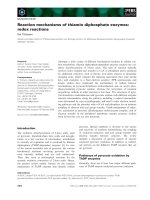
Fig. 1. Genetic organization and procession of HCV polyprotein. A schematic representation of HCV genome is given at the top. The HCV
genome is composed of ssRNA encoding a large ORF flanked by 5¢- and 3¢-UTR. Translation of the polyprotein precursor is mediated by the
IRES contained in the 5¢-UTR. The polyprotein is co- and post-translationally processed in 10 proteins by signal peptide peptidase (black solid
arrows), by NS2 ⁄ 3 autoprotease (black and large arrow) and by NS3 ⁄ 4A protease (black doted arrows). The F protein is generated by transla-
tion of an alternative reading frame but no functions have yet been attributed to this protein.
In vitro HCV infection models M. Re
´
geard et al.
4706 FEBS Journal 274 (2007) 4705–4718 ª 2007 The Authors Journal compilation ª 2007 FEBS
in chimpanzees [7]. In parallel, the replicon system has
allowed the selection of highly permissive cell clones
(Fig. 2Bb). Indeed, the subpopulation of Huh7 cells
that supports a high viral replication rate has been
cured from the replicon by long-lasting IFN treatment.
Two such cell lines have been generated and named
Huh7-Lunet and Huh7.5. Another subclone, Huh7.5.1,
has been generated similarly by curing Huh7.5 cells of
replicating HCV. All of these cell lines were shown to
support RNA replication to a much greater extent
than the parental cell line [8–10]. The efficient replica-
tion of HCV in these cells may be explained by partial
impairment of their antiviral defence system. Indeed,
Landford et al. [11] suggested that some steps in the
signalling pathway for detecting dsRNA were defective
in the parental Huh7 cells. Moreover, permissiveness
of HCV replication in Huh7.5 cells is probably rein-
forced by the presence of a defective mutation in the
RIG-I gene, which disturbs the antiviral immune
response [12]. The antiviral effect of IFNa observed
in vivo was nevertheless reproduced in this system
[4,11]. Thereafter, improvements to this model were
achieved with the efficient insertion of a reporter gene
into the viral genome facilitating measurement of the
replication activity of replicons [5,13–15]. Replicons of
other genotypes (1a and 2a) have also been developed
[16,17]. Genomic HCV replicons have also been gener-
ated and have enabled the selection of cells with stable
expression of the entire viral polyprotein (Fig. 2A).
However, replication efficiency was lower than that
observed with subgenomic replicons and no virus pro-
duction was observed in these cells [16,18,19]. This
defect could be due to the presence of adaptative
mutations that are detrimental to viral particle assem-
bly and secretion or to the lack of some critical HCV
partners in Huh7 cells. Some data argue for the former
hypothesis. On the one hand, inoculation of chimpan-
zees with a Con1 sequence containing these adaptative
mutations failed to establish a productive infection [7].
On the other hand, production of infectious particles
in Huh7 cells has been achieved with a clone named
Japanese fulminant hepatitis 1 (JFH1) constituting the
so-called model of HCV cellular clone (see below) [20].
Owing to its efficient replication rate, this in vitro
replicon model enabled investigation into the replica-
tion process, the replication complex and host–virus
interactions [21,22]. Its exploitation has also enabled
high-throughput screening of anti-HCV drugs targeting
the replication of various genotype replicons [23].
Although practical, only the replication step containing
in vitro adaptative mutations can be studied with this
system using viral genomes containing in vitro adaptive
A4SN
2SN
3SN
B5SNA5SN
oeN
RTU
’5 VCH
RTU’3 VCH
c
imon
egbu
S
no
c
il
p
er
cimoneG
noci
l
per
2E1EC B5SNA5SN3SN2SN
7
p
oeN
RTU’5 VCH
RTU’3 VCH
3SN
B5SNA5SN
o
eN
RTU’5 VCH
RTU’3 VCH
VMCE
SERI
d
n
anoitceles
8
14G
noisnapxe l
a
no
l
c
t
n
e
mta
e
rt
NF
I
fo noitar
o
por
tc
el
E
ht
i
wslle
c 7h
uH
A
senolcbussllec 7huH
V
CHg
nitacilpersllec 7huH
ro
c
i
mone
g
b
u
S
noci
lpe
r cimoneg
cimonegbuS
nocilper
B
VMCE
SERI
B4SN
A4SN
B4SN
A
4
S
N
B4SN
b
a
VMCE
SERI
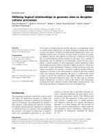
Fig. 2. Schematic representation of the rep-
licon system. Subgenomic and genomic
replicons are composed of the HCV 5¢-UTR,
the gene coding neomycin phosphotransfer-
ase (Neo
R
), the encephalomyocarditis virus
IRES, the region encoding HCV proteins and
the 3¢-UTR (A). Huh7 cells are electroporat-
ed with replicon RNA. Cell colonies effi-
ciently replicating the HCV replicon are
selected because of their resistance to
G418 (Ba). In parallel, Huh7 subclones highly
permissive to HCV replication can be
obtained by G418 treatment of cells trans-
fected with HCV subgenomic replicon. Cells
are then treated by IFN to eliminate the
HCV replicon (Bb).
M. Re
´
geard et al. In vitro HCV infection models
FEBS Journal 274 (2007) 4705–4718 ª 2007 The Authors Journal compilation ª 2007 FEBS 4707
mutations. Furthermore, it should be kept in mind
that, in this system, hundreds of RNA copies per cell
are present, in contrast to 5–50 copies in infected
hepatocytes. Therefore, results should be ascertained
using systems closer to HCV physiology.
Infection of primary cell cultures and
cell lines
Parallel to the development of this replication system,
intensive research has aimed to discover HCV infec-
tion systems. Until recently, sera obtained from
infected patients or chimpanzees were the only source
of HCV infectious particles. However, purification of
natural HCV particles from patient sera proved diffi-
cult because of the heterogeneity of their densities.
Viral RNA is detected in fractions ranging from 1.03
to 1.25 gÆmL
)1
in a sucrose gradient. Low-density
HCV particles are associated with either low- or very-
low-density lipoproteins [24–28] and have been shown
by assays of chimpanzee model to be the most
infectious fraction [29,30]. HCV particles of higher
densities correspond to particles associated with
immunoglobulin, free particles [28,30,31] or free
nucleocapsids [32].
Because hepatocytes are the main target of the virus
in vivo, several groups have attempted to infect pri-
mary hepatocytes or hepatic cells in vitro. Thus, adult
or fetal primary human hepatocytes have been shown
to support HCV infection and replication in vitro [33–
36]. Similarly, infections have also been conducted on
primary cells obtained from other mammals, including
chimpanzees and tree shrews [37,38]. In these models,
the replication rate was low, between 0.01 and 0.1
RNA copies per cell, depending on the experiment.
Therefore, highly sensitive assays were required to
detect viral RNA within infected cells or in cell-culture
supernatants. In order to demonstrate that infection
had taken place, researchers put forward supplemen-
tary data: detection of HCV negative-strand RNA,
which only appears during ongoing replication; the
sensitivity of replication to IFNa treatment; secretion
of neosynthesized virions able to infect naive cells; and
selection of quasispecies during the culture of infected
hepatocytes. To succeed in obtaining HCV infection in
primary human hepatocytes, an existing model of hep-
atitis B virus infection was used to determine optimal
infection conditions [39,40]. Rumin et al. pointed out
the need to reach high levels of cellular differentiation,
which they suggested may account for the ability of
these cells to support virus assembly and secretion [36].
Similarly, primary hepatocytes have recently been cul-
tivated in spheroid formation because this culture con-
dition maintains the differentiation state of the cell.
However, no real improvement in viral replication effi-
ciency was achieved with this new model [41]. It has
also been noted that, for unknown reasons, sera from
patients are not always infectious and no obvious cor-
relation can be drawn between infectivity and viral
RNA titre or with the presence of antibodies directed
against structural proteins [36]. Although primary
human hepatocytes infected with patient sera are the
most physiological in vitro model at present, the diffi-
culty of obtaining cells and intrinsic technical con-
straints make it hard to use in everyday experiments.
This may explain the limited number of studies based
on this model. However, recent work using this model
supported the involvement of the low-density lipopro-
tein receptor (LDLR) in the entry process of HCV.
The soluble form of LDLR, natural LDLR ligands
and antibodies directed against LDLR efficiently com-
peted with HCV infection, suggesting that LDLR
is probably involved in the entry process of native
HCV [42].
In parallel, some groups have focused their efforts on
developing an in vitro model based on hepatic cell lines
[43]. Among those tested, HepG2, Huh7 and PH5CH,
the latter was the most susceptible to infection and
replication. However, the replication rate in PH5CH
remained low as viral RNA could only be detected
using RT-nested PCR. Recently, Aly et al. immortal-
ized primary human hepatocytes with human papilloma
virus E6E7 genes [44]. These HPV18⁄ E6E7-immortal-
ized hepatocytes could maintain hepatic-specific mark-
ers in long-term culture and were susceptible to HCV
infection, as assayed by RT-QPCR detection of the
intracellular HCV positive RNA strand. However, viral
replication efficiency was low compared with the infec-
tion system based on primary human hepatocytes
(10
2
and 10
4
copies of viral RNA per microgram of
cellular RNA, respectively) [42]. mAbs directed against
CD81, another probable component of the receptor
complex, and IFNa treatment, inhibited infection and
replication, respectively. Interestingly, an interferon
regulatory factor-7-defective form of this cell line has
been engineered by stable expression of transdominant
mutant interferon regulatory factor-7. These deficient
cells were more susceptible to infection. Indeed,
hundred more copies of HCV RNA could be detected
inside these cells following infection, whatever the geno-
type (1b, 2 and 3). However no production of progeny
viruses was shown in this infection model.
In some patients infected with HCV, analysis of
HCV negative-strand RNA by RT-PCR indicated its
presence in both the liver and haematopoietic cells
[45]. Growing evidence supports the idea that HCV is
In vitro HCV infection models M. Re
´
geard et al.
4708 FEBS Journal 274 (2007) 4705–4718 ª 2007 The Authors Journal compilation ª 2007 FEBS
also lymphotropic and lymphocytes may be an HCV
reservoir. In fact, Cribier et al. reported the in vitro
infection of primary peripheral blood mononuclear
cells with high-titre sera. Despite the low replication
efficiency, HCV RNA was detected for a month in cell
culture [46]. Several other laboratories have shown that
HCV could infect a B-cell line (Daudi) and T-cell lines
(MT-2 and MOLT-4) in vitro [21]. In a more recent
study by Sung et al. , proof of replication in this cell
type was demonstrated by the establishment of a B-cell
lymphoma cell line derived from an HCV-infected
patient with type II-mixed cryoglobulinaemia [47].
This cell line persistently replicated the HCV genome
and produced virions that were infectious in primary
human hepatocytes and lymphocytes in vitro.
In summary, use of HCV-containing sera to recon-
stitute the entire life cycle of HCV in vitro has proved
to be very difficult. Although infection of primary cells
has been shown with convincing data, low replication
efficiency and inherent technical difficulties have lim-
ited their use. The development of the newly described
HPV18 ⁄ E6E7-immortalized hepatocytes might consti-
tute an easier model to conduct further analyses. Par-
allel to the intensive research discussed thus far, other
groups have developed surrogate models to investigate
specific steps of HCV life cycle.
HCV-like particles
HCV-like particles are generated by self-assembly of
the HCV structural proteins and are nonreplicative.
The first HCV-like particle model was described by
Baumert and collaborators in 1998, with particles pro-
duced in insect cells using a recombinant baculovirus
containing the cDNA of HCV structural proteins of
genotype 1b or 1a [48]. HCV-like particles were
observed by electron microscopy in intracellular com-
partments but were not secreted in the supernatant.
Consequently, purification of HCV-like particles was
achieved by cell lysis followed by sucrose-gradient
purification. HCV-like particles are described as being
40–60 nm in diameter and of a rather high density
( 1.17 gÆmL
)1
) that should correspond to the density
of the free viral particles contained in the serum of
infected patients [48]. Structural characterization of
HCV-like particle envelope proteins has been con-
ducted by analysing their antigenic properties. This
was done using a large panel of monoclonal and con-
formational antibodies directed against E1 and E2,
and sera from infected patients [49,50]. Results
suggested that E1 and E2 located at the surface of
HCV-like particles formed E1E2 heterodimers in a
virion-like conformation.
This model has essentially been used in two major
fields of research: binding process and vaccination
development. Specific binding of HCV-like particles
was obtained for various hepatic and lymphocyte cell
lines and also for dendritic cells, independently of
CD81 expression [51,52]. The limited involvement of
CD81 in the binding process was further illustrated by
the poor binding inhibition achieved in the presence of
mAbs directed against CD81. In contrast, heparin sul-
fate seemed to mediate this interaction [53]. Taking
advantage of this binding model, a recent study
showed that interaction of envelope glycoproteins with
the surface of HepG2 cells induces gene expression
modulations. This suggested that HCV binding might
induce changes in the cell that could favour HCV
infection [54]. In parallel, virus-like particles could con-
stitute attractive vaccine candidates for papillomavirus-
es and retroviruses because they could mimic some
properties of native viruses. Concerning HCV-like par-
ticles, it has been shown that they are able to induce
humoral and cellular immune responses in BALB ⁄ c
mice, baboons and chimpanzees [55–57]. Immunized
chimpanzees were thereafter inoculated with HCV of
homologous genotype. Although vaccinated chimpan-
zees became infected by HCV, the infection was
controlled quickly compared with unvaccinated
animals [55].
Although the structural, biophysical and antigenic
properties of HCV-like particles have been character-
ized and might partly mimic those of native HCV parti-
cles and be close to pseudotyped particles described
later, the binding of HCV-like particle does not require
CD81 participation. However, CD81 at the surface of
Huh7 cells has been shown to be a crucial receptor
involved in the infection process of pseudotyped viruses
and cellular clones of HCV (see below). Moreover, this
first HCV-like particle model does not permit investiga-
tion into HCV morphogenesis because viral budding
was not observed in insect cells. By contrast, Blanchard
et al. developed another HCV-like particle model that
could be used to investigate this issue. HCV-like parti-
cles were produced in mammalian cells (BHK-21) by
expression of HCV structural proteins in a Semliki for-
est virus vector [58]. Budding of HCV-like particles
with a diameter of 50 nm was observed using electron
microscopy and occurred at the endoplasmic reticulum
membrane towards the lumen. Although most of these
HCV-like particles seemed to display an abortive bud-
ding process, it was shown that the correctly processed
HCV core protein drives this event [59,60]. However,
due to the absence of complete budding, this model
cannot be used to study the following steps of HCV
assembly in eukaryotic cells.
M. Re
´
geard et al. In vitro HCV infection models
FEBS Journal 274 (2007) 4705–4718 ª 2007 The Authors Journal compilation ª 2007 FEBS 4709
HCV pseudo particles
A few years after the development of HCV-like parti-
cles, another model was created to specifically investi-
gate the entry process of HCV. This system is called
pseudo particles of HCV (HCVpp), as envelope glyco-
proteins of HCV are incorporated at the surface of
other enveloped viruses substituting their natural enve-
lope proteins. The first constructed pseudotyped
viruses were vesicular stomatitis virus (VSV) ⁄ HCV
pseudotypes expressing HCV E1 and ⁄ or HCV E2
chimeric proteins. These contain the transmembrane
and cytoplasmic domains of envelope protein G of
VSV [61]. Although pseudotyped virus infectivity was
neutralized by antibodies directed against E1 and E2
or by sera from HCV-infected chimpanzees or humans,
these HCVpp exhibited a surprising tropism with lim-
ited infectivity on primary human hepatocytes, and
better infectivity on kidney cell lines of human or non-
human origin [61–63]. One may speculate that the
broad tropism of these HCVpp might be influenced by
the background infectivity of VSV. Moreover, some of
these viruses harboured only one of the two glycopro-
teins suggesting that E1 and E2 could independently
carry out the entry process of these HCVpp. Because
E1 and E2 are thought to be assembled in hetero-
dimers at the surface of native viruses, it seems likely
that both E1 and E2 are required for the infection
process.
A second generation of HCVpp (Fig. 3) has been
developed using unmodified E1 and E2 envelope glyco-
proteins, which are exposed at the surface of retro-
viral particles carrying a genome with a marker gene.
Retrovirus budding occurs at the plasma membrane,
although some data indicate that it might also exist
intracellularly [64]. Despite the specific endoplasmic
reticulum retention of HCV envelope glycoproteins
[65,66], it has been shown that in overexpression sys-
tems, a small fraction of envelope glycoproteins could
be secreted to the surface membrane via the secretory
pathway. This led to Golgi-specific modification of gly-
cosylation in envelope proteins [67,68]. HCV envelope
glycoproteins at the surface of retroviral particles are
comprised mainly of correctly folded E1 and E2
assembled as heterodimers and a small fraction of E1
and E2 covalently linked in aggregates [68]. In this
model, expression of both E1 and E2 should derive
from a unique expression construct for optimal infec-
tivity [67,69]. Various HCVpp have been developed
with envelope proteins of genotypes 1a, 1b, 2a, 3a, 4a,
5a and 6a allowing analysis of cross- and genotype-
specific neutralization [67,70]. The presence of a mar-
ker gene packaged in these HCVpp has enabled easy
evaluation of infectivity mediated by HCV glycopro-
teins. Using this system, the tropism of HCVpp has
been studied and is, with few exceptions, liver specific
[67,69–71].
Because of this model, numerous questions regard-
ing the HCV entry process could be assessed. On the
one hand, the receptor candidates scavenger receptor
class B type 1 (SR-B1) and CD81 have been evaluated
in Huh7 cells. Whereas CD81 has been shown to be
directly involved in the entry process, SR-B1 influ-
enced HCVpp entry via its cholesterol-uptake activity
[68–74]. In fact, high-density lipoprotein was shown to
promote HCV entry and reduce the inhibitory effect of
HCV-neutralizing antibodies from infected patients
[73,75]. On the other hand, the route of HCVpp entry
has been assessed in Huh7 cells using specific drugs
that increase endosomal pH or disrupt clathrin vesi-
cles, with transdominant mutants of the GTPases
Rab5 and Rab7 and small interfering RNA directed
against the clathrin heavy chain. These results support
the hypothesis that HCVpp penetrate cells using
R
e
p
o
rt
e
r
2E1E-VCHVMC
VMC
lop-
gaG
retropeR
Ψ
RTLVMC
RTL
ca
b
n
oitcefsna
rT
T392 KEH
n
o
i
tcu
d
o
rp ppV
C
H
R
e
p
o
r
t
e
r
R
e
p
o
r
t
e
r
senilllec
a
mot
a
p
e
hfo no
i
tc
e
fn
I
nietorp ret
rop
erfosisy
la
n
a
dn
a
noisserpxe
Fig. 3. Production of HCV pseudoparticles. To produce recombinant
retroviruses 293T are transfected with three expression vectors.
The first (a) is the packaging construct that encodes for retroviral
Gag and Pol proteins. After translation of the second vector (b), the
RNA produced and which contains the sequence of a reporter gene
could be encapsidated in particles via the presence of the retroviral
encapsidation sequence (Y). The third vector (c) encodes HCV E1
and E2 glycoproteins. Recombinant viruses collected from the
supernatant are made up of a retroviral capsid containing a RNA
genome with the HCV glycoproteins at their surface. Their specific
infectivity on hepatoma cell lines was analysed by expression of
the reporter gene.
In vitro HCV infection models M. Re
´
geard et al.
4710 FEBS Journal 274 (2007) 4705–4718 ª 2007 The Authors Journal compilation ª 2007 FEBS
clathrin vesicles and passage in the early endosome is
necessary for fusion between the viral envelope and
an intracellular membrane [69,71,76,77]. This fusion
should be carried out by a fusion peptide present in
viral envelope protein(s). Recently, data obtained with
HCVpp suggested that E1 and E2 might both contain
membrane fusion determinants, underlying a potential
difference with the fusion process of other flaviviruses
[78,79].
Although this model has technical advantages (easy
culture system and read out) and has enabled large
advances in our knowledge of HCV, it represents only
one category of HCV form derived from the sera of
infected patients: free viruses not associated with either
lipoproteins or immunoglobulins. Moreover, the chi-
meric nature of the viruses and the intrinsic character-
istics of the Huh7 cell line (its nonpermissiveness to
infection with HCV-containing sera) limit absolute
extrapolation of the results to native HCV. Thus
results should be reproduced in a cell-culture system
closer to physiological conditions.
Cellular clone of HCV
A major breakthrough has been achieved in the in vitro
modelling of HCV propagation with the development
of a cellular clone of HCV (HCVcc). This system is
based on the utilization of a very particular HCV
molecular clone of genotype 2a obtained from a Japa-
nese patient with fulminant hepatitis (JFH1). First
results with this JFH1 clone were obtained using the
subgenomic replicon model. After transfection of Huh7
cells with this replicon RNA, 4.5 · 10
4
cfuÆlg
)1
RNA were counted, whereas only 9 · 10
2
cfuÆlg
)1
RNA were obtained with transfection of the con1 rep-
licon of genotype 1b harbouring in vitro adaptative
mutations. Moreover, no adaptative mutations seemed
to be required in the subgenomic JFH1 replicon for its
efficient replication in cell culture [17]. The JFH1 sub-
genome was also shown to replicate efficiently in other
human cell lines of hepatic or nonhepatic origin
(HepG2, IMY-N9, HeLa, HEK 293) and in mouse
embryonic fibroblasts [80–82]. Soon after the discovery
of the extraordinary replication potential of the subge-
nomic JFH1 replicon, Wakita et al. [20] described how
transfection of Huh7 cells with the whole JFH1 RNA
sequence led to the production of viruses shown to be
infectious in vitro on naive Huh7 cells and in vivo in
chimpanzees (Fig. 4). Use of Huh7.5 or Huh7.5.1 cell
lines further optimized the kinetics of replication and
HCVcc secretion [10,83]. Because of this model, it has
been possible to observe cell-derived HCV parti-
cles, which showed a diameter of 55 nm when analy-
sed by electron microscopy [20]. In parallel, density
analyses of infected-cell supernatant suggested that
HCVcc are associated with lipoproteins with an hetero-
geneous density peak correlated to specific infectivity
ranging from 1.05 to 1.1 gÆmL
)1
in sucrose gradients
[10,83,84]. However, in vitro association of HCVcc
with lipoproteins might not be as optimal as in vivo
passage of this virus in chimpanzees or in mice con-
taining human liver xenografts. In fact, enhanced virus
infectivity is observed after in vivo passage and is cor-
related with a lower density of HCV particles [85].
Further analyses are needed to characterize the associ-
ation between lipoprotein and HCVcc in cell culture
and determine whether it reflects that seen in patient
sera. Most importantly, the efficiency of this model is
entirely attributed to this specific JFH1 molecular
clone. Attempts to reproduce this model with other
HCV molecular clones of various genotypes showed
rather limited results with very low virus release
despite demonstration of their infectivity in chimpan-
zees [86–88]. To expand the studies to other genotypes,
intergenotypic and intragenotypic chimeras have been
constructed replacing the structural proteins of the
JFH1 clone with those of Con1, H77, J6 and HCV-
452 molecular clones of genotypes 1b, 1a, 2a and 3a,
respectively [89,90]. With the exception of the HCV-
452–JFH1 chimera, efficient virus production was
obtained. Crossover before or inside the NS2 sequence
RTU’3
C
1E
2E
7p
RTU’5
3SN
1HFJ A
NR VCH
ccVCH
noit
c
u
d
orp
7huHfo
n
oitcefnI
se
ni
l
l
l
ec
larivfosisylana noisserpxE
nietorp retroper
ro
nietorp
ni
noitaroportcelE
senilllec 7huH
A4SN
B4SN
2SN
B5SNA5SN
Fig. 4. The HCVcc model. Huh7 cell lines are electroporated with
the RNA transcripts of the JFH1 genome. A few days after trans-
fection, viruses are secreted in the supernatant of cells replicating
HCV JFH1 genome. Their specific infectivity and replicative poten-
tial can be assessed on Huh7 cell lines and analysed by the expres-
sion of viral or reporter proteins or the quantification of intracellular
viral RNA.
M. Re
´
geard et al. In vitro HCV infection models
FEBS Journal 274 (2007) 4705–4718 ª 2007 The Authors Journal compilation ª 2007 FEBS 4711
had no significant effect on replication efficiency but
was shown to be critical for efficient virus production
[89,90]. Concerning the H77–JFH1 chimera, mutations
affecting E1, p7, NS2 and ⁄ or NS3 have been detected
and have contributed to improved assembly and
release of viral particles [90]. In addition to the utility
of these chimeras to decipher the HCV entry process
and assess antibody-neutralization efficiency, these
experiments point to the involvement of p7 and NS2
in HCV morphogenesis and release.
This recent in vitro model reproducing the entire life
cycle of HCV in Huh7 cell lines has enabled extensive
research into various areas of HCV biology. In the
field of HCV entry, much effort has been employed to
confirm results obtained using the HCVpp model. For
example, HCV entry into target cells is mediated by
E2 [10], and CD81 has been shown to be critical for
HCVcc infectivity. In fact, antibodies directed against
CD81 and CD81 downregulation with RNA interfer-
ence inhibited HCVcc entry into Huh7 cell lines
[10,20,83,91]. Moreover, recent studies analysing the
differential permissiveness of hepatic cell lines have
shown that CD81 surface expression was a key
requirement for HCVcc infection [83,92]. SR-B1 is
another putative HCV receptor that has been exten-
sively studied with HCVpp. mAbs directed against SR-
B1 and oxidized low-density lipoproteins, a ligand of
high affinity for SR-B1, competed with HCVcc entry,
supporting the hypothesis that SR-B1 is involved in
HCV entry [93,94]. Most recently, Claudin-1, a compo-
nent of the tight junctions, has been proposed to be
involved in the entry process of HCVpp and HCVcc
[95]. In fact, exogenous complementation of human
cells expressing CD81 and SR-B1 (293T and SW13)
with Claudin-1 permitted their infection with HCVpp
or HCVcc. Events late in the entry process of HCV
have also been studied and various results supported
the hypothesis drawn up using the HCVpp model:
HCV uses a clathrin-dependant pathway to enter
Huh7 cell lines in a pH-dependant fashion [76,96,97].
In the field of virus morphogenesis, the HCVcc model
enabled to incriminate p7 and NS2 in the assembly of
infectious viral particles [89,90,98]. Using this model,
the association of lipoproteins with virions during their
egress has also been suggested [84,99]. However, only
limited investigation could be conducted into HCV
morphogenesis due to the very rare observation of
particles in producing cells [100]. Finally, this model
has been also used to study virus–host interactions. As
a consequence of long-term HCVcc propagation, resis-
tant cells are selected and adaptated viruses emerge,
displaying a better infectivity partly related to a single
mutation in E2 [101].
Although the HCVcc model is dependent on the spe-
cific sequence of JFH1 5¢-UTR, NS proteins and
3¢-UTR, its utilization has led to great advances in our
knowledge of HCV biology. Urgent information is
needed to determine the genetic specificity of this
JFH1 molecular clone which confers high replication
efficiency and virus release. Nevertheless, viruses
expressing exogenous marker are becoming useful tools
to investigate both HCV biology and the potency of
antiviral drugs [20,96,97,102]. One example of the con-
tribution of these constructs is the discovery of a pro-
tective effect that cells already replicating HCV possess
against HCV superinfection [102,103].
Concluding remarks
Table 1 presents all the in vitro HCV models, their
potential use and limitations. Increasing data about
viral life cycle mechanisms have been accumulated in
recent years, particularly regarding entry and replica-
tion processes. The recent HCVcc model has emerged
as the most useful research tool to date.
Several membrane receptors are involved in the HCV
entry process: CD81, SR-B1, LDLR and Claudin-1.
The multiplication of candidate receptors and the
potential synergistic role of CD81 and SR-B1 support
the hypothesis of a multistep entry pathway that may
involve different receptors. Moreover, one should keep
in mind that HCV particles exist in patient sera in vari-
ous forms, either free or associated with lipoproteins or
immunoglobulin. One can therefore speculate that,
depending on its form, HCV may take advantage of
different sets of receptors to enter into target cells.
Whereas the implications of CD81, SR-B1 and Clau-
din-1 have been ascertained using the HCVpp and the
HCVcc model, the role of LDLR has been assessed in
primary human hepatocytes with serum-derived HCV
particles. Further analyses are needed to define for each
form of HCV particle, the exact sequence of events and
the precise implications of these candidates as attach-
ment or as entry receptors. In parallel, growing
evidence supports an internalization of HCV by endo-
cytosis followed by passage in the early endosome.
However, neither the exact location of the fusion pep-
tide in the viral surface glycoproteins nor the fusion
process has been clearly documented. Given the hetero-
geneity of serum-derived HCV particles, it cannot
be excluded that multiple entry pathways may lead
to productive infection with potentially different effi-
ciencies.
In the field of HCV replication, a large break-
through was the development of subgenomic replicons
that allowed examination of the viral components of
In vitro HCV infection models M. Re
´
geard et al.
4712 FEBS Journal 274 (2007) 4705–4718 ª 2007 The Authors Journal compilation ª 2007 FEBS
the replication complex and identification of the cellu-
lar partners of HCV replication. Although discovery of
the adaptative mutations necessary for efficient in vitro
replication enabled high-throughput studies, it also
raised the as yet unsolved question of the mechanisms
of replication enhancement driven by these mutations.
More recently, the JFH1 subgenome has been shown
to replicate at high levels without any adaptative muta-
tions. Analysis has shown that this may be due to a
more efficient initiation of RNA synthesis by JFH1
NS5B compared with that of the Con1 clone [104].
Moreover, and despite the generation of intergenotypic
chimera, the HCVcc model is based on the extraordi-
nary replication efficiency of the unique JFH1 strain
and therefore requires minimal sequences of this pecu-
liar strain: the 5¢-UTR sequence and the C-terminus
NS2 or the NS3 sequence to the 3¢-UTR. More infor-
mation is needed to better understand this greater rep-
lication efficiency and the determinants necessary for
the production and release of infectious viruses.
Another limitation of both the replicon system and
the HCVcc model is their requirement for Huh7 cell
lines that possess an impaired innate antiviral
response. HCVcc has also been shown to be infectious
and able to replicate efficiently in the newly described
HPV18 ⁄ E6E7-immortalized hepatocytes. In this experi-
ment, interferon regulatory factor-7, a regulatory fac-
tor of IFN response, was downregulated. It would
therefore be interesting to evaluate HCVcc infectivity
in primary human hepatocytes that possess intact
antiviral responses. HCVcc infection studies would
then be assessed in a more physiological context and
could be compared with serum-derived HCV infection.
Moreover, it would be of great interest to better
understand all the antiviral mechanisms developed by
primary human hepatocytes that may explain their lim-
ited rate of infection.
Acknowledgements
We thank C. Gamble for her critical reading of the
manuscript. Our research is supported by grants from
the ANRS and the Ligue Nationale contre le Cancer.
CL and MT are supported by fellowships provided by
the Ministe
`
re de l’Education et de la Recherche and
the Re
´
gion Bretagne, respectively.
References
1 WHO (1997) Hepatitis C global prevalence. Wkly
Epidemiol Rec 72, 341–344.
2 Lohmann V, Korner F, Koch J, Herian U, Theilmann
L & Bartenschlager R (1999) Replication of subgenom-
ic hepatitis C virus RNAs in a hepatoma cell line.
Science 285, 110–113.
3 Blight KJ, Kolykhalov AA & Rice CM (2000) Efficient
initiation of HCV RNA replication in cell culture.
Science 290, 1972–1974.
Table 1. HCV in vitro models: their possible applications and limitations.
Reproduced viral steps Area of research Limitations
Subgenomic replicon Replication replication mechanisms
intracellular host defences
evasion mechanisms
antiviral screening
in vitro adaptative mutations
restricted to Huh7 cell lines
artificially high level of replication
Serum-derived HCV Entire life cycle entry process
replication mechanisms
intracellular host defences
antiviral screening
few cell lines support infection
technical difficulties of primary cell culture
HCV-LP Infection, morphogenesis cell attachment
vaccination
morphogenesis
no secretion of particles
independence of CD81 for entry
HCVpp Infection entry process no budding at the ER
exogenous core
no association of particles with lipoproteins
HCVcc Entire life cycle entry process
replication mechanisms
intracellular host defence
evasion mechanisms
virus production
antiviral screening
restricted to JFH1 NS sequence
restricted to Huh7 cell lines
M. Re
´
geard et al. In vitro HCV infection models
FEBS Journal 274 (2007) 4705–4718 ª 2007 The Authors Journal compilation ª 2007 FEBS 4713
4 Guo JT, Bichko VV & Seeger C (2001) Effect of alpha
interferon on the hepatitis C virus replicon. J Virol 75,
8516–8523.
5 Krieger N, Lohmann V & Bartenschlager R (2001)
Enhancement of hepatitis C virus RNA replication
by cell culture-adaptive mutations. J Virol 75, 4614–
4624.
6 Lohmann V, Korner F, Dobierzewska A & Bartensch-
lager R (2001) Mutations in hepatitis C virus RNAs
conferring cell culture adaptation. J Virol 75, 1437–
1449.
7 Bukh J, Pietschmann T, Lohmann V, Krieger N,
Faulk K, Engle RE, Govindarajan S, Shapiro M,
St Claire M & Bartenschlager R (2002) Mutations that
permit efficient replication of hepatitis C virus RNA in
Huh-7 cells prevent productive replication in chimpan-
zees. Proc Natl Acad Sci USA 99, 14416–14421.
8 Blight KJ, McKeating JA & Rice CM (2002) Highly
permissive cell lines for subgenomic and genomic
hepatitis C virus RNA replication. J Virol 76, 13001–
13014.
9 Friebe P, Boudet J, Simorre JP & Bartenschlager R
(2005) Kissing-loop interaction in the 3¢ end of the
hepatitis C virus genome essential for RNA replication.
J Virol 79, 380–392.
10 Zhong J, Gastaminza P, Cheng G, Kapadia S, Kato T,
Burton DR, Wieland SF, Uprichard SL, Wakita T &
Chisari FV (2005) Robust hepatitis C virus infection
in vitro. Proc Natl Acad Sci USA 102, 9294–9299.
11 Lanford RE, Guerra B, Lee H, Averett DR, Pfeiffer B,
Chavez D, Notvall L & Bigger C (2003) Antiviral
effect and virus–host interactions in response to alpha
interferon, gamma interferon, poly(I)–poly(C), tumor
necrosis factor alpha, and ribavirin in hepatitis C virus
subgenomic replicons. J Virol 77, 1092–1104.
12 Sumpter R Jr, Loo YM, Foy E, Li K, Yoneyama M,
Fujita T, Lemon SM & Gale M Jr (2005) Regulating
intracellular antiviral defense and permissiveness to
hepatitis C virus RNA replication through a cellular
RNA helicase, RIG-I. J Virol 79, 2689–2699.
13 Moradpour D, Evans MJ, Gosert R, Yuan Z,
Blum HE, Goff SP, Lindenbach BD & Rice CM
(2004) Insertion of green fluorescent protein into non-
structural protein 5A allows direct visualization of
functional hepatitis C virus replication complexes.
J Virol 78, 7400–7409.
14 Murray EM, Grobler JA, Markel EJ, Pagnoni MF,
Paonessa G, Simon AJ & Flores OA (2003) Persistent
replication of hepatitis C virus replicons expressing the
beta-lactamase reporter in subpopulations of highly
permissive Huh7 cells. J Virol 77, 2928–2935.
15 Yi M, Bodola F & Lemon SM (2002) Subgenomic
hepatitis C virus replicons inducing expression of a
secreted enzymatic reporter protein. Virology 304,
197–210.
16 Blight KJ, McKeating JA, Marcotrigiano J & Rice
CM (2003) Efficient replication of hepatitis C virus
genotype 1a RNAs in cell culture. J Virol 77, 3181–
3190.
17 Kato T, Date T, Miyamoto M, Furusaka A,
Tokushige K, Mizokami M & Wakita T (2003) Effi-
cient replication of the genotype 2a hepatitis C virus
subgenomic replicon. Gastroenterology 125, 1808–
1817.
18 Ikeda M, Yi M, Li K & Lemon SM (2002) Selectable
subgenomic and genome-length dicistronic RNAs
derived from an infectious molecular clone of the
HCV-N strain of hepatitis C virus replicate efficiently
in cultured Huh7 cells. J Virol 76, 2997–3006.
19 Pietschmann T, Lohmann V, Kaul A, Krieger N,
Rinck G, Rutter G, Strand D & Bartenschlager R
(2002) Persistent and transient replication of full-length
hepatitis C virus genomes in cell culture. J Virol 76,
4008–4021.
20 Wakita T, Pietschmann T, Kato T, Date T,
Miyamoto M, Zhao Z, Murthy K, Habermann A,
Krausslich HG, Mizokami M et al.
(2005) Production
of infectious hepatitis C virus in tissue culture from a
cloned viral genome. Nat Med 11, 791–796.
21 Bartenschlager R, Frese M & Pietschmann T (2004)
Novel insights into hepatitis C virus replication and
persistence. Adv Virus Res 63, 71–180.
22 Lindenbach BD & Rice CM (2005) Unravelling hepati-
tis C virus replication from genome to function. Nature
436, 933–938.
23 De Francesco R & Migliaccio G (2005) Challenges and
successes in developing new therapies for hepatitis C.
Nature 436, 953–960.
24 Andre P, Komurian-Pradel F, Deforges S, Perret M,
Berland JL, Sodoyer M, Pol S, Brechot C, Paranhos-
Baccala G & Lotteau V (2002) Characterization of
low- and very-low-density hepatitis C virus RNA-
containing particles. J Virol 76, 6919–6928.
25 Nielsen SU, Bassendine MF, Burt AD, Martin C,
Pumeechockchai W & Toms GL (2006) Association
between hepatitis C virus and very-low-density lipopro-
tein (VLDL) ⁄ LDL analyzed in iodixanol density gradi-
ents. J Virol 80, 2418–2428.
26 Prince AM, Huima-Byron T, Parker TS & Levine DM
(1996) Visualization of hepatitis C virions and putative
defective interfering particles isolated from low-density
lipoproteins. J Viral Hepat 3, 11–17.
27 Thomssen R, Bonk S, Propfe C, Heermann KH,
Kochel HG & Uy A (1992) Association of hepatitis C
virus in human sera with beta-lipoprotein. Med Micro-
biol Immunol 181, 293–300.
28 Thomssen R, Bonk S & Thiele A (1993) Density heter-
ogeneities of hepatitis C virus in human sera due to the
binding of beta-lipoproteins and immunoglobulins.
Med Microbiol Immunol 182, 329–334.
In vitro HCV infection models M. Re
´
geard et al.
4714 FEBS Journal 274 (2007) 4705–4718 ª 2007 The Authors Journal compilation ª 2007 FEBS
29 Bradley D, McCaustland K, Krawczynski K, Spelbring
J, Humphrey C & Cook EH (1991) Hepatitis C virus:
buoyant density of the factor VIII-derived isolate in
sucrose. J Med Virol 34, 206–208.
30 Hijikata M, Shimizu YK, Kato H, Iwamoto A, Shih
JW, Alter HJ, Purcell RH & Yoshikura H (1993) Equi-
librium centrifugation studies of hepatitis C virus: evi-
dence for circulating immune complexes. J Virol 67,
1953–1958.
31 Kanto T, Hayashi N, Takehara T, Hagiwara H, Mita
E, Naito M, Kasahara A, Fusamoto H & Kamada T
(1995) Density analysis of hepatitis C virus particle
population in the circulation of infected hosts: implica-
tions for virus neutralization or persistence. J Hepatol
22, 440–448.
32 Maillard P, Krawczynski K, Nitkiewicz J, Bronnert C,
Sidorkiewicz M, Gounon P, Dubuisson J, Faure G,
Crainic R & Budkowska A (2001) Nonenveloped
nucleocapsids of hepatitis C virus in the serum of
infected patients. J Virol 75, 8240–8250.
33 Castet V, Fournier C, Soulier A, Brillet R, Coste J,
Larrey D, Dhumeaux D, Maurel P & Pawlotsky JM
(2002) Alpha interferon inhibits hepatitis C virus repli-
cation in primary human hepatocytes infected in vitro.
J Virol 76, 8189–8199.
34 Lazaro CA, Chang M, Tang W, Campbell J, Sullivan
DG, Gretch DR, Corey L, Coombs RW & Fausto N
(2007) Hepatitis C virus replication in transfected and
serum-infected cultured human fetal hepatocytes. Am J
Pathol 170, 478–489.
35 Fournier C, Sureau C, Coste J, Ducos J, Pageaux G,
Larrey D, Domergue J & Maurel P (1998) In vitro
infection of adult normal human hepatocytes in pri-
mary culture by hepatitis C virus. J Gen Virol 79,
2367–2374.
36 Rumin S, Berthillon P, Tanaka E, Kiyosawa K, Tra-
baud MA, Bizollon T, Gouillat C, Gripon P, Guguen-
Guillouzo C, Inchauspe G et al. (1999) Dynamic analy-
sis of hepatitis C virus replication and quasispecies
selection in long-term cultures of adult human hepato-
cytes infected in vitro. J Gen Virol 80, 3007–3018.
37 Lanford RE, Sureau C, Jacob JR, White R & Fuerst
TR (1994) Demonstration of in vitro infection of chim-
panzee hepatocytes with hepatitis C virus using strand-
specific RT ⁄ PCR. Virology 202, 606–614.
38 Zhao X, Tang ZY, Klumpp B, Wolff-Vorbeck G,
Barth H, Levy S, von Weizsacker F, Blum HE &
Baumert TF (2002) Primary hepatocytes of Tupaia
belangeri as a potential model for hepatitis C virus
infection. J Clin Invest 109 , 221–232.
39 Gripon P, Diot C, Theze N, Fourel I, Loreal O,
Brechot C & Guguen-Guillouzo C (1988) Hepatitis B
virus infection of adult human hepatocytes cultured in
the presence of dimethyl sulfoxide. J Virol 62, 4136–
4143.
40 Rumin S, Gripon P, Le Seyec J, Corral-Debrinski M &
Guguen-Guillouzo C (1996) Long-term productive epi-
somal hepatitis C virus replication in primary cultures
of adult human hepatocytes infected in vitro . J Viral
Hepat 3, 227–238.
41 Chong TW, Smith RL, Hughes MG, Camden J, Rudy
CK, Evans HL, Sawyer RG & Pruett TL (2006) Pri-
mary human hepatocytes in spheroid formation to
study hepatitis C infection. J Surg Res 130, 52–57.
42 Molina S, Castet V, Fournier-Wirth C, Pichard-Garcia
L, Avner R, Harats D, Roitelman J, Barbaras R,
Graber P, Ghersa P et al. (2007) The low-density lipo-
protein receptor plays a role in the infection of primary
human hepatocytes by hepatitis C virus. J Hepatol
46,
411–419.
43 Bartenschlager R & Lohmann V (2001) Novel cell cul-
ture systems for the hepatitis C virus. Antiviral Res 52,
1–17.
44 Aly HH, Watashi K, Hijikata M, Kaneko H, Takada
Y, Egawa H, Uemoto S & Shimotohno K (2007)
Serum-derived hepatitis C virus infectivity in interferon
regulatory factor-7-suppressed human primary hepato-
cytes. J Hepatol 46, 26–36.
45 Lerat H, Berby F, Trabaud MA, Vidalin O, Major M,
Trepo C & Inchauspe G (1996) Specific detection of
hepatitis C virus minus strand RNA in hematopoietic
cells. J Clin Invest 97, 845–851.
46 Cribier B, Schmitt C, Bingen A, Kirn A & Keller F
(1995) In vitro infection of peripheral blood mononu-
clear cells by hepatitis C virus. J Gen Virol 76, 2485–
2491.
47 Sung VM, Shimodaira S, Doughty AL, Picchio GR,
Can H, Yen TS, Lindsay KL, Levine AM & Lai MM
(2003) Establishment of B-cell lymphoma cell lines
persistently infected with hepatitis C virus in vivo and
in vitro: the apoptotic effects of virus infection. J Virol
77, 2134–2146.
48 Baumert TF, Ito S, Wong DT & Liang TJ (1998) Hep-
atitis C virus structural proteins assemble into virus-
like particles in insect cells. J Virol 72, 3827–3836.
49 Baumert TF, Wellnitz S, Aono S, Satoi J, Herion D,
Tilman Gerlach J, Pape GR, Lau JY, Hoofnagle JH
et al. (2000) Antibodies against hepatitis C virus-like
particles and viral clearance in acute and chronic hepa-
titis C. Hepatology 32, 610–617.
50 Triyatni M, Vergalla J, Davis AR, Hadlock KG,
Foung SK & Liang TJ (2002) Structural features of
envelope proteins on hepatitis C virus-like particles as
determined by anti-envelope monoclonal antibodies
and CD81 binding. Virology 298, 124–132.
51 Barth H, Schafer C, Adah MI, Zhang F, Linhardt RJ,
Toyoda H, Kinoshita-Toyoda A, Toida T, Van Kuppe-
velt TH, Depla E et al. (2003) Cellular binding of hep-
atitis C virus envelope glycoprotein E2 requires cell
surface heparan sulfate. J Biol Chem 278, 41003–41012.
M. Re
´
geard et al. In vitro HCV infection models
FEBS Journal 274 (2007) 4705–4718 ª 2007 The Authors Journal compilation ª 2007 FEBS 4715
52 Triyatni M, Saunier B, Maruvada P, Davis AR, Ulia-
nich L, Heller T, Patel A, Kohn LD & Liang TJ
(2002) Interaction of hepatitis C virus-like particles and
cells: a model system for studying viral binding and
entry. J Virol 76, 9335–9344.
53 Barth H, Schnober EK, Zhang F, Linhardt RJ, Depla
E, Boson B, Cosset FL, Patel AH, Blum HE & Baum-
ert TF (2006) Viral and cellular determinants of the
hepatitis C virus envelope–heparan sulfate interaction.
J Virol 80, 10579–10590.
54 Fang X, Zeisel MB, Wilpert J, Gissler B, Thimme R,
Kreutz C, Maiwald T, Timmer J, Kern WV, Donauer
J et al. (2006) Host cell responses induced by hepatitis
C virus binding. Hepatology 43, 1326–1336.
55 Elmowalid GA, Qiao M, Jeong SH, Borg BB, Baumert
TF, Sapp RK, Hu Z, Murthy K & Liang TJ (2007)
Immunization with hepatitis C virus-like particles
results in control of hepatitis C virus infection in chim-
panzees. Proc Natl Acad Sci USA 104, 8427–8432.
56 Jeong SH, Qiao M, Nascimbeni M, Hu Z, Rehermann
B, Murthy K & Liang TJ (2004) Immunization with
hepatitis C virus-like particles induces humoral and cel-
lular immune responses in nonhuman primates. J Virol
78, 6995–7003.
57 Murata K, Lechmann M, Qiao M, Gunji T, Alter HJ
& Liang TJ (2003) Immunization with hepatitis C
virus-like particles protects mice from recombinant
hepatitis C virus–vaccinia infection. Proc Natl Acad Sci
USA 100, 6753–6758.
58 Blanchard E, Brand D, Trassard S, Goudeau A &
Roingeard P (2002) Hepatitis C virus-like particle mor-
phogenesis. J Virol 76, 4073–4079.
59 Ait-Goughoulte M, Hourioux C, Patient R, Trassard
S, Brand D & Roingeard P (2006) Core protein cleav-
age by signal peptide peptidase is required for hepati-
tis C virus-like particle assembly. J Gen Virol 87,
855–860.
60 Blanchard E, Hourioux C, Brand D, Ait-Goughoulte
M, Moreau A, Trassard S, Sizaret PY, Dubois F &
Roingeard P (2003) Hepatitis C virus-like particle bud-
ding: role of the core protein and importance of its
Asp111. J Virol 77, 10131–10138.
61 Lagging LM, Meyer K, Owens RJ & Ray R (1998)
Functional role of hepatitis C virus chimeric glycopro-
teins in the infectivity of pseudotyped virus. J Virol 72,
3539–3546.
62 Buonocore L, Blight KJ, Rice CM & Rose JK (2002)
Characterization of vesicular stomatitis virus recombi-
nants that express and incorporate high levels of hepa-
titis C virus glycoproteins. J Virol 76 , 6865–6872.
63 Matsuura Y, Tani H, Suzuki K, Kimura-Someya T,
Suzuki R, Aizaki H, Ishii K, Moriishi K, Robison CS,
Whitt MA et al. (2001) Characterization of pseudotype
VSV possessing HCV envelope proteins. Virology 286,
263–275.
64 Sandrin V, Boulanger P, Penin F, Granier C, Cosset
FL & Bartosch B (2005) Assembly of functional hepa-
titis C virus glycoproteins on infectious pseudoparticles
occurs intracellularly and requires concomitant incor-
poration of E1 and E2 glycoproteins. J Gen Virol 86,
3189–3199.
65 Deleersnyder V, Pillez A, Wychowski C, Blight K, Xu
J, Hahn YS, Rice CM & Dubuisson J (1997) Forma-
tion of native hepatitis C virus glycoprotein complexes.
J Virol 71, 697–704.
66 Duvet S, Cocquerel L, Pillez A, Cacan R, Verbert A,
Moradpour D, Wychowski C & Dubuisson J (1998)
Hepatitis C virus glycoprotein complex localization in
the endoplasmic reticulum involves a determinant for
retention and not retrieval. J Biol Chem 273, 32088–
32095.
67 Bartosch B, Dubuisson J & Cosset FL (2003) Infec-
tious hepatitis C virus pseudo-particles containing
functional E1–E2 envelope protein complexes. J Exp
Med 197, 633–642.
68 Op De Beeck A, Voisset C, Bartosch B, Ciczora Y,
Cocquerel L, Keck Z, Foung S, Cosset FL & Dubuis-
son J (2004) Characterization of functional hepatitis C
virus envelope glycoproteins. J Virol 78, 2994–3002.
69 Hsu M, Zhang J, Flint M, Logvinoff C, Cheng-Mayer
C, Rice CM & McKeating JA (2003) Hepatitis C virus
glycoproteins mediate pH-dependent cell entry of
pseudotyped retroviral particles. Proc Natl Acad Sci
USA 100, 7271–7276.
70 Meunier JC, Engle RE, Faulk K, Zhao M, Bartosch B,
Alter H, Emerson SU, Cosset FL, Purcell RH & Bukh
J (2005) Evidence for cross-genotype neutralization of
hepatitis C virus pseudo-particles and enhancement of
infectivity by apolipoprotein C1. Proc Natl Acad Sci
USA 102, 4560–4565.
71 Bartosch B, Vitelli A, Granier C, Goujon C, Dubuis-
son J, Pascale S, Scarselli E, Cortese R, Nicosia A &
Cosset FL (2003) Cell entry of hepatitis C virus
requires a set of co-receptors that include the CD81
tetraspanin and the SR-B1 scavenger receptor. J Biol
Chem 278, 41624–41630.
72 Bartosch B, Verney G, Dreux M, Donot P, Morice Y,
Penin F, Pawlotsky JM, Lavillette D & Cosset FL
(2005) An interplay between hypervariable region 1 of
the hepatitis C virus E2 glycoprotein, the scavenger
receptor BI, and high-density lipoprotein promotes
both enhancement of infection and protection against
neutralizing antibodies. J Virol 79, 8217–8229.
73 Voisset C, Callens N, Blanchard E, Op De Beeck A,
Dubuisson J & Vu-Dac N (2005) High density lipopro-
teins facilitate hepatitis C virus entry through the
scavenger receptor class B type I. J Biol Chem 280,
7793–7799.
74 Zhang J, Randall G, Higginbottom A, Monk P, Rice
CM & McKeating JA (2004) CD81 is required for
In vitro HCV infection models M. Re
´
geard et al.
4716 FEBS Journal 274 (2007) 4705–4718 ª 2007 The Authors Journal compilation ª 2007 FEBS
hepatitis C virus glycoprotein-mediated viral infection.
J Virol 78, 1448–1455.
75 Voisset C, Op de Beeck A, Horellou P, Dreux M, Gus-
tot T, Duverlie G, Cosset FL, Vu-Dac N & Dubuisson
J (2006) High-density lipoproteins reduce the neutraliz-
ing effect of hepatitis C virus (HCV)-infected patient
antibodies by promoting HCV entry. J Gen Virol 87,
2577–2581.
76 Blanchard E, Belouzard S, Goueslain L, Wakita T,
Dubuisson J, Wychowski C & Rouille Y (2006) Hepa-
titis C virus entry depends on clathrin-mediated endo-
cytosis. J Virol 80, 6964–6972.
77 Meertens L, Bertaux C & Dragic T (2006) Hepatitis C
virus entry requires a critical postinternalization step
and delivery to early endosomes via clathrin-coated
vesicles. J Virol 80, 11571–11578.
78 Drummer HE, Boo I & Poumbourios P (2007) Muta-
genesis of a conserved fusion peptide-like motif and
membrane-proximal heptad-repeat region of hepatitis
C virus glycoprotein E1. J Gen Virol 88, 1144–1148.
79 Lavillette D, Pecheur EI, Donot P, Fresquet J, Molle
J, Corbau R, Dreux M, Penin F & Cosset FL (2007)
Characterization of fusion determinants points to the
involvement of three discrete regions of both E1 and
E2 glycoproteins in the membrane fusion process of
hepatitis C virus. J Virol doi: 10.1128/JVI.02642-06.
80 Chang KS, Cai Z, Zhang C, Sen GC, Williams BR &
Luo G (2006) Replication of hepatitis C virus (HCV)
RNA in mouse embryonic fibroblasts: protein kinase R
(PKR)-dependent and PKR-independent mechanisms
for controlling HCV RNA replication and mediating
interferon activities. J Virol 80, 7364–7374.
81 Date T, Kato T, Miyamoto M, Zhao Z, Yasui K,
Mizokami M & Wakita T (2004) Genotype 2a hepatitis
C virus subgenomic replicon can replicate in HepG2
and IMY-N9 cells. J Biol Chem 279 , 22371–22376.
82 Kato T, Date T, Miyamoto M, Zhao Z, Mizokami M
& Wakita T (2005) Nonhepatic cell lines HeLa and 293
support efficient replication of the hepatitis C virus
genotype 2a subgenomic replicon. J Virol 79, 592–596.
83 Lindenbach BD, Evans MJ, Syder AJ, Wolk B,
Tellinghuisen TL, Liu CC, Maruyama T, Hynes RO,
Burton DR, McKeating JA et al. (2005) Complete
replication of hepatitis C virus in cell culture. Science
309, 623–626.
84 Gastaminza P, Kapadia SB & Chisari FV (2006) Dif-
ferential biophysical properties of infectious intracellu-
lar and secreted hepatitis C virus particles. J Virol 80,
11074–11081.
85 Lindenbach BD, Meuleman P, Ploss A, Vanwolleghem
T, Syder AJ, McKeating JA, Lanford RE, Feinstone
SM, Major ME, Leroux-Roels G et al. (2006) Cell cul-
ture-grown hepatitis C virus is infectious in vivo and
can be recultured in vitro. Proc Natl Acad Sci USA
103, 3805–3809.
86 Kato T, Matsumura T, Heller T, Saito S, Sapp RK,
Murthy K, Wakita T & Liang TJ (2007) Production of
infectious hepatitis C virus of various genotypes in cell
cultures. J Virol 81, 4405–4411.
87 Sakai A, Takikawa S, Thimme R, Meunier JC,
Spangenberg HC, Govindarajan S, Farci P, Emer-
son SU, Chisari FV, Purcell RH et al. (2007) In vivo
study of the HC-TN strain of hepatitis C virus recov-
ered from a patient with fulminant hepatitis: RNA
transcripts of a molecular clone (pHC-TN) are infec-
tious in chimpanzees but not in Huh7.5 cells. J Virol
81, 7208–7219.
88 Yi M, Villanueva RA, Thomas DL, Wakita T &
Lemon SM (2006) Production of infectious genotype
1a hepatitis C virus (Hutchinson strain) in cultured
human hepatoma cells. Proc Natl Acad Sci USA 103,
2310–2315.
89 Pietschmann T, Kaul A, Koutsoudakis G, Shavinskaya
A, Kallis S, Steinmann E, Abid K, Negro F, Dreux M,
Cosset FL & Bartenschlager R (2006) Construction
and characterization of infectious intragenotypic and
intergenotypic hepatitis C virus chimeras. Proc Natl
Acad Sci USA 103, 7408–7413.
90 Yi M, Ma Y, Yates J & Lemon SM (2007) Compensa-
tory mutations in E1, 7, NS2, and NS3 enhance yields
of cell culture-infectious intergenotypic chimeric hepati-
tis C virus. J Virol 81, 629–638.
91 Koutsoudakis G, Herrmann E, Kallis S, Bartenschlager
R & Pietschmann T (2007) The level of CD81 cell sur-
face expression is a key determinant for productive
entry of hepatitis C virus into host cells. J Virol 81,
588–598.
92 Akazawa D, Date T, Morikawa K, Murayama A,
Miyamoto M, Kaga M, Barth H, Baumert TF, Dub-
uisson J & Wakita T (2007) CD81 expression is impor-
tant for the permissiveness of huh7 cell clones for
heterogeneous hepatitis C virus infection. J Virol 81,
5036–5045.
93 Catanese MT, Graziani R, von Hahn T, Moreau M,
Huby T, Paonessa G, Santini C, Luzzago A, Rice CM,
Cortese R et al. A (2007) High-avidity monoclonal
antibodies against the human scavenger class B type I
receptor efficiently block hepatitis C virus infection in
the presence of high-density lipoprotein. J Virol 81,
8063–8071.
94 von Hahn T, Lindenbach BD, Boullier A, Quehenber-
ger O, Paulson M, Rice CM & McKeating JA (2006)
Oxidized low-density lipoprotein inhibits hepatitis C
virus cell entry in human hepatoma cells. Hepatology
43, 932–942.
95 Evans MJ, von Hahn T, Tscherne DM, Syder AJ,
Panis M, Wolk B, Hatziioannou T, McKeating JA,
Bieniasz PD & Rice CM (2007) Claudin-1 is a hepatitis
C virus co-receptor required for a late step in entry.
Nature 446, 801–805.
M. Re
´
geard et al. In vitro HCV infection models
FEBS Journal 274 (2007) 4705–4718 ª 2007 The Authors Journal compilation ª 2007 FEBS 4717
96 Koutsoudakis G, Kaul A, Steinmann E, Kallis S, Loh-
mann V, Pietschmann T & Bartenschlager R (2006)
Characterization of the early steps of hepatitis C virus
infection by using luciferase reporter viruses. J Virol
80, 5308–5320.
97 Tscherne DM, Jones CT, Evans MJ, Lindenbach BD,
McKeating JA & Rice CM (2006) Time- and tempera-
ture-dependent activation of hepatitis C virus for low-
pH-triggered entry. J Virol 80 , 1734–1741.
98 Jones CT, Murray CL, Eastman DK, Tassello J &
Rice CM (2007) Hepatitis C virus p7 and NS2 proteins
are essential for infectious virus production. J Virol
101128/JVI00690-07.
99 Huang H, Sun F, Owen DM, Li W, Chen Y, Gale M
Jr & Ye J (2007) Hepatitis C virus production by
human hepatocytes dependent on assembly and secre-
tion of very low-density lipoproteins. Proc Natl Acad
Sci USA 104, 5848–5853.
100 Rouille Y, Helle F, Delgrange D, Roingeard P, Voisset
C, Blanchard E, Belouzard S, McKeating J, Patel AH,
Maertens G et al. (2006) Subcellular localization of
hepatitis C virus structural proteins in a cell culture
system that efficiently replicates the virus. J Virol 80,
2832–2841.
101 Zhong J, Gastaminza P, Chung J, Stamataki Z, Isoga-
wa M, Cheng G, McKeating JA & Chisari FV (2006)
Persistent hepatitis C virus infection in vitro: coevolu-
tion of virus and host. J Virol 80, 11082–11093.
102 Schaller T, Appel N, Koutsoudakis G, Kallis S, Loh-
mann V, Pietschmann T & Bartenschlager R (2007)
Analysis of hepatitis C virus superinfection exclusion
by using novel fluorochrome gene-tagged viral
genomes. J Virol 81, 4591–4603.
103 Tscherne DM, Evans MJ, von Hahn T, Jones CT,
Stamataki Z, McKeating JA, Lindenbach BD & Rice
CM (2007) Superinfection exclusion in cells infected
with hepatitis C virus. J Virol 81, 3693–3703.
104 Binder M, Quinkert D, Bochkarova O, Klein R, Kez-
mic N, Bartenschlager R & Lohmann V (2007) Identifi-
cation of determinants involved in initiation of
hepatitis C virus RNA synthesis by using intergeno-
typic replicase chimeras. J Virol 81, 5270–5283.
In vitro HCV infection models M. Re
´
geard et al.
4718 FEBS Journal 274 (2007) 4705–4718 ª 2007 The Authors Journal compilation ª 2007 FEBS