Báo cáo khoa học: G protein-coupled receptor-induced Akt activity in cellular proliferation and apoptosis pptx
Bạn đang xem bản rút gọn của tài liệu. Xem và tải ngay bản đầy đủ của tài liệu tại đây (429.14 KB, 12 trang )
REVIEW ARTICLE
G protein-coupled receptor-induced Akt activity in cellular
proliferation and apoptosis
David C. New, Kelvin Wu, Alice W. S. Kwok and Yung H. Wong
Department of Biochemistry, the Molecular Neuroscience Center, and the Biotechnology Research Institute, Hong Kong University of
Science and Technology, Clear Water Bay, Kowloon, Hong Kong, China
Akt, also known as protein kinase B (PKB), is a ser-
ine ⁄ threonine protein kinase that plays a pivotal role
in many physiological processes, including metabolism,
development, cell cycle progression, migration and sur-
vival [1–4]. The Akt subfamily of protein kinases con-
sists of three isoforms – Akt1, Akt2 and Akt3 (also
termed PKBa, PKBb and PKBc) – which are the
products of distinct genes. All three proteins share a
conserved tertiary structure of an N-terminal pleckstrin
homology domain, a kinase domain and a C-terminal
regulatory domain containing the hydrophobic motif
phosphorylation site [5]. While the homology between
the three isoforms allows for a degree of functional
redundancy [1], there also seems to be considerable
scope for isoform-specific activation and substrate
specificity [3,6].
Akt plays an integral role in the phosphoinositide
3-kinase (PI3K) signaling pathways. PI3K pathways
are activated in response to extracellular signals medi-
ated by cell-surface receptors of the G protein-coupled
receptor (GPCR), integrin and growth factor ⁄ receptor
tyrosine kinase (RTK) superfamilies. Receptor-medi-
ated activation of PI3K results in the generation of
phosphatidylinositol (3,4,5)-trisphosphate from phos-
phatidylinositol (4,5)-bisphosphate, a reaction that is
reversed by the enzymes phosphatase and tensin homo-
logue (PTEN) and SH2-domain-containing inositol
polyphosphate 5-phosphatase (SHIP). Both Akt and
Keywords
Akt; protein kinase B; G protein; G protein-
coupled receptor; cell cycle; apoptosis
Correspondence
Y. H. Wong, Department of Biochemistry,
Hong Kong University of Science and
Technology, Clear Water Bay, Kowloon,
Hong Kong, China
Fax: +852 2358 1552
Tel: +852 2358 7328
E-mail:
(Received 17 August 2007, revised 17 Sep-
tember 2007, accepted 24 September 2007)
doi:10.1111/j.1742-4658.2007.06116.x
Akt (also known as protein kinase B) plays an integral role in many intra-
cellular signaling pathways activated by a diverse array of extracellular sig-
nals that target several different classes of membrane-bound receptors. Akt
plays a particularly prominent part in signaling networks that result in the
modulation of cellular proliferation, apoptosis and survival. Thus, the over-
expression of Akt subtypes has been measured in a number of cancer types,
and dominant-negative forms of Akt can trigger apoptosis and reduce the
survival of cancer cells. G protein-coupled receptors act as cell-surface
detectors for a diverse spectrum of biological signals and are able to acti-
vate or inhibit Akt via several direct and indirect means. In this review, we
shall document how G protein-coupled receptors are able to control Akt
activity and examine the resulting biochemical and physiological changes,
with particular emphasis on cellular proliferation, apoptosis and survival.
Abbreviations
CDK, cyclin-dependent kinase; EGF, epidermal growth factor; EGFR, epidermal growth factor receptor; ERK, extracellular signal-regulated
kinase; FH, forkhead; GPCR, G protein-coupled receptor; LPA, lysophosphatidic acid; MAPK, mitogen-activated protein kinase; mTOR,
mammalian target of rapamycin; NF-jB, nuclear factor jB; p70
S6K
, p70 ribosomal protein S6 kinase; PAR-2, protease-activated receptor-2;
PDGFRa, platelet-derived growth factor receptor a; PI3K, phosphoinositide 3-kinase; PKB, protein kinase B; PTEN, phosphatase and tensin
homologue; RTK, receptor tyrosine kinase; SHIP, SH2-domain-containing inositol polyphosphate 5-phosphatase; TNF, tumour necrosis factor;
TSC, tuberous sclerosis complex; TSH, thyroid-stimulating hormone receptor.
FEBS Journal 274 (2007) 6025–6036 ª 2007 The Authors Journal compilation ª 2007 FEBS 6025
phosphoinositide-dependent kinase are recruited to the
plasma membrane by phosphatidylinositol (3,4,5)-tris-
phosphate through their pleckstrin homology domains,
where phosphoinositide-dependent kinase phosphory-
lates Akt1 on residue Thr308 in its kinase domain [7].
A second phosphorylation takes place at Ser473 in the
hydrophobic motif region of Akt1. This phosphoryla-
tion event seems to be catalyzed by a number of differ-
ent kinases, which are probably stimulus- and ⁄ or cell
type-specific. This stabilizes the active conformation of
Akt and allows it to translocate to the cytoplasm or
nucleus to search for its many target proteins [8].
Akt’s role in physiology suggests that aberrant Akt
signaling may be a factor in disease states. Most nota-
bly, amplification of Akt isoform genes and Akt
mRNA overexpression has been observed in many
human cancers [9]. Akt activity in cancer cells may
also be enhanced by the amplification of genes encod-
ing PI3K or by a reduction in the activity of PTEN or
SHIP [9]. It is therefore to be expected that the inhibi-
tion of PI3K, Akt and their downstream effectors has
been targeted in the development of cancer therapies
[10]. The involvement of Akt in cancer is not surpris-
ing given the ability of Akt to promote cellular
proliferation through the direct and indirect phosphor-
ylation of a number of cell cycle regulatory proteins
[11], and its ability to inactivate pro-apoptotic factors,
such as Bad, caspase-9 and forkhead (FH) transcrip-
tion factors [12]. In contrast, it is thought that a reduc-
tion in Akt signaling may contribute to diabetes by
reducing the survival of pancreatic b cells [13].
GPCRs act as cell-surface detectors for
a diverse spectrum of biological signals
To date, over 200 GPCRs have been matched with a
ligand that activates the receptor to promote a wide
variety of intracellular biochemical changes [14], even
though it is estimated that the human genome encodes
between 800 and 1000 GPCR subtypes [15,16]. Their
pervasive influence, coupled with their cell-surface
accessibility, has resulted in GPCRs becoming the tar-
gets of as many as 45% of modern medicines [17],
which are used to treat conditions as diverse as inflam-
mation, incontinence, hypertension, depression and
pain [18].
GPCRs preferentially couple to heterotrimeric
G proteins (consisting of a, b and c subunits) that are
grouped into four classes, known as Ga
q ⁄ 11
,Ga
i ⁄ o
,Ga
s
and Ga
12 ⁄ 13
, based on the effector with which the
a-subunit primarily interacts. The activated G proteins
in turn promote the activation or inhibition of a variety
of intracellular events, including the activation of phos-
pholipases, mitogen-activated protein kinases (MAP-
Ks), activation ⁄ inhibition of adenylyl and guanylyl
cyclases, and the opening and closing of ion channels.
In this review, we shall investigate the ability of
GPCRs to activate Akt signaling pathways both
directly, through the interaction of Gbc subunits with
PI3K, and indirectly, through the GPCR transactiva-
tion of RTKs and integrins. We shall also examine the
downstream signaling and physiological consequences
of GPCR-induced Akt activation, paying particular
attention to the consequences for cellular proliferation,
survival and apoptosis.
GPCR activation of PI3K ⁄⁄ Akt signaling
pathways
GPCRs promote intracellular signaling through both
Ga and Gbc subunits, which can activate distinct,
complementary or antagonistic pathways. As we will
demonstrate, a large number of GPCRs, coupled to all
four classes of G protein, activate PI3K ⁄ Akt pathways
through either Ga or Gbc subunits (see below, Table 1
and Fig. 1). Gbc subunits are able to bind directly to
and activate PI3K heterodimeric proteins containing
either the p110b or the p110c subunits [19]. Further-
more, it has been demonstrated that muscarinic and
lysophosphatidic acid (LPA) GPCRs are only able to
activate Akt in cells expressing the p110b or p110c
subunits, and that this activation is mediated by Gbc
subunits, but not by Ga subunits [20]. Direct activa-
tion of PI3K by Ga subunits has not been specifically
measured but they can activate PI3K ⁄ Akt pathways by
transactivating integrins, RTKs and other growth fac-
tor receptors.
Numerous RTKs and integrins can independently
activate PI3K ⁄ Akt pathways [21] but it has also been
reported that they can be transactivated by GPCRs
through Ga-orGbc-dependent pathways [22,23]. For
example, ligands for the LPA, endothelin-1 and throm-
bin receptors all promote DNA synthesis in Rat1
fibroblasts by transactivating the epidermal growth
factor receptor (EGFR, an RTK). Such transactivation
requires the activation of matrix metalloproteases to
release EGF from its membrane-bound form, which
then stimulates the EGFR and downstream extracellu-
lar signal-regulated kinase (ERK) pathways [24].
PI3K ⁄ Akt pathways are also activated by a similar
method of transactivation [25]. In Swiss 3T3 cells,
bradykinin and bombesin promote cellular prolifera-
tion by an EGFR-dependent formation of a signaling
complex that activates PI3K ⁄ Akt cascades [26]. A
number of other RTKs are also transactivated by
GPCRs [27–29], and it has been demonstrated that
Regulation of Akt signaling pathways by GPCRs D. C. New et al.
6026 FEBS Journal 274 (2007) 6025–6036 ª 2007 The Authors Journal compilation ª 2007 FEBS
Table 1. GPCR-induced Akt activity and the consequences for cellular proliferation and apoptosis. A selection of examples is presented here
demonstrating the signaling components employed in connecting GPCR-initiated signals to downstream events regulated by Akt. ›, indicates
an increase in protein levels or activity; fl , indicates a decrease in protein levels or activity. AC, adenylyl cyclase; AFX, FOX04; ALXR, lipoxin
A
4
receptor; CDK, cyclin-dependent kinase; CREB, cAMP-response element binding; cyt c, cytochrome c; EGFR, epidermal growth factor
receptor; ERK, extracellular signal-regulated kinase; FKHR, forkhead in rhabdomyosarcoma; FSH, follicle stimulating hormone; IGF-1, insulin
growth factor-1; IGF-IRb, insulin-like growth factor receptor; IRS-1, insulin receptor substrate 1; GnRH, gonadotropin-releasing hormone;
GSK3b, glycogen synthase kinase 3b; LHRH, luteinizing hormone-releasing hormone; LPA, lysophophatidic acid; MMP, matrix metalloprotein-
ase; mTOR, mammalian target of rapamycin; NF-jB, nuclear factor-jB; p70
S6k
, p70 ribosomal protein S6 kinase; PAR-2, protease-activated
receptor-2; PDGF, platelet-derived growth factor; PDGFR, platelet-derived growth factor receptor; PDK1, phosphoinositide-dependent
kinase 1; PI3K, phosphoinositide 3-kinase; PKA, protein kinase A; PKC, protein kinase C; PP2A, protein phosphatase 2A; PTP, protein tyro-
sine phosphatase, ROCK, Rho-associated kinase; ROS, reactive oxygen species; SHP, Src homology 2-containing phosphatase; TSC2, tuber-
ous sclerosis complex 2; TSH, thyroid stimulating hormone; VPAC, vasoactive intestinal peptide receptor.
GPCR Intracellular pathway Functional consequences References
G
i ⁄ o
pathways
Adenosine A
2
›PI3K ⁄›Akt ⁄flTSC2 Survival [41]
Adenosine A
3
›PLC ⁄›PI3K ⁄›Akt ⁄flERK Anti-proliferation [56]
ALXR flPDGF ⁄›EGF ⁄›PI3K ⁄›Akt ⁄›p27
Kip1
⁄›p21
Cip1
⁄
flCDK2 ⁄flcyclin E
Anti-inflammatory effects,
antiproliferation
[47]
CXCR4 ›Ca
2+
⁄›Pyk2 ⁄›PI3K ⁄›ERK ⁄›Akt ›DNA synthesis, proliferation [34]
d-opioid ›PI3K ⁄›Akt ⁄flTSC2 Survival [50]
›PI3K ⁄›Akt ⁄flGSK3b ⁄flAFX ⁄flFKHR Survival, proliferation [81]
Dopamine D
2
›PP2A ⁄flAkt ⁄›GSK-3 Dopamine-induced neurological
responses
[37]
j-opioid ›PI3K ⁄›Akt ⁄flTSC2 Survival [50]
LPA ›MMP ⁄›EGFR ⁄›PI3K ⁄›Akt ›Cyclin D1,›DNA synthesis,
proliferation, survival
[24,25]
›PI3K ⁄›Akt ⁄›p70
S6k
Survival [70]
›PI3K ⁄›Akt ⁄›NF-jB Survival [80]
l-opioid ›PI3K ⁄›Akt ⁄flTSC2 Survival [50]
Muscarinic M
2
›PI3K ⁄›Akt Survival [63]
Muscarinic M
4
›PI3K ⁄›Akt ⁄flTSC2 Survival [40]
Pheromone V2R ›ERK ⁄›Akt ⁄›CREB Survival [64]
Somatostatin SST
2
flPI3K ⁄flAkt ⁄flNF-jB Anti-proliferative, apoptotic,
antiangiogenic and
anti-invasive
[36]
›Src ⁄›SHP-1 ⁄›SHP-2 ⁄›PI3K ⁄›Ras ⁄›ERK ⁄
›p27
Kip1
G
1
cell cycle arrest [57]
Somatostatin SST
2b
›PI3K ⁄›Akt ⁄›p70
S6K
›DNA synthesis, survival,
proliferation
[45]
G
q ⁄ 11
pathways
a-thrombin ›PI3Kb ⁄›Akt ⁄›cyclin D ⁄›CDK4 ›G1-S phase transition [46]
Angiotensin II type 1 ›MMP ⁄›EGFR ⁄›PI3K ⁄›Akt Survival, proliferation [25]
›EGFR ⁄›PI3K ⁄›Akt ⁄›mTOR ⁄›p70
S6K
›G1 cyclins, flp27
Kip1
, flp21
Cip1
,
proliferation
[52]
Apelin APJ ›PI3K ⁄›Akt ⁄flcaspase 8 ⁄flcyt c ⁄flcaspase 9 ⁄
flcaspase 3
Survival [68]
Bradykinin ›EGFR ⁄›PI3K ⁄›Akt ›Cyclin D1, ›Cyclin E, ›DNA
synthesis, proliferation
[26]
Bombesin ›EGFR ⁄›PI3K ⁄›Akt ›DNA synthesis, proliferation [26]
›IGF-IRb ⁄›Src ⁄›PI3K ⁄›Akt Survival [29]
Endothelin-1 ›IGF-IRb ⁄›Src ⁄›PI3K ⁄›Akt Survival [29]
›Src ⁄›PI3K ⁄›Akt Glut4 translocation [38]
GnRH ›PKC ⁄›Src ⁄›Pyk2 ⁄›MMP ⁄›EGFR ⁄›PI3K ⁄›Akt Survival, proliferation [25]
LPA ›PI3K ⁄›Akt ⁄›NF-jB Survival [80]
LHRH ›PKCa ⁄flAkt ⁄›GSK3 ⁄›
Bad ⁄›caspase 3 Apoptosis [75]
Muscarinic M
1
›PI3K ⁄›Akt Survival [63]
›PTP ⁄flphosphorylated-IRS-1 ⁄flPI3K ⁄flAkt ⁄›RhoA ⁄
flROCK-I
Apoptosis [73]
D. C. New et al. Regulation of Akt signaling pathways by GPCRs
FEBS Journal 274 (2007) 6025–6036 ª 2007 The Authors Journal compilation ª 2007 FEBS 6027
Table 1. (Continued).
GPCR Intracellular pathway Functional consequences References
Muscarinic subtypes ›Src ⁄›PI3K ⁄›Akt ⁄›ERK ⁄flGSK3b ⁄flcaspase 3 ⁄
›CREB
Survival, proliferation [67]
PAR-2 ›Ca
2+
⁄›PKC ⁄›Pyk2 ⁄›Src ⁄›PI3K ⁄›Akt Actin reorganization and cell
migration
[33]
Serotonin ›PI3K ⁄›ROS ⁄›Akt ⁄›mTOR ⁄›p70
S6K
Proliferation [53]
Vasopressin V
1
›Ca
2+
⁄›PKC ⁄›Pyk2 ⁄›Src ⁄›EGFR ⁄›PI3K ⁄›Akt ⁄
›mTOR ⁄›p70
S6K
Cell growth, proliferation [51]
G
s
pathways
Adenosine A
2A
›Ca
2+
⁄›Src ⁄›Trk ⁄›Akt Survival [65]
b-adrenergic ›AC ⁄›PKA ⁄›Src ⁄›EGFR ⁄›PI3K ⁄›Akt Mucin secretion [31]
FSH ›PI3K ⁄›Akt ⁄flFOXO1a Follicular survival and
development
[69]
TSH ›cAMP ⁄›PKA ⁄›PI3K ⁄›PDK1 ⁄›mTOR ⁄›p70
S6K
Proliferation, thyroid follicle
activity
[54]
›cAMP ⁄›PKA ⁄›PI3K-Ras complex ⁄flERK DNA synthesis, mitogenesis [55]
VPAC-1 ›TrkA ⁄›PI3K ⁄›Akt Survival, development,
differentiation
[28]
G
12 ⁄ 13
pathways
LPA ›Rho ⁄›p160ROCK ⁄›PDGFRa ⁄›PI3K ⁄›Akt ⁄flFKHR flTranscription of apoptotic and
antiproliferative genes
[30]
Thrombin ›Rho ⁄›p160ROCK ⁄›PDGFRa ⁄›PI3K ⁄›Akt ⁄flFKHR flTranscription of apoptotic and
antiproliferative genes
[30]
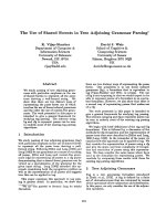
Fig. 1. Routes to Akt activation. G
12 ⁄ 13
-, G
i ⁄ o
-, G
q
-, and G
s
-coupled receptors are all known to activate the phosphoinositide 3-kinase
(PI3K) ⁄ Akt pathway through either Ga or Gbc subunits. Ga
q
and Ga
s
subunits utilize secondary messenger systems [Ca
2+
, cAMP, reactive
oxygen species (ROS)] to promote PI3K ⁄ Akt activation. Whilst Ca
2+
-induced PI3K activation is a feature of G
i ⁄ o
-coupled GPCRs, Gbc sub-
units released from these receptors are also able to directly bind to and activate PI3K. In addition to these mechanisms, all four classes of
GPCRs are able to activate the PI3K ⁄ Akt pathway by transactivating RTK at the plasma membrane either through matrix metalloproteinases
(G
i ⁄ o
-, G
q
-, and G
s
-coupled receptors) or through Rho ⁄ Rho-associated kinase (Rock)-mediated expression of RTK ligands (G
12 ⁄ 13
-coupled
receptors). The Rho ⁄ Rock pathway can also indirectly inhibit PI3K activity, although the signaling components involved have not yet been
elucidated (indicated by a dashed line).
Regulation of Akt signaling pathways by GPCRs D. C. New et al.
6028 FEBS Journal 274 (2007) 6025–6036 ª 2007 The Authors Journal compilation ª 2007 FEBS
constitutively active Ga
12
subunits activate PI3K⁄Akt
signaling via the transactivation of the platelet-derived
growth factor receptor a (PDGFRa) [30]. However,
it is not clear whether transactivation of RTKs by
GPCRs can also occur through the induced expression
of RTK ligands. Alternatively, the RTK ligand
requirement may be bypassed by the GPCR-induced
Src family tyrosine kinase activation of RTKs [27], as
evidenced by the Src kinase-dependent EGFR transac-
tivation promoted by the b-adrenergic receptor in gas-
tric mucosal cells [31].
Src-family kinases are firmly embedded in signal
transduction pathways activated by diverse extracellu-
lar stimuli playing a significant role in the crosstalk
between many pathways, including those that facilitate
the GPCR activation of Akt [32]. The protease-
activated receptor-2 (PAR-2) utilizes Ga
q
subunits to
promote the activation of protein kinase C and the
mobilization of intracellular Ca
2+
, leading to the
formation of a complex containing Src-family kinases,
the focal adhesion kinase, Pyk2, and PI3K [33].
Similar findings have been made for the G
i ⁄ o
-coupled
CXCR4 receptor, which promotes DNA synthesis via
a Pyk2 ⁄ PI3K ⁄ ERK pathway [34]. These complexes
may form as part of larger integrin ⁄ paxillin signaling
platforms that promote the phosphorylation and
activation of PI3K subunits [35].
GPCR activation may also lead to the inactivation
of Akt. It has been reported that somatostatin SST
2
receptors directly form a complex with the p85 regula-
tory subunit of PI3K. Agonist activation induced the
dissociation of this complex, preventing PI3K activa-
tion [36]. Following agonist-induced activation, dopa-
mine D
2
receptors are internalized and form a
multiprotein complex that includes b-arrestin, protein
phosphatase 2A and Akt. Protein phosphatase 2A
inactivates Akt, thereby relieving Akt’s inhibition of
glycogen synthase kinase 3b and allowing it to mediate
dopamine-induced neurological responses [37]. An
alternative mode of b-arrestin-mediated PI3K ⁄ Akt
inhibition is proposed to occur upon activation of the
Ga
q
-coupled PAR-2 receptor. Upon recruitment to the
PAR-2 receptor, b-arrestin forms a complex with PI3K
and spatially restricts its enzyme activity, thereby mod-
ulating the PAR-2 receptor activation of PI3K ⁄ Akt
mediated by Pyk2 and Src-family kinases (see the pre-
ceding discussion) [33]. In contrast, another study has
indicated that b-arrestins mediate the endothelin A
receptor activation of Akt by recruiting Src-family kin-
ases that phosphorylate and activate Ga
q
, ultimately
leading to PI3K ⁄ Akt pathway activation [38]. Never-
theless, the vital role of b-arrestins in modulating the
apoptotic events following the activation of some
GPCRs was highlighted by a study which showed that
in mouse embryonic fibroblasts devoid of b-arrestins
the N-formyl peptide receptor, vasopressin V
2
, chemo-
kine CXCR2 and the angiotensin II AT
1A
receptors all
promote apoptosis through the activation of PI3K,
MAPKs and Src kinases, leading to the activation of
caspase pathways [39]. Reconstituting the b-arrestins
prevented the GPCR-induced apoptosis, suggesting
that for some GPCRs b-arrestins constrain their apop-
totic abilities. The same study also demonstrated the
GPCR selectivity of these events because in the
absence of b-arrestins the CXCR4 and b
2
-adrenergic
receptors were unable to activate apoptosis.
Recent studies have indicated that constitutively
active Ga subunits of the Ga
q ⁄ 11
and Ga
12 ⁄ 13
subfami-
lies may actually inhibit the EGFR-mediated activa-
tion of Akt in transfected HEK-293 cells [40].
This contradicts the previously noted ability of
constitutively active Ga
12
subunits to potentiate
PDGFRa-mediated PI3K ⁄ Akt signaling [30]. It is not
immediately apparent whether these studies relate to
GPCR signaling because RTKs are able to utilize
heterotrimeric G protein pathways independently of
GPCR activation [41].
Akt mediation of GPCR-induced cell
cycle control
GPCRs have been widely reported to mediate mito-
genic signals leading to cellular proliferation [2,42],
and the overexpression or mutation of many GPCR
subtypes in numerous cell types is thought to contrib-
ute to deregulated growth and tumour development
[43,44]. The transmembrane and intracellular pathways
mediating the GPCR control of cell cycle progression
are extensive [2], with all pathways converging in the
nucleus to regulate the expression, localization, activity
or stability of a small number of cell cycle proteins
that are critical for the orderly progression from the
G1 to S phases of the cell cycle. Akt, in response to
GPCR activation, directly interacts with some of these
cell cycle proteins or exerts its effects through its
downstream partners (Fig. 2).
Evidence suggests that the GPCR activation of Akt
pathways can be either proliferative or antiprolifera-
tive, depending on the nature of the stimulus and the
cell type observed. Competing effects on cell cycle pro-
gression generated simultaneously by the same extra-
cellular signal have been observed, suggesting that the
final outcome of a signaling event relies on the balance
of several competing mechanisms. For example, activa-
tion of the SST
2a
receptor in Chinese hamster ovary
cells promotes the sustained activation of the MAPK
D. C. New et al. Regulation of Akt signaling pathways by GPCRs
FEBS Journal 274 (2007) 6025–6036 ª 2007 The Authors Journal compilation ª 2007 FEBS 6029
family member p38 and the up-regulation of the cell
cycle inhibitory protein p21
Cip1
. Conversely, activation
of the SST
2b
receptor resulted in the activation by
PI3K of both Akt and the p70 ribosomal protein S6
kinase (p70
S6K
), which led to cell cycle progression
[45], probably through induction of the expression of
cyclins (key proteins for the G1 to S phase transition)
[2]. Both somatostatin receptor subtypes were shown
to be activating the same Ga
i
subtypes but it was pos-
tulated that the Gbc subunit pairings may have been
receptor subtype selective [45]. Although we now know
that GPCR interactions with b-arrestins may also con-
trol PI3K ⁄ Akt activation (as discussed above), a study
on a -thrombin receptor signaling demonstrated that
this GPCR activated Akt in b-arrestin-dependent and
-independent ways. b-arrestin-independent activation
of Akt was more prolonged than b-arrestin-dependent
activation and led to cyclin D1 accumulation, cy-
clin D1-cyclin-dependent kinase (CDK) 4 activity and
cell cycle progression [46]. The intermediaries between
Akt and cyclin D1 accumulation were not determined
but it is known that the cyclin D1 protein is stabilized
by the Akt-mediated inactivation of glycogen synthase
kinase 3b, which normally phosphorylates and pro-
motes the degradation of cyclin D1. In addition, Akt
also phosphorylates and inactivates FH transcription
factors, which bind to and activate the p27
Kip1
pro-
moter (another cell cycle inhibitory protein). Akt may
also reduce the stability of p27
Kip1
, and Akt phosphor-
ylation of p27
Kip1
adversely affects its nuclear localiza-
tion [11]. Indeed, the anti-inflammatory lipoxins act
through GPCRs to inhibit the PDGFR-mediated acti-
vation of Akt and the subsequent decrease in the levels
of p21
Cip1
and p27
Kip1
, as well as inhibiting the
PDGFR-mediated cyclin E–CDK2 complex formation
and cell cycle progression [47].
Akt-induced phosphorylation of the tumour sup-
pressor tuberous sclerosis complex (TSC)2 (also known
as tuberin) causes the dissociation of TSC2 and TSC1
(also known as hamartin), relieving their inhibition of
the mammalian target of rapamycin (mTOR) kinase
[48]. Increased mTOR activity reduces the stability of
p27
Kip1
, releasing its restrictions on cell cycle progres-
sion. In addition, mTOR activates the proliferative
kinase p70
S6K
[11]. Some GPCRs have now been
shown to couple to this PI3K ⁄ Akt ⁄ tuberin ⁄ mTOR sys-
tem. In PC-12 and other neuronal cells, the G
i ⁄ o
-cou-
pled a
2
-adrenergic receptors, muscarinic M
4
receptors,
as well as the d-, j- and l-opioid receptors, all pro-
mote TSC2 phosphorylation via a PI3K ⁄ Akt-depen-
dent pathway [41,49,50]. Despite such evidence, a
direct role for a G
i
-coupled GPCR ⁄ Akt ⁄ mTOR signal-
ing axis in cellular proliferation has not been demon-
strated, as it has been for anti-apoptotic, pro-survival
pathways (see below). However, activation of the G
q
-
coupled vasopressin V
1
receptor in mesangial cells
potently stimulates cell growth and proliferation by a
Pyk2 ⁄ Src-dependent transactivation of EGFR followed
by an mTOR-dependent activation of p70
S6K
and cell
cycle progression [51]. A very similar proliferative
EGFR ⁄ PI3K ⁄ Akt ⁄ mTOR ⁄ p70
S6K
pathway is activated
by G
q
-coupled angiotensin II type 1 receptors in
Fig. 2. Targets of Akt phosphorylation. Acti-
vated (phosphorylated) Akt isoforms are
able to regulate key cellular and physiologi-
cal processes by phosphorylating a wide
range of substrates involved in cellular sur-
vival (blue), glucose metabolism (orange),
cell cycle progression (green), and protein
synthesis (pink). Dashed lines indicate a
translocation event.
Regulation of Akt signaling pathways by GPCRs D. C. New et al.
6030 FEBS Journal 274 (2007) 6025–6036 ª 2007 The Authors Journal compilation ª 2007 FEBS
mouse embryonic stem cells, leading to increases in the
expression levels of G1 cyclins and their CDK part-
ners, along with decreases in the levels of p21
Cip1
and
p27
Kip1
[52]. Mitogenic responses through these path-
ways have been reported for a number of other G
q
-
coupled receptors, including those for serotonin [53].
The proliferative actions of G
s
-coupled GPCRs
mediated by these pathways have not been reported.
Nevertheless, activation of the G
s
-coupled thyroid-
stimulating hormone receptor (TSH) in thyrocytes
results in proliferation via an Akt-independent path-
way activated by the TSH receptor interaction with
PI3K, leading to the activation of p70
S6K
and mTOR
[54]. A separate study has indicated that the TSH
receptor promotes PI3K pathway activation and DNA
synthesis by stimulating the association of PI3K with
Ras [55]. Ras is known to bind to and activate several
PI3K subtypes [19], and itself is a major target of
GPCR activity [2].
In relation to GPCR control of proliferation, Akt
control of ERK has also been recorded. For example,
agonist stimulation of the G
i
-coupled adenosine A
3
receptor expressed in human melanoma cells triggers
PI3K phosphorylation of Akt, leading to a reduction in
the levels of active, phosphorylated ERK1 ⁄ 2 and an
inhibition of cellular proliferation [56]. ERKs are
known to regulate the transcriptional activity of several
transcription factors that control the expression of G1
cyclins and CDK inhibitors [2]. A seemingly similar
PI3K ⁄ ERK-dependent pathway is activated by SST
2
receptors, leading to the induction of p27
Kip1
[57].
Akt mediation of GPCR-induced
survival and anti-apoptotic pathways
A key role of Akt is to facilitate cell survival and to
prevent apoptotic cell death. In fact, dominant nega-
tive alleles of Akt reduce the ability of growth factors,
extracellular matrix and other stimuli to support cell
survival. Conversely, the overexpression of Akt can
rescue cells from apoptosis [9]. This is achieved by the
phosphorylation and inactivation of pro-apoptotic
factors such as Bad, caspase-9 and FH transcription
factors.
Bad belongs to the Bcl2 family of apoptotic pro-
teins. In some cell types, unphosphorylated Bad forms
a complex with pro-survival members of the Bcl2
family at the mitochondrial membrane, inducing the
release of cytochrome c from the mitochondria and
triggering caspase-mediated apoptosis. Akt phosphory-
lation of Bad leads to its sequestration in the cytosol
by 14-3-3 proteins, preventing it from binding to its
partners at the mitochondrial membrane [9]. Likewise,
Akt also phosphorylates and inactivates caspase-9,
thereby inhibiting the terminal execution phase of
apoptosis [12,58]. In the absence of Akt activity, FH
family members are found in the nucleus where they
initiate apoptosis through the enhanced expression of
specific pro-apoptotic Bcl2 family members. Addition-
ally, FH transcription factors promote the expression
of the tumour necrosis factor (TNF) receptor-
associated death domain and of the TNF-related
apoptosis-inducing ligand, leading to the activation of
death-receptor signaling and caspase-mediated apopto-
sis [59]. Activated Akt phosphorylates FH family
members, which are then exported from the nucleus
and sequestered in the cytoplasm by their interaction
with 14-3-3 proteins [12]. Akt-dependent cell survival
may also be achieved by the activation of the nuclear
factor-jB (NF-jB) transcription factor and the direct
phosphorylation and activation of the cAMP-response
element binding protein. These two transcription fac-
tors have been implicated in the promotion of the
expression of genes encoding survival proteins, such as
c-myc, inhibitor-of-apoptosis proteins 1 ⁄ 2 and Bcl2
[9,60].
GPCR-mediated inhibition of apoptosis was
observed many years ago when, for example, the acti-
vation of muscarinic M
3
receptors endogenously
expressed in rat cerebellar granule neurons protected
the cells against K
+
-induced apoptosis [61]. In neuro-
nal PC12 cells, agonist activation of exogenously
expressed muscarinic M
1
receptors protected against
apoptosis induced by growth factor withdrawal [62].
The intracellular pathways responsible for mediating
these effects are gradually being revealed and it is now
clear that Akt-dependent signaling is a vital avenue for
the transmission of pro-survival, anti-apoptotic signals
emanating from GPCRs. For example, in transfected
COS-7 cells both G
q
-coupled M
1
and G
i
-coupled M
2
muscarinic GPCRs are able to activate Akt and pre-
vent UV-induced apoptosis [63], while the G
o
-coupled
V2R pheromone receptor promotes the survival of
vomeronasal stem cells via a pathway dependent on
Akt and cAMP-response element binding protein acti-
vation [64]. G
s
-coupled receptors have also been noted
to utilize Akt-dependent mechanisms to promote cell
survival. Adenosine acting through the A
2A
receptor
transactivates the Trk neurotrophin RTKs, which in
turn activate Akt and cell survival [65], while an
uncharacterized G
s
-coupled receptor for the peptide
hormone ghrelin protects pancreatic b-cells against
induced apoptosis via both Akt and MAPK pathways
[66].
The GPCR-mediated signaling events downstream
of Akt have also begun to be characterized. In oligo-
D. C. New et al. Regulation of Akt signaling pathways by GPCRs
FEBS Journal 274 (2007) 6025–6036 ª 2007 The Authors Journal compilation ª 2007 FEBS 6031
dendrocytes, carbachol (a nonselective muscarinic
receptor agonist) significantly reduces caspase-mediated
apoptosis by stimulating PI3K ⁄ Akt pathways [67]. The
role of caspases in GPCR-induced cell survival is fur-
ther confirmed by the ability of the peptide hormone
apelin to decrease the activation of caspase 9, as well
as caspases 3 and 8. In mouse osteoblasts, this inhibi-
tion of caspase activity and the apoptotic activity
induced by serum deprivation, steroids or TNF-a were
blocked by inhibitors of PI3K and Akt [68].
It is apparent that GPCRs also modify the expres-
sion and activity of members of the Bcl2 family of pro-
teins in order to regulate cell survival and apoptosis.
As discussed above, the Akt-mediated phosphorylation
of FH transcription factors removes them from the
nucleus, preventing them from promoting the expres-
sion of pro-apoptotic Bcl2 proteins. The initiation of
these pro-survival responses by GPCRs is little studied,
but there are indications that GPCR ⁄ Akt ⁄ FH tran-
scription factor ⁄ Bcl2 protein pathways are relevant to
cell survival. For example, follicle stimulating hormone
is thought to play a role in follicular survival and
development in the ovary. When expressed in HEK-
293 cells, the follicle stimulating hormone receptor rap-
idly promoted the phosphorylation and inactivation of
the FOXO1a FH transcription factor, probably by
Akt [69]. The consequences of FOXO1a inactivation
were not examined in this study but other work has
described the ability of GPCRs to inhibit the expres-
sion of Bcl2 proteins. In multiple cell types, the G
i
-
coupled LPA receptors activate PI3K ⁄ Akt ⁄ p70
S6K
pathways as part of their cell survival mechanism [70].
It is suspected that the activation of these pathways by
LPA and sphingosine 1-phosphate receptors results in
the suppression of the cellular levels of the pro-apopto-
tic Bcl2 family member Bax [71]. In PC12 cells, it was
determined that M
4
receptors induced a Gbc subunit-
dependent activation of Akt and were able to augment
nerve growth factor (NGF)-mediated cell survival [40].
This Akt activation was accompanied by the degrada-
tion of TSC2. While not directly measured in this
study, removal of TSC2 would be expected to promote
mTOR activity. mTOR is thought to affect apoptosis
and cell survival in several different ways, including by
regulating the expression levels of the anti-apoptotic
Bcl2 family member Bcl-X
L
[72].
It seems that given the correct conditions, GPCRs
can actually promote apoptosis. In HeLa cells, this can
be achieved by an M
1
receptor-mediated inhibition of
insulin receptor-stimulated Akt activation or by a
direct activation of caspase ⁄ RhoA ⁄ Rho-associated
kinase pathways [73], possibly by up-regulating the
expression of the pro-apoptotic Bax [74], in contrast to
the previously noted suppression of the cellular levels
of Bax by the LPA and sphingosine 1-phosphate
receptors [71]. An alternative approach to the induc-
tion of apoptosis has been adopted by the luteinizing
hormone-releasing hormone, which inhibits the insulin
growth factor-1 receptor-mediated activation of Akt.
Inhibition by the LHRH receptor in pituitary cells
results in a reduction in Bad phosphorylation and a
reduction in the ability of insulin growth factor-1 to
rescue cells from apoptosis [75].
It is clear that Akt-dependent survival pathways rep-
resent an attractive target for the development of anti-
cancer agents. In fact, inhibitors of mTOR not only
cause cell cycle arrest but also promote apoptosis
directly by sensitizing cells to the effects of DNA-dam-
aging agents [72,76]. The contribution that the regula-
tion of GPCR activity may make to the modulation of
these potentially therapeutic pathways is being inten-
sively investigated [77,78]. One approach to cancer
therapy has been to target nonselectively the activity
of heterotrimeric G proteins using compound BIM-
46174. A variety of biochemical assays indicate that
BIM-46174 inhibits the formation and ⁄ or dissociation
of the Ga ⁄ Gbc heterotrimeric complex. Exposure of a
variety of human cancer cells to BIM-46174 inhibits
their growth by inducing caspase-dependent apoptosis.
In mice, this drug seems to complement established
chemotherapeutic regimes [79]. Individual GPCRs have
also been targeted. For example, LPA receptors couple
to several different G protein subfamilies to activate
Akt and the transcriptional activity of NF-jB. In
androgen-insensitive prostate cancer PC3 cells, this
activation of Akt and NF-jB is required to escape cell
death. Therefore, it has been suggested that as NF-jB
is constitutively activated in prostate cancer, a strategy
of targeted disruption of the LPA ⁄ Akt ⁄ NF-jB path-
way may benefit androgen-insensitive prostate cancer
treatment [80]. In small cell lung cancer cells with con-
stitutively active Akt signaling pathways, the applica-
tion of the d-opioid receptor antagonist naltrindole
promotes apoptosis. This correlated with reduced
levels of phosphorylation and activity of PI3K, Akt,
glycogen synthase kinase 3b and FH transcription
factors, as well as the up-regulation of several pro-
apoptotic gene products [81].
Conclusions
There is little doubt that Akt is a crucial intermediary
in many intracellular signaling pathways initiated by
diverse extracellular stimuli acting at several classes
of membrane-bound receptors. Recent years have
produced a growing body of evidence that clearly
Regulation of Akt signaling pathways by GPCRs D. C. New et al.
6032 FEBS Journal 274 (2007) 6025–6036 ª 2007 The Authors Journal compilation ª 2007 FEBS
establishes GPCRs as key initiators of the modulation
of Akt-dependent signaling events. Furthermore, the
regulation of Akt-dependent cellular proliferation and
cellular survival in human cancer cells by numerous
GPCRs has opened up the possibility of controlling
cellular events through the use of ligands for a variety
of receptors. The investigation of the applicability of
such an approach for therapeutic benefit is in its
infancy but, as we have described, progress has been
made with efforts to control growth and trigger apop-
tosis in human cancer cells by targeting heterotrimeric
G proteins and individual GPCRs [79,80]. However,
experimental data obtained from transgenic and
knockout mice dictates that a cautious approach to
targeting Akt activity will be necessary. Mice defi-
cient in Akt2 display insulin resistance and type-II dia-
betes-like syndrome, while both Akt1 and Akt2 are
required for platelet activation [8]. Encouragingly,
expression of a dominant negative, kinase dead mutant
of Akt using an adenoviral vector selectively induced
apoptosis in tumor cells with elevated levels of Akt
activity but not in normal cells [82]. This suggests that
unlike normal cells, tumor cells are dependent on
increased Akt activity for survival, indicating that
short-term inhibition of Akt signaling may not be toxic
to normal cells.
As with many aspects of cellular signaling, much
remains to be uncovered in order to deepen our
understanding of how individual GPCRs functionally
interact with different G proteins to initiate a cascade
of events leading to the activation of different Akt
subtypes, which in turn trigger a multitude of down-
stream pathways. Many of these GPCR-initiated
events are likely to be cell-type specific and modu-
lated by the actions of a host of other extracellular
and intracellular cues that must ultimately be inte-
grated to achieve the required biochemical ⁄ physiologi-
cal outcomes.
Acknowledgements
This work was supported, in part, by grants from the
Research Grants Council of Hong Kong (HKUST
3 ⁄ 03C, HKUST 6443 ⁄ 06 m), the University Grants
Committee (AoE ⁄ B-15 ⁄ 01), and the Hong Kong
Jockey Club. YHW was a recipient of the Croucher
Senior Research Fellowship.
References
1 Dummler B & Hemmings BA (2007) Physiological roles
of PKB ⁄ Akt isoforms in development and disease.
Biochem Soc Trans 35, 231–235.
2 New DC & Wong YH (2007) Molecular mechanisms
mediating the G protein-coupled regulation of cell cycle
progression. J Mol Signal 2,2.
3 Stambolic V & Woodgett JR (2006) Functional distinc-
tions of protein kinase B ⁄ Akt isoforms defined by their
influence on cell migration. Trends Cell Biol 16, 461–
466.
4 Downward J (2004) PI 3-kinase, Akt and cell survival.
Semin Cell Dev Biol 15, 177–182.
5 Hanada M, Feng J & Hemmings BA (2004) Structure,
regulation and function of PKB ⁄ AKT - a major thera-
peutic target. Biochim Biophys Acta 1697, 3–16.
6 Okano J, Gaslightwala I, Birnbaum MJ, Rustgi AK &
Nakagawa H (2000) Akt ⁄ protein kinase B isoforms are
differentially regulated by epidermal growth factor stim-
ulation. J Biol Chem 275, 30934–30942.
7 Alessi DR, James SR, Downes CP, Holmes AB, Gaff-
ney PR, Reese CB & Cohen P (1997) Characterization
of a 3-phosphoinositide-dependent protein kinase which
phosphorylates and activates protein kinase Ba. Curr
Biol 7, 261–269.
8 Fayard E, Tintignac LA, Baudry A & Hemmings BA
(2005) Protein kinase B ⁄ Akt at a glance. J Cell Sci 118,
5675–5678.
9 Nicholson KM & Anderson NG (2002) The protein
kinase B ⁄ Akt signalling pathway in human malignancy.
Cell Signal 14, 381–395.
10 Chen YL, Law PY & Loh HH (2005) Inhibition of
PI3K ⁄ Akt signaling: an emerging paradigm for targeted
cancer therapy. Curr Med Chem Anti Canc Agents 5,
575–589.
11 Liang J & Slingerland JM (2003) Multiple roles of the
PI3K ⁄ PKB (Akt) pathway in cell cycle progression. Cell
Cycle 2, 339–345.
12 Vanhaesebroeck B & Alessi DR (2000) The PI3K-
PDK1 connection: more than just a road to PKB.
Biochem J 346 , 561–576.
13 Zdychova J & Komers R (2005) Emerging role of Akt
kinase ⁄ protein kinase B signaling in pathophysiology
of diabetes and its complications. Physiol Res 54, 1–16.
14 Pierce KL, Premont RT & Lefkowitz RJ (2002) Seven-
transmembrane receptors. Nat Rev Mol Cell Biol 3,
639–650.
15 Marchese A, George SR, Kolakowski LF Jr, Lynch KR
& O’Dowd BF (1999) Novel GPCRs and their endoge-
nous ligands: expanding the boundaries of physiology
and pharmacology. Trends Pharmacol Sci 20, 370–375.
16 Venter JC, Adams MD, Myers EW, Li PW, Mural RJ,
Sulton GG, Smith HO, Yandell M, Evans CA, Holt
RA et al. (2001) The sequence of the human genome.
Science 291, 1304–1351.
17 Drews J (2000) Drug discovery: a historical perspective.
Science 287, 1960–1964.
18 Nambi P & Aiyar N (2003) G protein-coupled receptors
in drug discovery. Assay Drug Dev Technol 1, 305–310.
D. C. New et al. Regulation of Akt signaling pathways by GPCRs
FEBS Journal 274 (2007) 6025–6036 ª 2007 The Authors Journal compilation ª 2007 FEBS 6033
19 Schwindinger WF & Robishaw JD (2001) Heterotrimer-
ic G-protein bc-dimers in growth and differentiation.
Oncogene 20, 1653–1660.
20 Murga C, Fukuhara S & Gutkind JS (2000) A novel
role for phosphatidylinositol 3-kinase b in signaling
from G protein-coupled receptors to Akt. J Biol Chem
275, 12069–12073.
21 Wymann MP, Zvelebil M & Laffargue M (2003) Phos-
phoinositide 3-kinase signalling – which way to target?
Trends Pharmacol Sci 24, 366–376.
22 Schafer B, Gschwind A & Ullrich A (2004) Multiple G-
protein-coupled receptor signals converge on the epider-
mal growth factor receptor to promote migration and
invasion. Oncogene 23, 991–999.
23 Ohtsu H, Dempsey PJ & Eguchi S (2006) ADAMs as
mediators of EGF receptor transactivation by G pro-
tein-coupled receptors. Am J Physiol Cell Physiol 291,
C1–C10.
24 Schafer B, Marg B, Gschwind A & Ullrich A (2004b)
Distinct ADAM metalloproteinases regulate G protein-
coupled receptor-induced cell proliferation and survival.
J Biol Chem 279, 47929–47938.
25 Shah BH, Neithardt A, Chu DB, Shah FB & Catt KJ
(2006) Role of EGF receptor transactivation in phos-
phoinositide 3-kinase-dependent activation of MAP
kinase by GPCRs. J Cell Physiol 206, 47–57.
26 Santiskulvong C, Sinnett-Smith J & Rozengurt E
(2001) EGF receptor function is required in late G1
for cell cycle progression induced by bombesin and
bradykinin. Am J Physiol Cell Physiol 281, C886–
C898.
27 Piiper A & Zeuzem S (2004) Receptor tyrosine kinases
are signaling intermediates of G protein-coupled recep-
tors. Curr Pharm Des 10, 3539–3545.
28 El Zein N, Badran BM & Sariban E (2007) The neuro-
peptide pituitary adenylate cyclase activating protein
stimulates human monocytes by transactivation of the
Trk ⁄ NGF pathway. Cell Signal 19, 152–162.
29 Sumitomo M, Milowsky MI, Shen R, Navarro D, Dai
J, Asano T, Hayakawa M & Nanus DM (2001) Neutral
endopeptidase inhibits neuropeptide-mediated transacti-
vation of the insulin-like growth factor receptor-Akt cell
survival pathway. Cancer Res 61, 3294–3298.
30 Kumar RN, Ha JH, Radhakrishnan R & Dhanasekaran
DN (2006) Transactivation of platelet-derived growth
factor receptor a by the GTPase-deficient activated
mutant of Ga
12
. Mol Cell Biol 26, 50–62.
31 Slomiany BL & Slomiany A (2005) Gastric mucin secre-
tion in response to b-adrenergic G protein-coupled
receptor activation is mediated by Src kinase-dependent
epidermal growth factor receptor transactivation.
J Physiol Pharmacol 56, 247–258.
32 Parsons SJ & Parsons JT (2004) Src family kinases, key
regulators of signal transduction. Oncogene 23, 7906–
7909.
33 Wang P & DeFea KA (2006) Protease-activated
receptor-2 simultaneously directs b-arrestin-1-dependent
inhibition and Ga
q
-dependent activation of
phosphatidylinositol 3-kinase. Biochemistry 45,
9374–9385.
34 Bajetto A, Barbero S, Bonavia R, Piccioli P, Pirani P,
Florio T & Schettini G (2001) Stromal cell-derived
factor-1a induces astrocyte proliferation through the
activation of extracellular signal-regulated kinases
1 ⁄ 2 pathway. J Neurochem 77, 1226–1236.
35 Luttrell DK & Luttrell LM (2004) Not so strange
bedfellows: G-protein-coupled receptors and Src family
kinases. Oncogene 23, 7969–7978.
36 Bousquet C, Guillermet-Guibert J, Saint-Laurent N,
Archer-Lahlou E, Lopez F, Fanjul M, Ferrand A,
Fourmy D, Pichereaux C, Monsarrat B et al. (2006)
Direct binding of p85 to sst2 somatostatin receptor
reveals a novel mechanism for inhibiting PI3K pathway.
EMBO J 25, 3943–3954.
37 Beaulieu JM, Gainetdinov RR & Caron MG (2007) The
Akt-GSK-3 signaling cascade in the actions of dopa-
mine. Trends Pharmacol Sci 28, 166–172.
38 Imamura T, Huang J, Dalle S, Ugi S, Usui I, Luttrell
LM, Miller WE, Lefkowitz RJ & Olefsky JM (2001) b-
Arrestin-mediated recruitment of the Src family kinase
Yes mediates endothelin-1-stimulated glucose transport.
J Biol Chem 276, 43663–43667.
39 Revankar CM, Vines CM, Cimino DF & Prossnitz ER
(2004) Arrestins block G protein-coupled receptor-medi-
ated apoptosis. J Biol Chem 279, 24578–24584.
40 Wu EH, Tam BH & Wong YH (2006) Constitutively
active a subunits of G
q ⁄ 11
and G
12 ⁄ 13
families inhibit
activation of the pro-survival Akt signaling cascade.
FEBS J 273, 2388–2398.
41 Wu EH & Wong YH (2005) Involvement of G
i ⁄ o
pro-
teins in nerve growth factor-stimulated phosphorylation
and degradation of tuberin in PC-12 cells and cortical
neurons. Mol Pharmacol 67, 1195–1205.
42 Moolenaar WH (1991) G protein-coupled receptors,
phosphoinositide hydrolysis, and cell proliferation. Cell
Growth Differ 2, 359–364.
43 Li S, Huang S & Peng SB (2005) Overexpression of G
protein-coupled receptors in cancer cells: involvement in
tumor progression. Int J Oncol 27, 1329–1339.
44 Schoneberg T, Schulz A, Biebermann H, Hermsdorf T,
Rompler H & Sangkuhl K (2004) Mutant G protein-
coupled receptors as a cause of human diseases. Phar-
macol Ther 104, 173–206.
45 Sellers LA, Alderton F, Carruthers AM, Schindler M &
Humphrey PP (2000) Receptor isoforms mediate oppos-
ing proliferative effects through Gbc-activated p38 or
Akt pathways. Mol Cell Biol 20 , 5974–5985.
46 Goel R, Phillips-Mason PJ, Raben DM & Baldassare JJ
(2002) a-Thrombin induces rapid and sustained Akt
phosphorylation by b-arrestin1-dependent and -indepen-
Regulation of Akt signaling pathways by GPCRs D. C. New et al.
6034 FEBS Journal 274 (2007) 6025–6036 ª 2007 The Authors Journal compilation ª 2007 FEBS
dent mechanisms, and only the sustained Akt phosphor-
ylation is essential for G1 phase progression. J Biol
Chem 277, 18640–18648.
47 Mitchell D, Rodgers K, Hanly J, McMahon B, Brady
HR, Martin F & Godson C (2004) Lipoxins inhibit
Akt ⁄ PKB activation and cell cycle progression in
human mesangial cells. Am J Pathol 164, 937–946.
48 Wu EH, Wu KK & Wong YH (2007) Tuberin: a stimu-
lus-regulated tumor suppressor protein controlled by a
diverse array of receptor tyrosine kinases and G pro-
tein-coupled receptors. Neurosignals 15, 217–227.
49 Wu EH & Wong YH (2006) Activation of muscarinic
M4 receptor augments NGF-induced pro-survival Akt
signaling in PC12 cells. Cell Signal 18, 285–293.
50 Wu EH & Wong YH (2005) Activation of d-, j-, and l-
opioid receptors induces phosphorylation of tuberin in
transfected HEK 293 cells and native cells. Biochem Bio-
phys Res Commun 334, 838–844.
51 Ghosh PM, Mikhailova M, Bedolla R & Kreisberg JI
(2001) Arginine vasopressin stimulates mesangial cell
proliferation by activating the epidermal growth factor
receptor. Am J Physiol Renal Physiol 280, F972–F979.
52 Han HJ, Han JY, Heo JS, Lee SH, Lee MY & Kim
YH (2007) ANG II-stimulated DNA synthesis is medi-
ated by ANG II receptor-dependent Ca
2+
⁄ PKC as well
as EGF receptor-dependent PI3K ⁄ Akt ⁄ mTOR ⁄
p70S6K1 signal pathways in mouse embryonic stem
cells. J Cell Physiol 211, 618–629.
53 Liu Y & Fanburg BL (2006) Serotonin-induced growth
of pulmonary artery smooth muscle requires activation
of phosphatidylinositol 3-kinase ⁄ serine-threonine pro-
tein kinase B ⁄ mammalian target of rapamycin ⁄ p70 ribo-
somal S6 kinase 1. Am J Respir Cell Mol Biol 34, 182–
191.
54 Suh JM, Song JH, Kim DW, Kim H, Chung HK,
Hwang JH, Kim JM, Hwang ES, Chung J, Han JH
et al. (2003) Regulation of the phosphatidylinositol 3-
kinase, Akt ⁄ protein kinase B, FRAP ⁄ mammalian target
of rapamycin, and ribosomal S6 kinase 1 signaling path-
ways by thyroid-stimulating hormone (TSH) and stimu-
lating type TSH receptor antibodies in the thyroid
gland. J Biol Chem 278, 21960–21971.
55 Ciullo I, Diez-Roux G, Di Domenico M, Migliaccio A
& Avvedimento EV (2001) cAMP signaling selectively
influences Ras effectors pathways. Oncogene 20, 1186–
1192.
56 Merighi S, Benini A, Mirandola P, Gessi S, Varani K,
Leung E, Maclennan S & Borea PA (2005) A
3
adeno-
sine receptor activation inhibits cell proliferation via
phosphatidylinositol 3-kinase ⁄ Akt-dependent inhibition
of the extracellular signal-regulated kinase 1 ⁄ 2 phos-
phorylation in A375 human melanoma cells. J Biol
Chem 280, 19516–19526.
57 Lahlou H, Saint-Laurent N, Esteve JP, Eychene A,
Pradayrol L, Pyronnet S & Susini C (2003) sst2
Somatostatin receptor inhibits cell proliferation through
Ras-, Rap1-, and B-Raf-dependent ERK2 activation.
J Biol Chem 278, 39356–39371.
58 Kumar S (2007) Caspase function in programmed cell
death. Cell Death Differ 14, 32–43.
59 Burgering BM & Medema RH (2003) Decisions on life
and death: FOXO Forkhead transcription factors are in
command when PKB ⁄ Akt is off duty. J Leukoc Biol 73,
689–701.
60 Li X & Stark GR (2002) NF-jB-dependent signaling
pathways. Exp Hematol 30, 285–296.
61 Yan GM, Lin SZ, Irwin RP & Paul SM (1995) Activa-
tion of muscarinic cholinergic receptors blocks apoptosis
of cultured cerebellar granule neurons. Mol Pharmacol
47, 248–257.
62 Lindenboim L, Pinkas-Kramarski R, Sokolovsky M &
Stein R (1995) Activation of muscarinic receptors inhib-
its apoptosis in PC12M1 cells. J Neurochem 64, 2491–
2499.
63 Murga C, Laguinge L, Wetzker R, Cuadrado A & Gut-
kind JS (1998) Activation of Akt ⁄ protein kinase B by G
protein-coupled receptors. A role for a and bc subunits
of heterotrimeric G proteins acting through phosphati-
dylinositol-3-OH kinase c. J Biol Chem 273, 19080–
19085.
64 Xia J, Sellers LA, Oxley D, Smith T, Emson P &
Keverne EB (2006) Urinary pheromones promote
ERK ⁄ Akt phosphorylation, regeneration and survival
of vomeronasal (V2R) neurons. Eur J Neurosci 24,
3333–3342.
65 Lee FS, Rajagopal R & Chao MV (2002) Distinctive
features of Trk neurotrophin receptor transactivation by
G protein-coupled receptors. Cytokine Growth Factor
Rev 13, 11–17.
66 Granata R, Settanni F, Biancone L, Trovato L, Nano
R, Bertuzzi F, Destefanis S, Annunziata M, Martinetti
M, Catapano F et al. (2007) Acylated and unacylated
ghrelin promote proliferation and inhibit apoptosis of
pancreatic b-cells and human islets: involvement of
3¢,5¢-cyclic adenosine monophosphate ⁄ protein kinase A,
extracellular signal-regulated kinase 1 ⁄ 2, and phosphati-
dyl inositol 3-kinase ⁄ Akt signaling.
Endocrinology 148,
512–529.
67 Cui QL, Fogle E & Almazan G (2006) Muscarinic ace-
tylcholine receptors mediate oligodendrocyte progenitor
survival through Src-like tyrosine kinases and PI3K ⁄ Akt
pathways. Neurochem Int 48, 383–393.
68 Tang SY, Xie H, Yuan LQ, Luo XH, Huang J, Cui
RR, Zhou HD, Wu XP & Liao EY (2007) Apelin
stimulates proliferation and suppresses apoptosis of
mouse osteoblastic cell line MC3T3–E1 via JNK and
PI3-K ⁄ Akt signaling pathways. Peptides 28, 708–718.
69 Nechamen CA, Thomas RM, Cohen BD, Acevedo G,
Poulikakos PI, Testa JR & Dias JA (2004) Human
follicle-stimulating hormone (FSH) receptor interacts
D. C. New et al. Regulation of Akt signaling pathways by GPCRs
FEBS Journal 274 (2007) 6025–6036 ª 2007 The Authors Journal compilation ª 2007 FEBS 6035
with the adaptor protein APPL1 in HEK 293 cells:
potential involvement of the PI3K pathway in FSH
signaling. Bio Reprod 71, 629–636.
70 Ye X, Ishii I, Kingsbury MA & Chun J (2002)
Lysophosphatidic acid as a novel cell survival ⁄ apoptotic
factor. Biochim Biophys Acta 1585, 108–113.
71 Goetzl EJ, Kong Y & Mei B (1999) Lysophosphatidic
acid and sphingosine 1-phosphate protection of T cells
from apoptosis in association with suppression of Bax.
J Immunol 162, 2049–2056.
72 Tirado OM, Mateo-Lozano S & Notario V (2005)
Rapamycin induces apoptosis of JN-DSRCT-1 cells by
increasing the Bax: Bcl-xL ratio through concurrent
mechanisms dependent and independent of its mTOR
inhibitory activity. Oncogene 24, 3348–3057.
73 Ueda H, Morishita R, Narumiya S, Kato K & Asano T
(2004) Ga
q ⁄ 11
signaling induces apoptosis through two
pathways involving reduction of Akt phosphorylation
and activation of RhoA in HeLa cells. Exp Cell Res
298, 207–217.
74 Del Re DP, Miyamoto S & Brown JH (2007) RhoA ⁄
Rho kinase up-regulate Bax to activate a mitochondrial
death pathway and induce cardiomyocyte apoptosis.
J Biol Chem 282, 8069–8078.
75 Rose A, Froment P, Perrot V, Quon MJ, LeRoith D &
Dupont J (2004) The luteinizing hormone-releasing hor-
mone inhibits the anti-apoptotic activity of insulin-like
growth factor-1 in pituitary aT3 cells by protein kinase
Ca-mediated negative regulation of Akt. J Biol Chem
279, 52500–52516.
76 Beuvink I, Boulay A, Fumagalli S, Zilbermann F, Ruetz
S, O’Reilly T, Natt F, Hall J, Lane HA & Thomas G
(2005) The mTOR inhibitor RAD001 sensitizes tumor
cells to DNA-damaged induced apoptosis through inhi-
bition of p21 translation. Cell 120, 747–759.
77 Youn BS, Yu KY, Oh J, Lee J, Lee TH & Broxmeyer
HE (2002) Role of the CC chemokine receptor 9 ⁄ TECK
interaction in apoptosis. Apoptosis 7, 271–276.
78 Fischer OM, Hart S, Gschwind A & Ullrich A (2003)
EGFR signal transactivation in cancer cells. Biochem
Soc Trans 31, 1203–1208.
79 Prevost GP, Lonchampt MO, Holbeck S, Attoub S,
Zaharevitz D, Alley M, Wright J, Brezak MC, Coulomb
H, Savola A et al. (2006) Anticancer activity of BIM-
46174, a new inhibitor of the heterotrimeric Ga ⁄ Gbc
protein complex. Cancer Res 66, 9227–9234.
80 Raj GV, Sekula JA, Guo R, Madden JF & Daaka Y
(2004) Lysophosphatidic acid promotes survival of
androgen-insensitive prostate cancer PC3 cells
via activation of NF-kappaB. Prostate 61, 105–
113.
81 Chen YL, Law PY & Loh HH (2004) Inhibition of
Akt ⁄ protein kinase B signaling by naltrindole in small
cell lung cancer cells. Cancer Res 64, 8723–8730.
82 Jetzt A, Howe JA, Horn MT, Maxwell E, Yin Z,
Johnson D & Kumar CC (2003) Adenoviral-mediated
expression of a kinase-dead mutant of Akt induces
apoptosis selectively in tumor cells and suppresses
tumor growth in mice. Cancer Res 63, 6697–
6706.
Regulation of Akt signaling pathways by GPCRs D. C. New et al.
6036 FEBS Journal 274 (2007) 6025–6036 ª 2007 The Authors Journal compilation ª 2007 FEBS