Báo cáo khoa học: Small heat shock protein Hsp27 prevents heat-induced aggregation of F-actin by forming soluble complexes with denatured actin docx
Bạn đang xem bản rút gọn của tài liệu. Xem và tải ngay bản đầy đủ của tài liệu tại đây (454.17 KB, 12 trang )
Small heat shock protein Hsp27 prevents heat-induced
aggregation of F-actin by forming soluble complexes
with denatured actin
Anastasia V. Pivovarova
1,2
, Natalia A. Chebotareva
1
, Ivan S. Chernik
3
, Nikolai B. Gusev
3
and
Dmitrii I. Levitsky
1,4
1 A. N. Bach Institute of Biochemistry, Russian Academy of Sciences, Moscow, Russia
2 School of Bioengineering and Bioinformatics, Moscow State University, Russia
3 Department of Biochemistry, School of Biology, Moscow State University, Russia
4 A. N. Belozersky Institute of Physico-Chemical Biology, Moscow State University, Russia
Actin is one of the most ubiquitous and abundant
proteins in nature. It is one of the main constituents
of the cell cytoskeleton, and its interaction with
myosin motor coupled with ATP hydrolysis is the
molecular basis of muscle contraction and a number
of other events in cell motility. Actin exists in mono-
meric (G) and polymeric (F) forms. Monomeric
G-actin is a globular protein with a molecular mass
Keywords
actin; analytical ultracentrifugation;
dynamic light scattering; size exclusion
chromatography; small heat shock proteins
Correspondence
D. I. Levitsky, A. N. Bach Institute of
Biochemistry, Russian Academy of
Sciences, Leninsky prosp. 33,
119071 Moscow, Russia
Fax: +7 495 954 2732
Tel: +7 495 952 1384
E-mail:
(Received 24 July 2007, revised 10 Septem-
ber 2007, accepted 24 September 2007)
doi:10.1111/j.1742-4658.2007.06117.x
Previously, we have shown that the small heat shock protein with apparent
molecular mass 27 kDa (Hsp27) does not affect the thermal unfolding of
F-actin, but effectively prevents aggregation of thermally denatured F-actin
[Pivovarova AV, Mikhailova VV, Chernik IS, Chebotareva NA, Levitsky
DI & Gusev NB (2005) Biochem Biophys Res Commun 331, 1548–1553],
and supposed that Hsp27 prevents heat-induced aggregation of F-actin by
forming soluble complexes with denatured actin. In the present work, we
applied dynamic light scattering, analytical ultracentrifugation and size
exclusion chromatography to examine the properties of complexes formed
by denatured actin with a recombinant human Hsp27 mutant (Hsp27–3D)
mimicking the naturally occurring phosphorylation of this protein at Ser15,
Ser78, and Ser82. Our results show that formation of these complexes
occurs upon heating and accompanies the F-actin thermal denaturation.
All the methods show that the size of actin–Hsp27-3D complexes decreases
with increasing Hsp27-3D concentration in the incubation mixture and that
saturation occurs at approximately equimolar concentrations of Hsp27-3D
and actin. Under these conditions, the complexes exhibit a hydrodynamic
radius of 16 nm, a sedimentation coefficient of 17–20 S, and a molecular
mass of about 2 MDa. It is supposed that Hsp27-3D binds to denatured
actin monomers or short oligomers dissociated from actin filaments upon
heating and protects them from aggregation by forming relatively small
and highly soluble complexes. This mechanism might explain how small
heat shock proteins prevent aggregation of denatured actin and by this
means protect the cytoskeleton and the whole cell from damage caused by
accumulation of large insoluble aggregates under heat shock conditions.
Abbreviations
DLS, dynamic light scattering; DSC, differential scanning calorimetry; Hsp27, recombinant human heat shock protein with apparent
molecular mass 27 kDa; Hsp27-3D, pseudophosphorylated Hsp27 with mutations S15D, S78D and S82D; R
h
, hydrodynamic radius;
sHSP, small heat shock protein; SEC, size exclusion chromatography.
FEBS Journal 274 (2007) 5937–5948 ª 2007 The Authors Journal compilation ª 2007 FEBS 5937
of 42 kDa. An important feature of actin is its abil-
ity to polymerize upon addition of neutral salts, with
formation of long, polar F-actin filaments.
Different types of stress, e.g. heat shock, can
induce actin unfolding, leading to disruption of actin
filaments and aggregation of fully or partially dena-
tured actin [1,2]. Accumulation of aggregated proteins
is dangerous for the cell, and this is especially impor-
tant in the case of abundant proteins, such as actin.
There are different mechanisms for preventing forma-
tion of insoluble aggregates, and the small heat shock
proteins (sHSPs) play an important role in this pro-
cess.
sHSPs comprise a large and diverse family of pro-
teins with molecular masses from 12 to 43 kDa. The
members of this protein family share the so-called
a-crystallin domain, consisting of 80–100 amino acids,
which is located in the C-terminal part of the protein,
whereas the N-terminal part differs in sequence and
length [3–5]. Almost all sHSPs assemble into large olig-
omeric complexes that vary in structure and number of
monomers [3,6,7]. In vitro, sHSPs act as molecular
chaperones in preventing unfolded proteins from
irreversible aggregation and insolubilization [5,8,9],
and their chaperone activity is dependent on the
quaternary structure [10,11]. Different protein kinases
phosphorylate sHSPs, and by this means might affect
their oligomeric structure and chaperone activity
[3,4,12].
Expression of some sHSPs is increased in response
to different kinds of injury, such as heat shock, and
their content is especially high in heart, striated and
smooth muscle [3,13,14], where the expression of actin
is also very high. It seems very likely that one of the
main functions of sHSPs in muscles is their interaction
with actin. Many investigations [4,15] have been
devoted to analyses of this interaction. The most con-
tradictory results were obtained in studies on the abil-
ity of sHSPs to affect actin polymerization and to
interact with native actin filaments. It was supposed
that some sHSPs (Hsp25, Hsp27) may act as actin-
capping proteins, which inhibit actin polymerization
depending on their oligomeric state and extent of
phosphorylation [16–18]. Recently published data indi-
cate that Hsp27 interacts with monomeric actin and by
this means might affect actin polymerization [19].
However, direct involvement of Hsp27 in the regula-
tion of actin polymerization still remains questionable,
and has not been confirmed in other publications
[20,21]. Another sHSP, Hsp20, was also claimed to be
a genuine actin-binding protein involved in the regula-
tion of smooth muscle contraction [22]. However,
more recently, it was found that Hsp20 does not
directly interact with actin filaments either in solution
or in myofibrils obtained from smooth, cardiac or skel-
etal muscle [20]. Thus, at present, it seems unlikely that
the sHSPs can act as genuine actin-binding proteins
under normal conditions.
It seems more likely that sHSPs interact with actin
only under unfavorable conditions. Disruption of actin
filaments is among the most immediate early effects of
various stresses. Multiple publications indicate that
different stress conditions, such as oxidative stress,
acidosis, energy depletion, heat shock, or excessive
contractile activity, might induce translocation of
sHSPs from cytosol to cytoskeleton and that this
translocation can result in stabilization of actin fila-
ments [23–27]. Very recently, it has been shown that,
under heat shock conditions (upon incubation at
43 °C), aB-crystallin, a member of the sHSP family,
directly interacts with actin in immunoprecipitation
experiments, and associates with actin filaments in liv-
ing cells, and that this in vivo interaction of aB-crystal-
lin prevents heat-induced disorganization of actin
filaments [28]. However, no effects of aB-crystallin
were observed in unstressed cells. These facts agree
with the data showing that in vitro sHSPs do not inter-
act with intact actin filaments [2,20], but prevent heat-
induced aggregation of actin [2,20,21,29]. Thus, it
seems probable that sHSPs interact with actin fila-
ments only under stress conditions, such as heat shock,
but the exact molecular mechanism of this interaction
is not clearly understood.
We have previously shown that, in solution, some
recombinant sHSPs (chicken Hsp24, human Hsp27,
and their 3D mutants mimicking phosphorylation)
have no influence on the thermal unfolding of F-actin
as measured by differential scanning calorimetry
(DSC), but they effectively prevent aggregation of
thermally denatured actin [2]. Furthermore, we ana-
lyzed in cosedimentation experiments the interaction of
denatured actin with the S15D ⁄ S78D ⁄ S82D mutant
construct of Hsp27, hereafter referred to as Hsp27-3D,
which has been proposed to mimic the properties of
phosphorylated Hsp27 in vitro [12]. It has been shown
that, after heating of F-actin in the presence of Hsp27-
3D, denatured actin does not precipitate upon high-
speed centrifugation and is found in the supernatant
together with Hsp27-3D, whereas both intact F-actin
and F-actin heated in the absence of Hsp27-3D fully
precipitate under the same conditions [2]. From these
data, we proposed that Hsp27-3D and other sHSPs
can form relatively small, stable and highly soluble
complexes with denatured actin, and this is the mecha-
nism by which sHSPs prevent the aggregation of
F-actin.
Stable complexes of small HSP with denatured actin A. V. Pivovarova et al.
5938 FEBS Journal 274 (2007) 5937–5948 ª 2007 The Authors Journal compilation ª 2007 FEBS
In the present work, we performed further studies
on the complexes of Hsp27-3D with denatured actin.
We applied dynamic light scattering (DLS), analytical
ultracentrifugation and size exclusion chromatography
(SEC) to examine some properties of these complexes,
such as their size and stoichiometry. Hsp27-3D is
especially useful for such experiments, due to its very
small size, it being much smaller than wild-type
human Hsp27 [10,11,30,31] and many other sHSPs,
which usually form large oligomers [3–7,9]. This
mutant imitates phosphorylation of Hsp27 by MAP-
KAP2 kinase [12], and this phosphorylation is
induced by different stimuli and stress conditions
[19,23,24]. Phosphorylation (or mutations) induces
dissociation of large oligomers of Hsp27 and forma-
tion of small dimers and tetramers that are much
more useful for investigation of interactions with
denatured actin than large, variably sized oligomers
formed by nonphosphorylated Hsp27. Our results
demonstrate that, upon heating, thermal unfolding of
F-actin is accompanied by formation of stable, solu-
ble complexes of Hsp27-3D with denatured actin that
contain roughly equal quantities of denatured actin
and Hsp27-3D.
Results
DLS studies of actin–Hsp27-3D complexes formed
upon thermal denaturation of F-actin
Previously, we have shown that Hsp27-D has no influ-
ence on the thermal unfolding of F-actin as measured
by DSC, but effectively prevents aggregation of ther-
mally denatured actin [2]. Here we applied DLS to
investigate in more detail the Hsp27-3D effects on
actin aggregation in the course of thermal denaturation
of F-actin. Previous studies have shown that the DLS
method allows determination of the size of particles
formed in the process of protein aggregation during
heating [32–35]. We performed the DLS experiments
under similar conditions and at the same heating rate
(1 C°Æmin
)1
) as used in the previously described DSC
measurements [2], except that a lower actin concentra-
tion (0.5 mgÆmL
)1
instead of 1.0 mgÆmL
)1
) was used.
Under these conditions, F-actin denatures within a
temperature range of 55–70 °C, with a maximum at
61 °C [1,2].
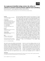
Before thermal denaturation (i.e. at temperatures up
to 55 °C), F-actin demonstrates, as expected, a very
random distribution of hydrodynamic radius (R
h
) val-
ues, from 10 nm to 1000 nm and even to a few
micrometers (Figs 1A and 2A). Obviously, real R
h
values cannot be obtained by DLS for long actin
filaments of different length. The R
h
distribution was
essentially the same for F-actin in the presence and
absence of Hsp27-3D (Fig. 2A). This agrees with our
previous results [2] showing that, in vitro, Hsp27-3D
does not interact with native actin filaments.
In the absence of Hsp-3D, thermal denaturation of
F-actin led to the formation of very large aggregates
with R
h
up to 10 lm (Fig. 2B). In contrast, in the
presence of Hsp27-3D, the F-actin thermal denatur-
ation was accompanied by complete disappearance of
large particles with high R
h
, and only small particles
with R
h
of 17 nm were detected (Fig. 1A). When
F-actin thermal denaturation was completed, at 70 °C
we observed a very narrow R
h
distribution, with an
average R
h
of 17 nm (Fig. 2B). These small particles
retained their size on following heating up to 80 °C,
and some slight increase in R
h
was only observed at
temperatures above 80 °C (Fig. 1A). The R
h
reached
40–50 nm at 84 °C (Fig. 1A), and this R
h
value
remained unchanged after cooling the sample to 25 °C
(Fig. 1B).
Similar DLS experiments were performed under con-
ditions when F-actin at a constant concentration of
0.5 mgÆmL
)1
was heated in the presence of Hsp27-3D
at various concentrations, from 0.015 to 0.5 mgÆmL
)1
.
The results show that the R
h
value for the complexes
of Hsp27-3D with denatured actin strongly depends on
the Hsp27-3D concentration in the sample (Fig. 3).
The R
h
of the complexes decreased from 53 to 16–
17 nm with an increase in the concentration of added
Fig. 1. Formation of the complexes of denatured actin with Hsp27-
3D as studied by DLS. (A) F-actin (0.5 mgÆmL
)1
) was heated at
a constant rate of 1 C°Æmin
)1
in the presence of Hsp27-3D
(0.125 mgÆmL
)1
), and R
h
was plotted as a function of temperature.
(B) After being heated to 85 °C, the sample was cooled and incu-
bated at 25 °C, and R
h
was plotted as a function of incubation time.
Other conditions: 30 m
M Hepes (pH 7.3), 100 mM NaCl, and 1 mM
MgCl
2
. DLS measurements were carried out at a scattering angle
of 90°.
A. V. Pivovarova et al. Stable complexes of small HSP with denatured actin
FEBS Journal 274 (2007) 5937–5948 ª 2007 The Authors Journal compilation ª 2007 FEBS 5939
Hsp27-3D from 0.015 to 0.125 mgÆ mL
)1
, and this R
h
value ( 16 nm) remained almost unchanged upon a
further increase in Hsp27-3D concentration up to
0.5 mgÆmL
)1
. These results suggest that the smallest
complexes of Hsp27-3D with denatured actin are
formed at Hsp27-3D concentrations above
0.125 mgÆmL
)1
, i.e. at an Hsp27-3D ⁄ actin weight ratio
higher than 1 : 4.
Thus, the results of DLS experiments clearly demon-
strate formation of stable complexes of Hsp27-3D with
denatured actin. The size of these complexes (average
R
h
of 16 nm under saturation conditions) is much
smaller than the corresponding values for native actin
filaments or actin aggregates formed upon thermal
denaturation of F-actin in the absence of Hsp27-3D.
Analytical ultracentrifugation of the Hsp27-3D
complexes with denatured actin
As already mentioned, the soluble complexes of
Hsp27-3D with denatured actin, which are formed in
the course of thermal denaturation of F-actin, retained
their size after cooling to room temperature (Fig. 1).
This property of the complexes allows their investiga-
tion in sedimentation velocity experiments.
F-actin (0.5 mgÆmL
)1
) was heated at a constant rate
of 1 C°Æmin
)1
up to 75 °C, i.e. to complete actin dena-
turation, in the presence of Hsp27-3D at different con-
centrations, from 0.1 to 0.4 mgÆmL
)1
. In all cases, we
did not observe any significant increase in light scat-
tering, which normally accompanies thermal denatur-
ation of F-actin in the absence of sHSPs, and this
indicated that Hsp27-3D formed soluble and relatively
small complexes with denatured actin. After cooling,
the samples were used for analytical ultracentrifuga-
tion to study the sedimentation behavior of these
complexes.
The differential distributions c(s, f ⁄ f
0
) of sedimenta-
tion coefficients (s) and for the actin–Hsp27-3D
Fig. 2. Distribution of the particles by their size (R
h
) for F-actin
(0.5 mgÆmL
)1
) in the absence or in the presence of Hsp27-3D
(0.125 mgÆmL
)1
) registered before F-actin thermal denaturation (at
30–35 °C) (A) and after F-actin denaturation (at 70 °C) (B). Condi-
tions were the same as in Fig. 1A. Each plot is an average of 10
distributions obtained within the temperature range 30–35 °Cin
Fig. 1A (A), or five distributions obtained within the range 69–71 °C
(B).
Fig. 3. Dependence of R
h
for the complexes of denatured actin
with Hsp27-3D on the Hsp27-3D concentration in the initial incuba-
tion mixture of Hsp27-3D with F-actin. The Hsp27-3D concentration
varied from 0.015 to 0.5 mgÆmL
)1
, and the F-actin concentration
was constant and equal to 0.5 mgÆmL
)1
. Other conditions were the
same as in Fig. 1A. The R
h
values were determined from the R
h
distributions obtained after F-actin thermal denaturation at 70 °C.
The Hsp27-3D ⁄ actin weight ratios in the initial mixture are indicated
for each point.
Stable complexes of small HSP with denatured actin A. V. Pivovarova et al.
5940 FEBS Journal 274 (2007) 5937–5948 ª 2007 The Authors Journal compilation ª 2007 FEBS
complexes obtained at different Hsp27-3D concentra-
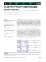
tions are shown in Fig. 4A–C. In all cases, the
c(s, f ⁄ f
0
) distributions of the complexes exhibit several
peaks whose sedimentation coefficients depend on the
Hsp27-3D concentration. At the lowest Hsp27-3D con-
centration (0.1 mgÆmL
)1
), the sample exhibits a sedi-
mentation coefficient in the range 10–45 S, and the
c(s, f ⁄ f
0
) distribution is represented by four main peaks
with maxima at 14–17 S, 22.5 S, 28 S, and 35 S
(Fig. 4A). Increasing the Hsp27-3D concentration in
the sample up to 0.2 mgÆ mL
)1
resulted in almost com-
plete disappearance of the fractions with s >30S
(Fig. 4B). In this case, the c(s, f ⁄ f
0
) distribution of the
complex is represented by the main, large-amplitude
peak with a maximum at 21.6 S, a small peak at
29.8 S, and several badly resolved peaks at 8–18 S. A
further increase in the Hsp27-3D concentration (up to
0.4 mgÆmL
)1
) resulted in full disappearance of all
peaks with s > 30 S, narrowing of the distribution
curve, and shifting of the distribution curve to the
lower s-values (Fig. 4C). Under these conditions, the
c(s, f ⁄ f
0
) distribution curve of the actin–Hsp27-3D
complex shows three peaks of similar amplitude, with
maxima at 14 S, 17 S, and 19.4 S, and several small
peaks at 9 S, 11.4 S, and 28.2 S.
Besides the above mentioned peaks, all c(s, f ⁄ f
0
)
distribution curves also contain the peak at 3.0–3.2 S
(Fig. 4A–C). This peak is assigned to Hsp27-3D
unbound to actin, in good agreement with previous
reports that unheated Hsp27-3D has a sedimentation
coefficient of 3 S [11,30,31], which is believed to
correspond to Hsp27-3D dimers. Previous studies
have shown that isolated Hsp27-3D denatures at
70 °C, and its thermal denaturation is completely
reversible [2]. Thus, under the conditions used here,
Hsp27-3D fully denatured when the samples were
heated to 75 °C, and then fully renatured upon cool-
ing prior to sedimentation experiments. The results
presented in Fig. 4 show that the denaturation–rena-
turation procedure had no significant influence on the
sedimentation behavior of Hsp27-3D. Increasing
Hsp27-3D concentration in the sample increases the
amplitude of the peak at 3.2 S (Fig. 4A–C), and this
indicates that the amount of actin-free Hsp27-3D,
increases with increasing concentration of added
Hsp27-3D.
Thus, the results of these experiments show that,
under the conditions used, a proportion of Hsp27-
3D is involved in the formation of stable complexes
with denatured actin, which exhibit a sedimentation
coefficient in the range 8–40 S, depending on the
Hsp27-3D concentration, with average s
20,w
of about
17–20 S. The remaining Hsp27-3D remains free and
sediments with s
20,w
¼ 3–3.2 S. Knowing the total
concentration of Hsp27-3D and determining the
quantity of free Hsp27-3D, we can calculate the
amount of Hsp27-3D involved in the complexes with
denatured actin, and by this means estimate the
stoichiometry Hsp27-3D ⁄ actin in these complexes.
Unfortunately, analytical ultracentrifugation cannot
provide exact data on the concentration of free
Hsp27-3D in the probes. For this purpose, we
applied SEC.
Fig. 4. Sedimentation velocity analysis of the complexes of Hsp27-
3D with thermally denatured actin. The complexes were obtained
by heating of F-actin (0.5 mgÆmL
)1
)to75°C at a constant rate of
1C°Æmin
)1
in the presence of 0.1 mgÆmL
)1
(A), 0.2 mgÆmL
)1
(B)
and 0.4 mgÆmL
)1
(C) Hsp27-3D. Differential sedimentation coeffi-
cient distributions [c(s, f ⁄ f
o
) versus s] were obtained at 20 °C (after
cooling the species) and saved as the one-dimensional c(s,*) distri-
butions. The rotor speed was 30 000 r.p.m. Other conditions were:
30 m
M Hepes (pH 7.3), 100 mM NaCl, and 1 mM MgCl
2
.
A. V. Pivovarova et al. Stable complexes of small HSP with denatured actin
FEBS Journal 274 (2007) 5937–5948 ª 2007 The Authors Journal compilation ª 2007 FEBS 5941
Stoichiometry of the Hsp27-3D–actin complexes
analyzed by SEC
In general, the probes for SEC experiments were pre-
pared by the same means as for analytical ultracentrif-
ugation, except that higher protein concentrations were
used. F-actin (1.0 mgÆmL
)1
) was heated at a constant
rate of 1 C°Æ min
)1
up to 68 °C in the absence or in the
presence of Hsp27-3D (0.25–1.0 mgÆmL
)1
). Under
these conditions, F-actin was fully denatured both in
the absence and in the presence of Hsp27-3D [2]. In
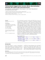
the absence of Hsp27-3D, thermal denaturation of
F-actin was accompanied by a strong increase in light
scattering, whereas in the presence of Hsp27-3D, only
a small increase in light scattering was observed
(Fig. 5A). The amplitude of light scattering was depen-
dent on the concentration of Hsp27-3D (Fig. 5A).
After cooling, the samples were subjected to high-
speed centrifugation (20 min at 140 000 g) to sediment
protein aggregates, and the supernatants thus obtained
(Fig. 5B) were subjected to SEC to separate soluble
complexes formed by Hsp27-3D with denatured actin
from actin-free Hsp27-3D.
F-actin heated up to 68 °C in the absence of Hsp27-
3D was fully precipitated upon ultracentrifugation,
and therefore no peaks were detected on the elution
profile (data not shown). When F-actin was heated in
the presence of Hsp27-3D, subjected to ultracentrifuga-
tion, and loaded on the SEC column, we detected two
peaks on the elution profile (Fig. 6A). According to
the data of SDS⁄ PAGE, the first asymmetric peak,
eluted close to the void volume (8–10 mL), contained
both actin and Hsp27-3D (data not shown), thus indi-
cating that this peak contains soluble complexes
formed by denatured actin and Hsp27-3D. Surpris-
ingly, the size of this peak on the elution profile was
constant and independent of the initial concentration
of Hsp27-3D (Fig. 6A). We suppose that the majority
of soluble complexes formed by denatured actin and
Hps27-3D were retarded on the column filter, and only
a small, nearly equal proportion of these complexes
entered the column and was detected in the first peak
on the elution profile.
The second peak, eluted at about 14.2 mL (apparent
molecular mass about 100 kDa), contained isolated
Hsp27-3D. The size of this peak was clearly increased
with increasing initial concentrations of Hsp27-3D in
the incubation mixture. Thus, knowing the initial total
concentration of Hsp27-3D and the concentration of
Hps27-3D remaining free, we were able to indirectly
estimate the concentration of Hps27-3D bound to
denatured actin. Plotting the concentration of Hsp27-
3D bound to denatured actin against the total concen-
tration of Hsp27-3D, we tried to determine the stoichi-
ometry of the complexes formed. Unfortunately, at
fixed F-actin concentration (24 lm) and Hsp27-3D
concentrations varying in the range 0–44 lm, we were
unable to achieve saturation (Fig. 6B). Probably, satu-
ration can be reached at higher Hsp27-3D concentra-
tions that were unattainable under the conditions used.
Therefore, we performed similar experiments under
different conditions, i.e. at a constant Hsp27-3D con-
centration of 1 mgÆmL
)1
( 44 lm) and various
F-actin concentrations (0.25–3.0 mgÆmL
)1
or 6–70 lm).
The probes containing different concentrations of
actin and fixed concentration of Hsp27-3D were heated
Fig. 5. Concentration-dependent effect of Hsp27-3D on the heat-
induced aggregation of F-actin. (A) F-actin (1.0 mgÆmL
)1
) was
heated at a constant rate of 1 C°Æmin
)1
in the absence (curve 1) or
in the presence (curves 2–4) of Hsp27-3D, and aggregation was fol-
lowed by light scattering at 350 nm. The Hsp27-3D concentration
was equal to 0.25, 0.5 and 1.0 mgÆmL
)1
for curves 2, 3, and 4,
respectively. Other conditions were the same as in Fig. 1A. After
being heated to 68 °C, the samples were cooled and subjected to
ultracentrifugation, and protein composition of supernatants was
analyzed by SDS ⁄ PAGE (B). Lanes 1 and 2 represent control
unheated F-actin (0.5 mgÆmL
)1
) and Hsp27-3D (0.5 mgÆmL
)1
),
respectively. Lanes 3–7: supernatants obtained from the samples
subjected to heating up to 68 °C and ultracentrifugation. Lanes 3–5:
F-actin in the presence of 1.0, 0.5 and 0.25 mgÆmL
)1
Hsp27-3D,
respectively. Lanes 6 and 7: F-actin alone and Hsp27-3D alone
(0.5 mgÆmL
)1
), respectively. Positions of actin and Hsp27-3D are
marked on the left.
Stable complexes of small HSP with denatured actin A. V. Pivovarova et al.
5942 FEBS Journal 274 (2007) 5937–5948 ª 2007 The Authors Journal compilation ª 2007 FEBS
to 68 °C under the above mentioned conditions. The
samples were cooled, and subjected to ultracentrifuga-
tion, and the supernatants obtained were loaded on
the Superdex 200 column. Again, two peaks were
detected on the elution profile (Fig. 7A). The first peak
contained soluble complexes containing denatured
F-actin and Hsp27-3D (Fig. 7B), whereas the second
peak contained isolated Hps27-3D (Fig. 7C). The first
peak was eluted close to the void volume (8–10 mL),
and its size was only slightly increased upon increase
of the initial F-actin concentration (Fig. 7, insert). For
instance, a 12-fold increase of initial F-actin concentra-
tion was accompanied by a less than two-fold increase
of the first peak. This is probably because a large pro-
portion of the complexes formed by denatured actin
and Hsp27-3D that remains in the supernatant after
ultracentrifugation was retarded on the column filter,
and only a small proportion of these complexes
entered the column and was detected in the first peak.
This means that the size of the first peak cannot be
directly used for correct determination of the quantity
of complexes formed by denatured actin and Hsp27-
3D.
In contrast, the size of the second peak correspond-
ing to isolated Hsp27-3D was strongly dependent on
the initial F-actin concentration, and an increase of
actin concentration was accompanied by significant
decrease in the Hsp27-3D remaining free. Thus, the
size of this peak provides information on the quantity
of actin-free Hsp27-3D. Isolated Hsp27-3D
(1 mgÆmL
)1
) was either kept on ice or heated up to
70 °C in the absence of F-actin, and after ultracentri-
fugation was subjected to SEC (curves 1 and 2 in
Fig. 7A). The size of the peaks was not dependent on
prior heating, thus indicating high thermal stability of
isolated Hsp27-3D. Measuring the size of this peak
and comparing it with the size of corresponding peaks
obtained in the presence of variable concentrations of
F-actin, we were able to determine the concentration
of Hsp27-3D remaining free at different actin concen-
trations. The concentration of Hsp27-3D bound to
denatured actin was determined by subtracting the
concentration of free Hps27-3D from the total concen-
tration of Hsp27-3D. Plotting the concentration of
actin-bound Hps27-3D against the F-actin ⁄ Hsp27-3D
molar ratio in the initial mixture (Fig. 7D), we found
that saturation was achieved at a molar ratio close to
1 : 1. This means that under conditions of saturation,
denatured actin and Hsp27-3D form equimolar com-
plexes.
Discussion
This article expands our knowledge of the mechanism
by which Hsp27-3D and probably other mammalian
sHSPs protect F-actin from heat-induced aggregation.
Previous work has clearly demonstrated that sHSPs
Fig. 6. Analysis of actin–Hsp27-3D complexes by SEC. The com-
plexes were obtained at a constant F-actin concentration of
1.0 mgÆmL
)1
and different Hsp27-3D concentrations as shown in
Fig. 5. (A) Equal volumes (500 lL) of each sample were sub-
jected to SEC on a Superdex 200 HR 10 ⁄ 30 column. Curve-1
corresponds to Hsp27-3D alone (0.5 mgÆmL
)1
). Curves 2–4 corre-
spond to the actin–Hsp29-3D complexes obtained at Hsp27-3D
concentrations of 1.0, 0.5 and 0.25 mgÆmL
)1
, respectively
(lanes 3–5 in Fig. 5B). (B) Dependence of molar concentration of
Hsp27-3D bound to denatured actin in their complexes obtained
at a constant F-actin concentration on the concentration of added
Hsp27-3D. The concentration of bound Hsp27-3D was calculated
as the difference between the concentration of added Hsp27-3D
and that of actin-free Hsp27-3D in the samples as determined
from (A).
A. V. Pivovarova et al. Stable complexes of small HSP with denatured actin
FEBS Journal 274 (2007) 5937–5948 ª 2007 The Authors Journal compilation ª 2007 FEBS 5943
have no effect on the F-actin thermal unfolding mea-
sured by DSC, but they effectively prevent aggregation
of thermally denatured actin [2]. Based on previous
results of cosedimentation experiments [2], we have
proposed that Hsp27-3D and probably other sHSPs
prevent heat-induced aggregation of F-actin by form-
ing relatively small, stable and highly soluble com-
plexes with denatured actin. In the present work, we
studied the properties of these complexes using DLS,
SEC, and analytical ultracentrifugation. For this
purpose, we used Hsp27-3D, as this Hsp27 mutant
mimicking naturally occurring phosphorylation is
known to exist in vitro in small-size oligomers that are
much smaller than the large oligomers of many other
sHSPs [3–5,7,9–11,30,31].
Comparison of the DLS results shown here
(Fig. 1A) with DSC data obtained earlier [2] clearly
shows that actin–Hsp27-3D complexes are formed
during the course of F-actin thermal denaturation.
All the methods used here show that the size of
these complexes depends on the Hsp27-3D ⁄ actin
ratio in the initial mixture of Hsp27-3D and F-actin
(Figs 3, 4 and 7A). Each method (DLS, SEC, sedi-
mentation velocity analysis) has some advantages
and drawbacks [36]. However, all the methods
clearly show that the size (and mass) of the actin–
Hsp27-3D complexes decreases with increase in the
Hsp27-3D content in the initial mixture. Saturation
of the complexes with Hsp27-3D molecules occurs at
approximately equimolar concentrations of Hsp27-3D
and actin (Figs 3 and 7D). This agrees with previous
studies on the sHSP complexes with various dena-
tured proteins, suggesting a maximum binding capac-
ity of one protein subunit per one sHSP subunit
[37]. Under these conditions, the actin–Hsp27-3D
Fig. 7. SEC analysis of the actin–Hsp27-3D complexes obtained at
a constant Hsp27-3D concentration. The complexes were obtained
by heating an F-actin–Hsp27-3D mixture to 70 °C at a constant rate
of 1 C°Æmin
)1
. Experiments were performed with a constant
Hsp27-3D concentration equal to 1 mgÆmL
)1
and different F-actin
concentrations, varying from 0.25 to 3.0 mgÆmL
)1
. After being
cooled, the samples were subjected to ultracentrifugation, and
equal volumes of the supernatants (500 lL) were analyzed by SEC.
(A) SEC curves 1 and 2 correspond, respectively, to control
unheated Hsp27-3D and Hsp27-3D heated to 70 °C, both at a con-
centration of 0.5 mgÆmL
)1
. Curves 3–7 correspond to Hsp27-3D
(1 mgÆmL
)1
) heated in the presence of 0.25, 0.5, 1.0, 2.0 and
3.0 mgÆmL
)1
F-actin, respectively. The inset expands the region of
8–11.5 mL elution volume for clarity. (B) SDS ⁄ PAGE for the actin–
Hsp27-3D complexes obtained at a constant Hsp27-3D concentra-
tion (1.0 mgÆmL
)1
) and different F-actin concentrations: 0.25 (3),
0.5 (4), 1.0 (5), 2.0 (6) and 3.0 mgÆmL
)1
(7). The lane numbers cor-
respond to the numbers of SEC curves in (A). In all cases, fractions
with elution volumes from 8.5 to 9.0 mL in (A) were collected,
combined, and subjected to SDS ⁄ PAGE. (C) SDS ⁄ PAGE of free
Hsp27-3D (1, 2) and in the presence of denatured actin (3–7) [frac-
tions with an elution volume of 14 mL in (A)]. The lane numbers
correspond to those for SEC curves in (A). (D) Dependence of
molar concentration of Hsp27-3D bound to denatured actin in their
complexes obtained at a constant Hsp27-3D concentration (44 l
M)
on the F-actin ⁄ Hsp27-3D molar ratio in the initial incubation mix-
ture. The concentration of actin-bound Hsp27-3D was calculated as
in Fig. 6A, using SEC data from (A) and a molecular mass of
Hsp27-3D monomer equal to 22.8 kDa.
Stable complexes of small HSP with denatured actin A. V. Pivovarova et al.
5944 FEBS Journal 274 (2007) 5937–5948 ª 2007 The Authors Journal compilation ª 2007 FEBS
complexes exhibit R
h
of 16 nm and s
20,w
of about
17–20 S (Figs 3 and 4). It should be noted that
Hsp27-3D (Fig. 5) and other sHSPs [2] effectively
prevent F-actin thermal aggregation, even at rather
low sHSP concentrations, when the sHSP ⁄ actin
molar ratio is much lower than 1 : 1.
It is noteworthy that the complexes of a similar
size, with R
h
of about 16 nm and s
20,w
of about 17–
20 S, were observed previously in DLS and analytical
ultracentrifugation experiments not only with Hsp27-
3D, but also after heating of F-actin in the presence
of a-crystallin [38]. In that case, however, we could
not clearly separate the complexes of a-crystallin with
denatured actin from actin-free a-crystallin, which
formed large oligomers with R
h
11 nm [33] and
s
20,w
¼ 18 ± 2 S [39,40]. (Incidentally, this was the
reason why we used only Hsp27-3D in the present
work). Nevertheless, the similarity between Hsp27-3D
and a-crystallin in the size of their complexes with
denatured actin suggests that this parameter of the
complexes is mainly determined by the target protein
(denatured actin), but independent of the sHSP used.
This agrees with previous studies showing that mouse
Hsp25 and yeast Hsp26, the two members of the
sHSP family that significantly differ in their quater-
nary structure, form similar complexes with various
denatured proteins, and the size of these complexes is
dependent only on the target protein [37]. Taken
together, all these results support a viewpoint that the
formation of soluble complexes with non-native pro-
teins is a conserved feature of the sHSP family of
chaperones, and the morphology of these complexes
is substrate-dependent, but independent of the sHSP
used [37].
Previous electron microscopy studies showed spheri-
cal, regularly shaped particles formed by Hsp25 or
Hsp26 with various denatured target proteins [37].
Assuming a spherical shape for the actin–Hsp27-3D
complexes, we can estimate the apparent molecular
mass of the complex using an empirical relationship
between the relative molecular mass and the hydro-
dynamic radius: M
r
¼ (1.68R
h
)
2.3394
[41]. According to
this estimation, the particles with R
h
of 16 nm (i.e.
the actin–Hsp27-3D complexes formed under satura-
tion conditions) have a molecular mass of about
2 MDa. If we take into account equal amounts of
actin (42 kDa) and Hsp27-3D (molecular mass of the
monomer 22.8 kDa) in their complexes, then the com-
plexes with R
h
of 16 nm should contain about 30
denatured actin monomers and an equal quantity of
Hsp27-3D monomers.
Thus, the number of denatured actin molecules in
their complexes with Hsp27-3D is much lower than
in intact actin filaments, which contain hundreds and
even thousands of actin subunits. This is consistent
with a recently proposed dissociative mechanism of
F-actin thermal denaturation [1]. One of the main
features of this mechanism is that the actin filament
denatures not as a whole, but as separate monomers
or short oligomers that dissociate from the filament
during heating. In the absence of sHSP, denatured
actin monomers (or short oligomers) easily aggregate,
and during this process even undamaged actin fila-
ments become trapped and are precipitated. The
results presented here, together with the data
obtained earlier [2], suggest that sHSPs bind to dena-
tured actin monomers or short oligomers and protect
them from aggregation by forming relatively small
and highly soluble complexes, whose size is much less
than that of intact F-actin. We suppose that this is
the mechanism by which sHSPs prevent the aggrega-
tion of F-actin during its thermal denaturation. In
many respects, this mechanism is similar to that pos-
tulated earlier for different soluble enzymes [37,42,43].
Generally, sHSPs cannot protect target proteins from
denaturation and cannot refold denatured substrate.
However, sHSPs prevent the aggregation of denatured
target proteins, forming a reservoir of folding inter-
mediates that can either be refolded by the network
of cell chaperones or passed to proteasomes for de-
gradation. Our results suggest that this reservoir, in
the case of F-actin, is presented as soluble and rela-
tively small complexes formed by sHSPs with dena-
tured actin molecules obtained during the heating of
F-actin.
In conclusion, the analysis of the properties of the
complexes formed between sHSP and denatured actin,
as performed by different methods, provides new
insights into the mechanism by which sHSPs prevent
the aggregation of F-actin induced by its thermal dena-
turation. This mechanism may explain how sHSPs pro-
tect the cytoskeleton and the whole cell from damage
caused by accumulation of large, insoluble aggregates
under heat shock conditions.
Experimental procedures
Proteins
Rabbit skeletal actin was prepared by the method of
Spudich & Watt [44]. Its concentration was determined by
its absorbance at 290 nm, using an E
1%
of 6.3 cm
)1
.
Monomeric G-actin in G buffer (2 mm Tris ⁄ HCl, pH 8.0,
0.2 mm ATP, 0.2 mm CaCl
2
, 0.5 mm b-mercaptoethanol,
1mm NaN
3
) was polymerized into F-actin filaments by
the addition of MgCl
2
to a final concentration of 2 mm.
A. V. Pivovarova et al. Stable complexes of small HSP with denatured actin
FEBS Journal 274 (2007) 5937–5948 ª 2007 The Authors Journal compilation ª 2007 FEBS 5945
Prior to experiments, F-actin was diluted to a final con-
centration (from 0.25 to 3.0 mgÆmL
)1
) with 30 mm Hepes
(pH 7.3), containing 100 mm NaCl and 1 mm MgCl
2
.
Recombinant human Hsp27-3D was cloned, expressed and
purified as described previously [21,45]. All proteins were
homogeneous according to SDS ⁄ PAGE [46]. Soluble com-
plexes of Hsp27-3D with denatured actin were formed by
heating the mixture of F-actin and Hsp27-3D at a con-
stant rate of 1 C°Æmin
)1
up to a temperature at which full,
irreversible denaturation of F-actin occurred (above 68 °C)
[2]. Insoluble aggregates were removed, if necessary, by
high-speed centrifugation of the samples (20 min at
140 000 g).
F-actin aggregation
Thermally induced aggregation of F-actin was detected by
changes in light scattering at 90° as described previously
[1,2]. The measurements were performed on a Cary Eclipse
fluorescence spectrophotometer (Varian Australia Pty Ltd,
Mulgrave, Victoria, Australia) equipped with a temperature
controller and thermoprobes. F-actin in the absence or in
the presence of Hsp27-3D was heated at a constant rate of
1C°Æ min
)1
from 30 °C up to 68–75 °C. The light scattering
at 350 nm was measured with excitation and emission slits
of 2.5 and 1.5 nm, respectively. When the heating was com-
pleted, the samples were cooled, and the aliquots were with-
drawn and subjected to ultracentrifugation at 140 000 g for
20–30 min on a Beckman airfuge (Beckman Instruments
Inc., Palo Alto, CA, USA). The protein composition of the
supernatants and pellets was determined by SDS ⁄ PAGE
[46].
DLS
DLS measurements were performed on a Photocor Com-
plex apparatus (Photocor Instruments Inc., College Park,
MD, USA) equipped with a temperature controller [33,34].
The sample protein solution was illuminated by a 633 nm
laser light, and the scattering signal was observed at an
angle of 90°. During the course of measurements, the tem-
perature fluctuations were approximately ± 0.1 °C. DLS
data were accumulated and analyzed with the multifunc-
tional real-time correlator Photocor-FC. dynals software
(Alango, Tirat Carmel, Israel) was used for polydisperse
analysis of DLS data. The mean hydrodynamic radius of
the particles, R
h
, was calculated from the Stokes–Einstein
equation: D ¼ k
B
T ⁄ 6pgR
h
, where D is the diffusion
coefficient obtained from the DLS measurements, k
B
is
Boltzmann’s constant, T is the absolute temperature, and g
is the shear viscosity of the solvent. The viscosity of the
solutions was measured on an AMVn Automated Micro
Viscosimiter (Anton Paar, Graz, Austria). The data were
further analyzed and plotted using origin 7.0 software
(OriginLab Corp., Northampton, MA, USA).
Analytical ultracentrifugation
Sedimentation velocity experiments were carried out in a
model E analytical ultracentrifuge (Beckman) equipped
with absorbance optics, a photoelectric scanner, a
monochromator, and a computer on-line. A four-hole rotor
An-F Ti and 12 mm double sector cells were used. The sed-
imentation profiles of the actin–Hsp27-3D complexes were
recorded by measuring the absorbance at 280 nm. All cells
were scanned simultaneously. The time interval between
scans was 3 min. The sedimentation coefficients were esti-
mated from the differential sedimentation coefficient distri-
bution [c(s, f ⁄ f
0
) versus s], which was analyzed using the
sedfit program [47,48].
SEC
Analytical SEC was carried out on a Super-
dex 200 HR 10 ⁄ 30 column using the ACTA-FPLC system
(Amersham Pharmacia, Biotech Europe GmbH, Helsinki,
Finland). The column was equilibrated with 30 mm
Hepes ⁄ KOH (pH 7.3) containing 100 mm NaCl and 1 mm
MgCl
2
. The samples (500 lL) were loaded on the column
and eluted at a rate of 0.5 mLÆmin
)1
. The column was
calibrated with the following molecular mass markers:
thyroglobulin (669 kDa), catalase (240 kDa), glyceralde-
hyde-3-phosphate dehydrogenase (122 kDa), BSA (68 kDa),
and ovalbumin (43 kDa).
Acknowledgements
This work was supported by the Russian Foundation
for Basic Research (grants 06-04-48343 to D. I. Levit-
sky and 07-04-00115 to N. B. Gusev), the Program
‘Molecular and Cell Biology’ of the Russian Academy
of Sciences, and by INTAS (grant 03-51-4813).
References
1 Mikhailova VV, Kurganov BI, Pivovarova AV & Levit-
sky DI (2006) Dissociative mechanism of F-actin ther-
mal denaturation. Biochemistry (Moscow) 71, 1261–
1269.
2 Pivovarova AV, Mikhailova VV, Chernik IS, Chebotar-
eva NA, Levitsky DI & Gusev NB (2005) Effects of
small heat shock proteins on the thermal denaturation
and aggregation of F-actin. Biochem Biophys Res
Commun 331, 1548–1553.
3 Haslbeck M (2002) sHsps and their role in the chaper-
one network. Cell Mol Life Sci 59, 1649–1657.
4 Gusev NB, Bogatcheva NV & Marston SB (2002)
Structure and properties of small heat shock proteins
(sHsp) and their interaction with cytoskeleton proteins.
Biochemistry (Moscow) 67, 511–519.
Stable complexes of small HSP with denatured actin A. V. Pivovarova et al.
5946 FEBS Journal 274 (2007) 5937–5948 ª 2007 The Authors Journal compilation ª 2007 FEBS
5 Haslbeck M, Franzmann T, Weinfurtner D & Buchner
J (2005) Some like it hot: the structure and function of
small heat-shock proteins. Nat Struct Mol Biol 12, 842–
846.
6 Van Monfort RL, Bashs E, Friedrich KL, Slingsby C &
Vierling E (2001) Crystal structure and assembly of a
eukaryotic small heat shock protein. Nat Struct Biol 8,
1025–1030.
7 Kim KK, Kim R & Kim SH (1998) Crystal structure of
a small heat shock protein. Nature 394, 595–599.
8 Jakob U, Gaestel M, Engel K & Buchner J (1993) Small
heat shock proteins are molecular chaperones. J Biol
Chem 268, 1517–1520.
9 Ganea E (2001) Chaperone-like activity of a-crystallin
and other small heat shock proteins. Curr Prot Pept Sci
2, 205–225.
10 Shashidharamurthy R, Koteiche HA, Dong J &
Mchaourab HS (2005) Mechanism of chaperone func-
tion in small heat shock proteins. Dissociation of the
Hsp27 oligomer is required for recognition and binding
of destabilized T4 lysozyme. J Biol Chem 280, 5281–
5289.
11 Lelj-Garolla B & Mauk AG (2006) Self association and
chaperone activity of Hsp27 are thermally activated.
J Biol Chem 281, 8169–8174.
12 Rogalla T, Ehrnsperger M, Preville X, Kotlyarov A,
Lutsch G, Ducasse C, Paul C, Wieske M, Arrigo AP,
Buchner J et al. (1999) Regulation of Hsp27 oligo-
merization, chaperone function, and protective activity
against oxidative stress ⁄ tumor necrosis factor a by
phosphorylation. J Biol Chem 274, 18947–18956.
13 Lutsch G, Vetter R, Offhauss U, Wieske M, Grone H-
J, Klemenz R, Shimke I, Stahl J & Benndorf R (1997)
Abundance and location of the small heat shock pro-
teins HSP25 and alpha B-crystallin in rat and human
heart. Circulation 96, 3466–3476.
14 Frank E, Madsen O, van Rheele T, Ricard G, Huyden
MA & de Jong WW (2004) Evolutionary diversity of
vertebrate small heat shock proteins. J Mol Evol 59 ,
792–805.
15 Mounier N & Arrigo A-P (2002) Actin cytoskeleton and
small heat shock proteins: how do they interact? Cell
Stress Chaperones 7, 167–176.
16 Miron T, Vancompernolle K, Vandekerckhove J, Wilc-
khek M & Geiger B (1991) A 25-kDa inhibitor of actin
polymerization is a low molecular mass heat shock pro-
tein. J Cell Biol 114, 255–261.
17 Benndorf R, Hayess K, Ryazantsev S, Wieske M,
Behlke J & Lutsch G (1994) Phosphorylation and supra-
molecular organization of murine small heat shock pro-
tein HSP25 abolish its actin polymerization-inhibiting
activity. J Biol Chem 269, 20780–20784.
18 Butt E, Immler D, Meyer HE, Kotlyarov A, Laass K &
Gaestel M (2001) Heat shock protein 27 is a substrate
of cGMP-dependent protein kinase in intact human
platelet. J Biol Chem 276, 7108–7113.
19 During RL, Gibson BG, Li W, Bishai EA, Sidhu GS,
Landry J & Southwick FS (2007) Anthrax lethal toxin
paralyzes actin-based motility by blocking Hsp27 phos-
phorylation. EMBO J 26, 2240–2250.
20 Bukach OV, Marston SB & Gusev NB (2005) Small
heat shock protein with apparent molecular mass 20
kDa (Hsp20, HspB6) is not a genuine actin-binding
protein. J Muscle Res Cell Motil 26
, 175–191.
21 Panasenko OO, Kim MV, Marston SB & Gusev NB
(2003) Interaction of the small heat shock protein with
molecular mass 25 kDa (hsp25) with actin. Eur J
Biochem 270, 892–901.
22 Brophy CM, Lamb S & Graham A (1999) The small
heat shock-related protein-20 is an actin-associated pro-
tein. J Vasc Surg 29, 326–333.
23 Huot J, Houle F, Spitz DR & Landry J (1996) Hsp27
phosphorylation-mediated resistance against actin frag-
mentation and cell death induced by oxidative stress.
Cancer Res 56, 273–279.
24 Lavoie JN, Lambert H, Hickey E, Weber LA & Landry
J (1995) Modulation of cellular thermoresistance and
actin filament stability accompanies phosphorylation-
induced changes in the oligomeric structure of heat
shock protein 27. Mol Cell Biol 15, 505–516.
25 Bryantsev AL, Loktionova SA, Ilyinskaya OP, Tararak
EM, Kampinga HH & Kabakov AE (2002) Distribu-
tion, phosphorylation, and activities of Hsp25 in
heat-stressed H9c2 myoblasts: a functional link to
cytoprotection. Cell Stress Chaperones 7, 146–155.
26 Van Why SK, Mann AS, Ardito T, Thulin G, Ferris S,
Macleod MA, Kashgarian M & Seigel NJ (2003) Hsp27
associates with actin and limits injury in energy depleted
renal epithelia. J Am Soc Nephrol 14, 98–106.
27 Koh TJ & Escobedo J (2003) Cytoskeletal disruption
and small heat shock protein translocation immediately
after lengthening contraction. Am J Physiol Cell Physiol
286, C713–C722.
28 Singh BN, Rao KS, RamakrishnaT, Rangaraj N & Rao
CM (2007) Association of aB-crystallin, a small heat
shock protein, with actin: role in modulating actin fila-
ment dynamics in vivo. J Mol Biol 366, 756–767.
29 Wang K & Spector A (1996) a-Crystallin stabilizes actin
filaments and prevents cytochalasin-induced depolymer-
ization in a phosphorylation-dependent manner. Eur J
Biochem 242, 56–66.
30 Lelj-Garolla B & Mauk AG (2005) Self association of a
small heat shock protein. J Mol Biol 345, 631–642.
31 Chernik IS, Panasenko OO, Li Y, Marston SB & Gusev
NB (2004) pH-induced changes of the structure of small
heat shock proteins with molecular mass 24 ⁄ 27 kDa
(HspB1). Biochem Biophys Res Commun 324, 1199–
1203.
A. V. Pivovarova et al. Stable complexes of small HSP with denatured actin
FEBS Journal 274 (2007) 5937–5948 ª 2007 The Authors Journal compilation ª 2007 FEBS 5947
32 Khanova HA, Markossian KA, Kurganov BI, Samoilov
AM, Kleimenov SYu, Levitsky DI, Yudin IK, Timofe-
eva AC, Muranov KO & Ostrovsky MA (2005) Mecha-
nism of chaperone-like activity. Suppression of thermal
aggregation of b
L
-crystallin by a-crystallin. Biochemistry
44, 15480–15487.
33 Khanova HA, Markossian KA, Kleimenov SYu, Levit-
sky DI, Chebotareva NA, Golub NV, Asryants RA,
Muronetz VI, Saso L, Yudin IK et al. (2007) Effect of
a-crystallin on thermal denaturation and aggregation of
rabbit muscle glyceraldehydes-3-phosphate dehydroge-
nase. Biophys Chem 125, 521–531.
34 Markossian KA, Khanova HA, Kleimenov SYu, Levit-
sky DI, Chebotareva NA, Asryants RA, Muronetz VI,
Saso L, Yudin IK & Kurganov BI (2006) Mechanism of
thermal aggregation of rabbit muscle glyceraldehyde-3-
phosphate dehydrogenase. Biochemistry 45, 13375–
13384.
35 Meremyanin AV, Eronina TB, Chebotareva NA, Klei-
menov SYu, Yudin IK, Muranov KO, Ostrovsky MA
& Kurganov BI (2007) Effect of a-crystallin on thermal
aggregation of glycogen phosphorylase b from rabbit
skeletal muscle. Biochemistry (Moscow) 72, 518–528.
36 Philo JS (2006) Is any measurement method optimal for
all aggregate sizes and types? AAPS J 8, E564–E571.
37 Stromer T, Ehrnsperger M, Gaestel M & Buchner J
(2003) Analysis of the interaction of small heat shock
proteins with unfolding proteins. J Biol Chem 278,
18015–18021.
38 Pivovarova AV, Mikhailova VV, Chernik IS, Gusev NB
& Levitsky DI (2005) Small heat-shock proteins prevent
thermally induced aggregation of actin filaments by
formation of soluble complexes with denatured actin.
FEBS J 272 (Suppl. 1), 356.
39 Chebotareva NA, Meremyanin AV, Makeeva VF &
Kurganov BI (2006) Self association of phosphorylase
kinase under molecular crowding conditions. Progr Col-
loid Polym Sci 131, 83–92.
40 Markossian K, Kurganov B, Levitsky D, Khanova H,
Chebotareva N, Samoilov A, Eronina T, Fedurkina N,
Mitskevich L, Merem’yanin A et al. (2006) Mechanisms
of chaperone-like activity. In Protein Folding: New
Research (Obalinsky TR, ed.), pp. 89–171. Nova Science
Publishers Inc., New York.
41 Creighton TE (1993) Proteins. Structures and Molecular
Properties. W. H. Freeman & Co., New York.
42 Ehrnsperger M, Graber S, Gaestel M & Buchner J
(1997) Binding of non-native proteins to Hsp25 during
heat shock creates a reservoir of folding intermediates
for reactivation. EMBO J 16, 221–229.
43 Veinger L, Diamant S, Buchner J & Goloubinoff P
(1998) The small heat-shock protein IbpB from Escheri-
chia coli stabilizes stress-denatured proteins for subse-
quent refolding by a multichaperone network. J Biol
Chem 273, 11032–11037.
44 Spudich JA & Watt S (1971) The regulation of rabbit
skeletal muscle contraction. Biochemical studies of the
interaction of the tropomyosin–troponin complex with
actin and the proteolytic fragments of myosin. J Biol
Chem 246, 4866–4871.
45 Bukach OV, Seit-Nebi AS, Marston SB & Gusev NB
(2004) Some properties of human small heat shock pro-
tein Hsp20 (HspB6). Eur J Biochem 271, 291–302.
46 Laemmli UK (1970) Cleavage of structural proteins
during the assembly of the head of bacteriophage T4.
Nature 227, 680–685.
47 Schuck P (2000) Size distribution analysis of macromol-
ecules by sedimentation velocity ultracentrifugation and
Lamm equation modeling. Biophys J 78, 1606–1619.
48 Beown PH & Schuck P (2006) Macromolecular size-
and-shape distribution by sedimentation velocity analyt-
ical ultracentrifugation. Biophys J 90, 4651–4661.
Stable complexes of small HSP with denatured actin A. V. Pivovarova et al.
5948 FEBS Journal 274 (2007) 5937–5948 ª 2007 The Authors Journal compilation ª 2007 FEBS