Báo cáo khoa học: Ferrous Campylobacter jejuni truncated hemoglobin P displays an extremely high reactivity for cyanide – a comparative study ppt
Bạn đang xem bản rút gọn của tài liệu. Xem và tải ngay bản đầy đủ của tài liệu tại đây (401.53 KB, 13 trang )
Ferrous Campylobacter jejuni truncated hemoglobin P
displays an extremely high reactivity for cyanide –
a comparative study
Alessandro Bolli
1
, Chiara Ciaccio
2,3
, Massimo Coletta
2,3
, Marco Nardini
4
, Martino Bolognesi
4
,
Alessandra Pesce
5
, Michel Guertin
6
, Paolo Visca
1,7
and Paolo Ascenzi
1,7
1 Dipartimento di Biologia and Laboratorio Interdipartimentale di Microscopia Elettronica, Universita
`
‘Roma Tre’, Italy
2 Dipartimento di Medicina Sperimentale e Scienze Biochimiche, Universita
`
di Roma ‘Tor Vergata’, Italy
3 Consorzio Interuniversitario di Ricerca in Chimica dei Metalli nei Sistemi Biologici, Bari, Italy
4 Dipartimento di Scienze Biomolecolari e Biotecnologie and CNR-INFM, Universita
`
di Milano, Italy
5 Dipartimento di Fisica, CNR-INFM, and Centro di Eccellenza per la Ricerca Biomedica, Universita
`
di Genova, Italy
6De
´
partement de Biochimie et de Microbiologie, Universite
´
Laval, Faculte
´
des Sciences et de Ge
´
nie, Que
´
bec, Canada
7 Istituto Nazionale per le Malattie Infettive IRCCS ‘Lazzaro Spallanzani’, Roma, Italy
Over the last decade, three types of hemoglobins (Hbs)
have been identified in microorganisms. The first type
comprises flavohemoglobins (flavoHbs), which are
characterized by the classic 3-on-3 a-helical sandwich
globin domain, hosting the heme, covalently linked
to a flavin reductase domain. The second Hb type
Keywords
Campylobacter jejuni truncated
hemoglobin P; cyanide binding; kinetics;
Mycobacterium tuberculosis truncated
hemoglobin N and O; thermodynamics
Correspondence
P. Ascenzi, Dipartimento di Biologia,
Universita
`
‘Roma Tre’, Viale G. Marconi 446,
I-00146 Roma, Italy
Fax: +39 06 5517 6321
Tel: +39 06 5517 3200(2)
E-mail:
(Received 7 September 2007, revised 28
November 2007, accepted 7 December
2007)
doi:10.1111/j.1742-4658.2007.06223.x
Campylobacter jejuni hosts two hemoglobins (Hbs). The Camplylobacter je-
juni single-domain Hb (called Cgb) is homologous to the globin domain of
flavohemoglobin, and it has been proposed to protect the bacterium against
nitrosative stress. The second Hb is called Ctb (hereafter Cj-trHbP),
belongs to truncated Hb group III, and has been hypothesized to be
involved in O
2
chemistry. Here, the kinetics and thermodynamics of
cyanide binding to ferric and ferrous Cj-trHbP [Cj-trHbP(III) and
Cj-trHbP(II), respectively] are reported and analyzed in parallel with those
of related heme proteins, with particular reference to those from Mycobac-
terium tuberculosis. The affinity of cyanide for Cj-trHbP(II) is higher than
that reported for any known (in)vertebrate globin by more than three
orders of magnitude (K ¼ 1.2 · 10
)6
m). This can be fully attributed to the
highest (ever observed for a ferrous Hb) cyanide-binding association rate
constant (k
on
¼ 3.3 · 10
3
m
)1
Æs
)1
), even though the binding process dis-
plays a rate-limiting step (k
max
¼ 9.1 s
)1
). Cj-trHbP(III) shows a very high
affinity for cyanide (L ¼ 5.8 · 10
)9
m); however, cyanide association kinet-
ics are independent of cyanide concentration, displaying a rate-limiting step
(l
max
¼ 2.0 · 10
)3
s
)1
). Values of the first-order rate constant for cyanide
dissociation from Cj-trHbP(II)–cyanide and Cj-trHbP(III)–cyanide (k
off
¼
5.0 · 10
)3
s
)1
and l
off
‡ 1 · 10
)4
s
)1
, respectively) are similar to those
reported for (in)vertebrate globins. The very high affinity of cyanide for
Cj-trHbP(II), reminiscent of that of horseradish peroxidase(II), suggests
that this globin may participate in cyanide detoxification.
Abbreviations
Cj-trHbP, Campylobacter jejuni truncated hemoglobin P; flavoHb, flavohemoglobin; Hb, hemoglobin; HbC, hemoglobin C; HbI, hemoglobin I;
Mb, myoglobin; Mt-trHbN, Mycobacterium tuberculosis truncated hemoglobin N; Mt-trHbO, Mycobacterium tuberculosis truncated
hemoglobin O; trHb, truncated hemoglobin.
FEBS Journal 275 (2008) 633–645 ª 2008 The Authors Journal compilation ª 2008 FEBS 633
comprises single-domain globins homologous to the
globin domain of flavoHbs. In contrast to flavoHbs,
they are devoid of the reductase domain. The third
Hb type comprises truncated hemoglobins (trHbs),
which display a smaller globin domain and the typical
2-on-2 a-helical sandwich fold. On the basis of phylo-
genetic analyses, trHbs have been divided into three
groups (N or I, O or II, and P or III) [1–9].
Campylobacter jejuni is the most common bacterial
zoonosis and the main cause of bacterial gastroenteritis
in the Western world. C. jejuni is a common colonizer
of the intestinal tract of wild and domestic animals, pri-
marily birds and cattle, where it can persist at high cell
density and from which it can be transmitted to humans
through the orofecal route [10–12]. C. jejuni contains
two Hbs, i.e. Cgb and Ctb (the latter named Cj-trHbP
hereafter). Cgb, belonging to the second Hb type, has
been proposed to protect C. jejuni against nitrosative
stress, probably via an NO dioxygenase reaction [13,14].
Cj-trHbP belongs to trHb group III, and displays an
extremely high O
2
affinity, making it unlikely to be an
O
2
carrier. On the basis of the polarity of the heme dis-
tal cavity, reminiscent of that found in cytochrome c
peroxidase, Cj-trHbP has been proposed to be involved
in (pseudo)enzymatic O
2
chemistry [15–17].
The structure of Cj-trHbP has been solved by X-ray
crystallography [16], and the surroundings of the heme
distal pocket have been characterized by resonance
Raman spectroscopy [17]. Cj-trHbP shows the typical
2-on-2 a-helical sandwich fold, despite the partial
absence of the Gly-based sequence motifs that were
considered necessary for the attainment of the trHb
fold. Unique structural features characterize the
C–E region and the FG helical hinge, indicating that
the heme group is more deeply buried in the protein
moiety than in other Hbs. In ferric Cj-trHbP [Cj-
trHbP(III)], the heme-bound cyanide is stabilized by
direct hydrogen bonding to TyrB10 and TrpG8. The
HisE7 residue, which is about 4.5 A
˚
from the ligand,
has been observed in two conformations that have
been defined as open and closed. Although the gating
role of HisE7 in the modulation of ligand access into
and out of the heme pocket is openly debated [16,17],
this mechanism is in keeping with the absence of a
protein matrix tunnel ⁄ cavity system in Cj-trHbP, in
contrast to what has been observed for group I trHbs
[16,18]. The very high affinity of O
2
for Cj-trHbP(II)
has been attributed to the proposed network of hydro-
gen bonds that would stabilize the heme-bound O
2
through residues TyrB10 and TrpG8, resulting in a
very low ligand dissociation rate [17].
Being a normal inhabitant of the intestinal tract
of bovines and birds [10–12], C. jejuni is likely to
require cyanide detoxification system(s) when tran-
siently exposed to breakdown products of cyanogenic
glucosides ingested with the animal diet [19]. Here, the
kinetics and thermodynamics of cyanide (the term cya-
nide refers to all forms of KCN ⁄ HCN present in the
buffered aqueous solution [20]) binding to ferric and
ferrous Cj-trHbP [Cj-trHbP(III) and Cj-trHbP(II),
respectively] are reported and analyzed in parallel
with those of related heme proteins, with particular
reference to trHbs from Mycobacterium tuberculosis
(i.e. Mt-trHbN and Mt-trHbO).
Results
Cyanide binding to Mt-trHbN(III), Mt-trHbO(III),
and Cj-trHbP(III)
Over the whole cyanide concentration range explored
(from 1.0 · 10
)6
m to 1.0 · 10
)3
m), the time course
for cyanide binding to Mt-trHbN(III), Mt-trHbO(III)
and Cj-trHbP(III) corresponds to a single exponential
for more than 90% of its course between 350 nm and
460 nm (Figs 1A and 2A–C; see Eqns 1,3a,3b) [21].
Values of l
obs
for cyanide binding to Mt-trHbN(III)
and Mt-trHbO(III) are wavelength-independent but
ligand concentration-dependent (Fig. 2A,B,D). The
plot of l
obs
versus cyanide concentration for ligand
binding to Mt-trHbN(III) and Mt-trHbO(III) is linear
(Fig. 2D; see Eqn 2) with a y-intercept close to 0,
indicating that l
off
£ 1 · 10
)3
s
)1
; the slope of the
plot of l
obs
versus cyanide concentration corre-
sponds to l
on
¼ (3.8 ± 0.4) · 10
2
m
)1
Æs
)1
and l
on
=
(3.2 ± 0.3) · 10
2
m
)1
Æs
)1
, respectively (see Scheme 1;
Table 1) [18]. In contrast, values of the observed rate
constant for the formation of the Cj-trHbP(III)–cya-
nide species (i.e. l
obs
) are wavelength-independent and
ligand concentration-independent (Figs 1A and 2C,E).
This suggests that at cyanide concentrations
‡ 1 · 10
)6
m, a rate-limiting conformational change(s)
affects cyanide binding to Cj-trHbP(III) [l
obs
=
l
max
¼ (2.0 ± 0.3) · 10
)3
s
)1
]. According to saturation
kinetics (see Scheme 2 and Eqn 4) [22], values
of L
pre
£ 1 · 10
)7
m and l
on
(= l
max
⁄ L
pre
) ‡ 2 · 10
4
m
)1
Æs
)1
were estimated (Table 1).
Cyanide binding to Mt-trHbN(III), Mt-trHbO(III)
and Cj-trHbP(III) follows a simple equilibrium (see
Schemes 1 and 3 and Eqn 5; Fig. 3) [21,23]; values
of L are (1.8 ± 0.2) · 10
)6
m, (1.1 ± 0.1) · 10
)6
m,
and (5.8 ± 0.6) · 10
)9
m, respectively (Table 1). As
expected for simple systems [21], values of the Hill
coefficient n for cyanide binding to Mt-trHbN(III),
Mt-trHbO(III) and Cj-trHbP(III) are 1.01 ± 0.04,
1.00 ± 0.05, and 0.99 ± 0.04, respectively.
Cyanide binding to Camplylobacter jejuni trHbP A. Bolli et al.
634 FEBS Journal 275 (2008) 633–645 ª 2008 The Authors Journal compilation ª 2008 FEBS
From values of l
on
and L, values of l
off
(= L · l
on
)
for cyanide dissociation from Mt-trHbN(III)–cyanide,
Mt-trHbO(III)–cyanide and Cj-trHbP(III)–cyanide
(6.8 · 10
)4
s
)1
, 3.5 · 10
)4
s
)1
, and ‡ 1 · 10
)4
s
)1
,
respectively) were estimated (Table 1).
Cyanide binding to Mt-trHbN(II), Mt-trHbO(II),
and Cj-trHbP(II)
Over the whole cyanide concentration range explored
(from 3.1 · 10
)5
m to 1.6 m), the time course for cya-
nide binding to Mt-trHbN(II), Mt-trHbO(II) and Cj-
trHbP(II) conforms to a single-exponential decay for
more than 95% of its course between 350 nm and
500 nm (Figs 1B and 4A–C; see Eqns 6,8a,8b) [21].
Values of the pseudo-first-order rate constant for
the formation of the Mt-trHbN(II)–cyanide, Mt-
trHbO(II)–cyanide and Cj-trHbP(II)–cyanide species
(i.e. k
obs
) are wavelength-independent, at fixed cyanide
concentration (Fig. 1B).
The plots of k
obs
versus cyanide concentration for
ligand binding to Mt-trHbN(II) and Mt-trHbO(II) are
linear (see Scheme 4 and Eqn 7) [21] (Fig. 4D) with the
y-intercept at (1.3 ± 0.2) · 10
)2
s
)1
, corresponding to
k
off
(Table 1). Data analysis according to Eqn (7) [21]
yielded values of k
on
of (5.0 ± 0.6) · 10
)2
m
)1
Æs
)1
and
(8.5 ± 0.9) · 10
)2
m
)1
Æs
)1
for cyanide binding to Mt-
trHbN(II) and Mt-trHbO(II), respectively (Table 1). In
contrast, the plot of k
obs
versus cyanide concentration
for ligand binding to Cj-trHbP(II) is hyperbolic (see
Scheme 5 and Eqns 9,10) [22] (Fig. 4E) with the y-inter-
cept close to 0, indicating that k
off
£ 1 · 10
)2
s
)1
.
Data analysis according to Eqns (9,10) [22] yielded
k
on
¼ (3.3 ± 0.4) · 10
3
m
)1
Æs
)1
, K
pre
¼ (2.8 ± 0.3) ·
10
)3
m, and k
max
¼ 9.1 ± 0.8 s
)1
(Table 1). The hyper-
bolic plot of k
obs
versus cyanide concentration indicates
that conformational transition(s) compete(s) with ligand
binding to Cj-trHbP(II) at cyanide concentrations
>5· 10
)4
m (see Eqns 9,10) [22].
Cyanide binding to Mt-trHbN(II), Mt-trHbO(II)
and Cj-trHbP(II) follows a simple equilibrium (see
Scheme 6 and Eqn 11) [21,23] (Fig. 5); values of K
are (2.4 ± 0.3) · 10
)1
m, (1.6 ± 0.2) · 10
)1
m, and
(1.2 ± 0.2) · 10
)6
m, respectively (Table 1). As
expected for simple systems [18], values of the Hill
coefficient n for cyanide binding to Mt-trHbN(II),
Mt-trHbO(II) and Cj-trHbP(II) are 1.00 ± 0.03,
0.99 ± 0.03, and 1.02 ± 0.03, respectively.
From values of k
on
and K, the value of k
off
(= K · k
on
[21]) for cyanide dissociation from Cj-
trHbP(II)-cyanide (4.0 · 10
)3
s
)1
) was estimated.
Values of K for cyanide binding to Mt-trHbN(II),
Mt-trHbO(II) and Cj-trHbP(II) obtained at equilib-
rium [(2.4 ± 0.3) · 10
)1
m, (1.6 ± 0.2) · 10
)1
m, and
(1.2 ± 0.2) · 10
)6
m, respectively] are in excellent
agreement with those calculated from kinetic parame-
ters (K ¼ k
off
⁄ k
on
¼ 2.4 · 10
)1
m, 1.5 · 10
)1
m, and
1.7 · 10
)6
m, respectively) (Table 1). Values of k
off
for
cyanide dissociation from Mt-trHbN(II)–cyanide, Mt-
trHbO(II)–cyanide and Cj-trHbP(II)–cyanide obtained
from the kinetics and thermodynamics of cyanide
binding to trHb(II) [(1.3 ± 0.1) · 10
)2
s
)1
,
(1.3 ± 0.1) · 10
)2
s
)1
, and 4.0 · 10
)3
s
)1
, respectively]
correspond to those determined by dithionite-mediated
reduction of trHb(II)–cyanide (1.2 · 10
)2
s
)1
,
1.3 · 10
)2
s
)1
, and 5.0 · 10
)3
s
)1
, respectively) [16,18]
(Table 1).
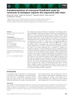
Fig. 1. Wavelength-independent kinetics of cyanide binding to Cj-
trHbP(III) and Cj-trHbP(II). (A) Normalized time course for cyanide
binding to Cj-trHbP(III) at k ¼ 410 nm (trace a) and k ¼ 420 nm
(trace b). The cyanide concentration was 1.0 · 10
)5
M. The time
course analysis according to Eqns (3a,3b) [18,21] yielded the follow-
ing values of l
obs
: (1.9 ± 0.2) · 10
)3
s
)1
(trace a, k ¼ 410 nm) and
(1.8 ± 0.2) · 10
)3
s
)1
(trace b, k ¼ 420 nm), respectively. (B) Nor-
malized time course for cyanide binding to Cj-trHbP(II) at k ¼
431 nm (trace a) and k ¼ 436 nm (trace b). The cyanide concentra-
tion was 1.0 · 10
)3
M. The time course analysis according
to Eqns (8a,8b) [18,21] yielded the following values of k
obs
:
3.8 ± 0.4 s
)1
(trace a, k ¼ 431 nm) and 3.9 ± 0.4 s
)1
(trace b, k ¼
436 nm), respectively. The protein concentration was 3.5 · 10
)6
M.
The absorbance change ranges between 0.1 and 0.3 according
to k. All data were obtained at pH 7.0 and 20.0 °C. For details, see
text.
A. Bolli et al. Cyanide binding to Camplylobacter jejuni trHbP
FEBS Journal 275 (2008) 633–645 ª 2008 The Authors Journal compilation ª 2008 FEBS 635
Discussion
It is well known that the heme–Fe(III)–cyanide
complexes are very stable, values of the dissociation
equilibrium constant being lower than 2 · 10
)5
m
[18,20,21,24–30] (Table 1). The different stabilities of
heme–Fe(III)–cyanide complexes in heme proteins are
primarily determined by the rate of ligand dissociation;
values of l
off
range between 3 · 10
)3
s
)1
and
1 · 10
)7
s
)1
[18,20,21,25,27–32] (Table 1), with the
exception of horseradish peroxidase and cytochrome c
peroxidase (l
off
¼ 2.8 · 10
)1
s
)1
and 9.0 · 10
)1
s
)1
,
respectively) [24,26]. Values of l
on
for cyanide binding
to most heme(III) proteins range between
1 · 10
2
m
)1
Æs
)1
and 5 · 10
2
m
)1
Æs
)1
[18,20,21,25,27–32].
In contrast, Glycera dibranchiata HbC displays an l
on
value of 4.9 · 10
)1
m
)1
Æs
)1
[20], whereas Cj-trHbP(III),
as well as horseradish peroxidase and cytochrome c
peroxidase [24,26] show l
on
‡ 2 · 10
4
m
)1
Æs
)1
(Table 1).
However, it must be remarked that the kinetics of cya-
nide binding to Cj-trHbP(III) appear to be limited by
Fig. 2. Kinetics of cyanide binding to Mt-trHbN(III), Mt-trHbO(III),
and Cj-trHbP(III). (A) Normalized averaged time courses for cyanide
binding to Mt-trHbN(III). The cyanide concentration was
1.0 · 10
)4
M (trace a), 2.0 · 10
)4
M (trace b), and 5.0 · 10
)4
M (tra-
ce c). The time course analysis according to Eqn (1) [18,21] yielded
the following values of l
obs
: 4.0 · 10
)2
s
)1
(trace a), 8.1 · 10
)2
s
)1
(trace b), and 1.9 · 10
)1
s
)1
(trace c). (B) Normalized averaged time
courses for cyanide binding to Mt-trHbO(III). The cyanide concen-
tration was 1.0 · 10
)4
M (trace a), 2.0 · 10
)4
M (trace b), and
5.0 · 10
)4
M (trace c). The time course analysis according to
Eqn (1) [18,21] yielded the following values of l
obs
: 2.4 · 10
)2
s
)1
(trace a), 5.9 · 10
)2
s
)1
(trace b), and 1.6 · 10
)1
s
)1
(trace c).
(C) Normalized averaged time courses for cyanide binding to Cj-
trHbP(III). For clarity, trace a and trace b have been upshifted by
0.6 and 0.3, respectively. The cyanide concentration was
1.0 · 10
)6
M (trace a), 1.0 · 10
)5
M (trace b), and 1.0 · 10
)3
M (tra-
ce c). The time course analysis according to Eqns (3a,3b) [21]
yielded the following values of l
obs
: 1.8 · 10
)3
s
)1
(trace a),
1.9 · 10
)3
s
)1
(trace b), and 2.1 · 10
)3
s
)1
(trace c). (D) Depen-
dence of the pseudo-first-order rate constant l
obs
for cyanide bind-
ing to Mt-trHbN(III) (squares) and Mt-trHbO(III) (circles) on ligand
concentration (i.e. cyanide concentration). The analysis of data
for cyanide binding to Mt-trHbN(III) and Mt-trHbO(III) according
to Eqn (2) [18,21] yielded the following values of l
on
:
(3.8 ± 0.4) · 10
2
M
)1
Æs
)1
and (3.2 ± 0.4) · 10
2
M
)1
Æs
)1
, respectively.
(E) Dependence of the pseudo-first-order rate constant l
obs
for
cyanide binding to Cj-trHbP(III) on ligand concentration (i.e.
cyanide concentration). The pH-independent value of l
obs
is
(1.9 ± 0.3) · 10
)3
s
)1
. Data referring to cyanide binding to Mt-
trHbN(III) and Mt-trHbO(III) were obtained from Milani et al. [18].
The protein concentration ranged between 2.0 · 10
)7
M and
5.0 · 10
)6
M. All data were obtained at pH 7.0 and 20.0 °C. For
details, see text.
Cyanide binding to Camplylobacter jejuni trHbP A. Bolli et al.
636 FEBS Journal 275 (2008) 633–645 ª 2008 The Authors Journal compilation ª 2008 FEBS
conformational transition(s) [l
max
¼ (2.0 ± 0.3) ·
10
)3
s
)1
, independent of the ligand concentration]
(Fig. 2), a feature never observed within heme(III)
proteins.
The reaction of cyanide with heme(II) proteins has
received little attention, due to the low stability of the
heme–Fe(II)–ligand complexes (K ‡ 5.8 · 10
)2
m)
[18,27,30,33–40]. Cj-trHbP(II) and horseradish peroxi-
dase are two exceptions in this respect, as values of
the cyanide dissociation equilibrium constant (i.e. K)
are 1.2 · 10
)6
m and 5.0 · 10
)4
m [35], respectively
(Table 1). Values of k
off
range between 5 · 10
)3
s
)1
and 1.5 s
)1
, whereas values of k
on
range between
5 · 10
)2
m
)1
Æs
)1
and 3.3 · 10
3
m
)1
Æs
)1
[18,27,30,33,
35,36,38–40]; in this context, Cj-trHbP(II) shows the
highest and the lowest values for k
on
and k
off
, respec-
tively (Table 1). As reported for Cj-trHbP(III) (Fig. 2),
the kinetics of cyanide binding to Cj-trHbP(II) (Fig. 4)
are limited by conformational transition(s), the appar-
ent rate constant tending to be independent of the
ligand concentration at cyanide concentrations
> 3.0 · 10
)3
m (i.e. k
max
¼ 9.1 s
)1
) (Fig. 4).
Values of K for cyanide binding to Scapharca ina-
equivalvis HbI(II) and horse heart myoglobin (Mb)(II)
measured in equilibrium experiments are about 10-fold
lower than those obtained from the ratio of the asso-
ciation and dissociation rate constants (Table 1),
possibly reflecting the formation of metastable inter-
mediate(s) [30,36,41,42]. In contrast, the excellent
Table 1. Values of kinetic and thermodynamic parameters for cyanide binding to ferric and ferrous heme-proteins. Values in italic were cal-
culated according to the following equations: L ¼ l
off
⁄ l
on
and K ¼ k
off
⁄ k
on
.
(Non)vertebrate globin
Fe(III) Fe(II)
l
on
(M
)1
Æs
)1
) l
off
(s
)1
) L (M) k
on
(M
)1
Æs
)1
) k
off
(s
)1
) K (M)
Mt-trHbN 3.8 · 10
2a
6.8 · 10
)4
1.8 · 10
)6b
5.0 · 10
)2b
1.3 · 10
)2b
1.2 · 10
)2a
2.4 · 10
)1b
2.4 · 10
)1
Mt-trHbO 3.2 · 10
2a
3.5 · 10
)4
1.1 · 10
)6b
8.5 · 10
)2b
1.3 · 10
)2b
1.3 · 10
)2a
1.6 · 10
)1b
1.5 · 10
)1
Cj-trHbP ‡ 2 · 10
4b
‡ 1 · 10
)4
5.8 · 10
)9b
3.3 · 10
3b
4.0 · 10
)3
5.0 · 10
)3c
1.2 · 10
)6b
1.7 · 10
)6
Sperm whale Mb 1.8 · 10
2d
8.0 · 10
)4d
4.3 · 10
)6
– 2.1 · 10
)2e
4.0 · 10
)1f
Horse heart Mb 1.7 · 10
2g
3.0 · 10
)3
1.8 · 10
)5g
2.5
h
1.5 · 10
)1i
4.0 · 10
)1h
5.8 · 10
)2
S. inaequivalvis HbI
h
2.3 · 10
2
6.2 · 10
)6
2.7 · 10
)8
2.7 1.1 · 10
)2
5.8 · 10
)2
4.0 · 10
)3
Human Hb 1.1 · 10
2j
1.4 · 10
)7
1.3 · 10
)9k
–
–
(R-state) 1.2 · 10
)1l
(T-state) 1.5
l
1
m
–
Horseradish peroxidase 9.0 · 10
4n
2.8 · 10
)1n
2.4 · 10
)6n
2.9 · 10
)6
2.9 · 10
1o
2.5 · 10
)2o
5.0 · 10
)4o
8.6 · 10
)4
a
pH 7.0, 20.0 °C [18].
b
pH 7.0, 20.0 °C (present study).
c
pH 7.0, 20.0 °C [16].
d
pH 6.6, 25.0 °C [27].
e
pH 7.0, 20.0 °C [37].
f
pH 9.3,
20.0 °C [27].
g
pH 7.0, 22.0 °C [21].
h
pH 9.2, 20.0 °C [30].
i
pH 8.2, 25.0 °C [36].
j
pH 6.05, 20.0 °C [25].
k
pH 7.0, 20 °C [21].
l
pH 7.0,
20.0 °C [38].
m
pH 10.6; the temperature is unknown [33].
n
pH 7.05, 25.0 °C [24].
o
pH 9.1, 20.0 °C [35].
Fig. 3. Ligand-binding isotherms for cyanide association with Mt-
trHbN(III) (A, squares), Mt-trHbO(III) (A, circles), and Cj-trHbP(III)
(B). The analysis of data for cyanide association with Mt-trHbN(III),
Mt-trHbO(III) and Cj-trHbP(III) according to Eqn (5) [23] yielded the
following values of L: (1.8 ± 0.2) · 10
)6
M, (1.1 ± 0.1) · 10
)6
M,
and (5.8 ± 0.6) · 10
)9
M, respectively. The protein concentration
ranged between 2.0 · 10
)7
M and 2.2 · 10
)7
M. All data were
obtained at pH 7.0 and 20.0 °C. For details, see text.
A. Bolli et al. Cyanide binding to Camplylobacter jejuni trHbP
FEBS Journal 275 (2008) 633–645 ª 2008 The Authors Journal compilation ª 2008 FEBS 637
agreement between values of K obtained at equilibrium
and from the ratio of the association and dissociation
rate constants for cyanide binding to Mt-trHbN(II),
Mt-trHbO(II) and Cj-trHbP(II) (Table 1) excludes the
occurrence of metastable intermediate(s) in the forma-
tion and dissociation of the trHb(II)–cyanide species.
Cj-trHbP(II) shows ligand-binding properties remi-
niscent of those of horseradish peroxidase(II). In fact,
even though horseradish peroxidase(II) shows a rela-
tively high reactivity towards cyanide [35] when com-
pared to that of ferrous 2-on-2 and 3-on-3 globins
[16,18,27,30,33,36–38], it turns out to be 100-fold
slower than what was observed for Cj-trHbP(II)
(Table 1). Furthermore, values of the second-order
rate constant for O
2
, CO and cyanide binding to
horseradish peroxidase(II) (5.7 · 10
4
m
)1
Æs
)1
[43]),
4.0 · 10
3
m
)1
Æs
)1
[44,45], and 2.9 · 10
1
m
)1
Æs
)1
[35],
respectively) span over three orders of magnitude, as
observed for Cj-trHbP(II) [9.1 · 10
5
m
)1
Æs
)1
[16];
1.1 · 10
5
m
)1
Æs
)1
(Coletta M & Guertin M, unpub-
lished results); and 3.3 · 10
3
m
)1
Æs
)1
(present study)].
In contrast, values of kinetic and thermodynamic
parameters for O
2
, CO and cyanide binding to ferrous
2-on-2 and 3-on-3 globins [e.g. Mt-trHb(II) and sperm
Fig. 4. Kinetics of cyanide binding to Mt-trHbN(II), Mt-trHbO(II),
and Cj-trHbP(II). (A) Normalized averaged time courses for cyanide
binding to Mt-trHbN(II). The cyanide concentration was
4.0 · 10
)1
M (trace a), 8.0 · 10
)1
M (trace b), and 1.6 M (trace c).
The time course analysis according to Eqn (6) [21] yielded the fol-
lowing values of k
obs
: 3.3 · 10
)2
s
)1
(trace a), 5.9 · 10
)2
s
)1
(tra-
ce b), and 9.8 · 10
)2
s
)1
(trace c). (B) Normalized averaged time
courses for cyanide binding to Mt-trHbO(II). The cyanide concentra-
tion was 4.0 · 10
)1
M (trace a), 8.0 · 10
)1
M (trace b), and 1.6 M
(trace c). The time course analysis according to Eqn (6) [21] yielded
the following values of k
obs
: 4.6 · 10
)2
s
)1
(trace a), 7.9 · 10
)2
s
)1
(trace b), and 1.5 · 10
)1
s
)1
(trace c). (C) Normalized averaged time
courses for cyanide binding to Cj-trHbP(II). The cyanide concentra-
tion was 2.5 · 10
)4
M (trace a), 1.0 · 10
)3
M (trace b), and
2.0 · 10
)3
M (trace c). The time course analysis according to
Eqns (8a,8b) [21] yielded the following values of k
obs
: 7.1 · 10
)1
s
)1
(trace a), 2.3 s
)1
(trace b), and 3.8 s
)1
(trace c). (D, E) Dependence
of the pseudo-first-order rate-constant k
obs
for cyanide binding to
Mt-trHbN(II) (D, squares), Mt-trHbO(II) (D, circles) and Cj-trHbP(II)
(E) on the ligand concentration (i.e. cyanide concentration). The
analysis of data for cyanide binding to Mt-trHbN(II) and Mt-trHbO(II)
according to Eqn (7) (dashed line) [21] yielded the following values
of k
on
: (5.0 ± 0.6) · 10
)2
M
)1
Æs
)1
and (8.5 ± 0.9) · 10
)2
M
)1
Æs
)1
,
respectively. The value of k
off
for cyanide dissociation from
Mt-trHbN(II)–cyanide and Mt-trHbO(II)–cyanide is (1.3 ± 0.2) ·
10
)2
s
)1
. The analysis of data for cyanide binding to Cj-trHbP(II)
according to Eqn (9) (solid line) [22] and Eqn (10) (dashed line) [22]
yielded K
pre
¼ (2.8 ± 0.3) · 10
)3
M), k
max
¼ 9.1 ± 0.8 s
)1
, and
k
on
¼ k
max
⁄ K
pre
¼ (3.3 ± 0.4) · 10
3
M
)1
Æs
)1
. The protein concentra-
tion ranged between 2.9 · 10
)6
M and 3.6 · 10
)6
M. All data were
obtained at pH 7.0 and 20.0 °C. For details, see text.
Cyanide binding to Camplylobacter jejuni trHbP A. Bolli et al.
638 FEBS Journal 275 (2008) 633–645 ª 2008 The Authors Journal compilation ª 2008 FEBS
whale Mb(II)] span over nine orders of magnitude
[1,21,46,47]. Therefore, Cj-trHbP(II) and horseradish
peroxidase discriminate among different ligands much
less than do ferrous 2-on-2 and 3-on-3 globins [e.g.
Mt-trHb(II) and sperm whale Mb(II)]. Such observa-
tions might be in keeping with the postulated involve-
ment of Cj-trHbP in O
2
chemistry, like peroxidase,
rather than in O
2
transport, which may require specific
adaptations to different environmental conditions [17].
The affinity of cyanide for heme(III) proteins
appears to depend on the presence of heme distal site
proton acceptor and donor group(s) that may assist
the deprotonation of the incoming ligand, or the pro-
tonation of the outgoing cyanide anion [18]. This inter-
pretation is in agreement with the very slow kinetics
of cyanide binding to Glycera dibranchiata monomeric
HbC(III), whose heme distal site lacks residue(s) capa-
ble of catalyzing proton exchange, and with the effects
shown by changes in the polarity of the heme distal
pocket of mutated human, pig and sperm whale Mbs
[1,20,21,48].
Concerning Cj-trHbP(III), the crystal structure
shows that the stabilization of the heme-bound cyanide
is achieved through direct hydrogen bonds of the
ligand to residue TyrB10 (phenolic OH group) and to
the indole nitrogen atom of TrpG8 (Fig. 6). Such
interactions, together with the presence of a water
molecule, trapped in the heme distal site and hydro-
gen-bonded to TyrB10 (Fig. 6), may assist the proton
exchange processes required for efficient heme–ligand
association ⁄ dissociation. In a dynamic protein context,
the contribution of HisE7, shown to adopt different
conformations in Cj-trHbP(III) crystals, might also be
considered either in direct ligand interactions or in
affecting the conformation of neighboring polar resi-
dues [16,17].
The crystal structure of the cyanide derivative of
Mt-trHbN(III) shows that only one direct hydrogen
bond (to TyrB10) stabilizes the heme-bound cyanide; a
second hydrogen-bonding contribution may be pro-
vided by the nearby GlnE11 residue (Fig. 6), substitut-
ing for a generally apolar residue (Val, Ile, and Leu) at
this site in vertebrate Hbs. The access to the heme
distal site of Mt-trHb through the E7-gate appears
to be precluded by the location of the E-helix and by
residue LeuE7 [49]. However, heme ligands may
diffuse though (apolar) protein matrix tunnels,
which have been mapped in the crystal structures of
Mt-trHbN xenon derivatives [50]. Stabilization of the
heme-bound cyanide in group II Mt-trHbO(III) takes
place through two hydrogen bonds, provided by the
side chain of TyrCD1 and by the indole nitrogen atom
of TrpG8 (in a dynamic context, TyrB10 may also be
part of such a ligand hydrogen-bonded network)
(Fig. 6). Access to the heme distal site through the
E7-gate is possible in Mt-trHbO, given the small size
of residue AlaE7 [51].
The comparison of the crystal structures of the cya-
nide derivatives of Mt-trHbN(III), Mt-trHbO(III) and
Cj-trHbP(III) suggests that diverse ligand diffusion
paths and binding mechanisms are active in the three
trHb groups. Although in all three groups the heme
ligand eventually becomes part of a hydrogen-bonded
network involving heme distal residues, the nature and
the involvement of residues at sites CD1, E7, E11
and G8 varies in a group-specific fashion, giving rise
to different stabilization patterns for the heme-bound
cyanide (Fig. 6) [16,49,51].
It appears worth noticing that Cj-trHbP displays the
highest affinity as well as the fastest combination and
the slowest dissociation rate for cyanide binding of the
known members of the Hb superfamily. Furthermore,
as the kinetics of cyanide binding to Cj-trHbP appear
to be limited by conformational transition(s) with first-
order rate constants dependent on the oxidation state
of the heme iron atom, Cj-trHbP may represent a
Fig. 5. Ligand-binding isotherms for cyanide association with
Mt-trHbN(II) (A, squares), Mt-trHbO(II) (A, circles), and Cj-trHbP(II)
(B). The analysis of data for cyanide association with Mt-trHbN(II),
Cj-trHbP(II) and Mt-trHbO(II) according to Eqn (11) [23] yielded the
following values of K: (2.4 ± 0.3) · 10
)1
M, (1.6 ± 0.2) · 10
)1
M,
and 1.2 ± 0.2) · 10
)6
M, respectively. The protein concentration
ranged between 2.3 · 10
)6
M and 3.5 · 10
)6
M. All data were
obtained at pH 7.0 and 20.0 °C. For details, see text.
A. Bolli et al. Cyanide binding to Camplylobacter jejuni trHbP
FEBS Journal 275 (2008) 633–645 ª 2008 The Authors Journal compilation ª 2008 FEBS 639
reference system for investigating the interplay between
the redox state of the heme iron atom and conforma-
tional transition(s) modulating trHb reactivity.
Finally, the very high affinity of cyanide for Cj-trHbP
suggests that this globin may participate in cyanide
detoxification, facilitating the survival of C. jejuni.
Indeed, the intestinal localization of C. jejuni in herbi-
vores suggests that this organism could be exposed to
cyanide generated from the enzymatic breakage of
cyanogenic glycosides of ingested plants [19]. Interest-
ingly, inspection of the C. jejuni NCTC11168 genome
( reveals that this bacterium
lacks proteins that have been annotated as canonical
double-domain rhodaneses, although it contains two
putative proteins with a rhodanese (RHOD) module
(NCBI accession numbers CAL34666 and CAL34648).
Moreover, C. jejuni expresses a cyanide-resistant low-
affinity terminal oxidase (not of the cytochrome bd
type) encoded by cydAB genes [52], which could facili-
tate survival in cyanide-containing environments.
Experimental procedures
Materials
Cloning, expression and purification of Cj-trHbP were
performed as previously reported [16]. Mt-TrHbN and
Mt-trHbO were cloned, expressed and purified as previ-
ously reported [53,54]. Mt-trHbN(III), Mt-trHbO(III) and
Cj-trHbP(III) were prepared by adding a few grains of fer-
ricyanide to the trHb solution [21]. Mt-trHbN(II), Mt-
trHbO(II) and Cj-trHbP(II) were prepared by adding a few
grains of dithionite to the trHb solution, under anaerobic
conditions [21]. All chemicals (from Merck AG, Darmstadt,
Germany) were of analytical grade and were used without
further purification.
Kinetics of cyanide binding to Mt-trHbN(III) and
Mt-trHbO(III)
The kinetics of cyanide binding to Mt-trHbN(III) and Mt-
trHbO(III) were measured by mixing a protein-buffered
solution (2.0 · 10
)6
m and 5.0 · 10
)6
m, respectively)
Fig. 6. View of the heme distal pocket of the cyanide derivative of
Mt-trHbN(III) (Protein Data Bank code: 1RTE [18]), Mt-trHbO(III)
(Protein Data Bank code: 1NGH [51]), and Cj-trHbP(III) (Protein Data
Bank code: 2IG3 [16]), displaying part of the surrounding protein
structure (ribbon), the heme group (red), the cyanide ligand, and
key residues stabilizing the heme Fe-bound cyanide. Hydrogen
bonds stabilizing the heme Fe-bound cyanide are represented by
dashed red lines. All pictures were drawn with
MOLSCRIPT [55]. For
details, see text.
Cyanide binding to Camplylobacter jejuni trHbP A. Bolli et al.
640 FEBS Journal 275 (2008) 633–645 ª 2008 The Authors Journal compilation ª 2008 FEBS
with a cyanide-buffered solution (from 5.0 · 10
)5
m to
1.0 · 10
)3
m). The reaction was monitored between 380 nm
and 460 nm (Table 2), using the SFM-20 rapid-mixing
stopped-flow apparatus (Bio-Logic SAS, Claix, France). No
gaseous phase was present [18].
Values of the first-order rate constant for cyanide binding
to Mt-trHbN(III) and Mt-trHbO(III) (l
obs
) were calculated
according to Eqn (1) [18,21]:
½Mt-trHbðIIIÞ
t
¼½Mt-trHbðIIIÞ
i
e
l
obs
t
ð1Þ
The dependence of l
obs
on cyanide concentration for ligand
binding to Mt-trHbN(III) and Mt-trHbO(III) was analyzed
according to the minimum reaction mechanism shown in
Scheme 1 [18]:
Mt-trHb(III) þcyanide $
l
on
l
off
Mt-trHb(III)cyanide ðScheme 1Þ
where l
on
is the second-order rate constant for cyanide
binding to Mt-trHbN(III) and Mt-trHbO(III) (i.e. for the
formation of Mt-trHbN(III)–cyanide and Mt-trHbO(III)–
cyanide), and l
off
is the first-order rate constant for
cyanide dissociation from Mt-trHbN(III)–cyanide and
Mt-trHbO(III)–cyanide.
Values of l
on
were obtained according to Eqn (2) [18,21]:
l
obs
¼ l
on
½cyanideð2Þ
Kinetics of cyanide binding to Cj-trHbP(III)
The kinetics of cyanide binding to Cj-trHbP(III) were mea-
sured by mixing a protein-buffered solution (2.0 · 10
)7
m)
with a cyanide-buffered solution (from 1.0 · 10
)6
m to
1.0 · 10
)3
m). The reaction was followed spectrophotomet-
rically between 350 nm and 460 nm (see Table 2). Absor-
bance spectra were recorded every 3 min. No gaseous phase
was present [18].
Values of the first-order rate constant for cyanide binding
to Cj-trHbP(III) (l
obs
) were calculated according to Eqn (3)
[21]:
½Cj-trHbPðIIIÞ
t
¼½Cj-trHbPðIIIÞ
i
e
l
obs
t
ð3aÞ
½Cj-trHbPðIIIÞ
t
¼½Cj-trHbPðIIIÞ
i
ð1 e
l
obs
t
Þð3bÞ
The dependence of l
obs
on cyanide concentration for
ligand binding to Cj-trHbP(III) was analyzed according
to the minimum reaction mechanism shown in Scheme 2
[22]:
Cj-trHbP(III) þ cyanide $
l
þ1
l
1
ðCj-trHbP(III)–cyanideÞ
1
$
l
þ2
l
2
ðCj-trHbP(III)–cyanideÞ
2
ðScheme 2Þ
where l
+1
(= l
on
¼ l
max
⁄ L
pre
) is the second-order rate
constant for cyanide binding to Cj-trHbP(III) [i.e. for the
formation of the transient (Cj-trHbP(III)–cyanide)
1
species],
l
–1
⁄ l
+1
(= L
pre
) is the pre-equilibrium constant, l
+2
(= l
max
)
represents the asymptotic value of l
obs
for cyanide con-
centration ‡ 10 · L
pre
, and l
–2
(= l
off
) is the first-order rate
constant for cyanide dissociation from the final Cj-
trHbP(III)–cyanide complex, [i.e. Cj-trHbP(III)–cyanide)
2
].
Step 1 of Scheme 2 (characterized by l
+1
and l
–1
) is not a
simple process but represents a multistep reaction reflecting
the dynamic pathway of the ligand from the bulk solvent to
the heme pocket, where it reacts with the heme Fe(III) atom
(i.e. step 2 of (Scheme 2), characterized by l
+2
and l
–2
).
Values of l
on
, l
max
and L
pre
were estimated according to
Eqn (4) [22]:
l
obs
¼ l
max
½cyanide=ðL
pre
þ½cyanideÞ ð4Þ
Thermodynamics of cyanide binding to
Mt-trHbN(III), Mt-trHbO(III), and Cj-trHbP(III)
The thermodynamics of cyanide binding to Mt-trHbN(III),
Mt-trHbO(III) and Cj-trHbP(III) were determined by
adding a cyanide-buffered solution (from 4.1 · 10
)8
m to
1.8 · 10
)5
m) to a protein-buffered solution ([Mt-
trHbN(III)] ¼ 2.2 · 10
)7
m,[Mt-trHbO(III)] ¼ 2.1 · 10
)7
m, and [Cj-trHbP(III)] ¼ 2.0 · 10
)7
m). The reaction was
followed spectrophotometrically between 350 nm and
460 nm (see Table 2). Absorbance spectra were recorded
after achieving the equilibrium (the equilibration time ranged
between 1 h and 48 h). No gaseous phase was present.
The dependence of the molar fraction of cyanide-bound
trHb(III) (i.e. a) on cyanide concentration was analyzed
according to the minimum reaction mechanism shown in
Scheme 3 [21]:
trHb(III) þ cyanide $
l
on
l
off
trHb(III)–cyanide ðScheme 3Þ
Values of the dissociation equilibrium constant for
cyanide binding to Mt-trHbN(III), Mt-trHbO(III) and
Cj-trHbP(III) (L ¼ l
off
⁄ l
on
) were calculated according to
Eqn (5) [23]:
Table 2. Values of k
max
and e of the absorption spectra in the
Soret region of ferric [i.e. Fe(III) and Fe(III)–cyanide] and ferrous
[i.e. Fe(II) and Fe(II)–cyanide] derivatives of Mt-trHbN, Mt-trHbO,
and Cj-trHbP. Values of k
max
(nm) are in italic and values
of e (m
M
)1
cm
)1
) are in bold.
Protein Fe(III) Fe(III)–cyanide Fe(II) Fe(II)–cyanide
Mt-trHbN
a
406
141
418
102
432
103
435
142
Mt-trHbO
b
409
104
419
105
429
92
436
144
Cj-trHbP 410
c
141
c
420
d
112
d
433
d
119
d
434
d
174
d
a
pH 7.0 and 20.0 °C [18].
b
pH 7.0 and 20.0 °C [18].
c
pH 7.0 and
20.0 °C (present study).
d
pH 7.0 and 20.0 °C [16].
A. Bolli et al. Cyanide binding to Camplylobacter jejuni trHbP
FEBS Journal 275 (2008) 633–645 ª 2008 The Authors Journal compilation ª 2008 FEBS 641
a ¼ ðð½cyanideþL þ½trHb(IIIÞÞ þ
p
ðð½cyanideþL
þ½trHbðIIIÞÞ
2
4 ½cyanide½trHbðIIIÞÞÞ=
ð2 ½trHbðIIIÞÞ ð5Þ
Kinetics of cyanide binding to Mt-trHbN(II) and
Mt-trHbO(II)
The kinetics of cyanide binding to Mt-trHbN(II) and Mt-
trHbO(II) were measured by mixing a protein-buffered
solution (2.9 · 10
)6
m and 3.6 · 10
)6
m, respectively) with
a cyanide-buffered solution (from 2.0 · 10
)1
m to 1.6 m).
The reaction was monitored between 350 nm and 460 nm
(see Table 2) using the SFM-20 rapid-mixing stopped-flow
apparatus (Bio-Logic SAS, Claix, France). No gaseous
phase was present.
Values of the first-order rate constant for cyanide binding
to Mt-trHbN(II) and Mt-trHbO(II) (k
obs
) were calculated
according to Eqn (6) [21]:
½Mt-trHbðIIÞ
t
¼½Mt-trHbðIIÞ
i
e
k
obs
t
ð6Þ
The dependence of k
obs
on cyanide concentration for
ligand binding to Mt-trHbN(II) and Mt-trHbO(II) was
analyzed according to the minimum reaction mechanism
shown in Scheme 4 [21]:
Mt-trHb(II) þcyanide $
k
on
k
off
Mt-trHb(II)–cyanide ðScheme 4Þ
where k
on
is the second-order rate constant for cyanide
binding to Mt-trHbN(II) and Mt-trHbO(II) [i.e. for the for-
mation of Mt-trHbN(II)–cyanide and Mt-trHbO(II)–cya-
nide], and k
off
is the first-order rate constant for cyanide
dissociation from Mt-trHbN(II)–cyanide and Mt-
trHbO(II)–cyanide.
Values of k
on
and k
off
were obtained according to
Eqn (7) [21]:
k
obs
¼ k
on
½cyanideþk
off
ð7Þ
Kinetics of cyanide binding to Cj-trHbP(II)
The kinetics of cyanide binding to Cj-trHbP(II) were mea-
sured by mixing a protein-buffered solution (3.5 · 10
)6
m)
with a cyanide-buffered solution (from 3.1 · 10
)5
m to
2.0 · 10
)3
m). The reaction was monitored between 390 nm
and 500 nm (see Table 2) using the rapid-mixing SX.18MV
stopped-flow apparatus equipped with the PDA.1 photo-
diode array accessory (Applied Photophysics, Salis-
bury, UK). No gaseous phase was present.
Values of the first-order rate constant for cyanide binding
to Cj-trHbP(II) (k
obs
) were calculated according to Eqn (8)
[21]:
½Cj-trHbP(II)
t
¼½Cj-trHbP(II)
i
e
k
obs
t
ð8aÞ
½Cj-trHbPðIIÞ
t
¼½Cj-trHbPðIIÞ
i
ð1 e
k
obs
t
Þð8bÞ
The dependence of k
obs
on cyanide concentration for
ligand binding to Cj-trHbP(II) was analyzed according to
the minimum reaction mechanism shown in Scheme 5 [22]:
Cj-trHbP(II) þ cyanide $
k
þ1
k
1
ðCj-trHbP(II)–cyanideÞ
1
$
k
þ2
k
2
ðCj-trHbP(II)–cyanideÞ
2
ðScheme 5Þ
where k
+1
(= k
on
¼ k
max
⁄ K
pre
) is the second-order rate
constant for cyanide binding to Cj-trHbP(II) [i.e. for the
formation of the transient (Cj-trHbP(II)-cyanide)
1
species],
k
)1
⁄ k
+1
(= K
pre
) is the pre-equilibrium constant,
k
+2
(= k
max
) represents the asymptotic value of k
obs
for cyanide concentration ‡ 10 · K
pre
, and k
–2
(= k
off
)is
the first-order rate constant for cyanide dissociation from
the final Cj-trHbP(II)–cyanide complex [i.e. (Cj-trHbP(II)–
cyanide)
2
]. Step 1 of Scheme 5 (characterized by k
+1
and
k
)1
) is not a simple process but represents a multistep reac-
tion reflecting the dynamic pathway of the ligand from the
bulk solvent to the heme pocket, where it reacts with the
heme Fe(II) atom (i.e. step 2 of Scheme 5, characterized by
k
+2
and k
–2
).
Values of k
on
, k
max
and K
pre
were obtained according to
Eqn (9) [22]:
k
obs
¼ k
max
½cyanide=ðK
pre
þ½cyanideÞ ð9Þ
Under conditions where the cyanide concentration
£ 10 · K
pre
, Eqn (9) approximates to Eqn (10) [22]:
k
obs
¼ k
on
½cyanideð10Þ
Thermodynamics of cyanide binding to
Mt-trHbN(II), Mt-trHbO(II), and Cj-trHbP(II)
The thermodynamics of cyanide binding to Mt-trHbN(II),
Mt-trHbO(II) and Cj-trHbP(II) were determined by adding
a cyanide-buffered solution (from 2.5 · 10
)7
m to 1.3 m)
to a protein-buffered solution ([Mt-trHbN(II)] =
2.9 · 10
)6
m,[Mt-trHbO(II)] ¼ 2.3 · 10
)6
m, and
[Cj-trHbP(II)] ¼ 3.5 · 10
)6
m). The reaction was followed
spectrophotometrically between 350 nm and 460 nm (see
Table 2). Absorbance spectra were recorded after achieving
the equilibrium (the equilibration time ranged between
10 min and 12 h). No gaseous phase was present.
The dependence of the molar fraction of cyanide-bound
trHb(II) (i.e. a) on cyanide concentration was analyzed
according to the minimum reaction mechanism shown in
Scheme 6 [21]:
trHb(II) þ cyanide $
k
on
k
off
trHb(II)–cyanide ðScheme 6Þ
Cyanide binding to Camplylobacter jejuni trHbP A. Bolli et al.
642 FEBS Journal 275 (2008) 633–645 ª 2008 The Authors Journal compilation ª 2008 FEBS
Values of the dissociation equilibrium constant for
cyanide binding to Mt-trHbN(II), Mt-trHbO(II) and
Cj-trHbP(II) (K ¼ k
off
⁄ k
on
) were calculated according to
Eqn (11) [23]:
a ¼ ðð½cyanideþK þ½trHb(II)Þ þ
p
ðð½cyanideþK
þ [trHb(II)]Þ
2
4 ½cyanide½trHb(II)ÞÞ=
ð2 ½trHb(II)Þ ð11Þ
Data analysis
Data analysis was performed with the program matlab 7.0
(MathWorks Inc., South Natick, MA, USA). All data
(obtained at pH 7.0, 1.0 · 10
)1
m phosphate buffer, and
20.0 °C) were determined at least in quadruplicate.
Acknowledgements
Professor Martino Bolognesi is grateful to CIMAINA
(Milano, Italy) for continuous support. Part of this
study was supported by grants from the Natural Sci-
ences and Engineering Research Council of Canada
(grant 46306-01, 2005–2010) to M. Guertin, the
National Institute of Health of the USA (grant 1-R01-
AI052258, 2004–2007) to M. Guertin, the Ministry for
University and Scientific Research of Italy (FIRB Pro-
ject ‘Biologia Strutturale’, contract RBLA03B3KC) to
M. Bolognesi, the Ministry of Health of Italy (INMI-
IRCCS ‘Lazzaro Spallanzani’, Roma, Italy, Ricerca
corrente 2006) to P. Ascenzi, and University ‘Roma
Tre’ (Roma, Italy, CLAR-2006) to P. Ascenzi.
References
1 Bolognesi M, Bordo D, Rizzi M, Tarricone C &
Ascenzi P (1997) Nonvertebrate hemoglobins: structural
bases for reactivity. Prog Biophys Mol Biol 68, 29–68.
2 Poole RK & Hughes MN (2000) New functions for
the ancient globin family: bacterial responses to
nitric oxide and nitrosative stress. Mol Microbiol 36,
775–783.
3 Wittenberg JB, Bolognesi M, Wittenberg BA & Guertin
M (2002) Truncated hemoglobins: a new family of
hemoglobins widely distributed in bacteria, unicellular
eukaryotes, and plants. J Biol Chem 277, 871–874.
4 Wu G, Wainwright LM & Poole RK (2003) Microbial
globins. Adv Microb Physiol 47, 255–310.
5 Frey AD & Kallio PT (2003) Bacterial hemoglobins
and flavohemoglobins: versatile proteins and their
impact on microbiology and biotechnology. FEMS
Microbiol Rev 27, 525–545.
6 Egawa T & Yeh SR (2005) Structural and functional
properties of hemoglobins from unicellular organisms as
revealed by resonance Raman spectroscopy. J Inorg
Biochem 99, 72–96.
7 Vinogradov SN, Hoogewijs D, Bailly X, Arredondo-
Peter R, Gough J, Dewilde S, Moens L & Vanfleteren
JR (2006) A phylogenomic profile of globins. BMC
Evol Biol 6, 31.
8 Vuletich DA & Lecomte JT (2006) A phylogenetic and
structural analysis of truncated hemoglobins. J Mol
Evol 62, 196–210.
9 Ascenzi P, Bolognesi M, Milani M, Guertin M & Visca
P (2007) Mycobacterial truncated hemoglobins: from
genes to functions. Gene 398, 42–51.
10 Altekruse SF, Stern NJ, Fields PI & Swerdlow DL
(1999) Campylobacter jejuni – an emerging foodborne
pathogen. Emerg Infect Dis 5, 28–35.
11 Moore JE, Corcoran D, Dooley JS, Fanning S, Lucey
B, Matsuda M, McDowell DA, Megraud F, Millar BC,
O’Mahony R et al. (2005) Campylobacter. Vet Res 36,
351–382.
12 Young KT, Davis LM & DiRita VJ (2007) Campylo-
bacter jejuni: molecular biology and pathogenesis. Nat
Rev Microbiol 5, 665–679.
13 Elvers KT, Wu G, Gilberthorpe NJ, Poole RK & Park
SF (2004) Role of an inducible single-domain hemoglo-
bin in mediating resistance to nitric oxide and nitrosa-
tive stress in Campylobacter jejuni and Campylobacter
coli. J Bacteriol 186, 5332–5341.
14 Lu C, Mukai M, Lin Y, Wu G, Poole RK & Yeh SR
(2007) Structural and functional properties of a single
domain hemoglobin from the food-borne pathogen
Campylobacter jejuni. J Biol Chem 282, 25917–25928.
15 Wainwright LM, Elvers KT, Park SF & Poole RK
(2005) A truncated haemoglobin implicated in oxygen
metabolism by the microaerophilic food-borne pathogen
Campylobacter jejuni. Microbiology 151, 4079–4091.
16 Nardini M, Pesce A, Labarre M, Richard C, Bolli A,
Ascenzi P, Guertin M & Bolognesi M (2006) Structural
determinants in the group III truncated hemoglobin
from Campylobacter jejuni. J Biol Chem 281, 37803–
37812.
17 Lu C, Egawa T, Wainwright LM, Poole RK & Yeh SR
(2007) Structural and functional properties of a trun-
cated hemoglobin from a food-borne pathogen Cam-
pylobacter jejuni. J Biol Chem 282, 13627–13636.
18 Milani M, Ouellet Y, Ouellet H, Guertin M, Boffi A,
Antonini G, Bocedi A, Mattu M, Bolognesi M & As-
cenzi P (2004) Cyanide binding to truncated hemoglo-
bins: a crystallographic and kinetic study. Biochemistry
43, 5213–5221.
19 Hughes MA, Sharif AL, Dunn MA & Oxtoby E (1988)
The molecular biology of cyanogenesis. Ciba Found
Symp 140, 111–130.
20 Mintorovitch J, van Pelt D & Satterlee JD (1989)
Kinetic study of the slow cyanide binding to Glycera
A. Bolli et al. Cyanide binding to Camplylobacter jejuni trHbP
FEBS Journal 275 (2008) 633–645 ª 2008 The Authors Journal compilation ª 2008 FEBS 643
dibranchiata monomer hemoglobin components III and
IV. Biochemistry 28, 6099–6104.
21 Antonini E & Brunori M (1971) Hemoglobin and myo-
globin in their reactions with ligands. North Holland
Publishing Co., Amsterdam.
22 Antonini E, Ascenzi P, Menegatti E & Guarneri M
(1983) Multiple intermediates in the reaction of bovine
b-trypsin with bovine pancreatic trypsin inhibitor
(Kunitz). Biopolymers 22, 363–375.
23 Claro E (2006) Analyzing ligand depletion in a satura-
tion equilibrium binding experiment. Biochem Mol Biol
Educ 34, 428–431.
24 Ellis WD & Dunford HB (1968) The kinetics of cyanide
and fluoride binding by ferric horseradish peroxidase.
Biochemistry 7, 2054–2062.
25 Gibson QH, Parkhurst LJ & Geraci G (1969) The reac-
tion of methemoglobin with some ligands. J Biol Chem
244, 4668–4676.
26 Erman JE (1974) Kinetic and equilibrium studies of
cyanide binding by cytochrome c peroxidase. Biochemis-
try 13, 39–44.
27 Cox RP & Hollaway MR (1977) The reduction by
dithionite of Fe(III) myoglobin derivatives with differ-
ent ligands attached to the iron atom. A study by
rapid-wavelength-scanning stopped-flow spectrophoto-
metry. Eur J Biochem 74 , 575–587.
28 Job D, Zeba B, Puppo A & Rigaud J (1980) Kinetic
studies of the reaction of ferric soybean leghemoglobins
with hydrogen peroxide, cyanide and nicotinic acid. Eur
J Biochem 107, 491–500.
29 Nardini M, Tarricone C, Rizzi M, Lania A, Desideri A,
De Sanctis G, Coletta M, Petruzzelli R, Ascenzi P,
Coda A et al. (1995) Reptile heme protein structure:
X-ray crystallographic study of the aquo-met and
cyano-met derivatives of the loggerhead sea turtle
(Caretta caretta) myoglobin at 2.0 A
˚
resolution. J Mol
Biol 247, 459–465.
30 Boffi A, Ilari A, Spagnuolo C & Chiancone E (1996)
Unusual affinity of cyanide for ferrous and ferric Scaph-
arca inaequivalvis homodimeric hemoglobin. Equilibria
and kinetics of the reaction. Biochemistry 35, 8068–
8074.
31 Bolognesi M, Boffi A, Coletta M, Mozzarelli A, Pesce
A, Tarricone C & Ascenzi P (1999) Anticooperative
ligand binding properties of recombinant ferric Vitreos-
cilla homodimeric hemoglobin: a thermodynamic, kinetic
and X-ray crystallographic study. J Mol Biol 291, 637–
650.
32 Bolognesi M, Rosano C, Losso R, Borassi A, Rizzi M,
Wittenberg JB, Boffi A & Ascenzi P (1999) Cyanide
binding to Lucina pectinata hemoglobin I and to sperm
whale myoglobin: an X-ray crystallographic study.
Biophys J 77, 1093–1099.
33 Stitt F & Coryell CD (1939) The magnetic study of the
equilibrium between ferrohemoglobin, cyanide ion, and
cyanide ferrohemoglobin. J Am Chem Soc 61, 1263–
1266.
34 Keilin D & Hartree EF (1955) Cyanide compounds of
ferroperoxidase and myoglobin and their reversible pho-
todissociation. Biochem J 61, 153–171.
35 Phelps CF, Antonini E & Brunori M (1971) The binding
of cyanide to ferroperoxidase. Biochem J 122, 79–87.
36 Olivas E, De Waal DJ & Wilkins RG (1977) Reduction
of metmyoglobin derivatives by dithionite ion. J Biol
Chem 252, 4038–4042.
37 Bellelli A, Antonini G, Brunori M, Springer BA & Sli-
gar SG (1990) Transient spectroscopy of the reaction of
cyanide with ferrous myoglobin. J Biol Chem 265,
18898–18901.
38 Brunori M, Antonini G, Castagnola M & Bellelli A
(1992) Cooperative cyanide dissociation from ferrous
hemoglobin. J Biol Chem 267, 2258–2263.
39 Antonini G, Bellelli A, Concetti A, Falcioni G & Brun-
ori M (1994) Cyanide dissociation from the hemoglobin
of Parascaris equorum. Biochim Biophys Acta 1025,
252–257.
40 Antonini G, Bellelli A, Brunori M & Falcioni G (1996)
Kinetic and spectroscopic properties of the cyanide
complexes of ferrous haemoglobins I and IV from trout
blood. Biochem J 314, 533–540.
41 Boffi A, Chiancone E, Takahashi S & Rousseau DL
(1997) Stereochemistry of the Fe(II)- and Fe(III)-cya-
nide complexes of the homodimeric Scapharca inaequi-
valvis hemoglobin. A resonance Raman and FTIR
study. Biochemistry 36, 4505–4509.
42 Boffi A, Chiancone E, Peterson ES, Wang J, Rousseau
DL & Friedman JM (1997) Dynamics of cyanide bind-
ing to ferrous Scapharca inaequivalvis homodimeric
hemoglobin. Biochemistry 36, 4510–4514.
43 Wittenberg JB, Noble RW, Wittenberg BA, Antonini E,
Brunori M & Wyman J (1967) Studies on the equilibria
and kinetics of the reactions of peroxidase with ligands:
II. The reaction of ferroperoxidase with oxygen. J Biol
Chem 242, 626–634.
44 Kertesz D, Antonini E, Brunori M, Wyman J & Zito R
(1965) Studies on the equilibria and kinetics of the reac-
tions of peroxidases with ligands. I. The reaction of fer-
roperoxidases with carbon monoxide. Biochemistry 4,
2672–2676.
45 Coletta M, Ascoli F, Brunori M & Traylor TG (1986)
pH dependence of carbon monoxide binding to ferrous
horse radish peroxidase. J Biol Chem 261, 9811–9814.
46 Brunori M, Coletta M, Ascenzi P & Bolognesi M
(1989) Kinetic control of ligand binding processes in
hemoproteins. J Mol Liq 42, 175–193.
47 Springer BA, Sligar SG, Olson JS & Phillips JN (1994)
Mechanisms of ligand recognition in myoglobin. Chem
Rev 94, 699–714.
48 Brancaccio A, Cutruzzola
`
F, Travaglini Allocatelli C,
Brunori M, Smerdon SJ, Wilkinson AJ, Dou Y,
Cyanide binding to Camplylobacter jejuni trHbP A. Bolli et al.
644 FEBS Journal 275 (2008) 633–645 ª 2008 The Authors Journal compilation ª 2008 FEBS
Keenan D, Ikeda-Saito M, Brantley RE Jr et al. (1994)
Structural factors governing azide and cyanide binding
to mammalian metmyoglobins. J Biol Chem 269,
13843–13853.
49 Milani M, Pesce A, Ouellet Y, Ascenzi P, Guertin M &
Bolognesi M (2001) Mycobacterium tuberculosis
hemoglobin N displays a protein tunnel suited for O
2
diffusion to the heme. EMBO J 20, 3902–3909.
50 Milani M, Pesce A, Ouellet Y, Dewilde S, Friedman J,
Ascenzi P, Guertin M & Bolognesi M (2004) Heme-
ligand tunneling in group I truncated hemoglobins.
J Biol Chem 279, 21520–21525.
51 Milani M, Savard PY, Ouellet H, Ascenzi P, Guertin M
& Bolognesi M (2003) A TyrCD1 ⁄ TrpG8 hydrogen
bond network and a TyrB10TyrCD1 covalent link
shape the heme distal site of Mycobacterium tuberculosis
hemoglobin O. Proc Natl Acad Sci USA 100,
5766–5771.
52 Jackson RJ, Elvers KT, Lee LJ, Gidley MD, Wain-
wright LM, Lightfoot J, Park SF & Poole RK (2007)
Oxygen reactivity of both respiratory oxidases in
Campylobacter jejuni: the cydAB genes encode a cya-
nide-resistant, low-affinity oxidase that is not of the
cytochrome bd type. J Bacteriol 189, 1604–1615.
53 Couture M, Yeh S, Wittenberg BA, Wittenberg JB,
Ouellet Y, Rousseau DL & Guertin M (1999) A
cooperative oxygen-binding hemoglobin from Myco-
bacterium tuberculosis. Proc Natl Acad Sci USA 96,
11223–11228.
54 Mukai M, Savard PY, Ouellet H, Guertin M & Yeh SR
(2002) Unique ligand–protein interactions in a new
truncated hemoglobin from Mycobacterium tuberculosis.
Biochemistry 41, 3897–3905.
55 Kraulis PJ (1991) Molscript: a program to produce
both detailed and schematic plots of protein structures.
J Appl Crystallogr 24, 946–950.
A. Bolli et al. Cyanide binding to Camplylobacter jejuni trHbP
FEBS Journal 275 (2008) 633–645 ª 2008 The Authors Journal compilation ª 2008 FEBS 645