Báo cáo khoa học: Characterization of the glutamyl endopeptidase from Staphylococcus aureus expressed in Escherichia coli pptx
Bạn đang xem bản rút gọn của tài liệu. Xem và tải ngay bản đầy đủ của tài liệu tại đây (519.79 KB, 15 trang )
Characterization of the glutamyl endopeptidase from
Staphylococcus aureus expressed in Escherichia coli
Takayuki K. Nemoto
1
, Yuko Ohara-Nemoto
1
, Toshio Ono
1
, Takeshi Kobayakawa
1
, Yu Shimoyama
2
,
Shigenobu Kimura
2
and Takashi Takagi
3
1 Department of Oral Molecular Biology, Course of Medical and Dental Sciences, Nagasaki University Graduate School of Biomedical
Sciences, Japan
2 Department of Oral Microbiology, Iwate Medical University School of Dentistry, Morioka, Japan
3 Department of Developmental Biology and Neurosciences, Graduate School of Life Sciences, Tohoku University, Sendai, Japan
Staphylococcus aureus produces extracellular proteases,
which are regarded as important virulence factors. One
of the classically defined exoproteases is a serine prote-
ase, GluV8, also known as V8 protease ⁄ SspA [1].
GluV8 contributes to the growth and survival of this
microorganism in animal models [2], and plays a key
role in degrading the cell-bound Staphylococcus surface
adhesion molecules of fibronectin-binding proteins and
protein A [3]. This protease specifically cleaves the
peptide bond after the negatively charged residues Glu
and, less potently, Asp, and belongs to the glutamyl
endopeptidase I (EC 3.4.21.19) family [4]. The nucleo-
tide sequence encodes a protein of 336 amino acids
that includes a prepropeptide consisting of 68 residues
(Met1-Asn68) and a C-terminal tail of 52 residues con-
sisting of a 12-fold repeat of the tripeptide Pro-
Asp ⁄ Asn-Asn [5]. Drapeau [6] first reported that the
activation of the GluV8 precursor is achieved by a
neutral metalloprotease. Shaw et al. [7] have recently
demonstrated that the GluV8 activation process
Keywords
chaperone; glutamyl endopeptidase;
Staphylococcus aureus; Staphylococcus
epidermidis; V8 protease
Correspondence
T. K. Nemoto, Department of Oral
Molecular Biology, Course of Medical and
Dental Sciences, Nagasaki University,
1-7-1 Sakamoto, Nagasaki 852-8588, Japan
Fax: +81 95 819 7642
Tel: +81 95 819 7640
E-mail:
(Received 2 November 2007, revised 6
December 2007, accepted 7 December
2007)
doi:10.1111/j.1742-4658.2007.06224.x
V8 protease, a member of the glutamyl endopeptidase I family, of Staphy-
lococcus aureus V8 strain (GluV8) is widely used for proteome analysis
because of its unique substrate specificity and resistance to detergents. In
this study, an Escherichia coli expression system for GluV8, as well as its
homologue from Staphylococcus epidermidis (GluSE), was developed, and
the roles of the prosegments and two specific amino acid residues, Val69
and Ser237, were investigated. C-terminal His
6
-tagged proGluSE was
successfully expressed from the full-length sequence as a soluble form. By
contrast, GluV8 was poorly expressed by the system as a result of autode-
gradation; however, it was efficiently obtained by swapping its preproseg-
ment with that of GluSE, or by the substitution of four residues in the
GluV8 prosequence with those of GluSE. The purified proGluV8 was con-
verted to the mature form in vitro by thermolysin treatment. The proseg-
ment was essential for the suppression of proteolytic activity, as well as for
the correct folding of GluV8, indicating its role as an intramolecular chap-
erone. Furthermore, the four amino acid residues at the C-terminus of the
prosegment were sufficient for both of these roles. In vitro mutagenesis
revealed that Ser237 was essential for proteolytic activity, and that Val69
was indispensable for the precise cleavage by thermolysin and was involved
in the proteolytic reaction itself. This is the first study to express quantita-
tively GluV8 in E. coli, and to demonstrate explicitly the intramolecular
chaperone activity of the prosegment of glutamyl endopeptidase I.
Abbreviations
CBB, Coomassie brilliant blue; GluSE, GluV8 homologue of Staphylococcus epidermidis; GluV8, glutamyl endopeptidase I of Staphylococcus
aureus.
FEBS Journal 275 (2008) 573–587 ª 2008 The Authors Journal compilation ª 2008 FEBS 573
involves the proteolytic cascade of the major extracel-
lular pathogenic proteases of S. aureus, including me-
talloprotease ⁄ aureolysin, GluV8 ⁄ SspA and the cysteine
protease SspB.
The expression of recombinant GluV8 in Escherichi-
a coli would be useful in order to elucidate in detail
the roles of the prepro- and C-terminal repeated seg-
ments, as well as specific amino acid residues, involved
in the processing and enzymatic activity. One expres-
sion study has been reported to date [8], in which
mature GluV8 was expressed as a sandwiched fusion
protein and recovered from inclusion bodies. The
mature protein was obtained by cleavage of the exoge-
nous peptides, denaturation–renaturation and purifica-
tion by ion chromatography. Using this expression
system, it was shown that GluV8 with its prepro- and
C-terminal repeated sequences deleted was able to fold
by itself, although the yield at the denaturation–rena-
turation step was limited to 20%. In addition to
E. coli expression, the expression of a GluV8 family
protease from Bacillus licheniformis was achieved
using Bacillus subtilis as a host [9], and from Strepto-
myces griseus using a Streptomyces lividans expression
system [10].
A prosegment of proteases is known to function as
an intramolecular chaperone as well as an inhibitor of
protease activity. Winther and Sørensen [11] reported
that the prosequence of carboxypeptidase Y functions
as a chaperone and reduces the rate of nonproductive
folding or aggregation. O’Donohue and Beaumont [12]
demonstrated dual roles of the prosequence of thermo-
lysin in enzyme inhibition and folding in vitro. This
group further demonstrated that the prosequence of
thermolysin acts as an intramolecular chaperone, even
when expressed in trans with the mature sequence in
E. coli [13]. For GluV8, Drapeau [6] demonstrated that
proteolytically inactive GluV8 precursor accumulates
in mutants of an S. aureus strain V8 lacking the metal-
loprotease. This study strongly suggests an inhibitory
function of the GluV8 prosequence. However, there is
no direct evidence demonstrating the role of the
GluV8 prosequence in enzyme inhibition. The intramo-
lecular chaperone activity of the GluV8 propeptide has
been characterized in even less detail. A study by Yab-
uta et al. [8] demonstrated the renaturation of GluV8
without the propeptide, which could be interpreted to
indicate that the preprosequence is not required for the
folding of GluV8 [4]. The establishment of a system
for the appropriate expression and activation of a
latent form of GluV8 in vitro would help to resolve
these issues.
A major extracellular protease of Staphylococcus epi-
dermidis, designated GluSE, has been characterized
previously [14]. Subsequently, Ohara-Nemoto et al.
[15] and Dubin et al. [16] cloned its structural gene,
gseA. GluSE consists of 282 amino acids, composed of
a preprosequence (Met1-Ser66) and mature portion
(Val67-Gln282). Amongst the glutamyl endopeptidase
family members, the amino acid sequence of mature
GluSE is most similar to that of GluV8 (59.1%),
whereas the prepropeptide has only limited similarity,
i.e. 23.5% [15]. In this study, it is shown that it is pos-
sible to express the C-terminal His
6
-tagged GluV8 in
E. coli, if its preprosegment is swapped for that of
GluSE. Furthermore, using this expression system, the
roles of the propeptide and specific amino acid residues
of GluV8 were investigated. The method described
herein should be valuable for studying the properties
of glutamyl endopeptidase I in detail.
Results
Expression of the full-length forms of GluSE and
GluV8 in E. coli
In order to minimize the modification of the N-termi-
nal preprosequence, the expression vector pQE60 was
used, which encodes an affinity tag, [Gly-Ser-Arg-Ser-
(His)
6
], at the C-terminus of the expressed protein. In
addition, Gly-Gly-Ser, derived from the vector, was
present between Met1 and Lys2 of the N-terminal
prepropeptide (Fig. 1). When the full-length GluSE
was expressed in E. coli, 29–32 kDa bands were abun-
dant in the purified fraction on protein staining on
SDS-PAGE (Fig. 2A, lane 6). For large-scale prepara-
tion, it was purified by one-step Talon affinity chroma-
tography, and approximately 18 mg of the
recombinant protein was obtained from a 1 L culture
(Fig. 3A).
When the full-length GluV8 was expressed on a
small scale (10 mL) and batch purified by affinity chro-
matography, a 40 kDa band was found on the immu-
noblot (Fig. 2B, lane 2). This 40 kDa species was
discernible as one of the bands from the purified frac-
tion (Fig. 2A, lane 7). However, our trial of large-scale
purification resulted in poor recovery of the GluV8
recombinant protein, i.e. < 0.1 mg ÆL
)1
of culture
(Fig. 3A), and the purity was only approximately 50%
(Fig. 3B). Therefore, there was a crucial difference in
the recovery between recombinant GluSE and GluV8.
Expression of the preproGluSE-mature GluV8
(proGluSE-matGluV8) chimeric protein in E. coli
By contrast with the kinship of the mature portion
between GluV8 and GluSE, the similarity in their
V8 protease prosegment as a chaperone and enzyme suppressor T. K. Nemoto et al.
574 FEBS Journal 275 (2008) 573–587 ª 2008 The Authors Journal compilation ª 2008 FEBS
preprosequences was restricted (23.5%), as shown in
Fig. 1 [15]. Thus, it was suspected that alteration
within the preprosequence was responsible for the poor
expression of GluV8. Thus, it was reasoned that swap-
ping of the preprosequence of GluV8 with that of
GluSE might overcome this difficulty. To test this sup-
position, the chimeric protein proGluSE-matGluV8
was expressed (Fig. 1). On SDS-PAGE, it migrated to
the 44 kDa position, indicating an apparent molecular
mass larger than the 40 kDa of the wild-type GluV8
(Fig. 2B, lane 8). Moreover, the Coomassie brilliant
blue (CBB)-stained band intensity was increased
(Fig. 2A, compare lanes 7 and 8). Indeed, in large-
scale preparation, it was purified by one-step Talon
affinity chromatography, and 3–6 mg of the recombi-
nant protein was obtained from a 1 L culture. The
purified fraction contained 44 kDa major and 42 kDa
minor species (Fig. 4A, lane 1).
Expression of the full-length form of GluV8 with
amino acid substitutions
Why was proGluSE-matGluV8 more stably expressed
than the genuine GluV8 full-length form in E. coli?It
is noteworthy that Glu62 and Glu65 are localized near
the processing site Asn68-Val69 of GluV8, and are
converted to Gln60 and Ser63, respectively, in GluSE.
Therefore, if a small amount of active GluV8 is pro-
duced during expression, the Glu62-Gln63 and Glu65-
His66 bonds may be autoproteolysed. The resulting
products, which carry a few residues of the propeptide,
potentially may acquire proteolytic activity, and the
cascade activation of the protease may be toxic to host
cells.
To test this hypothesis, the full-length form of GluV8
was expressed with substitutions of Glu62 and Glu65
by the amino acids of GluSE at equivalent positions,
i.e. Gln and Ser, respectively (designated GluV8 2mut).
The appearance of the 40, 42 and 44 kDa forms in
GluV8 2mut did not vary qualitatively from that of
intact GluV8 (Fig. 2B, compare lanes 7 and 9), but the
42 kDa form was predominant rather than the 40 kDa
form in wild-type GluV8 (lane 9). Thus, these muta-
tions prevented the degradation of GluV8.
By reference to the prosequence of GluSE, two addi-
tional substitutions were introduced, Ala67 to Pro and
Asn68 to Ser, into GluV8 2mut. The resulting form
possessed four substitutions: from Glu62, Glu65,
Ala67 and Asn68 of GluV8 to Gln, Ser, Pro and Ser,
respectively, of GluSE (Fig. 1B, asterisks, designated
GluV8 4mut). Consequently, a 44 kDa species, identi-
cal to that of proGluSE-matGluV8, was detected on
the immunoblot and was even obvious on CBB stain-
ing (Fig. 2A,B, lane 10). From the electrophoretic pro-
files, it was concluded that the proteolysis of GluV8
was most efficiently suppressed in GluV8 4mut, fol-
lowed by proGluSE-matGluV8 and then GluV8 2mut.
It was assumed that the proteolytic degradation of
GluV8 caused its activation and toxicity to host cells.
To confirm this assumption, the growth rates of E. coli
expressing the full-length form of GluV8 and its three
derivatives were compared (Fig. 2C). The cells express-
ing wild-type GluV8 proliferated most slowly at 30 °C.
The growth was partially accelerated by two amino
acid substitutions in the GluV8 propeptide (GluV8 2-
mut), and further by four substitutions (GluV8 4mut).
The cells with the proGluSE-GluV8 chimeric form
showed an intermediate growth rate between GluV8
2mut and GluV8 4mut. This result of bacterial growth
was in accord with the degree of suppression on auto-
proteolytic degradation, indicating the toxicity of the
activated proteases for E. coli cells.
A
B
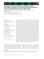
Fig. 1. Comparison of the amino acid sequences of GluSE and
GluV8. (A) The sequences of GluSE, GluV8 and proGluSE-matGluV8
(SE-V8) are illustrated schematically. Open and shaded boxes repre-
sent amino acid sequences derived from GluV8 and GluSE, respec-
tively. Closed areas at the N- and C-termini represent three and ten
amino acids, respectively, derived from the vector pQE60. pre, pre-
sequence; pro, prosequence; mature, mature sequence; repeat,
C-terminal 12-fold repeat of a tripeptide (Pro-Asp ⁄ Asn-Ala). (B)
Alignment of the amino acids of GluSE and GluV8 preprosequenc-
es. Lower case letters (ggs) represent amino acids derived from
the vector; hyphens represent deletions introduced for maximum
matching. Identical amino acids between GluSE and GluV8 are
underlined. Amino acid numbers on the top are for GluSE, and
those in the middle are for GluV8. Proteolytic sites observed in the
purified preparation and thermolysin-treated sample of proGluSE-
matGluV8 (SE-V8) are indicated by arrowheads (see Table 1).
Asterisks indicate amino acids substituted in this study.
T. K. Nemoto et al. V8 protease prosegment as a chaperone and enzyme suppressor
FEBS Journal 275 (2008) 573–587 ª 2008 The Authors Journal compilation ª 2008 FEBS 575
GluV8 4mut and proGluSE-matGluV8 were puri-
fied by large-scale preparation, yielding approxi-
mately 3–6 mgÆL
)1
of culture. From these data, it
was concluded that the full-length form of GluV8
could be recovered quantitatively by the suppression
of self-degradation, either by the use of the GluSE
prepropeptide or the GluV8 prepropeptide with four
amino acid substitutions. In subsequent experiments,
proGluSE-matGluV8 and GluV8 4mut were used as
the source of recombinant GluV8. Essentially identi-
cal results were obtained on enzyme activity with
both of these recombinant GluV8 species. However,
most data presented herein were obtained from
proGluSE-matGluV8, because this protein became
available at the early stage of our study.
Maturation processing of proGluSE-matGluV8
and GluV8 4mut
It has been reported that native GluV8 is processed to
its mature form through cleavage by a thermolysin
family metalloprotease, aureolysin [6,17]. Hence,
proGluSE-matGluV8 was incubated with serial doses
of thermolysin. As a result, the 44 kDa protein was
converted to a 42 kDa species and, finally, to 38 and
40 kDa species (Fig. 4A). The 42 kDa band appearing
at a small dose of thermolysin (lane 3) was composed
of multiple species with the N-termini of Asn43, Val46
and Ile56, and that at a large dose (lane 6) consisted
of a single species with the N-terminus of Ile56
(Table 1). The N-terminus of the 38 and 40 kDa forms
was Val69, which coincided with the N-terminus of
native GluV8 [5].
Thermolysin-processed recombinant proteins were
then subjected to zymography. The caseinolytic activity
emerged in a thermolysin dose-dependent manner
(Fig. 4B). The major band with caseinolytic activity
was at 33 kDa (Fig. 4B), indicating that the nonheated
sample of mature GluV8 migrated faster than the
heated sample on SDS-PAGE. This phenomenon is
examined further below (see Fig. 7). The proteolytic
activity towards the peptide substrate also emerged on
A B
C
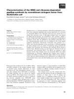
Fig. 2. SDS-PAGE of GluSE, GluV8 and their
derivatives. The lysates (lanes 1–5) and
batch-purified fractions (lanes 6–10) of
recombinant GluSE (lanes 1 and 6), GluV8
(lanes 2 and 7), proGluSE-matGluV8 (lanes 3
and 8), GluV8 2mut (lanes 4 and 9) and
GluV8 4mut (lanes 5 and 10) were prepared.
Aliquots (10 lL) were separated by PAGE
and stained with CBB (A) or immunoblotted
with anti-penta-His monoclonal IgG (B).
M, molecular mass markers. The apparent
molecular masses of major products are
shown on the left (A) and right (B). (C)
Growth curves of GluV8 (open circles),
proGluSE-matGluV8 (filled circles), GluV8 2-
mut (filled squares) and GluV8 4mut (open
squares) cultured at 30 °C in the presence
of 0.2 m
M isopropyl b-D-thiogalactopyrano-
side.
A
B
Fig. 3. Talon affinity chromatography of
recombinant proteins. (A) The bacterial
lysate (50 mL) of a 500 mL culture express-
ing the full-length form of GluSE (open cir-
cles) or GluV8 (filled circles) was separated
on a Talon affinity resin (1 · 5 cm) as
described in Experimental procedures. One
microlitre fractions were collected. (B) Aliqu-
ots (10 lL) of the eluates of GluV8 were
separated by SDS-PAGE and stained with
CBB. L, bacterial lysate expressing GluV8.
M, low-molecular-mass markers.
V8 protease prosegment as a chaperone and enzyme suppressor T. K. Nemoto et al.
576 FEBS Journal 275 (2008) 573–587 ª 2008 The Authors Journal compilation ª 2008 FEBS
thermolysin treatment (Fig. 4C). Thermolysin itself did
not possess these activities, even at the maximum dose
used (Fig. 4B,C). Therefore, it was concluded that the
40 kDa form represents the mature form. The 38 kDa
form that possessed an identical N-terminus seemed to
be processed further at the C-terminal end. It was sus-
pected that the Glu279-Asp280 bond of GluV8 was
degraded by an autoproteolytic process. Taken
together, these findings indicate that the GluV8 mature
peptide fuses to the correctly folded GluSE proseg-
ment, and thus is correctly processed to the mature
form by thermolysin in vitro.
Next, the biochemical properties and proteolytic
activities of native and recombinant mature forms of
GluV8 were compared. Native GluV8 was present as
two forms: 38 and 40 kDa (Fig. 5A). The profile of
recombinant GluV8 was essentially identical to that
of native GluV8, except for the presence of the non-
degraded 41–44 kDa bands of the recombinant form,
presumably as a result of insufficient cleavage with
thermolysin.
The N-terminal sequence of the 44 kDa GluV8 4mut
was also determined. Its N-terminus was Leu30
(Table 1), which is equivalent to the N-terminus
(Lys28) of the 44 kDa proGluSE-matGluV8. The
Ala27-Lys28 bond of proGluSE-matGluV8 and the
Ala29-Leu30 bond of GluV8 4mut appeared to match
with the recognition site of signal peptidase I [18].
However, because the borders between the pre- and
A
B
C
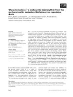
Fig. 4. In vitro processing of proGluSE-matGluV8 by thermolysin. proGluSE-matGluV8 was incubated at 0 °C (lane 1) or 37 °C (lane 2)
without protease or at 37 °C with 1 ng (lane 3), 3 ng (lane 4), 10 ng (lane 5), 30 ng (lane 6), 0.1 lg (lane 7), 0.3 lg (lane 8) or 1 lg (lane 9)
of thermolysin. As a control, thermolysin (1 lg) was incubated in the absence of GluV8 (lane Th ⁄ 35 kDa). Aliquots (0.5 lg) of thermolysin-
treated samples were separated by SDS-PAGE and stained with CBB (A) or visualized by zymography (B). M, molecular mass markers. The
apparent molecular masses of the major bands are indicated. (C) After incubation with thermolysin as described in Experimental procedures,
the proteolytic activity towards Z-Leu-Leu-Glu-MCA was measured (open circles). The activities (fluorescence units, FU) of the sample
incubated at 0 °C (open square) and thermolysin without GluV8 at 37 °C (filled circle) were also measured.
Table 1. N-terminal sequences of GluV8 derivatives. The N-termi-
nal sequences of the bands of proGluSE-matGluV8, obtained by
SDS-PAGE (Fig. 4A), and those of GluV8 4mut were determined.
Italic letters represent the amino acids derived from the preprose-
quence of GluSE.
Species Detected amino acids Determined sequence
proGluSE-matGluV8
44 kDa (Fig. 3, lane 1)
a
a KTDTESHNHS A27 ⁄ K28TDTESHNHS
b NKNVLDINSS E42 ⁄ N43KNVLDINSS
c SSLGTENKNV H36 ⁄ S37SLGTENKNV
42 kDa (lane 3)
a
a VLDINSSSHN N45 ⁄ V46 LDINSSSHN
b IKPSQNKSYP N55 ⁄ I56KPSQNKSYP
c NKNVLDINSS E42 ⁄ N43KNVLDINSS
42 kDa (lane 6) IKPSQNKSYP N55 ⁄ I56KPSQNKSYP
40 kDa VILPNNDRHQ S66 ⁄ V69ILPNNDRHQ
38 kDa VILPNNDRHQ S66 ⁄ V69ILPNNDRHQ
GluV8 4mut
44 kDa LSSKAMDNHP A29 ⁄ L30SSKAMDNHP
40 kDa VILPPNN S68 ⁄ V69ILPNN
b
a
A mixture of three fragments; their amounts were a > b >> c.
b
Ser68 was the amino acid of GluV8 4mut substituted by Asn68.
T. K. Nemoto et al. V8 protease prosegment as a chaperone and enzyme suppressor
FEBS Journal 275 (2008) 573–587 ª 2008 The Authors Journal compilation ª 2008 FEBS 577
prosequences of GluSE and GluV8 remain to be estab-
lished, it should be determined that these sites are
actually processed in GluSE and GluV8 expressed in
S. epidermidis and S. aureus, respectively.
Role of the prosequence
In order to investigate the role of the propeptide,
GluV8 was expressed with a series of truncated pro-
peptides of GluSE. Their N-termini started from Ile49,
Ile56, Asn61, Ser63, Pro65 or Ser66 (Fig. 5A). The
minimal propeptide possessed the last amino acid
(Ser66) of the GluSE propeptide. The expression levels
varied amongst the constructs, with the forms starting
from Pro65 and Ser66 being poorly recovered.
However, all were purified to near homogeneity as 40–
44 kDa bands. The proteolytic activities of the nonpro-
cessed molecules were trivial in all cases (Fig. 6D).
When the recombinant proteins were processed with
thermolysin, the 38 and 40 kDa mature forms were
produced in most cases (Fig. 6B, lanes 1–5, Th+). The
exceptions were GluSE Pro65-matGluV8 and GluSE
Ser66-matGluV8, which were thoroughly degraded by
thermolysin treatment (lanes 6 and 7, Th+). This find-
ing may cause the low expression of GluSE Pro65-mat-
GluV8 and GluSE Ser66-matGluV8. After thermolysin
processing, the truncated molecules containing the
sequences from Ile49, Ile56, Asn61 or Ser63 to the last
amino acid residue Ser66 of the GluSE prosegment
acquired protease activities comparable with that
of proGluSE-matGluV8. By contrast, GluSE Pro65-
matGluV8 showed significantly lower activity, and
GluSE Ser66-matGluV8 hardly possessed any activity
(Fig. 6C). Therefore, the C-terminal tetrapeptide of
the propeptide (Ser63-Tyr-Pro-Ser66), which was suffi-
cient for the suppression of protease activity, was also
AB
Fig. 5. Comparison of the active forms of native and recombinant
GluV8. (A) Aliquots (0.5 lg) of native GluV8 (lane 1) and recombi-
nant GluV8 treated with thermolysin (lane 2) were separated by
SDS-PAGE. M, low-molecular-mass markers. (B) The proteolytic
activities of native GluV8 (column 1) and recombinant GluV8 (col-
umn 2) were measured with 10 l
M Z-Leu-Leu-Glu-MCA. Values are
the means ± standard deviation (n = 3). Samples for columns 1
and 2 are identical to those for lanes 1 and 2, respectively, in (A).
AB
DC
Fig. 6. Minimal region of the prosequence responsible for chaperoning and enzyme inhibition. (A) N-terminal sequences of proGluSE-mat-
GluV8 and its N-terminally truncated forms. proGluSE-matGluV8 was expressed as the full-length form, but its N-terminus was processed up
to K
28
. (B) Recombinant proteins shown in (A) were incubated without protease at 0 °C (–) or with thermolysin (1 lg) at 37 °C (+) as
described in Experimental procedures. Thereafter, aliquots (0.5 lg) were separated by SDS-PAGE. (C) The Glu-specific protease activity of
aliquots (0.25 lg) pretreated with thermolysin. Values are the means ± standard deviation (n = 4). (D) The Glu-specific protease activity of
aliquots (1 lg) incubated without thermolysin. Values are the means ± standard deviation (n = 4). Numbers 1–7 are identical in (A)–(D).
V8 protease prosegment as a chaperone and enzyme suppressor T. K. Nemoto et al.
578 FEBS Journal 275 (2008) 573–587 ª 2008 The Authors Journal compilation ª 2008 FEBS
adequate for the intramolecular chaperone function.
GluSE Ser66-matGluV8 was also expressed with the
long N-terminal tag (Met-Arg-Gly-Ser-His
6
-Gly)
encoded by the pQE9 expression vector. The recombi-
nant protein possessed trace proteolytic activity both
before and after thermolysin treatment (data not
shown). Thus, the length of the propeptide was not
critical, but the sequence itself was important for
folding and suppression of the activity of the mature
portion.
When analysed carefully, the proteolytic activities of
the nonprocessed forms were not entirely zero. In par-
ticular, the activities of GluV8 with shorter propep-
tides, i.e. Asn61-Ser66 and Ser63-Ser66, could not be
ignored (Fig. 6C, columns 4 and 5). Concerning this
result, it should be noted that the recombinant GluSE
Asn61-matGluV8 and GluSE Ser63-matGluV8 were
expressed in consideration of the autoproteolytic sites
of the GluV8 propeptide, i.e. Glu62-Gln63 and Glu65-
His66 bonds, respectively (Fig. 1B). Accordingly,
GluV8 autoprocessed at these sites may possess weak
proteolytic activity, as postulated in the experiment of
Fig. 2.
Mutation of the essential amino acid Ser237
Establishment of the E. coli expression system of
GluV8 enabled the roles of certain amino acids com-
prising the protease to be investigated by in vitro muta-
genesis. As an initial approach, two key amino acids
were chosen: Ser237 and Val69. GluV8 is a serine pro-
tease, the active site of which consists of the His119,
Asp161 and Ser237 triad [19]. To confirm the role of
Ser237, its substitution by Ala was introduced to
proGluSE-matGluV8 (designated GluV8 Ser237Ala).
As a result, GluV8 Ser237Ala showed no caseinolytic
or Glu-specific activity (Fig. 7B,C).
As described in Fig. 4, the mobility of mature
GluV8 on SDS-PAGE was altered by heating of the
samples in SDS sample buffer. Unprocessed GluV8
Ser237Ala, as well as the wild-type, migrated to the
44 kDa position (Fig. 7A). After thermolysin treat-
ment, the mobility of the wild-type was shifted to 33
and 38 ⁄ 40 kDa under nonheated and heated condi-
tions in the presence of SDS, respectively (Fig. 7A).
The profile of GluV8 Ser237Ala was similar to that of
the wild-type, although 35 kDa (lane 7) and 41 kDa
A
B
C
Fig. 7. Effect of the amino acid substitution at Ser237 on the proteolytic activity. proGluSE-matGluV8 (wt), or its mutant (Ser237Ala), was
incubated at 0 °C without protease (–) or at 37 °C with 0.3 lg of thermolysin (+). Thereafter, aliquots (1 lg) were separated by SDS-PAGE
and stained with CBB (A) or subjected to zymography (B). Samples were mixed with a half volume of SDS sample buffer and subjected to
SDS-PAGE without heat (heat –) or after heat denaturation (heat +). M, low-molecular-mass markers. The apparent molecular masses of
major bands are indicated on the left. (C) Aliquots of the thermolysin-treated samples were subjected to the protease assay using Z-Leu-
Leu-Glu-MCA. Values are the means ± standard deviation (n = 3).
T. K. Nemoto et al. V8 protease prosegment as a chaperone and enzyme suppressor
FEBS Journal 275 (2008) 573–587 ª 2008 The Authors Journal compilation ª 2008 FEBS 579
(lane 8) intermediate forms were also observed. The
faster migration of processed and nonheated GluV8
strongly suggests its more compact conformation.
However, this conformation was not a prerequisite for
renaturation of the protein, because GluV8 exposed to
heat could renature under the conditions of zymo-
graphy (Fig. 7B, lane 4). This finding indicates that,
although the zymography experiment used nonheated
samples, the mature form of GluV8 could be renatured
even after exposure to heat in the presence of SDS.
Role of N-terminal Val69 in processing of the
GluV8 proform
Finally, the role of N-terminal Val69 of mature GluV8
was investigated. It has been proposed that the a-amino
group of N-terminal Val69 of mature GluV8 interacts
with the c-carboxyl group of Glu of a substrate peptide
[19]. If so, as any N-terminal residue, except the imino
acid Pro, possesses an a-amino group, it can be specu-
lated that Val69 is simply required for processing with
thermolysin, which hydrolyses the amino-side peptide
bond of hydrophobic amino acids. To test this, Val69
of proGluSE-matGluV8 was substituted by Phe. In
addition, Val69 was replaced by Ala and Gly, as therm-
olysin cleavage of peptide bonds with these amino acid
residues has been reported [20]. The 44 kDa mutant
forms, as well as the wild-type, were processed to
42 kDa intermediate forms, and further to 40 kDa,
indicating that the mutation does not modify the steric
structure of GluV8. However, these molecules showed
no proteolytic activity (Fig. 8). Strikingly, it was found
that the N-termini of the processed forms were not the
69th substituted amino acids, but entirely Ile70. These
results show that thermolysin attacks the Xaa69-Ile70
bond of the mutant rather than the Ser-Xaa69
(Xaa ” Phe, Gly or Ala) bond. As a consequence, it
was found unexpectedly that Val69 was indispensable
for correct processing by thermolysin at the Ser-Val69
bond, and that GluV8 with N-terminal Ile70 had essen-
tially no proteolytic activity.
Role of N-terminal Val69 in the proteolytic
activity
As Val69 was indispensable for precise processing at
the Ser66-Val69 bond, it was impossible to investigate
the role of Val69 in the enzymatic reaction. To over-
come this difficulty, mutant proGluSE-matGluV8 was
prepared, with Ser66 replaced by Arg (designated
proGluSE Arg66-matGluV8), because the peptide
bond between Arg66 and Val69 can be degraded by
A
B
Fig. 8. Effect of amino acid substitutions at Val69 on thermolysin processing. proGluSE-matGluV8 or its mutants at Val69 were incubated at
0 °C without protease (lane 1) or at 37 °C with 0.03 lg (lane 2), 0.1 lg (lane 3), 0.3 lg (lane 4), 1 lg (lane 5) or 3 lg (lane 6) of thermolysin.
Thereafter, aliquots (0.5 lg) were separated by SDS-PAGE (A) or subjected to the protease assay with Z-Leu-Leu-Glu-MCA (B). M, low-
molecular-mass markers. The apparent molecular masses of major bands and 35 kDa thermolysin are indicated. Symbol designations in (B):
Val69 (open circles), Val69Phe (filled circles), Val69Ala (open squares) and Val69Gly (open triangles; identical to Val69Phe).
V8 protease prosegment as a chaperone and enzyme suppressor T. K. Nemoto et al.
580 FEBS Journal 275 (2008) 573–587 ª 2008 The Authors Journal compilation ª 2008 FEBS
trypsin. Indeed, trypsin processing of proGluSE
Arg66-matGluV8 faithfully mimicked the thermolysin
processing of proGluSE-matGluV8 (Fig. 9A, compare
lanes 2 and 6). Concomitantly, its Glu-specific proteo-
lytic activity was enhanced (Fig. 9B). Although thermo-
lysin treatment of proGluSE Arg66-matGluV8 also
increased the activity (Fig. 8B, column 3), the effi-
ciency was less than that of trypsin treatment (col-
umn 4), reflecting the predominance of the
nondegraded 42 kDa intermediate (Fig. 9A, lane 5).
This should be the result of the substitution of the
P1¢ site Ser66 by nonfavourable Arg. Hence, it is possi-
ble to utilize trypsin as the processing enzyme.
Trypsin cleavage of proGluSE Arg66-matGluV8,
with Val69 substituted by Ala, Phe, Gly or Ser, gener-
ated the 40 kDa form with the designed N-termini
(data not shown). Their Glu-specific proteolytic activi-
ties were 4.5% (Ala), 1.4% (Phe), 1.1% (Gly) and
0.6% (Ser) of that of Val69 (Fig. 9B). Therefore, it
was concluded that Val69 plays an important role in
the enzyme reaction itself, although other amino acids,
such as Ala, may partially substitute for Val69.
Discussion
In this study, for the first time, GluV8 has been suc-
cessfully expressed as a soluble proform in E. coli. Pos-
sible reasons for the poor expression of GluV8 in
E. coli previously have been found. The propeptide of
GluV8 possesses Glu at positions 62 and 65; their
C-terminal ends undergo autoproteolysis and the
resultant GluV8 with truncated propeptides (Gln63-
Asn68 and His66-Asn68) is partially active. This may
induce the cascade reaction of GluV8 activation,
because recombinant proteins remain inside E. coli
cells, instead of being secreted from S. aureus. The
conversion of amino acids adjacent to the processing
site from Ala67-Asn68 to Pro-Ser further suppresses
the degradation. It is currently speculated that an
endogenous protease in E. coli cleaves the Ala67-
Asn68 or Asn68-Val69 bond of GluV8. The substitu-
tion of Asn67 by Pro can prevent this proteolysis,
because Pro-Xaa and Xaa-Pro bonds (Xaa ” any
amino acid) are highly resistant to most proteases.
A chimeric protease has been expressed previously
on a pro-aminopeptidase-processing protease, i.e. a
thermolysin-like metalloprotease produced by Aeromo-
nas caviae T64 [21]. The propeptide of the protease
could be replaced by that of vibriomysin, a homologue
of the protease, which shared 36% amino acid identity.
In the present study, it was demonstrated that the pro-
peptide of GluV8 could be replaced by that of GluSE,
although the similarity (15.4%) of their prosequences
was much lower than the case of the thermolysin-like
protease. Therefore, it can be proposed that the amino
acid requirement of prosequences for assistance in pro-
tein folding and inhibition of catalytic activity is lower
than the requirement for the proteolytic entity. This is
further indicated by the finding that the last four
residues of the propeptide of GluSE, which are com-
pletely different from those of GluV8, are sufficient for
the role of the propeptide of GluV8 (Fig. 1B).
A B
Fig. 9. Involvement of Val69 in protease activity. (A) Ser66 of proGluSE-matGluV8 was substituted by Arg (GluSE Arg66-GluV8). proGluSE-
matGluV8 (wt) and proGluSE Arg66-matGluV8 (Ser66Arg) were incubated at 0 °C without protease (lanes 1 and 4), at 37 °C with 0.3 lgof
thermolysin (lanes 2 and 5) or at 37 °C with 0.3 lg of trypsin (lanes 3 and 6), as described in Experimental procedures. As controls, 0.3 lg
of thermolysin (lane 7 ⁄ Th) and trypsin (lane 8 ⁄ Tr) were incubated without recombinant protein. Thereafter, aliquots (0.75 lg) were separated
by SDS-PAGE. M, low-molecular-mass markers. The apparent molecular masses of the major bands are indicated on the left. (B) Val69 of
proGluSE Arg66-matGluV8 was mutated, and the Glu-specific protease activity of the mutated forms was measured using aliquots of the
samples after incubation with thermolysin or trypsin. wt, proGluSE-matGluV8 (columns 1 and 2). Val69Xaa: amino acid at position 69 of
GluSE Arg66-GluV8 was substituted by Val (columns 3 and 4), Ala (columns 5 and 6), Phe (columns 7 and 8), Gly (columns 9 and 10) or Ser
(columns 11 and 12). Values are the means ± standard deviation (n = 3).
T. K. Nemoto et al. V8 protease prosegment as a chaperone and enzyme suppressor
FEBS Journal 275 (2008) 573–587 ª 2008 The Authors Journal compilation ª 2008 FEBS 581
Amongst the glutamyl endopeptidase family mem-
bers, GluV8 and GluSE are processed by a thermoly-
sin family metalloprotease, aureolysin [6,17,22]. By
contrast, the N-terminus of the Glu-specific endopepti-
dase from Bacillus licheniformis is Ser, indicating the
processing of the Lys-Ser bond by a protease with
trypsin-like specificity [9]. This may not be surprising,
because the processing enzyme can be changed from
thermolysin to trypsin by substitution of Ser66 of
proGluSE-matGluV8 by Arg66 (Fig. 9). This result
indicates that any proteolytic enzyme can activate the
glutamyl endopeptidase if it can properly cleave the
processing site.
GluV8 is a serine protease, the His119, Asp161 and
Ser237 residues of which form an active triad. Indeed,
Ser237 is essential for the protease reaction. Because
GluV8 Ser237Ala is normally processed by thermoly-
sin, its overall structure does not appear to be altered
from the active form. Therefore, to elucidate the mech-
anism of suppression of the protease activity and the
alteration in the proteolytic activity between the two
proteases, crystallographic analyses are now under way
in our laboratory using GluV8 Ser237Ala and GluSE
Ser235Ala.
The prosegment of bacterial proteases, such as
thermolysin [12,13] and subtilisin [23], is indispensable
for the suppression of protease activity and for correct
folding of the protease. An inhibitory role of the pro-
peptide has also been postulated for GluV8, because
the GluV8 precursor is specifically activated by the
metalloprotease, aureolysin [6]. However, direct evi-
dence has not been presented to date. The present
study has confirmed this role. By contrast, the intra-
molecular chaperone activity of the GluV8 propeptide
has not been investigated in detail previously, primar-
ily because of a lack of an appropriate expression sys-
tem for GluV8. A previous study has indicated that
the prosequence of GluV8 is dispensable for folding,
as the active enzyme is recovered after denaturation–
renaturation of a mature polypeptide [8]. However, in
the present study, the intramolecular chaperone activ-
ity of the GluSE propeptide towards the mature por-
tion of GluV8 was clearly demonstrated. Moreover, it
was demonstrated that only four residues of the pro-
peptide (Ser63-Tyr-Pro-Ser66) are sufficient for chaper-
one function. It was impossible to segregate the
regions responsible for the dual roles completely, indi-
cating that the two functions may be tightly connected
with each other. With regard to the two roles of the
propeptide, the inhibitory effect on protease activity
may be explained by the propeptide amino acids
attached to N-terminal Val69, because of the essential
role of the a-amino group of the N-terminal amino
acid [19]. However, it remains unknown how the pro-
sequence, especially the tetrapeptide (Ser63-Tyr-Pro-
Ser66) of the GluSE propeptide, supports the folding
of the mature portion of GluV8. It is supposed that
the tetrapeptide may form a scaffold for the folding of
the mature sequence. For example, it has been
reported that the intrinsically unstructured propeptide
of subtilisin adopts an arranged structure only in the
presence of the mature form of the protease [23].
Whether or not a similar mechanism is responsible for
the folding of the glutamyl endopeptidase family
should be investigated.
Our result on zymography reproduced the renatur-
ation of the mature polypeptide reported by Yabuta
et al. [8]. However, this finding does not exclude the
need for the intramolecular chaperone activity of the
propeptide. Similar results were observed on proteins
folded by general molecular chaperones. Thus, even if
a protein can fold spontaneously under in vitro condi-
tions, it may be unable to fold under in vivo conditions
without molecular chaperones. In particular, the fold-
ing of nascent polypeptides is substantially distinct
from the renaturation process of a polypeptide in vitro.
Like the general molecular chaperone Hsp70, which
immediately binds to nascent polypeptides [24], the
GluV8 propeptide may associate with subsequently
synthesized nascent polypeptide, and suppress the mis-
folding of the mature portion. By contrast, the entire
mature portion of GluV8 may be ready to fold sponta-
neously under in vitro denaturation and renaturation
conditions.
Mature GluV8 polypeptide was more resistant than
the nonprocessed form to denaturation in the presence
of SDS. The faster electrophoretic mobility of mature
GluV8 indicates a more compact structure. This
strongly suggests that the conformation of nonpro-
cessed GluV8 is distinct from the simple summation of
the pro- and mature polypeptides. Hence, the propep-
tide seems to prevent the mature polypeptide from
converting to a more compact structure. Noncovalent
association of an intramolecular chaperone propeptide
with the mature portion has been reported for subtili-
sin [23] and furin [25].
Prasad et al. [19] have proposed that the positively
charged a-amino group of the N-terminus is involved
in the substrate recognition of GluV8. In the same
context, Popowicz et al. [26] have reported that a
recombinant form of SplB, a GluV8 family member,
possesses proteolytic activity, whereas that carrying
an additional Gly-Ser dipeptide is devoid of activity;
no data were presented to substantiate this conclu-
sion. The present study clearly demonstrated the
inhibitory effect of the prosegment on the proteolytic
V8 protease prosegment as a chaperone and enzyme suppressor T. K. Nemoto et al.
582 FEBS Journal 275 (2008) 573–587 ª 2008 The Authors Journal compilation ª 2008 FEBS
activity. The proteolytic activities of GluV8 with
truncated GluSE propeptides, i.e. Ser63-Ser66 and
Asn61-Ser66, were not completely zero. By contrast,
the proteolytic activities of GluV8 with longer GluSE
propeptides (Ile56-Ser66, Ile49-Ser66 and Ser33-Ser66)
were more rigorously inhibited. The a-amino group of
the N-terminus of shorter propeptides may function
as a weak acceptor of the negative charge of a sub-
strate peptide.
In the present study, the role of Val69 was investi-
gated. Val69 was essential for precise processing at the
peptide bond between Ser66 of the GluSE propeptide
and Val69 of the GluV8 mature sequence for protease
maturation. When N-terminal Val69 was substituted
by Ala, Phe, Gly or Ser in GluV8 (with Arg66 substi-
tuted by Ser66), low but substantial protease activities
were found, i.e. 0.6–4.5% of the wild-type. Therefore,
the enzyme activity varied according to the N-terminal
amino acid, and was much lower than that with Val69.
Furthermore, it was demonstrated that GluV8 starting
from Ile70 was inactive. These findings indicate that
Val69 is more than just a supplier of an a-amino
group for substrate recognition, and is important, if
not essential, for the proteolytic reaction.
N-terminal Val is conserved amongst GluV8, GluSE
and Glu-specific proteases from Streptomyces griseus
[10] and Streptomyces fradiae [27]. By contrast, the
N-terminus of the six serine proteases Spl from S. aur-
eus is Glu [28]. Although a glutamyl endopeptidase
from Bacillus licheniformis possesses the sequence
Lys94-Ser-Val-Ile-Gly98 around the processing site, a
sequence similar to that of GluSE (Pro65-Ser-Val-Ile-
Leu71), the N-terminus of the mature form is reported
to be Ser95 rather than Val96, presumably being
dependent on the processing enzyme [9]. Moreover,
Kawalec et al. [29] reported that the processed glutam-
yl endopeptidase of Enterococcus faecalis with an addi-
tional Ser-1 possesses a much higher proteolytic
activity than that starting from Leu1. Therefore, the
requirement of Val at the N-terminus might be depen-
dent on the conformation of each protease. It would
be interesting to test whether or not the substitution of
the N-terminal amino acids of non-Val-type Glu-spe-
cific proteases by Val enhanced the proteolytic activity.
Glutamyl endopeptidases from Streptomyces fradiae
[27] and Streptomyces griseus [10] are assumed to be
activated through autoproteolysis at Glu-Xaa bonds.
Similarly, bacterial proteases, e.g. Arg- and Lys-spe-
cific proteases, from Porphyromonas gingivalis [30–32]
appear to be autoprocessed by the cleavage at Arg-
Xaa or Lys-Xaa bonds. Therefore, as shown in the
present study, the modification of the processing sites
by in vitro mutagenesis may be useful for the suppres-
sion of the autoproteolytic cascade of these proteases
for their expression in E. coli.
Experimental procedures
Materials
The materials used and their sources were as follows:
expression vector pQE60 and plasmid pREP4 from Qiagen
Inc. (Chatsworth, CA, USA); low-molecular-mass markers
from GE Healthcare (Milwaukee, WI, USA); kaleidoscope
prestained molecular standard from Bio-Rad (Richmond,
CA, USA); restriction enzymes and DNA-modifying
enzymes from Nippon Gene (Tokyo, Japan); KOD plus
DNA polymerase from Toyobo (Tokyo, Japan); fluorescent
peptide, Z-Leu-Leu-Glu-MCA, from Peptide Institute
(Osaka, Japan); trypsin and azocasein from Sigma-Aldrich
(St Louis, MO, USA); protease V8 ⁄ GluV8 from S. aureus V8
strain (Roche Diagnostics, Mannheim, Germany); Talon
metal affinity resin from Clontech Laboratories Inc. (Palo
Alto, CA, USA); anti-penta-His monoclonal antibody from
Qiagen Inc.; and alkaline phosphatase-conjugated rabbit
anti-mouse Ig(G + A + M) from Zymed Laboratories
Inc. (San Francisco, CA, USA). Oligonucleotide primers
were purchased from Genenet (Fukuoka, Japan).
Bacterial expression vector for GluSE
GluSE was expressed in E. coli with a histidine hexamer
tag at the C-terminus using the pQE60 expression vector
(Qiagen Inc.). The DNA fragment carrying the full-length
GluSE (Met1-Gln282) was amplified with a pair of prim-
ers: 5¢-TATGGATCCAAAAAGAGATTTTTATCTATATG
TAC-3¢ and 5¢-ATTGGATCCCTGAATATTTATATCAG
GTATATTG-3¢. BamHI sites introduced in the primers are
indicated in italic. Genomic DNA of S. epidermidis
(ATCC 14990) was used as a template. PCR was performed
for 30 cycles using the KOD plus system, which did not tag
any nucleotide at the 3¢-OH end of the PCR fragments. A
PCR product was then cut with BamHI and inserted into a
BamHI site of pQE60. Y1090[pREP4] cells were trans-
formed with the plasmid (designated as pQE60-GluSE),
and the transformants were selected on LB broth agar
plates containing 50 lgÆmL
)1
of ampicillin and 25 lgÆmL
)1
of kanamycin.
Expression vectors for the full-length form and
chimeric form of GluV8
The DNA fragment encoding the full-length form of GluV8
(Met1-Ala336) was amplified with a pair of primers (5¢-
ATGGGATCCAAAGGTAAATTTTTAAAAGTTAGTT
CT-3¢ and 5¢-ATTGGATCCCTGAATATTTATATCAGG
TATATTG-3¢) and then processed as described above.
T. K. Nemoto et al. V8 protease prosegment as a chaperone and enzyme suppressor
FEBS Journal 275 (2008) 573–587 ª 2008 The Authors Journal compilation ª 2008 FEBS 583
BamHI sites introduced in the primers are indicated in
italic. Genomic DNA of S. aureus V8 strain was used
as a template. The resulting plasmid was designated as
pQE60-GluV8.
A DNA fragment encoding a chimeric protein, i.e. the
prepropeptide of GluSE (Met1-Ser66) and the mature
sequence of GluV8 (Val69-Ala336), was amplified with a
pair of primers: 5¢-GTTATATTACCAAATAACGATCGT
CACC-3¢ and 5¢-ACTTGGGTAACTTTTATTTTGACTTG
GT-3¢. The former targeted the mature sequence of GluV8
(Val69-Ala336) and the latter the prepropeptide of GluSE
(Met1-Ser66). A mixture of pQE60-GluSE and pQE60-
GluV8 (45 ng each) was used as template. During the PCR
cycle, a 5 kb PCR fragment encoding the vector and the
GluSE Met1-Ser66 ⁄ GluV8 Val69-Ala336 chimeric protein
became predominant. After DpnI digestion of the templates,
the 5¢-end of the fragment was phosphorylated by T4 poly-
nucleotide kinase and self-ligated by T4 DNA ligase simulta-
neously. Y1090[pREP4] cells were transformed with the
resulting plasmid (designated pQE60-proGluSE-matGluV8).
Production of the chimeric plasmid was confirmed by DNA
sequencing.
Expression vectors for truncated forms of GluV8
Expression plasmids encoding the mature protein of GluV8
(Val69-Ala336) fused to truncated propeptides of GluSE at
the N-terminus, i.e. Ile49-Ser66, Ile56-Ser66, Asn61-Ser66,
Ser63-Ser66, Pro65-Ser66 and Ser66, were amplified by
PCR with appropriate primers carrying BamHI sites using
pQE60-proGluSE-matGluV8 as template (Fig. 6). The
amplified fragments were inserted into a BamHI site of
pQE60 as described above.
In vitro mutagenesis by PCR
In vitro mutagenesis was performed by the PCR technique,
as described above, using the following mutated primers
with the altered nucleotides indicated in italic. (a) Nucleo-
tides (GAA) encoding Glu at positions 62 and 65 of
pQE60-GluV8 were substituted with nucleotides encoding
Gln and Ser, respectively. The plasmid pQE60-GluV8 was
used as a template. A sense primer (5¢-CGTAGTCAC
GCAAATGTTATATTCCCAAATAACG-3¢) and an anti-
sense primer (5¢-TTGTTGTAATGGTTTGTTACCGCC
TTTTT-3¢) were used as PCR primers. The resulting plas-
mid was designated pQE60-GluV8 2mut. (b) Nucleotides
(GCAAAT) encoding Ala67-Asn68 of GluV8 were further
substituted with those (CCAAGT) encoding Pro65-Ser66 of
GluSE at equivalent positions. The plasmid pQE60-
GluV8 2mut was used as a template. A sense primer
(5¢-CGTAGTCACGCAAATGTTATATTCCCAAATAA
CG-3¢) and an antisense primer (5¢-ACTTGGGTGACTA
CGTTGTTGTAATGGTTT-3¢) were used as PCR primers.
The resulting plasmid was designated pQE60-GluV8 4mut.
(c) Nucleotides (TCA) encoding Ser237 of pQE60-GluV8
4mut were substituted with those encoding Ala with a sense
primer (5¢-GGTTCACCTGTATTTAATGAAAAAA-3¢)
and an antisense primer (5¢-TGCATTACCACCAG
TTGTACTTAAATC-3¢). (d) Nucleotides (AGT) encoding
Ser66 of pQE60-proGluSE-matGluV8 were substituted
with CGT encoding Arg. A sense primer (5¢-GTT
ATATTACCAAATAACGATCGTCACC-3¢) and an anti-
sense primer (5¢-ACGTGGGTAACTTTTATTTTGAC
TTGGTTTG-3¢) were used as PCR primers. The resulting
plasmid was designated pQE60-proGluSE Arg
66
-matGluV8.
(e) Nucleotides (GTT) encoding Val69 of pQE60-pro-
GluSE-matGluV8 and pQE60-GluSE Arg
66
-matGluV8 were
substituted with those encoding Phe (TTT), Ala (GCG),
Gly (GGT) or Ser (AGC) with appropriate primers.
Expression and purification of recombinant
proteins
His
6
-tagged recombinant proteins were expressed and puri-
fied as described previously [33]. Briefly, Y1090[pREP4]
carrying pQE9- or pQE60-derived expression plasmids was
cultured in LB broth containing 50 lgÆmL
)1
of ampicillin
and 25 lgÆmL
)1
of kanamycin at 37 °C overnight. Protein
expression was induced by dilution of the culture with
two volumes of LB broth containing 0.2 mm isopropyl b-
d-thiogalactopyranoside and incubation at 30 °C for 3 h.
Bacterial cells were harvested by centrifugation and lysed
with lysis ⁄ washing buffer (20 mm Tris ⁄ HCl, pH 8.0, 0.1 m
NaCl containing 10 mm imidazole) to which 0.5 mgÆmL
)1
of lysozyme and 10 lgÆmL
)1
of leupeptin had been added.
Recombinant proteins were recovered in the cell lysate
fraction and purified by affinity chromatography with
Talon metal affinity resin (Clontech Laboratories Inc.)
according to the manufacturer’s protocol, except that 10 mm
imidazole was included in the lysis ⁄ washing buffer. After
extensive washing, the bound proteins were eluted with 0.1 m
imidazole (pH 8.0) containing 10% (v ⁄ v) glycerol. The
purified proteins were stored at )80 °C until use.
In vitro processing and measurement of the
protease activity
Unless otherwise stated, the in vitro processing of recombi-
nant proteins and subsequent protease assay were per-
formed as follows. Recombinant proteins (10 lg ⁄ 0.1 mL)
were incubated in 10 mm sodium borate, pH 8.0, 0.005%
(v ⁄ v) Triton X 100 containing 2 mm CaCl
2
with thermoly-
sin (0.3 or 1 lg) at 37 °C for 4 h. Thereafter, aliquots were
incubated with 10 mm Z-Leu-Leu-Glu-MCA in 0.2 mL of
50 mm Tris ⁄ HCl (pH 8.0) and 5 mm EDTA at 25 °C for
2 h. Fifty-seven picomoles of proteins (0.18 lg for 32 kDa
GluSE proform and 0.25 lg for 44 kDa GluV8 proform)
were used for each protease assay unless otherwise stated.
V8 protease prosegment as a chaperone and enzyme suppressor T. K. Nemoto et al.
584 FEBS Journal 275 (2008) 573–587 ª 2008 The Authors Journal compilation ª 2008 FEBS
EDTA was added to the reaction mixture to inactivate
thermolysin [34]. The fluorescence was measured (excita-
tion, 380 nm; emission, 460 nm) with a fluorescence pho-
tometer F-4000 (Hitachi, Tokyo, Japan). The activity was
presented as fluorescence units (FU).
SDS-PAGE and zymography
Samples (0.5 or 1 lg) were separated by electrophoresis in
the presence of 0.1% SDS at a polyacrylamide concentra-
tion of 12.5% (w ⁄ v), and then stained with CBB. For
zymography, SDS-PAGE was performed using 12% poly-
acrylamide gels containing 1 mgÆmL
)1
of azocasein [35].
Samples (1 lg) were loaded onto the gel without heat treat-
ment unless otherwise stated. After SDS-PAGE, the gel
was incubated twice at 25 °C with 100 mL of 2.5% (w ⁄ v)
Triton X 100 for 20 min each time, and then twice at the
same temperature with 100 mL of 50 mm Tris ⁄ HCl,
pH 7.8, containing 30 m m NaCl, for 10 min each time.
Thereafter, the gel was incubated in 100 mL of the latter
buffer at 37 °C overnight. Finally, nonhydrolysed azocasein
in the polyacrylamide gel was stained with CBB.
Immunoblotting
Bacterial lysates containing recombinant proteins were pre-
pared as reported previously [36]. The purified fraction used
for immunoblotting was obtained by batch purification of
1 mL of bacterial lysate with 30 mL of a suspension
(resin ⁄ buffer, 1 : 1) of Talon affinity resin pre-equilibrated
with lysis buffer, followed by five washings with 1 mL of
washing buffer. The bound proteins were then extracted
and denatured with 30 mL of 3 · SDS sample buffer. The
bacterial lysate or an affinity-purified fraction (5 mL) was
loaded onto a polyacrylamide gel. Following electrophoresis
and the transfer of proteins to a polyvinylidene difluoride
membrane (Immobilon-P; Millipore, Bedford, MA, USA),
the membrane was incubated with 0.2 lgÆmL
)1
of anti-
penta-His monoclonal IgG (Qiagen Inc.), and then with
rabbit anti-mouse Ig(G + A + M)–alkaline phosphatase
conjugate at 0.1 lgÆmL
)1
(Zymed Laboratories Inc.). Blots
on the membrane were visualized by immersion in a mix-
ture of 5-bromo-4-chloro-3-indolyl phosphate and nitroblue
tetrazolium (Nakarai, Kyoto, Japan).
N-terminal amino acid sequencing
N-terminal amino acid sequences were determined after
separation of the recombinant proteins by SDS-PAGE and
transfer to a polyvinylidene difluoride membrane (Sequi-
Blot PVDF Membrane; Bio-Rad). After staining with CBB,
the bands were excised and sequenced directly with a model
Precise 49XcLC protein sequencer (ABI, Foster City, CA,
USA).
Protein concentration
Protein concentrations were determined by the CBB dye
method (Bio-Rad).
Acknowledgements
This work was supported by a Grant-in-Aid for Scien-
tific Research from the Ministry of Education, Culture,
Sports, Science, and Technology of Japan (to TKN).
References
1 Drapeau GR, Boily Y & Houmard J (1972) Role
of a metalloprotease in activation of the precursor
of staphylococcal protease. J Biol Chem 247, 6720–
6726.
2 Coulter SN, Schwan WR, Ng EY, Langhorne MH,
Ritchie HD, Westbrock-Wadman S, Hufnagle WO, Fol-
ger KR, Bayer AS & Stover CK (1998) Staphylococcus
aureus genetic loci impacting growth and survival in
multiple infection environments. Mol Microbiol 30, 393–
404.
3 Karlsson A, Saravia-Otten P, Tegmark K, Morfeldt E
& Arvidson S (2001) Decreased amounts of cell wall-
associated protein A and fibronectin-binding proteins in
Staphylococcus aureus sarA mutants due to up-regula-
tion of extracellular protease. Infect Immun 69, 4742–
4748.
4 Stennicke HR & Breddam K (1998) Glutamyl endopep-
tidase I. In: Handbook of Proteolytic Enzymes (Barrett
AJ, Rawlings ND & Woessner FF Jr, eds), pp. 243–
246. Academic Press, San Diego, CA.
5 Carmona C & Gray GL (1987) Nucleotide sequence of
the serine protease gene of Staphylococcus aureus, strain
V8. Nucleic Acids Res 15, 6757.
6 Drapeau GR (1978) Role of a metalloprotease in acti-
vation of the precursor of staphylococcal protease.
J Bacteriol 136, 607–613.
7 Shaw LN, Golonka E, Szmyd G, Foster SJ, Travis J &
Potempa J (2005) Cytoplasmic control of premature
activation of a secreted protease zymogen: deletion of
staphostatin B (SspC) in Staphylococcus aureus 8325-4
yields a profound pleiotropic phenotype. J Bacteriol
187, 1751–1762.
8 Yabuta M, Ochi N & Ohsuye K (1995) Hyperproduc-
tion of a recombinant fusion protein of Staphylococcus
aureus V8 protease in Escherichia coli and its processing
by OmpT protease to release an active V8 protease
derivative. Appl Microbiol Biotechnol 44, 118–125.
9 Kakudo S, Kikuchi N, Kitadokoro K, Fujiwara T,
Nakamura E, Okamoto H, Shin M, Tamaki M,
Teraoka H, Tsuzuki H et al. (1992) Purification,
characterization, cloning, and expression of a glutamic
T. K. Nemoto et al. V8 protease prosegment as a chaperone and enzyme suppressor
FEBS Journal 275 (2008) 573–587 ª 2008 The Authors Journal compilation ª 2008 FEBS 585
acid-specific protease from Bacillus licheniformis ATCC
14580. J Biol Chem 267, 23782–23788.
10 Suzuki Y, Yabuta M & Ohsuye K (1994) Cloning and
expression of the gene encoding the glutamic acid-spe-
cific protease from Streptomyces griseus ATCC10137.
Gene 150, 149–151.
11 Winther JR & Sørensen P (1991) Propeptide of car-
boxypeptidase Y provides a chaperone-like function as
well as inhibition of the enzymatic activity. Proc Natl
Acad Sci USA 88, 9330–9334.
12 O’Donohue MJ & Beaumont A (1996) The roles of the
prosequence of thermolysin in enzyme inhibition and
folding in vitro. J Biol Chem 271, 26477–26481.
13 Marie-Claire C, Ruffet E, Beaumont A & Roques BP
(1999) The prosequence of thermolysin acts as an intra-
molecular chaperone when expressed in trans with the
mature sequence in Escherichia coli. J Mol Biol 285 ,
1911–1915.
14 Sasaki M, Ohara-Nemoto Y, Tajika S, Kobayashi M,
Ikeda Y, Kaneko M & Takagi T (1998) Purification
and characterization of a glutamic acid-specific protease
from Staphylococcus aureus. Jpn J Oral Biol 40, 542–
548.
15 Ohara-Nemoto Y, Ikeda Y, Kobayashi M, Sasaki M,
Tajika S & Kimura S (2002) Characterization and
molecular cloning of a glutamyl endopeptidase from
Staphylococcus epidermidis. Microb Pathog 33, 33–41.
16 Dubin G, Chmiel D, Mak P, Rakwalska M, Rzychon
M & Dubin A (2001) Molecular cloning and biochemi-
cal characterization of proteases from Staphylococcus
epidermidis. Biol Chem 382, 1575–1582.
17 Shaw L, Golonka E, Potempa J & Foster SJ (2004) The
role and regulation of the extracellular proteases of
Staphylococcus aureus. Microbiology 150 (Pt 1), 217–
228.
18 Chang CN, Blobel G & Model P (1978) Detection of
prokaryotic signal peptidase in an Escherichia coli
membrane fraction: endoproteolytic cleavage of nas-
cent f1 pre-coat protein. Proc Natl Acad Sci USA 75,
361–365.
19 Prasad L, Leduc Y, Hayakawa K & Delbaere LT
(2004) The structure of a universally employed enzyme:
V8 protease from Staphylococcus aureus. Acta Crystal-
logr 60, 256–259.
20 Heinrikson R (1977) Applications of thermolysin in pro-
tein structural analysis. Methods Enzymol 47, 175–189.
21 Tang B, Nirasawa S, Kitaoka M, Marie-Claire C &
Hayashia K (2001) General function of N-terminal pro-
peptide on assisting protein folding and inhibiting cata-
lytic activity based on observations with a chimeric
thermolysin-like protease. Biochem Biophys Res Com-
mun 301, 1093–1098.
22 Sehab SA (1976) Purification and characterization of an
extracellular protease of Staphylococcus aureus inhibited
by EDTA.
Biochimie 58, 793–804.
23 Subbian E, Yabuta Y & Shinde UP (2005) Folding
pathway mediated by an intramolecular chaperone:
intrinsically unstructured propeptide mediates stochastic
activation of subtilisin. J Mol Biol 347, 367–383.
24 Beckmann RP, Mizzen LE & Welch WJ (1990) Interac-
tion of Hsp 70 with newly synthesized proteins: implica-
tions for protein folding and assembly. Science 248,
850–854.
25 Feliciangeli SF, Thomas L, Scott GK, Subbian E, Hung
C-H, Molloy SS, Jean F, Shinde U & Thomas G (2006)
Identification of a pH sensor in the furin propeptide
that regulates enzyme activation. J Biol Chem 281,
16108–16116.
26 Popowicz GM, Dubin G, Stec-Niemczyk J, Czarny A,
Dubin A, Potempa J & Holak TA (2006) Functional
and structural characterization of Spl proteases from
Staphylococcus aureus. J Mol Biol 358, 270–279.
27 Kitadokoro K, Nakayama E, Tamaki M, Horii T,
Okamoto H, Shin M, Sato T, Fujiwara T, Tsuzuki H,
Yoshida N et al. (1993) Purification, characterization,
and molecular cloning of an acidic amino acid-specific
proteinase from Streptomyces fradiae ATCC14544.
Biochim Biophys Acta 1163, 149–157.
28 Reed SB, Wesson CA, Liou LE, Trumble WR, Schie-
vert PM, Bohach GA & Bayles KW (2001) Molecular
cloning of a novel Staphylococcus aureus serine protease
operon. Infect Immun 69, 1521–1527.
29 Kawalec M, Potempa J, Moon JL, Travis J & Murray
BE (2005) Molecular diversity of a putative virulence
factor: purification and characterization of isoforms of
an extracellular serine glutamyl endopeptidase of
Enterococcus faecalis with different enzymatic activities.
J Bacteriol 187, 266–275.
30 Pavloff N, Pemberton PA, Potempa J, Chen W-CA,
Pike RN, Prochazka V, Kiefer MC, Travis J & Barr PJ
(1997) Molecular cloning and characterization of Por-
phyromonas gingivalis Lys-specific gingipain. J Biol
Chem 272, 1595–1600.
31 Pavloff N, Potempa J, Pike RN, Prochazka V, Kiefer
MC, Travis J & Barr PJ (1995) Molecular cloning and
structural characterization of the Arg-gingipain protein-
ase of Porphyromonas gingivalis: biosynthesis as a pro-
teinase-adhesin polyprotein. J Biol Chem 270, 1007–
1010.
32 Mikolajczyk-Pawlinska J, Kordula T, Pavloff N, Pem-
berton PA, Chen WC, Travis J & Potempa J (1998)
Genetic variation of Porphyromonas gingivalis genes
encoding gingipains, cysteine proteinases with arginine
or lysine specificity. J Biol Chem 379, 205–211.
33 Nemoto TK, Fukuma Y, Yamada S, Kobayakawa T,
Ono T & Ohara-Nemoto Y (2004) The region, adjacent
to the highly immunogenic site and shielded by the mid-
dle domain, is responsible for self-oligomerization ⁄ client
binding of HSP90 molecular chaperone. Biochemistry
43, 7628–7636.
V8 protease prosegment as a chaperone and enzyme suppressor T. K. Nemoto et al.
586 FEBS Journal 275 (2008) 573–587 ª 2008 The Authors Journal compilation ª 2008 FEBS
34 Fontana A (1988) Structure and stability of thermo-
philic enzymes: studies on thermolysin. Biophys Chem
29, 181–193.
35 Rice K, Peralta R, Bast D, Azavedo J & McGavin M
(2001) Description of Staphylococcus serine protease
(ssp) operon in Staphylococcus aureus and nonpolar
inactivation of sspA-encoded serine protease. Infect
Immun 69, 159–169.
36 Matsumoto S, Tanaka E, Nemoto TK, Ono T, Takagi
T, Imai J, Kimura Y, Yahara I, Kobayakawa T, Ayuse
T et al. (2002) Interaction between the N-terminal and
middle regions is essential for the in vivo function of
HSP90 molecular chaperone. J Biol Chem 277, 34959–
34966.
T. K. Nemoto et al. V8 protease prosegment as a chaperone and enzyme suppressor
FEBS Journal 275 (2008) 573–587 ª 2008 The Authors Journal compilation ª 2008 FEBS 587