Báo cáo khoa học: pyr RNA binding to the Bacillus caldolyticus PyrR attenuation protein – characterization and regulation by uridine and guanosine nucleotides potx
Bạn đang xem bản rút gọn của tài liệu. Xem và tải ngay bản đầy đủ của tài liệu tại đây (475.76 KB, 16 trang )
pyr RNA binding to the Bacillus caldolyticus PyrR
attenuation protein – characterization and regulation
by uridine and guanosine nucleotides
Casper M. Jørgensen
1,
*, Christopher J. Fields
1
, Preethi Chander
2
, Desmond Watt
1
,
John W. Burgner II
2,3
, Janet L. Smith
2,4
and Robert L. Switzer
1
1 Department of Biochemistry, University of Illinois, Urbana, USA
2 Department of Biological Sciences, Purdue University, Lafayette, IN, USA
3 Bindley Bioscience Center, Purdue University, West Lafayette, IN, USA
4 Life Sciences Institute and Department of Biological Chemistry, University of Michigan, Ann Arbor, USA
The PyrR protein regulates expression of the genes of
de novo pyrimidine nucleotide biosynthesis (pyr genes)
in nearly all Gram-positive and many other bacteria
by a transcription attenuation mechanism [1]. PyrR
acts by binding to a segment of pyr mRNA with con-
served sequence and secondary structure [1,2]. When
PyrR is bound, a downstream antiterminator stem-
loop structure is prevented from forming, and forma-
tion of a transcription terminator is permitted. The
affinity of PyrR for pyr mRNA is increased by uridine
nucleotides [2,3], so an elevated pyrimidine level in the
cells results in greater termination of transcription at
sites upstream of the ORF of the pyr genes. Three sites
of PyrR binding and transcription attenuation have
been identified in the pyr operons of Bacillus subtilis
and related Bacillus species [1]. These are located in
the 5¢ untranslated leader of the operon (binding
loop 1 or BL1), between the first cistron of the operon,
pyrR, and the second cistron pyrP (BL2), and between
pyrP and the third cistron pyrB (BL3) (Fig. 1A).
All of the initial genetic [4–7] and biochemical
[2,3,8,9] studies of the regulation of pyr genes by PyrR
in our laboratory were conducted with B. subtilis
strains and PyrR purified from B. subtilis. However,
Keywords
pyrimidine nucleotides; PyrR; regulation of
attenuation; RNA binding to proteins;
ultracentrifugation
Correspondence
R. L. Switzer, Department of Biochemistry,
University of Illinois, 600 South Mathews,
Urbana, IL 61801, USA
Fax: +1 217 244 5858
Tel: +1 217 333 3940
E-mail:
*Present address
Bioneer A ⁄ S, Hørsholm, Denmark
(Received 1 November 2007, revised 30
November 2007, accepted 10 December
2007)
doi:10.1111/j.1742-4658.2007.06227.x
The PyrR protein regulates expression of pyrimidine biosynthetic (pyr)
genes in many bacteria. PyrR binds to specific sites in the 5¢ leader RNA
of target operons and favors attenuation of transcription. Filter binding
and gel mobility assays were used to characterize the binding of PyrR from
Bacillus caldolyticus to RNA sequences (binding loops) from the three
attenuation regions of the B. caldolyticus pyr operon. Binding of PyrR to
the three binding loops and modulation of RNA binding by nucleotides
was similar for all three RNAs. The apparent dissociation constants at
0 °C were in the range 0.13–0.87 nm in the absence of effectors; dissocia-
tion constants were decreased by three- to 12-fold by uridine nucleotides
and increased by 40- to 200-fold by guanosine nucleotides. The binding
data suggest that pyr operon expression is regulated by the ratio of intra-
cellular uridine nucleotides to guanosine nucleotides; the effects of nucleo-
side addition to the growth medium on aspartate transcarbamylase (pyrB)
levels in B. subtilis cells in vivo supported this conclusion. Analytical ultra-
centrifugation established that RNA binds to dimeric PyrR, even though
the tetrameric form of unbound PyrR predominates in solution at the
concentrations studied.
Abbreviation
ATCase, aspartate transcarbamylase.
FEBS Journal 275 (2008) 655–670 ª 2008 The Authors Journal compilation ª 2008 FEBS 655
PyrR from the closely related thermophilic organism,
Bacillus caldolyticus, offers advantages for biochemical
studies. Bacillus caldolyticus PyrR is more stable than
the B. subtilis homologue. At the concentrations exam-
ined, B. caldolyticus PyrR exists in solution as a single
aggregation state (i.e. the tetramer) and forms crystals
that are highly suitable for X-ray crystallographic
analysis [10,11]. Bacillus caldolyticus offers an excellent
alternative system for studies of PyrR-dependent regu-
lation of the pyr operon. The organization and regula-
tion of the B. caldolyticus pyr operon is essentially the
same as in B. subtilis [12,13]. Plasmid-borne B. caldo-
lyticus pyrR restores normal regulation by pyrimidines
to a B. subtilis strain in which the pyrR gene was
deleted [13]. The structures of PyrR proteins from both
species have been determined at high resolution [8,10]
and the subunit and dimeric structures of the two
homologues are essentially identical, although B. sub-
tilis PyrR crystallizes as a hexamer or as a dimer,
whereas B. caldolyticus PyrR is a tetramer [10]. The
recent determination of the structure of B. caldolyticus
PyrR with bound nucleotides led to the unexpected
finding that both UMP and GMP bind to equivalent
sites on the PyrR dimer [10]. The nucleotide binding
sites do not overlap with the likely RNA binding site
on PyrR. A preliminary RNA binding study demon-
strated that guanosine nucleotides have effects on
RNA binding by PyrR that are opposite to the effects
of uridine nucleotides [10]. That is, GMP and GTP
decrease the affinity of PyrR for pyr RNA, whereas
UMP and UTP increase its affinity for RNA.
In the present study, we conducted a detailed inves-
tigation of the binding of B. caldolyticus PyrR to the
three RNA sequences to which it binds in B. caldolyti-
cus, which we called BcBL1, BcBL2 and BcBL3, and
the effects of nucleotides on RNA binding. A rapid
and convenient filter binding assay [14] was used for
many of these experiments. Electrophoretic mobility
shift assays and sedimentation velocity experiments
were also used to characterize binding of PyrR to
A
B
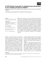
Fig. 1. (A) Map of the 5¢-end of the B. caldolyticus pyr operon. The thin bent arrow represents the transcriptional start site; ORFs are repre-
sented as thick arrows; the untranslated regions containing the three attenuator regions are shown as lines of medium thickness. (B)
Sequence of the three pyr mRNA species (binding loops) bound by PyrR that were examined. The BcBL1, BcBL2 and BcBL3 sequences
were derived from portions of the DNA sequence of attenuator regions 1, 2 and 3, respectively, shown in (A). Numbers refer to the nucleo-
tide number in the B. caldolyticus pyr transcript with +1 as the transcriptional start site [13]. The secondary structures were predicted by
MFOLD version 3.1 ( [32]. Three nucleotides in each binding loop that are not part of the wild-
type pyr mRNA sequence are underlined: The two first G residues in each transcript are specified by the T7 promoter and the terminal A
residue is added by Taq polymerase when used for preparation of templates for in vitro transcription by T7 polymerase. Arrows indicate
three single-base substitution RNA variants in BcBL2 examined.
RNA binding to PyrR C. M. Jørgensen et al.
656 FEBS Journal 275 (2008) 655–670 ª 2008 The Authors Journal compilation ª 2008 FEBS
RNA. Use of the filter binding assay was frustrated in
previous studies with B. subtilis PyrR because that pro-
tein tended to aggregate and failed to bind quantita-
tively to various hydrophobic filters. However, the
filter binding method can be used to study RNA bind-
ing to PyrR from B. caldolyticus, possibly because this
protein has a lower overall negative electrostatic sur-
face potential than the B. subtilis homologue [10] and
does not aggregate. The present study leads to a more
refined characterization of the PyrR–RNA interaction,
a definition of binding stoichiometry as one RNA
binding loop per PyrR dimer and a definition of the
specificity of nucleotide effects on RNA binding. The
implications of the the current findings for the physio-
logical regulation of pyrimidine biosynthesis are pre-
sented; the most important of these is that regulation
of pyr operon expression by PyrR relies on shifts in
the ratio of uridine nucleotides to guanosine nucleo-
tides, and not the intracellular concentration of uridine
nucleotides alone.
Results
Uridine and guanosine nucleotides modulate
PyrR binding to all three pyr mRNA binding
loops
The predicted secondary structures of the three
B. caldolyticus pyr mRNA binding loops (BcBL1,
BcBL2 and BcBL3) examined in the present study are
shown in Fig. 1B. All three binding loops contain seg-
ments that are conserved in PyrR binding loops from
homologous regulatory systems in other bacteria [2].
Conserved features include the predicted stem-loop
structure with a purine-rich internal bulge, a terminal
hexaloop containing the CNGNGA consensus
sequence, and the UUUAA consensus sequence in the
lower stem and internal bulge. Filter binding was used
to estimate the affinity of the B. caldolyticus PyrR pro-
tein to each of the three binding loops (Fig. 2A–C).
Binding was specific for pyr RNA, as shown by the
failure of a control RNA (i.e. the antisense strand to
BcBL1) to bind to any concentration of PyrR tested
(Fig. 2A).
Binding of PyrR to BcBL2 and BcBL3 in standard
binding buffer in the absence of effectors followed a
binding curve (sigmoid on a semi-log plot of PyrR
concentration versus % of total RNA bound) that was
indicative of a simple PyrR–RNA binding isotherm
(Fig. 2B,C). However, the binding curve for BcBL1
deviated consistently from the fitted curve (Fig. 2A).
On the other hand, in the presence of 0.5 mm UMP,
which stimulated binding for all three binding loops,
PyrR binding to BcBL1 resembled the binding
observed for the other two binding loops. The appar-
ent dissociation constant (K
d
) values for RNA binding
are shown in Table 1. When no nucleotides were pres-
ent, PyrR bound most tightly to BcBL2 and BcBL3
(K
d
of 0.13 ± 0.02 nm and 0.2 ± 0.08 nm, respec-
tively). The K
d
value for PyrR binding to BcBL1
(0.9 ± 0.3 nm) corresponds to slightly looser binding.
Addition of 0.5 mm UMP, UDP or UTP resulted in
tighter binding, yielding K
d
values in the range 0.04–
0.09 nm for the three RNAs. PRPP and dUMP also
stimulated binding, although not as effectively as
UMP.
The apparent K
d
values for binding of B. caldolyti-
cus pyr binding loops BcBL1, BcBL2 and BcBL3 to
PyrR were increased in the presence of GMP by 90-,
40- and 200-fold, respectively, relative to their values
in the absence of effector, indicative of a reduced
affinity for RNA (Table 1). However, these constants
were difficult to determine precisely because the bind-
ing data were not adequately fitted by a simple bind-
ing equation (Fig. 2A–C). GDP, GTP and dGMP
also inhibited binding, although they were less
effective at saturating concentrations than GMP
(Table 1).
Because all three binding loops bound with similar
affinity to PyrR and the effects of nucleotides on RNA
binding were similar for all three RNAs, we conducted
most of the subsequent studies with a single RNA
(BcBL2) because the binding of the homologous
B. subtilis RNA (BsBL2) was thoroughly investigated
in a previous study [2].
Concentrations of nucleotides required for
activation or inhibition of PyrR binding to
pyr binding loops
The concentrations of nucleotides that modulate PyrR
binding to RNA in vitro were determined so that these
values could be compared with likely intracellular con-
centrations of the nucleotides. Measurements of the
binding of RNA to PyrR over a wide range of nucleo-
tide concentrations in the filter binding assay yielded
the concentration at which the effect of the nucleotide
was half-maximal (Table 2). As a function of concen-
tration, UMP was ten-fold more effective than UTP at
stimulating binding of PyrR to BcBL1 and 100-fold
more effective than UTP at stimulating binding to
BcBL2. Additionally, the UTP concentration necessary
for activation of PyrR was almost ten-fold lower for
BcBL1 than for BcBL2. As a function of concentra-
tion, GTP was a much more effective inhibitor of
RNA binding than GMP. Even though addition of a
C. M. Jørgensen et al. RNA binding to PyrR
FEBS Journal 275 (2008) 655–670 ª 2008 The Authors Journal compilation ª 2008 FEBS 657
saturating GMP concentration resulted in a higher
apparent dissociation constant for RNA than did GTP
(Table 1), the concentration required to achieve this
inhibition was much higher for GMP (Table 2). The
high concentration of GMP needed to affect RNA
binding, as compared to GTP, suggests that GTP is
the more likely physiological regulator, especially given
that nucleoside triphosphate levels are usually several-
fold higher than levels of the corresponding nucleoside
monophosphate in vivo.
The guanosine to uridine nucleotide ratio
governs PyrR binding to pyr RNA
Table 3 shows the effects of varying the ratio between
effectors that increase and effectors that decrease
0.0001 0.001 0.01 0.1 1 10 100 1000 10 000 0.0001 0.001
100 000
0.0001 0.001
0.01 0.1 1 10 100 1000 10 000
100 000
0.01 0.1 1 10 100 1000 10 000
0.0001 0.001 0.01 0.1 1 10 100 1000 10 000
0.0001 0.001 0.01 0.1 1 10 100 1000 10 000
Fraction RNA bound
0.0
0.1
0.2
0.3
0.4
0.5
0.6
AD
BE
C
Fraction RNA bound
0.0
0.1
0.2
0.3
0.4
0.5
0.6
0.0
0.1
0.2
0.3
0.4
0.5
0.0
0.1
0.2
0.3
0.4
0.5
0.7
0.6
PyrR (n
M
)
PyrR (n
M
)
Fraction RNA bound
0.0
0.1
0.2
0.3
0.4
0.5
0.6
0.7
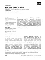
syassa tfihs leGstnemirepxe gnidnib retliF
Fig. 2. Representative filter binding experiment of the
32
P-labeled PyrR binding loops, BcBL1 (A), BcBL2 (B) and BcBL3 (C), to various con-
centrations of PyrR in the absence of effector (open circles), with 500 l
M UMP (closed circles) or 500 lM GMP (closed triangles). Binding to a
control RNA (the antisense strand of BcBL1) is indicated by open diamonds (A). Representative eletrophoretic gel mobility shift assay with
32
P-
labeled BcBL1 (D) and BcBL2 (E) in the absence of effector (open circles), with 500 l
M UMP (closed circles) or 500 lM GMP (closed triangles).
RNA binding to PyrR C. M. Jørgensen et al.
658 FEBS Journal 275 (2008) 655–670 ª 2008 The Authors Journal compilation ª 2008 FEBS
binding of PyrR to BcBL2 RNA with the total concen-
tration of the two effectors held constant. When the
GMP to UMP ratio was increased from 0.11 to 19,
the apparent dissociation constant for RNA increased
18-fold, demonstrating the antagonism of the two
effectors. When the ratio of GTP to UTP was varied
over the same range, the effects were similar to the
effects of GMP and UMP; the values for the apparent
K
d
for BcBL2 varied over a ten-fold range. The effects
of PRPP on RNA binding to PyrR were similar to
those of uridine nucleotides (Table 1); GMP and GTP
also antagonized the effects of PRPP (data not shown),
as would be expected if PRPP and the nucleotides bind
at the same site. From these observations, we predict
that the most important factor regulating the affinity
of PyrR for target pyr RNA sites in vivo is the intracel-
lular ratio of guanosine nucleotides to uridine nucleo-
tides, rather than the concentration of the individual
nucleotides.
Structural requirements of effectors for affecting
PyrR binding to BcBL2
To learn more about how PyrR distinguishes purine
and pyrimidine nucleotides, we tested the ability of
purine and pyrimidine nucleotide structural variants to
activate or inhibit binding of BcBL2 to PyrR (see sup-
plementary Table S1). In general, RNA binding to
PyrR was activated by pyrimidine nucleotides regard-
less of structure, whereas the specificity of purine
nucleotide effects on RNA binding indicated that both
the exocyclic oxo and amino groups of the purine ring
and the 2¢-hydroxyl group of ribose in GMP contrib-
ute significantly to its action. These observations sug-
gest specific interactions between PyrR and the purine
ring of purine nucleotides that do not occur with
pyrimidine nucleotides, even though such interactions
have not been observed in the presently available
X-ray structures of PyrR-nucleotide complexes [10,11].
Effects of Mg
2+
, pH and temperature on binding
of PyrR to BcBL2
Experiments characterizing the effects of Mg
2+
ion
concentration, pH and temperature on the binding of
BcBL2 RNA to PyrR in the filter binding assay are
shown in detail in the Supplementary Material. Three
important conclusions were derived from these studies.
First, Mg
2+
ions at a concentration of 10 mm or
higher were essential for tight binding of RNA. Inclu-
sion of Mg
2+
ions in the electrophoresis gel was subse-
quently found to be crucial for obtaining tight binding
of RNA in the gel shift assay. Second, the affinity of
PyrR for BcBL2 RNA was 50-fold higher at pH 7.5
than at pH 5.5, and the effect of GMP on RNA bind-
ing was strongly pH dependent, whereas the effect of
Table 1. Apparent RNA dissociation constants (K
d
values) from fil-
ter binding determinations of PyrR binding to the three pyr operon
binding loops. The effectors were present at 0.5 m
M. The data are
averages of at least three independent determinations and include
standard deviations of the mean value.
K
d
values (nM)
BcBL1 BcBL2 BcBL3
No effector 0.87 ± 0.3 0.13 ± 0.02 0.21 ± 0.08
UMP 0.07 ± 0.02 0.04 ± 0.01 0.08 ± 0.05
UDP 0.07 ± 0.02 0.04 ± 0.01 ND
UTP 0.09 ± 0.02 0.04 ± 0.01 ND
dUMP 0.16 ± 0.08 0.05 ± 0.01 ND
PRPP 0.11 ± 0.03 0.06 ± 0.01 ND
GMP 79 ± 17 5.2 ± 2.9 49 ± 25
GDP 37 ± 12 2.7 ± 1.6 ND
GTP 12 ± 3 1.1 ± 0.2 ND
dGMP 9 ± 5 0.73 ± 0.01 ND
ND, not determined.
Table 2. Half-maximum concentrations of nucleotides or PRPP
required for either activation (UMP, UTP and PRPP) or inhibition
(GMP and GTP) of binding of PyrR to BcBL1 and BcBL2. Data are
the average of at least two independent determinations.
Half-maximum concentration (l
M)
BcBL1 BcBL2
UMP 0.04 ± 0.02 0.02 ± 0.01
UTP 0.3 ± 0.2 2.4 ± 0.7
PRPP 0.7 ± 0.3 2.0 ± 1.4
GMP 269 ± 143 232 ± 162
GTP 18 ± 6 8 ± 6
Table 3. Effects of the ratio of guanosine to uridine nucleotide con-
centrations on binding of PyrR to BcBL2. The total concentration of
nucleotides was held constant at 1 m
M.
Concentration
of nucleotide
(l
M)
K
d
value for
RNA (n
M)
Concentration
of nucleotide
(l
M)
K
d
value for
RNA (n
M)
GMP UMP GTP UTP
0 0 0.13 0 0 0.13
0 1000 0.06 0 1000 0.05
100 900 0.06 50 950 0.06
250 750 0.05 150 850 0.09
500 500 0.11 250 750 0.11
750 250 0.29 500 500 0.17
900 100 0.84 750 250 0.40
950 50 1.1 900 100 0.61
1000 0 9.8 1000 0 0.75
C. M. Jørgensen et al. RNA binding to PyrR
FEBS Journal 275 (2008) 655–670 ª 2008 The Authors Journal compilation ª 2008 FEBS 659
UMP was much less so (see supplementary Fig. S1A).
Ionization of one of four histidine residues in B. caldo-
lyticus PyrR may mediate the pH dependence of the
GMP effect on RNA binding. Finally, the binding
studies in the present study were conducted at 0 °Cto
ensure stability of the components, for convenience in
maintaining a constant temperature, and for compari-
son with the results of previous gel shift studies. How-
ever, an increase in temperature promotes dissociation
of a protein–RNA complex; an increase in temperature
from 0 °Cto50°C, which is close to the growth tem-
perature for B. caldolyticus, increased the apparent K
d
for BcBL2 binding to PyrR by approximately 40-fold
to 4.5 ± 0.2 nm (see supplementary Fig. S1B).
Direct comparison of the filter binding and
electrophoretic mobility shift methods with
BcBL1 and BcBL2
It was desirable to confirm the fundamental conclu-
sions of the preceding RNA filter binding studies using
an alternative method. Previous studies [2] of pyr
RNA binding by B. subtilis PyrR used an electropho-
retic gel mobility shift method. Some of these prior
findings were different from those described for bind-
ing of pyr RNA by B. caldolyticus PyrR (see Discus-
sion). Therefore, it was important to compare directly
the two methods for measuring RNA binding. Bacillus
caldolyticus PyrR and radiolabeled B. caldolyticus
BcBL1 and BcBL2 were used for this comparison
because B. subtilis PyrR cannot be used for the filter
binding method due to this protein not being quantita-
tively retained by hydrophobic filters.
Inclusion of 1 mm Mg
2+
-acetate in the electrophore-
sis gel was necessary to observe binding of either
BcBL1 or BcBL2 to B. caldolyticus PyrR at concentra-
tions up to 100 lm protein, even though 10 mm Mg
2+
was included in the binding mixture prior to electro-
phoresis, the electrophoresis buffer contained 1 mm
Mg
2+
, and the gel was subjected to prior electrophore-
sis for 90 min before loading the samples. With this
modification of the previously used method [2], tight
binding of B. caldolyticus PyrR to BcBL1 and BcBL2
was observed by the gel shift method (Figs 2D,E and
3, Table 4). The binding of BcBL1 to PyrR was clearly
resolved into two phases (Fig. 2D), one corresponding
to tight binding (K
d1
in Table 4) and another that was
detected only at high concentrations of PyrR, well
above those that could be studied in the filter binding
studies. The significance of the species observed at
PyrR concentrations greatly in excess of those needed
to saturate the RNA is questionable because non-spe-
cific binding to RNA cannot be excluded. The binding
of BcBL2 was described by a single tight binding curve
although, in the presence of 0.5 mm GMP, the binding
curve was broad and fitted less well to a simple bind-
ing equation (Fig. 2E), as was observed on filter bind-
ing of BcBL2 under the same conditions (Fig. 2B). In
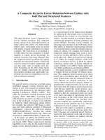
AB
Fig. 3. Analysis of the binding of
32
P-labeled BcBL2 to PyrR by the electrophoretic gel mobility shift method in the absence of effector (A)
and in the presence of 500 l
M GMP (B). The concentration of PyrR (nM subunit) present in each lane is indicated below. The apparent disso-
ciation constants derived from these experiments are shown in Table 4. The presence of the unbound BcBL2 RNA, the PyrR-BcBL2 complex
as well as a more slowly migrating secondary band are indicated on the side of each gel.
RNA binding to PyrR C. M. Jørgensen et al.
660 FEBS Journal 275 (2008) 655–670 ª 2008 The Authors Journal compilation ª 2008 FEBS
addition, a second, more slowly migrating PyrR-
BcBL2 complex was detected at high concentrations of
PyrR when 0.5 mm GMP was present (Fig. 3B). In the
absence of nucleotide (Fig. 3A) or when 0.5 mm UMP
was present (data not shown), this species was barely
detectable. Again, the significance of this loosely bind-
ing complex is open to question. Importantly, the val-
ues for K
d
(K
d1
for BcBL1) and the effects of UMP
and GMP (Table 4) agreed reasonably with the corre-
sponding values obtained with the filter binding
method (Table 1). We also found that addition of
Mg
2+
to the gel was necessary to obtain tight binding
of BcBL2 (K
d
=4nm)toB. subtilis PyrR (data not
shown). Thus, if care was taken to include 1 mm
Mg
2+
in the electrophoresis gel, similar results for the
tight binding RNA curves were obtained by both
methods, a finding that provides confidence in their
validity.
Binding of BcBL2 structural variants to PyrR
The binding of RNA to B. caldolyticus PyrR exhibits
high RNA sequence specificity, as expected from previ-
ous genetic and biochemical studies with B. subtilis
PyrR [2,6]. This was established by filter binding assay
of B. caldolyticus PyrR to three variants of B. caldolyt-
icus BL2 containing single base substitutions (Fig. 1).
Analogous variants of B. subtilis BL2 were observed in
previous gel shift studies with B. subtilis PyrR to have
very different apparent K
d
values relative to native
BL2 [2]. With two of the three structural variants
tested, the data (see supplementary Table S2) indicated
that a single base substitution in a highly conserved
portion of the binding loop RNA (G723A) caused
reduced binding to PyrR, whereas a substitution in a
non-conserved nucleotide (G726A) did not. However,
with a third structural variant, A724C, the binding
observed by filter binding was much tighter than that
detected by the gel mobility shift method (Supplemen-
tary material). Binding of this structural variant, how-
ever, was clearly altered from the wild-type RNA and
additional experiments indicated that the A724C vari-
ant RNA differs from the wild-type BcBL2 in its inter-
action with Mg
2+
(Supplementary material).
Effects of uridine and guanosine supplementation
on pyr gene expression in vivo
If PyrR-mediated regulation of the pyr operon in
Bacillus species is largely responsive to the ratio of uri-
dine to guanosine nucleotides, as suggested by the
effects of these nucleotides on binding of PyrR to
binding loop RNA in vitro, then addition of guanosine
or uridine to the bacterial growth medium would be
expected to stimulate or repress, respectively, the
expression of pyr genes. Assays of aspartate transcar-
bamylase (ATCase), the enzyme encoded by pyrB, the
third cistron of the operon, provided a convenient
measure of operon expression in such experiments.
Inclusion of guanosine in the growth medium
increased the level of ATCase in B. subtilis cells by
approximately 45% compared to a control culture
without supplementation; inclusion of uridine
decreased ATCase levels by almost two-fold (Table 5).
When both uridine and guanosine were included in the
medium in equal amounts, the ATCase level was lar-
gely repressed, but expression increased substantially
as the ratio of guanosine to uridine was increased. The
results demonstrate competition between the effects of
guanosine and uridine in the medium. As expected, the
effects of nucleoside addition were not observed in a
mutant strain of B. subtilis [4] in which the pyrR gene
was deleted. These observations demonstrate that the
effects of nucleotides on RNA binding to PyrR in vitro
correlate with their predicted effects on pyr gene
expression in vivo.
It should be noted that the effects of guanosine on
ATCase expression shown in Table 5 were obtained
Table 4. Apparent RNA dissociation constants (K
d
values) in elec-
trophoretic gel shift assays of binding of BcBL1 and BcBL2 to PyrR.
UMP and GMP were present at 0.5 m
M. Data are the average of
three to four independent determinations.
BcBL1 BcBL2
K
d1
(nM) K
d2
(nM) K
d
(nM)
No effector 0.18 ± 0.04 7650 ± 2500 0.11 ± 0.04
UMP 0.06 ± 0.02 6300 ± 4900 0.07 ± 0.01
GMP 19 ± 8 16800 ± 7400 3.3 ± 1.9
Table 5. Effects of nucleoside supplementation in the growth med-
ium on the expression of B. subtilis ATCase.
Strain
Addition to the medium
(lgÆmL
)1
)
ATCase specific
activity
(nmolÆmin
)1
Æmg
)1
)Guanosine Uridine
DB104 None None 86 ± 5
DB104 50 None 120 ± 11
DB104 None 50 56 ± 4
DB104 50 50 47 ± 6
DB104 50 10 110 ± 14
DB104 50 2 130 ± 9
DB104 DpyrR None None 1300 ± 390
DB104 DpyrR 50 None 1300 ± 390
DB104 DpyrR None 50 1400 ± 420
C. M. Jørgensen et al. RNA binding to PyrR
FEBS Journal 275 (2008) 655–670 ª 2008 The Authors Journal compilation ª 2008 FEBS 661
with cells grown with succinate as the carbon source.
Similar, but even larger, effects could be observed with
glucose-grown cells only in cultures harvested at the
end of exponential growth on limiting glucose; if the
cells were harvested during growth on excess glucose,
the stimulation of ATCase levels by guanosine was not
observed, although strong repression by uridine was
observed. These results indicate that guanosine uptake
and ⁄ or conversion to nucleotides is repressed by
growth on glucose [15], which masks the effect of gua-
nosine on pyr operon expression under such condi-
tions.
Studies of RNA binding to PyrR by analytical
ultracentrifugation
The quaternary structure of B. caldolyticus PyrR in
solution was determined from both sedimentation
velocity and equilibrium sedimentation experiments at
high and low protein concentrations and in the pres-
ence and absence of 0.1 m NaCl. The results of the
sedimentation velocity studies are summarized in the
(supplementary Table S3). The calculated weight aver-
age mass was in the range 83–101 kDa for native PyrR
and 94–99 kDa for the His-tagged PyrR used in sedi-
mentation velocity studies of RNA binding described
below. The masses calculated from the sequences of
the native and His-tagged PyrR in the tetrameric forms
are 79.8 and 91.2 kDa, respectively. Since these weight
average masses are calculated from the change in
shape of moving boundary during the run, and the
data are susceptible to various systematic errors, the
variation observed in the mass shown in Table S3 is
within experimental error.
Data from a sedimentation equilibrium study and
an approach to equilibrium analysis of native PyrR
over the concentration range of the 0.25–25 lm sub-
unit (see supplementary Figs S3 and S4) fit ade-
quately to sedimentation of a single tetrameric
species with a calculated weight average mass of
78.3 kDa, although an alternative fit of the data to a
model for sedimentation of a dimeric and tetrameric
species in equilibrium could not be excluded (Supple-
mentary material). A similar sedimentation equilib-
rium study with His-tagged PyrR (0.25–25 lm)
provided results similar to native PyrR except that
the fitted weight average mass was 91.6 kDa. Alto-
gether, the sedimentation velocity and equilibrium
studies show that both native and His-tagged PyrR
exist largely as tetramers in solution at concentrations
greater than 1 lm, which is in accordance with previ-
ous results obtained with size exclusion chromatogra-
phy and X-ray crystallography [10]. These data and
conclusions are discussed in greater detail in the
Supplementary material.
Sedimentation velocity was also used to analyze the
binding of RNA to PyrR. Purified His-tagged PyrR
was used for these studies because the native PyrR
contained traces of ribonuclease, which might have
degraded the RNA during the 3-day duration of the
titration experiment. As shown in Fig 4A, a 36 nt pyr
binding loop RNA derived from BcBL2 sedimented
as a single RNA species (s20,w = 2.63 S, molecular
mass = 12 900 Da) (molecular mass calculated from
sequence = 11 600 Da). This BcBL2 sample was
titrated by adding aliquots of concentrated PyrR
(Fig. 4B–E), so that up to six equivalents of monomer
were added without significant dilution (< 7%) of
the RNA. Species analysis using either the basic
F
E
D
C
B
A
Fig. 4. A size-distribution analysis of sedimenting species observed
during a titration of pyr binding loop RNA with increasing amounts
of PyrR. A plot of c(s) distributions against the uncorrected sedi-
mentation coefficient, s, is shown for RNA only (A), for molar ratios
RNA to PyrR subunit (B–E), and for PyrR (F) from absorbance data
collected at 260 nm. The c(s) values in the PyrR panel (F) were
multiplied by factors of 10 (dotted line) and 100 (dashed line) to
make them visible on the same scale as used for the other panels.
The initial concentration of RNA was 0.3 l
M for (A) and four sepa-
rate aliquots of PyrR were added to give the final ratios shown. A
concentration of PyrR of1.2 l
M was used in (F). The sedimentation
distributions, c(s), were calculated using
SEDPHAT; 72 scans were
collected at 3-min intervals. Further experimental details are given
in the Experimental procedures. The vertical dotted line relates the
protein peak to the other panels and the vertical dashed line does
the same for the PyrR–RNA complex.
RNA binding to PyrR C. M. Jørgensen et al.
662 FEBS Journal 275 (2008) 655–670 ª 2008 The Authors Journal compilation ª 2008 FEBS
non-interacting model of sedphat [16,17] or the more
powerful hybrid local continuous distribution ⁄ global
discrete species model [16,17] showed that only two
sedimenting species were present at significant concen-
trations in the range of 0.1–5 S for each aliquot
added (see supplementary Table S4). The first of these
(s20,w = 2.6 S) corresponds to the free RNA. A sec-
ond species appeared (s20,w = 4.9 S) that must corre-
spond to an RNA–PyrR complex because added
PyrR will not contribute more than 1–2% to the total
260 nm absorbance at the concentrations added. On
titrating the RNA with increasing amounts of PyrR,
the loading concentration of the peak corresponding
to free RNA declined, and that for the second peak
increased, as shown by the area under the peaks in
the c(s) distribution shown in Fig. 4. An additional
shoulder at 3 S, whose shape and position are
somewhat variable, is evident in Fig. 4E (see supple-
mentary Fig. S5), where the protein concentration is
approximately three-fold greater than that necessary
to saturate the RNA with the PyrR dimer. Based on
the species analysis above, we strongly suspect that
this shoulder is an artifact that results from the sensi-
tivity of the c(s) distribution to boundary effects. As
with the filter binding assays, some of the RNA
( 30%) remained unbound at greater than saturating
concentrations of PyrR. In Fig 4F, the PyrR stock
solution was diluted to 1.2 lm subunits into the same
buffer and centrifuged under the same conditions as
used for the other panels in Fig. 4. Most of the pro-
tein sedimented as a tetramer with an s20,w = 5.5 S
with a minor species at approximately 10% of the tet-
ramer concentration with an s20,w = 2.3 S and an
estimated molecular mass of 18 000 Da, which is
likely a nonparticipating PyrR monomer (sequence
molecular mass = 22 800 Da). The sedimentation
coefficients and buoyant mass variation observed with
increasing PyrR concentrations are summarized in the
(supplementary Table S4). The s values in Table S4
for the free RNA peak decreased significantly with
increasing PyrR concentration. We demonstrated that
the pyr RNA appeared to be electrophoretically intact
following the 3-day experiment at 20 °C (data not
shown), so the decrease in s value for the RNA is not
the result of RNA degradation. The sedimentation
coefficient of the new species (4.6–4.9 S) is signifi-
cantly lower than that of free PyrR (5.4 S); a complex
of RNA with the PyrR tetramer would be expected to
have a larger s value than free PyrR, barring a large,
unexpected increase in the hydrodynamic radius.
Thus, the complex of RNA with PyrR must involve
association with the protein in a form smaller than
the tetramer. If the buoyant mass of the RNA is
subtracted from that of the complex and the molecu-
lar weight of the remaining protein calculated, using a
partial specific volume of 0.74 (determined from the
amino acid composition of PyrR) and a solvent den-
sity of 1.0 gÆmL
)1
, the value obtained is 37 100 for
the protein component. This is in reasonable agree-
ment with the mass of a His-tagged PyrR dimer
(44 000). Finally, Fig. 5 shows a plot of the free
RNA remaining against the ratio of PyrR subunit
concentration to the initial RNA concentration (see
supplementary, Table S4). The trend in the data is
that of a typical stoichiometry plot where the RNA
concentration is in large excess of its dissociation
constant for PyrR. The data are consistent with a
stoichiometry of one RNA molecule per PyrR dimer
in the complex with approximately 30% of the RNA
that does not bind under these conditions. Thus, we
conclude that the complex has the composition of
(PyrR)
2
-RNA.
Fig. 5. A plot of A
260
for the free RNA peak, which is obtained by
integrating the area under the peak in the s range of 2–2.6 S,
against the molar ratio of PyrR to RNA in the sample. The data
were obtained from the analytical ultracentrifugation experiment
described in Fig. 4. The free RNA values were corrected for a slight
loss of total A
260
in the range 5–20%, of which a maximum of 5%
was due to dilution. The dotted line shows a least-squares fit
through the first three points. The horizontal dotted line shows the
concentration of non-binding RNA from Fig. 4D, in which the RNA
peak is clearly defined. The intersection between the dashed and
dotted lines indicates that two subunits of PyrR bind to one RNA
stem-loop.
C. M. Jørgensen et al. RNA binding to PyrR
FEBS Journal 275 (2008) 655–670 ª 2008 The Authors Journal compilation ª 2008 FEBS 663
Discussion
Complexity of RNA binding to PyrR
The complex binding curves for BcBL1 and for all
three binding loop RNAs in the presence of guanosine
nucleotides (Fig. 2) indicate that the binding of RNA
to PyrR cannot be fully described by a simple binding
equilibrium. Although the biophysical basis for the
complexity of the RNA binding curves is not estab-
lished, we suggest that it arises from multiple PyrR
conformational and ⁄ or aggregation states that differ in
their affinity for RNA and possibly also for nucleo-
tides. PyrR conformation is implicated because the
heterogeneity in RNA binding is strongly affected by
uridine and guanosine nucleotides, which are known to
bind to the UPRTase active site of PyrR [10]. The sim-
plest model that fits our observations posits the exis-
tence of two PyrR conformations, with one having a
higher affinity for RNA than the other. The high affin-
ity state is favored by binding of either uridine nucleo-
tides or binding of RNA itself in the case of BcBL1.
The low affinity state is favored by the binding of gua-
nosine nucleotides. Thus, RNA binding involves at
least two coupled reactions: RNA binding to PyrR
and nucleotide binding to PyrR.
The demonstration by analytical ultracentrifugation
that the PyrR tetramer dissociates into dimers when
RNA binds adds yet another reaction that is likely
coupled to the RNA and nucleotide binding reactions
discussed above. It is likely that, at high dilution, tet-
rameric PyrR dissociates to dimers in the absence of
RNA, but this could not be conclusively demonstrated
at the lowest concentration (1 lm) that could be ana-
lyzed by analytical ultracentrifugation. We note, how-
ever, that all of the filter binding experiments were
conducted at PyrR concentrations well below this
value, where some or all of the PyrR may be present
in dimeric form. We propose that the tetrameric form
of PyrR has low affinity for RNA because the likely
RNA binding site is known from the crystal structures
to be occluded in the center of the tetramer [10,11].
The dimeric form of PyrR, in which the RNA binding
site would be exposed to the solvent, is likely to have
higher affinity for RNA. Coupling of the dimer–tetra-
mer equilibrium to the equilibria for PyrR–RNA bind-
ing and PyrR–nucleotide binding could explain the
complex binding curves observed in the present study,
especially when RNA binding in the presence of gua-
nosine nucleotides was examined.
The involvement of multiple coupled equilibria (i.e.
PyrR tetramer–dimer association together with binding
of RNA and nucleotides to dimer and tetramer with
different affinities for each state of aggregation) in the
experimentally observed RNA binding in the present
study dictates that one should not regard the apparent
K
d
values for RNA or the half-maximal values for
nucleotide effects on RNA binding as simple equilib-
rium constants. Hence, we have consistently used the
term ‘apparent K
d
’ to describe the concentrations of
PyrR that yielded half-maximal RNA binding in our
experiments.
Correlations between results of filter binding
studies and electrophoretic mobility shift studies
of RNA binding
Direct comparison of the binding of BcBL1 and BcBL2
to B. caldolyticus PyrR by the filter binding and gel shift
methods demonstrated that, as long as Mg
2+
was
included in the electrophoresis gel, there was good
agreement between the two methods. However, agree-
ment was much poorer with the A724C structural vari-
ant of BcBL2 RNA, even with a high Mg
2+
concentration in the gel. The sensitivity to Mg
2+
and to
the structure of the RNA studied suggests that the gel
shift method can give highly misleading results in some
cases. An RNA that dissociates rapidly from PyrR may
appear to bind poorly, or not to bind at all, in the gel
shift assay. We conclude that, for protein–RNA binding
studies in general, it would be prudent to confirm elec-
trophoretic mobility shift conclusions whenever possible
by an alternative method, such as a filter binding assay.
In light of our current observations on the impor-
tance of Mg
2+
in gel shift assays with PyrR and pyr
binding loop RNAs, the studies of the specificity of
RNA binding of B. subtilis PyrR should be re-exam-
ined. Our findings with the native and the G723A and
G726A sequence variants of BcBL2 indicate that the
effects on affinity observed previously for native
BsBL2 and its structural variants [2] are valid, at least
qualitatively. However, previous observations indicat-
ing that B. subtilis PyrR binding to BsBL1 and BsBL3
was weak and barely affected by uridine nucleotides
are misleading and probably resulted from the dissoci-
ation of the required cation Mg
2+
from these two
RNAs, but not BsBL2, during electrophoresis. We
now have evidence that PyrR from both Bacillus spe-
cies binds tightly to all three binding loop RNAs from
both species and that binding of all three RNAs is sig-
nificantly modulated by nucleotides (data not shown).
Physiological implications of these studies
The data provided in Tables 1–3 indicate that uridine
nucleotides and guanosine nucleotides are the primary
RNA binding to PyrR C. M. Jørgensen et al.
664 FEBS Journal 275 (2008) 655–670 ª 2008 The Authors Journal compilation ª 2008 FEBS
metabolite modulators of PyrR binding to pyr attenua-
tor region RNA, and hence the primary regulators of
pyrimidine biosynthesis. Uridine nucleotide stimulation
of RNA binding is easily understood in terms of feed-
back regulation (i.e. end-product repression of the pyr
operon) because binding of PyrR to RNA leads to
increased termination of transcription prior to tran-
scription of pyr genes coding for biosynthetic enzymes.
UMP and UTP exert their effects on RNA binding
in vitro in the micromolar concentration range, well
below their estimated concentrations in exponentially
growing cells (i.e. 1 mm for UTP and 0.1–0.3 mm for
UMP) [18]. GTP is probably the primary physiological
guanosine nucleotide modulator because it is effective
at a much lower concentration than GMP (Table 2)
and is present in growing cells at approximately
0.6 mm [19], well above the concentrations at which it
exerts its effects on RNA binding to PyrR. The effects
of GMP are unlikely to be significant in vivo because
intracellular GMP levels [19,20] do not reach the con-
centration required to antagonize RNA binding.
Of the other metabolites studied, none appears as
being likely to serve as a physiologically important
modulator of pyr operon expression. In particular, in
previous publications [4,9], we suggested that PRPP
might may be a feed-forward regulator because it
would be expected to compete at uridine nucleotide
binding sites. However, the data obtained in the pres-
ent study indicate that PRPP actually behaves like
UMP and UTP to stimulate RNA binding to PyrR.
PRPP also fails to antagonize the effects of GTP on
RNA binding nearly as effectively as UMP or UTP
(data not shown). Thus, the effects of PRPP do not
appear to be physiologically important in the regula-
tion of pyr expression.
The intracellular concentrations of uridine nucleo-
tides exceed the half-maximal concentrations required
for activation of PyrR binding to RNA by two or
three orders of magnitude (Table 2), even allowing for
an increase of up to 40-fold in the K
d
values for PyrR
binding to RNA measured at 0 °C when shifted to the
physiological growth temperature for B. caldolyticus
(see supplementary Fig. S1B). The values of the appar-
ent dissociation constants suggest that PyrR functions
as a regulator of pyr gene expression largely under
conditions where the affinity of PyrR for pyr attenua-
tor sites is substantially reduced by the antagonistic
effect of guanosine nucleotides. Because guanosine and
uridine nucleotides can compete for binding to the
same sites on PyrR [10], the affinity of the protein for
pyr RNA will be determined by the ratio of their intra-
cellular concentrations, and not by the concentration
of the individual nucleotides. Table 3 illustrates how
the affinity of PyrR for BcBL2 RNA varied over a
ten- to 20-fold range as a function of the GMP ⁄ UMP
or GTP ⁄ UTP ratios. The experiments in Table 3 were
conducted at a total nucleotide concentration of 1 mm,
which is near their physiological concentration and
well above the concentrations at which the individual
nucleotides exert their effects on RNA binding. Most
importantly, investigations of the effects of nucleoside
addition to the growth medium on B. subtilis pyrB
(ATCase) expression in vivo (Table 5) support the con-
clusion that the ratio of guanosine to uridine nucleo-
tides dominates PyrR action. The crucial role of
guanosine nucleotides in the regulation of pyr genes in
bacilli was not fully appreciated before these studies
were conducted.
The cross-regulation of pyrimidine biosynthesis by
guanosine nucleotides is part of a more general phe-
nomenon. The accumulation of high levels of intracellu-
lar guanosine nucleotides indicates that cells have
adequate carbon, nitrogen and energy sources for RNA
and DNA synthesis. Thus, accumulated purine nucleo-
tides constitute appropriate feed-forward metabolites
to stimulate pyrimidine nucleotide biosynthesis. Other
examples of such feed-forward activation of pyrimidine
biosynthesis in Bacillus species include the activation by
GTP of the pyrimidine-repressible carbamyl phosphate
synthetase [21], of UMP kinase [22] and of CTP synthe-
tase [23]. Furthermore, GTP levels are used as regula-
tory signals governing a variety of more global
regulatory circuits in Bacillus cells. Examples of this
regulatory function of the GTP pool in B. subtilis
include the regulation of ribosomal RNA synthesis [24],
initiation of sporulation [25], activation of the CodY
regulon in nutrient-starved cells [26], and conversion of
GTP to ppGpp and pppGpp during the stringent
response to amino acid starvation [25]. A reduction in
intracellular GTP without a corresponding decline in
uridine nucleotide pools would result in much tighter
binding of PyrR to attenuation region RNA and
reduced expression of the pyr operon, which is an
appropriate response to the original nutrient limitation.
Experimental procedures
In vitro transcription and purification of
32
P-labeled RNA
For synthesis of radiolabeled RNA by in vitro transcrip-
tion, plasmids containing templates that contained the
T7 RNA polymerase promoter followed by DNA specify-
ing one of the three PyrR binding loops (BcBL1, BcBL2
and BcBL3) from the B. caldolyticus pyr operon were con-
structed. The isolated plasmids were used as templates in
C. M. Jørgensen et al. RNA binding to PyrR
FEBS Journal 275 (2008) 655–670 ª 2008 The Authors Journal compilation ª 2008 FEBS 665
PCR reactions to generate products of approximately
150 bp in length that were used as templates for in vitro
transcription with T7 RNA polymerase. The PCR tem-
plates were purified using the GFX purification kit prior to
in vitro transcription using the MaxiScript kit from Ambion
(Austin, TX, USA). The procedure recommended by the
manufacturer was used, except that the reactions were incu-
bated for 2 h at 37 °C, and the RNA was labeled by
including 10 lm [a-
32
P]GTP (800 CiÆmmol
)1
; MP Biomedi-
cals Inc., Irvine, CA, USA) in the reaction mix together
with 500 l m of each of ATP, UTP and CTP. Unincorpo-
rated nucleotides were separated by running the sample
through a G-50 Micro Column from Amersham Bioscienc-
es ⁄ GE Healthcare (Piscataway, NJ, USA). The labeled
RNA was purified using denaturing (8 m urea) 15% poly-
acrylamide gel electrophoresis as described previously [10].
Following phenol extraction of RNA eluted from a cut-out
piece of gel, the RNA was precipitated over night at
)20 °C with 1 lg of yeast tRNA as carrier RNA and resus-
pended in 200 lL buffer (25 mm Tris-acetate pH 7.5,
50 mm K-acetate and 1 mm EDTA). The concentration of
the RNA was determined by duplicate liquid scintillation
counting of a 10-fold dilution of the RNA sample. Prior to
use, the labeled RNA was denatured at 85 °C for 15 min
and allowed to refold for 15 min at 37 °C, after which
Mg-acetate was added to a final concentration of 10 mm.
Filter binding assays of RNA binding to PyrR
A double-filter method for filter binding [14] was used as
described previously [10]. Unless noted otherwise, the reac-
tion mixture contained 25 mm Tris-acetate (pH 7.5), 50 mm
K-acetate, 10 mm Mg-acetate, 25 pm
32
P-labeled RNA,
0.04 UÆlL
)1
of RNase inhibitor (Superase-In; Ambion),
100 lgÆmL
)1
( 4 lm) of yeast tRNA, 50 lgÆmL
)1
of acety-
lated BSA, nucleotides at the indicated concentrations
and various concentrations of native B. caldolyticus PyrR,
which was purified as described previously [10]. The con-
centration of the PyrR protein was determined using the
Bradford assay obtained from Bio-Rad (Hercules, CA.
USA). The 50 lL reaction mixtures were allowed to incu-
bate for 40 min on ice prior to filtering through the two
membranes followed by a single wash with 50 lL of bind-
ing buffer. Filtration and washing were complete within
60 s. For some experiments, the pH was varied by use of a
25 mm Tris-25 mm 2-(N-morpholino)ethanesulfonate-ace-
tate buffer. For experiments where the temperature was
varied, the pH of binding buffer was adjusted at each tem-
perature examined to the pH of binding buffer at room
temperature. The filters and the washing buffer were all
equilibrated at the relevant temperature for at least 30 min
prior to the experiments and the filtration apparatus was
used at the temperature indicated. After overnight exposure
of the membranes to a PhosphorImager screen, the radioac-
tivity was determined using a PhosphorImager and quanti-
fied using imagequant software (Molecular Dynamics ⁄ GE
Healthcare, Sunnyvale, CA, USA). Dissociation constants
were determined by fitting the data to a simple binding
equation using sigmaplot 9.0 (Systat Software, Inc San
Jose, CA, USA) as described previously [10] or, when two
phases were clearly identified, to a two-state binding curve.
Since the RNA concentration of 25 pm was close in some
cases to the apparent K
d
, these data were also fitted to a
quadratic binding equation [27], but the calculated apparent
K
d
values obtained were not significantly different from
those obtained with the simple binding equation. To deter-
mine the concentration of nucleotides required for half-
maximum effect on PyrR binding to the RNA, the obtained
K
d
values from at least two independent experiments were
plotted against the nucleotide concentration, and the half-
maximum concentration and standard deviation were calcu-
lated by sigmaplot 9.0. Only approximately 50–60% of the
total radioactive RNA was bound by saturating levels of
PyrR; this was consistently observed for all three binding
loop RNAs (Fig. 2) and a similar result was noted in sedi-
mentation velocity experiments. We suggest that the
unbound RNA consists of species that do not fold into the
native secondary structure required for binding.
Electrophoretic gel mobility shift assays of RNA
binding to PyrR
Gel shift analysis was performed essentially as described
previously [2] with the following modifications. The gels
were run in a Bio-Rad Protean IIxi apparatus cooled to
4 °C and contained 6% acrylamide (37.5 : 1 acrylamide ⁄ bis
solution), 12.5 mm Tris-acetate (pH 7.5), 2.5% glycerol and
1mm Mg-acetate. The running buffer contained 12.5 mm
Tris-acetate (pH 7.5) and 1 mm Mg-acetate. The gels were
pre-run for 1.5 h at 150 V followed immediately by loading
of 15 lL of sample, electrophoresis for 15 min at 50 V and
for 3 h at 300 V. After electrophoresis, the gels were dried
on filter paper and exposed overnight to a PhosphorImager
screen. Data were analyzed as described above. The RNA
binding mixture (50 lL) used for gel shifts was the same as
that employed in the the filter binding experiments, except
that BSA was omitted and 0.0125% loading dye (xylene
cyanol) and 5% glycerol were added. Binding of RNA to
appreciably higher concentrations of PyrR could be better
characterized by the gel shift method than by the filter
binding method described above because concentrations of
PyrR in excess of 1 lm were not quantitatively retained by
the nitrocellulose filter.
Preparation of ribonuclease-free PyrR for
analytical ultracentrifugation
His-tagged B. caldolyticus PyrR was used in the ultracen-
trifugation experiments. Direct comparison of the binding
RNA binding to PyrR C. M. Jørgensen et al.
666 FEBS Journal 275 (2008) 655–670 ª 2008 The Authors Journal compilation ª 2008 FEBS
of BcBL2 RNA to His-tagged PyrR and to native PyrR
under identical conditions by the filter binding methods
demonstrated that binding of RNA to His-tagged PyrR
was modulated by nucleotides in the same manner as native
PyrR, but the apparent K
d
for RNA was approximately
100-fold larger. For preparation of His-tagged PyrR, the
PyrR coding sequence was amplified from pSHCO2, a plas-
mid containing B. caldolyticus PyrR [10], and ligated into
plasmid pET412b, which is a pET41 derivative encoding a
C-terminal octa-histidine tag preceded by the cleavage site
for tobacco etch virus protease (E. Harms, Purdue Univer-
sity, Lafayette, IN, USA, personal communication); the
resultant plasmid is pCalCHIS. Escherichia coli strain
BL21-DE3 was transformed with pCalCHIS for pyrR
expression. Cultures of E. coli BL21-DE3 ⁄ pCalCHIS were
grown in LB medium at 37 °C until an absorbance of 0.8
was reached at A
600
, induced with 0.4 mm isopropyl thio-b-
d-galactoside and grown for 4 h at 37 °C. Cells were har-
vested and lysed by sonication in buffer containing 20 mm
Tris (pH 7.5), 500 mm NaCl. The lysate was centrifuged at
25 000 g at 4 °C for 30 min. Because PyrR at high concen-
tration is insoluble in the high-salt buffers used for Ni-affin-
ity purification, the purification protocol employed high
ionic strength prior to elution, and low ionic strength dur-
ing elution. Following filtration through a 0.4 lm mem-
brane, the supernatant was batch-bound to Ni-Sepharose
resin (Amersham) in buffer containing 20 mm Tris
(pH 7.5), 500 mm NaCl supplemented with 100 mm imidaz-
ole to minimize binding of E. coli proteins. The resin was
washed in 20 mm Tris (pH 7.5) and 100 mm imidazole,
packed into a Bio-Rad Econo-Column and eluted in 20 mm
Tris (pH 7.5) with a 0.1–1.0 m imidazole gradient. The pro-
tein eluted at approximately 600 mm imidazole. Fractions
were pooled and dialyzed against 20 mm Tris (pH 7.5), 5%
glycerol. Remaining traces of RNase contaminants were by
chromatography of purified PyrR on an Amersham Super-
dex 200 HiLoad16 ⁄ 60 column, eluting with 0.1 m Tris-HCl
(pH 7.5) and 0.15 m NaCl. The pooled fractions were dia-
lyzed against buffer containing 20 mm Tris (pH 7.5). The
RNase-free status of purified PyrR was confirmed by detec-
tion of no degraded RNA species on gel electrophoresis of
samples that had been incubated with radiolabeled RNA
for 2 weeks at room temperature.
Analytical ultracentrifugation
Sedimentation equilibrium and velocity experiments using
either native PyrR or His-tagged PyrR were run on a Beck-
man XL-I Analytical Ultracentrifuge (Beckman-Coulter,
Fullerton, CA, USA) at 20 °C. Carbon filled epon two sec-
tor centerpieces and sapphire windows were used for all
experiments. In all experiments, a buffer of 20 mm Tris-Cl
(pH 7.5) and, in some cases, 10 mm MgCl
2
and 0.1 m NaCl
were included. The cell and rotor were equilibrated under
vacuum and at 0 r.p.m. for at least 1 h prior to the start of
the run so that the system could reach thermal equilibrium.
For the velocity experiments, the centrifuge was started
from 0 r.p.m. and data collection began immediately with
scans collected every 3 min. Both absorbance and interfer-
ence data were usually collected. For the equilibrium exper-
iments, both approach to equilibrium data and data at
equilibrium were collected and the samples were pre-equili-
brated as described above. The condition of equilibrium
was tested using the program winmatch 0.99 from the
rasmb software archive ( />The sample took 40 h to initially reach equilibrium and
24 h between speed changes.
Multiwavelength sedimentation velocity experiments were
undertaken to define the stoichiometry of the RNA–PyrR
complex in solution. However, only data obtained at
260 nm had sufficient signal to noise ratio to be used for the
entire range of the titration. His-tagged PyrR was prepared
by dialysis against buffer containing 20 mm Tris-Cl
(pH 7.5), 10 mm MgCl
2
. BcBL2 RNA (nucleotides 702–737;
Fig. 1B), which was chemically synthesized by Integrated
DNA Technologies (Coralville, IA, USA), was prepared in
the same buffer. These experiments were run in a Beckman
Optima XL-I ultracentrifuge. A 12 mm path length cell con-
taining a double-sector carbon-filled epon centerpiece with
sapphire windows was used for all experiments, and this cell
was treated with RNaseZAP (Ambion) for 24 h to remove
nucleases. The reference and sample sectors were filled with
400 lL of dialysis buffer and sample, containing 0.3 lm
RNA, respectively. After thermal equilibration of the sample
(at 20 °C for at least 1 h at 0 r.p.m.), all sedimentation
velocity experiments were run at 50 000 r.p.m. at 20 °C and
scans were recorded at 3-min intervals using both absor-
bance (at 278 nm and 260 nm) and interference optics.
The initial sedimentation velocity analysis contained only
0.3 lm RNA, which has a calculated molecular mass of
11 564 Da. Immediately after completion of this first experi-
ment, a 5 lL aliquot of dialysis buffer was added to the ref-
erence sector, and a 5 lL aliquot of concentrated PyrR
protein added to the RNA in the sample chamber. This equi-
molar mix of PyrR subunit to RNA was sedimented as well.
Subsequent sedimentation velocity analyses were performed
by titrating the RNA with increasing amounts of PyrR in
varying molar ratios. The total time of the binding experi-
ment was 3 days. A final experiment was run in the absence
of added RNA at a PyrR subunit concentration of 1.2 lm.
Data were considered to be fitted when plots of the fit-
ting residuals were observed to be evenly distributed around
zero and the rmsd was lower than the signal to noise deter-
mined from the plateau region of the first scan. Data were
usually analyzed using the hybrid local continuous ⁄ global
discrete model [16] with zero to three discrete species
included. For all discrete species, the buoyant mass was cal-
culated, so that masses from molecules with dissimilar par-
tial specific volumes (protein and RNA) would be additive.
PyrR has no tryptophan residues and, at the concentrations
C. M. Jørgensen et al. RNA binding to PyrR
FEBS Journal 275 (2008) 655–670 ª 2008 The Authors Journal compilation ª 2008 FEBS 667
used in these experiments (A
280
and A
260
) would be no
more than 0.0075 AU at the highest protein concentration
(1.8 lm).
Solvent densities and viscosities were calculated to be
1.00 gÆmL
)1
A and 0.01 poise, respectively, for the Tris buf-
fer and 1.0039 gÆmL
)1
and 0.01 poise for the NaCl-contain-
ing buffer using sednterp [28]. A partial specific volume of
0.7398 cm
3
Æg
)1
was also calculated with sednterp from the
sequence of His-tagged PyrR and a partial specific volume
of 0.7450 cm
3
Æg
)1
was calculated from the sequence of
the native protein; an approximate value for RNA
(0.51 cm
3
Æg
)1
) was obtained from Table 2 of Durchschlag
and Zipper [29]. The data were fitted to various models
using the appropriate algorithms in sedfit 9.3b [30]
and sedphat 4.1b [16] (obtained from http://www.
analyticalultracentrifugation.com).
Growth of B. subtilis cells and ATCase assay
To investigate the effects of supplementation of the growth
medium with nucleosides on ATCase levels in B. subtilis,
cultures strains DB104 and DB104DpyrR [4] were grown on
buffered minimal medium [5], which included 0.1% (w ⁄ v)
casamino acids (Becton Dickinson ⁄ Difco, Franklin Lakes,
NJ, USA), 50 mgÆmL
)1
histidine and 6 mgÆmL
)1
disodium
succinate, supplemented with nucleosides as shown in
Table 5. Cells were grown at 37 °C with aeration, harvested
in the exponential growth phase by centrifugation, and rup-
tured by sonication. Cell debris was removed by a 10 min
centrifugation (14 000 g) and the supernatant was used in
assays.
ATCase activity was determined by a radiometric proce-
dure [31]. Assays were performed at 30 °Cina100lL final
volume containing the following: 50 mm Tris-acetate
(pH 8.3), 50 mm [
14
C]-aspartate (final specific activity of
10 lCiÆmmol
)1
; Amersham), and 10 mm carbamoyl phos-
phate (Sigma, St Louis, MO, USA). Reactions were initi-
ated by addition of enzyme and stopped with 900 lL 0.2 m
acetic acid after 30 min. A 950 lL portion of each reaction
was added to a column containing Dowex AG-50W-X8
(200–400 mesh hydrogen form; Bio-Rad) to retain unreact-
ed aspartate. Columns were washed four times with 400 lL
0.2 m acetic acid with the eluate collected in scintillation
vials. Aquasol-2 (Packard Research ⁄ Perkin-Elmer, Meriden,
CT, USA) was added to the collected eluate, total counts
were determined using liquid scintillation counting and used
to calculate ATCase activity. Protein concentrations were
determined using the BCA protein assay kit (Pierce, Rock-
ford, IL, USA), using BSA as the protein standard.
Acknowledgements
This research was supported by United States Public
Health Service Grants GM47112 to R.L.S., DK42303
to J.L.S. and from the Purdue University Bindley Bio-
science Center to J.W.B.
References
1 Switzer RL, Turner RJ & Lu Y (1999) Regulation of
the Bacillus subtilis pyrimidine biosynthetic operon by
transcriptional attenuation: control of gene expression
by an mRNA-binding protein. Prog Nucl Acids Res
Mol Biol 62, 329–367.
2 Bonner ER, D’Elia JN, Billips BK & Switzer RL (2001)
Molecular recognition of pyr mRNA by the Bacillus
subtilis attenuation regulatory protein PyrR. Nucleic
Acids Res 29, 4851–4865.
3 Turner RJ, Bonner ER, Grabner GK & Switzer RL
(1998) Purification and characterization of Bacillus sub-
tilis PyrR, a bifunctional pyr mRNA-binding attenua-
tion protein ⁄ uracil phosphoribosyltransferase. J Biol
Chem 273, 5932–5938.
4 Turner RJ, Lu Y. & Switzer RL (1994) Regulation of
the Bacillus subtilis pyrimidine biosynthetic (pyr) gene
cluster by an autogenous transcriptional attenuation
mechanism. J Bacteriol 176, 3708–3722.
5 Lu Y, Turner RJ & Switzer RL (1995) Roles of the
three transcriptional attenuators of the Bacillus subtilis
pyrimidine biosynthetic operon in the regulation of its
expression. J Bacteriol 177, 1315–1325.
6 Ghim S-Y & Switzer RL (1996) Characterization of cis-
acting mutations in the first attenuator region of the
B. subtilis pyr operon that are defective in regulation of
expression by pyrimidines. J Bacteriol 178, 2351–2355.
7 Ghim S-Y & Switzer RL (1996) Mutations in B. subtilis
PyrR, the pyr regulatory protein with defects in regula-
tion by pyrimidines. FEMS Microbiol Lett 137, 13–18.
8 Tomchick DR, Turner RJ, Switzer RL & Smith JL
(1998) Adaptation of an enzyme to regulatory function:
structure of Bacillus subtilis PyrR, a bifunctional pyr
RNA-binding attenuation protein and uracil phospho-
ribosyltransferase. Structure 6, 337–350.
9 Lu Y & Switzer RL (1996) Transcriptional attenuation
of the Bacillus subtilis pyr operon by the PyrR regula-
tory protein and uridine nucleotides in vitro. J Bacteriol
178, 7206–7211.
10 Chander P, Halbig KM, Miller JK, Fields CJ, Bonner
HKS, Grabner GK, Switzer RL & Smith JL (2005)
Structure of the nucleotide complex of PyrR, the pyr
attenuation protein from Bacillus caldolyticus, suggests
dual regulation by pyrimidine and purine nucleotides.
J Bacteriol 187, 1773–1782.
11 Chander P (2006) Structural analysis of the regulatory
protein of pyrimidine biosynthesis, PyrR. PhD Thesis,
Purdue University, West Lafayette, IN.
12 Ghim S-Y, Nielsen P & Neuhard J (1994) Molecular
characterization of pyrimidine biosynthesis genes from
RNA binding to PyrR C. M. Jørgensen et al.
668 FEBS Journal 275 (2008) 655–670 ª 2008 The Authors Journal compilation ª 2008 FEBS
the thermophile Bacillus caldolyticus. J Gen Microbiol
140, 479–491.
13 Ghim S-Y & Neuhard J (1994) The pyrimidine biosyn-
thesis operon of the thermophile Bacillus caldolyticus
includes genes for uracil phosphoribosyltransferase and
uracil permease. J Bacteriol 176, 3698–3707.
14 Wong L & Lohmann TM (1993) A double-filter method
for nitrocellulose-filter binding: application to protein-
nucleic acid interactions. Proc Nat Acad Sci USA 90,
5428–5432.
15 Schuch R, Garibian A, Saxild HH, Piggot PJ & Nyg-
aard P (1999) Nucleosides as a carbon source in Bacillus
subtilis: characterization of the drm-pupG operon.
Microbiology (Reading) 145, 2957–2966.
16 Schuck P. (2003) On the analysis of protein self-associa-
tion by sedimentation velocity analytical ultracentrifu-
gation. Anal Biochem 320, 104–124.
17 Balbo A, Minor KH, Velikovsky CA, Mariuzza RA,
Peterson CB & Schuck P (2005) Studying multiprotein
complexes by multisignal sedimentation velocity analyti-
cal ultracentrifugation. Proc Nat Acad Sci USA 102,
81–86.
18 Møllgaard H & Neuhard J (1978) Deoxycytidylate
deaminase from Bacillus subtilis. J Biol Chem 253,
3536–3542.
19 Saxild HH & Nygaard P (1991) Regulation of levels of
purine biosynthetic enzymes in Bacillus subtilis: effects
of changing purine nucleotide pools. J Gen Microbiol
137, 2387–2394.
20 Bernlohr DA & Switzer RL (1983) Regulation of Bacil-
lus subtilis glutamine phosphoribosylpyrophosphate
amidotransferase inactivation in vivo. J Bacteriol 153,
937–949.
21 Paulus TJ & Switzer RL (1979) Synthesis and inactiva-
tion of the carbamyl phosphate synthetase isozymes of
Bacillus subtilis during growth and sporulation. J Bacte-
riol 140, 769–773.
22 Gagyi C, Burcurenci N, Sirbu O, Labesse G, Ionescu
M, Ofiteru A, Assairi L, Landais S, Danchin A, Barzu
O et al. (2003) UMP kinase from the Gram-positive
bacterium Bacillus subtilis is strongly dependent on
GTP for optimal activity. Eur J Biochem 270, 3196–
3204.
23 Willemo
¨
es M & Sigurskjold BW (2002) Steady-state
kinetics of the glutaminase reaction of CTP synthase
from Lactococcus lactis. the role of the allosteric activa-
tor GTP in coupling between glutamine hydrolysis and
CTP synthesis. Eur J Biochem 19, 4772–4779.
24 Krasny L & Gourse RL (2004) An alternative strategy
for bacteria ribosome synthesis:
Bacillus subtilis rRNA
transcription regulation. EMBO J 23, 4473–4483.
25 Lopez JM, Dromerick A & Freese E (1981) Response
of guanosine 5’-triphosphate concentration to nutri-
tional changes and its significance for Bacillus subtilis
sporulation. J Bacteriol 146, 605–613.
26 Ratnayake-Lecamwasam M, Serror P, Wong KW &
Sonenshein AL (2001) Bacillus subtilis CodY represses
early-stationary-phase genes by sensing GTP levels.
Genes Dav 15, 1093–1103.
27 Schaak JE, Yakhnin H, Bevilqcqua PC & Babitzke P
(2003) A Mg
2+
-dependent RNA tertiary structure
forms in the Bacillus subtilis trp operon leader
transcript and appears to interfere with trpE translation
control by inhibiting TRAP binding. J Mol Biol 332,
555–574.
28 Laue TM, Shah BD, Ridgeway TM & Pelletier SL
(1992) Computer-aided interpretation of analytical
sedimentation data for proteins. In Analytical
Ultracentrifugation in Biochemistry and Polymer Science
(Harding SE, Rowe AJ & Horton JC eds), pp. 90–125.
The Royal Society of Chemistry, Cambridge, UK.
29 Durschlag H & Zipper P (1997) Calculation of partial
specific volumes and other volumetric properties of
small molecules and polymers. J Appl Cryst 30, 803–
807.
30 Schuck P (2000) Size distribution analysis of
macromolecules by sedimentation velocity ultracentrifu-
gation and Lamm equation modeling. Biophys J 78,
1606–1619.
31 Perbal B & Herve G (1972) Biosynthesis of Escherichia
coli aspartate transcarbamylase I. parameters of gene
expression and sequential biosynthesis of the subunits.
J Mol Biol 70, 511–529.
32 Zuker M (2003) Mfold web server for nucleic acid fold-
ing and hybridization prediction. Nucleic Acids Res 31,
3406–3415.
Supplementary material
The following supplementary material is available
online:
Doc. S1. Supplementary results and discussion.
Fig. S1. Effects of the pH of the binding buffer on
reactions incubated at 0 °C and of temperature on
reactions at pH 7.5 on binding of BcBL2 to PyrR as
determined by filter binding.
Fig. S2. Sedimentation velocity studies on native and
His-tagged PyrR at moderate ionic strength.
Fig. S3. An approach to equilibrium study with the
native PyrR at low ionic strength.
Fig. S4. Equilibrium sedimentation studies of native
and His-tagged PyrR at low ionic strength and concen-
trations shown.
Table S1. Structural variants of purine and pyrimidine
nucleotides that affect binding of PyrR to BcBL2 as
determined by filter binding.
Table S2. Apparent RNA dissociation constants
(K
d
values) for binding of structural variants of BcBL2
to PyrR.
C. M. Jørgensen et al. RNA binding to PyrR
FEBS Journal 275 (2008) 655–670 ª 2008 The Authors Journal compilation ª 2008 FEBS 669
Table S3. Sedimentation velocity analysis of native
and His-tagged PyrR.
Table S4. Sedimentation velocity constants from the
titration of BcBL2 with PyrR.
This material is available as part of the online article
from
Please note: Blackwell Publishing are not responsible
for the content or functionality of any supplementary
materials supplied by the authors. Any queries (other
than missing material) should be directed to the corre-
sponding author for the article.
RNA binding to PyrR C. M. Jørgensen et al.
670 FEBS Journal 275 (2008) 655–670 ª 2008 The Authors Journal compilation ª 2008 FEBS