Báo cáo khoa học: Ubiquitination of E3 ubiquitin ligase TRIM5a and its potential role pdf
Bạn đang xem bản rút gọn của tài liệu. Xem và tải ngay bản đầy đủ của tài liệu tại đây (1.17 MB, 16 trang )
Ubiquitination of E3 ubiquitin ligase TRIM5a and its
potential role
Keiko Yamauchi, Keiji Wada, Kunikazu Tanji, Makoto Tanaka and Tetsu Kamitani
Department of Cardiology, The University of Texas M. D. Anderson Cancer Center, Houston, TX, USA
Host cell barriers to the early phase of immunodefi-
ciency virus replication explain the current distribution
of these viruses among humans and nonhuman
primate species [1,2]. HIV-1, the cause of AIDS in
humans, can efficiently enter the cells of Old World
monkeys but encounters a block before reverse tran-
scription. Recently, this species-specific restriction at
the postentry stage was shown to be mediated mainly
by TRIM5a, a member of the tripartite motif (TRIM)
family [3,4]. However, the precise mechanism of this is
still unknown, because the molecular function of
TRIM5a has not been defined. TRIM proteins contain
RING, B-box and coiled-coil domains [5]. In addition,
some TRIM proteins, including TRIM5a and Ro52
(also called TRIM21), possess a B30.2 (SPRY) domain
at their C-terminus. Although the domain structure of
the TRIM family is known, the functions of most
TRIM proteins have not been determined. Recently,
however, we defined the function of Ro52, showing
that it is an enzyme for the ligation of ubiquitin
[6–10].
Ubiquitin, a 76 amino acid polypeptide, is highly
conserved in evolution, with only three amino acid dif-
ferences between the human and yeast homologs [11].
The C-terminus of ubiquitin contains a conserved Gly
residue, which is activated to form a thiol–ester linkage
with the Cys residue of the E1 ubiquitin-activating
enzyme. Activated ubiquitin is then transferred to the
E2 ubiquitin-conjugating enzyme to form another
thiol–ester linkage. Subsequently, with the aid of E3
Keywords
ligase; Ro52; TRIM5; ubiquitin; YopJ
Correspondence
T. Kamitani, Department of Cardiology, The
University of Texas M.D. Anderson Cancer
Center, 1515 Holcombe Blvd., Unit 1101,
Houston, TX 77030, USA
Fax: +1 713 563 0424
Tel: +1 713 563 0413
E-mail:
(Received 7 December 2007, revised 24
January 2008, accepted 30 January 2008)
doi:10.1111/j.1742-4658.2008.06313.x
HIV-1 efficiently infects susceptible cells and causes AIDS in humans.
Although HIV can also enter the cells of Old World monkeys, it encoun-
ters a block before reverse transcription. Data have shown that this
species-specific restriction is mediated by tripartite motif (TRIM)5a, whose
molecular function is still undefined. Here, we show that TRIM5a func-
tions as a RING-finger-type E3 ubiquitin ligase both in vitro and in vivo
and ubiquitinates itself in cooperation with the E2 ubiquitin-conjugating
enzyme UbcH5B. In addition to the self-ubiquitination, we show that
TRIM5a is ubiquitinated by another E3 ubiquitin ligase, Ro52, and
deubiquitinated by YopJ, one of the pathogenic proteins derived from
Yersinia species. Thus, the ubiquitination of TRIM5a is catalyzed by itself
and Ro52 and downregulated by YopJ. Unexpectedly, although TRIM5a
is ubiquitinated, our results have revealed that the proteasome inhibitors
MG115 and MG132 do not stabilize it in HeLa cells, suggesting that the
ubiquitination of TRIM5a does not lead to proteasomal degradation.
Importantly, TRIM5a is clearly conjugated by a single ubiquitin molecule
(monoubiquitination). Our monoubiquitin-fusion assay suggests that mono-
ubiquitination is a signal for TRIM5a to translocate from cytoplasmic
bodies to the cytoplasm.
Abbreviations
DAPI, 4¢,6-diamidino-2-phenylindole; EGFP, enhanced green fluorescent protein; HA, hemagglutinin; HEK, human embryonic kidney;
HIF, hypoxia-inducible factor; MBP, maltose-binding protein; RH, RGS-poly-His; TRIM, tripartite motif; UbG, truncated ubiquitin.
1540 FEBS Journal 275 (2008) 1540–1555 ª 2008 The Authors Journal compilation ª 2008 FEBS
ubiquitin ligase, ubiquitin becomes covalently attached
to the Lys residues of target proteins through the
formation of isopeptide bonds [11]. The internal Lys
residue at position 48 of ubiquitin can also form an
isopeptide bond with the C-terminal Gly residue of
another ubiquitin molecule to create a polyubiquitin
chain in some cases. This chain serves as a pro-
teasome-targeting signal [11]. In the proteasome,
polyubiquitinated proteins are degraded in an ATP-
dependent manner [11]. By targeting polyubiquitinated
proteins to the proteasome for degradation, ubiquitina-
tion plays a critical role in many biological events [11].
Ubiquitination is negatively regulated by deubiquiti-
nating enzymes, which remove ubiquitin from target
proteins [12].
As described above, Ro52 is a RING-finger protein
that belongs to a TRIM family [4]. Previous results
from several laboratories indicated that the RING-
finger proteins recruit E2 ubiquitin-conjugating
enzymes and act as E3 ubiquitin ligases [13,14].
Recently, we showed that Ro52 functions as an E3
ubiquitin ligase in a RING-finger-dependent manner as
well as other RING-type E3 ligases and that Ro52 is
ubiquitinated by itself (self-ubiquitination) through its
ligase activity [6–9]. Furthermore, we showed that the
self-ubiquitinated Ro52 is selectively deubiquitinated
by UnpEL (also known as Usp4) [8,10], which is a
deubiquitinating enzyme. Because of the structural sim-
ilarity between Ro52 and TRIM5a, we hypothesized
that TRIM5a also has E3 ligase activity, which enables
it to conjugate ubiquitin to itself (self-ubiquitination)
and Ro52 (cross-ubiquitination), and that the ubiquiti-
nated TRIM5a is selectively deubiquitinated by
UnpEL. Indeed, TRIM5d, an isoform generated by
alternative splicing, was previously shown to have E3
ubiquitin ligase activity in vitro [15]. Although TRIM5d
lacks the C-terminal B30.2 domain, it possesses other
domains found in TRIM5a, suggesting that TRIM5a
has E3 ubiquitin ligase activity. In this study, we tested
the hypotheses described above to characterize the
molecular function of TRIM5a and its regulator.
Results
TRIM5a and Ro52 are phylogenetically and
structurally similar
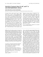
On human chromosome 11p15, the trim5 gene is
located with a cluster of other trim genes, including
ro52, trim68, trim6, trim34, trim22 and Trim3.Itisof
particular interest that trim6, trim34, trim5 and trim22
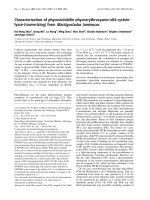
are assembled at adjacent loci [16] (Fig. 1A). This
chromosomal localization suggests that these trim
genes were generated by amplification from a single
gene on chromosome 11p15. To investigate the respec-
tive molecular evolution of these gene products, a phy-
logenetic study was performed (Fig. 1B). As expected,
TRIM5a, TRIM6, TRIM34, and TRIM22, the genes
for which are clustered at the chromosomal loci, are
also clustered in the phylogenetic tree. Importantly,
A
B
C
Fig. 1. Relationship between Ro52 and TRIM5. (A) Loci of trim
genes on human chromosome 11p15. (B) Phylogenetic tree of
TRIM family members encoded by genes on human chromo-
some 11p15. Amino acid sequences of TRIM family members
were aligned using
CLUSTAL W. The alignment was then used to
build trees in
MEGA3.1, using the neighbor-joining method. The scale
bar represents evolutionary distance in substitutions ⁄ amino acid
residues. (C) Schematic representation of domain structure of
human Ro52 and TRIM5a.
K. Yamauchi et al. Ubiquitination of TRIM5a and its role
FEBS Journal 275 (2008) 1540–1555 ª 2008 The Authors Journal compilation ª 2008 FEBS 1541
the evolutionary distance of TRIM5a is very close to
that of Ro52, TRIM6, and TRIM34, suggesting that
TRIM proteins such as Ro52, TRIM5a, TRIM6 and
TRIM34 have a similar function. Next, we investigated
the domain structure of TRIM5a and Ro52. As shown
in Fig. 1C, both proteins possess RING-finger and
B-box domains in the N-terminal region. In the central
region, Ro52 contains two separated coiled-coil
domains, whereas TRIM5a contains a fused coiled-coil
domain. In the C-terminal region, both proteins con-
tain a B30.2 domain. Thus, the domain structure of
TRIM5a is almost identical to that of Ro52, implying
that the two proteins have similar functions.
TRIM5a is ubiquitinated in the presence of
UbcH5B in vitro
Because TRIM5a was phylogenetically and structurally
similar to Ro52, which is a RING-type E3 ubiquitin
ligase, we hypothesized that TRIM5a also functions as
an E3 enzyme. However, this hypothesis raised the
question as to what the substrate of the TRIM5a-med-
iated ubiquitination is. Previously, we found that Ro52
acts as an E3 enzyme and ubiquitinates itself (self-
ubiquitination) [6–8], suggesting that TRIM5a likewise
acts as an E3 enzyme and ubiquitinates itself. We
therefore performed an in vitro ubiquitination assay to
test this possibility.
In the assay, maltose-binding protein (MBP)-fused
TRIM5a was expressed in bacteria and purified using
amylose resin beads. MBP–TRIM5a immobilized on
the beads was then incubated with recombinant E1
enzyme and different recombinant E2 enzymes
(UbcH2, UbcH5B, UbcH7, UbcH10, and hCDC34,
which were produced in bacteria) in the presence of
RGS-poly-His (RH)-tagged ubiquitin. In this in vitro
system, MBP–TRIM5a served as both a potential sub-
strate and a potential E3 enzyme for its self-ubiquitina-
tion. After the incubation, MBP–TRIM5a was
solubilized and analyzed by western blotting, using
antibodies to RH and to MBP. As shown in Fig. 2A,
the incubation of MBP–TRIM5a in the reaction mix-
ture containing UbcH2, UbcH7, UbcH10 or hCDC34
did not result in the ubiquitination of MBP–TRIM5a,
whereas the incubation of MBP–TRIM5a in the reac-
tion mixture containing UbcH5B resulted in both
the monoubiquitination and polyubiquitination of
MBP–TRIM5a. These results indicate that TRIM5a is
ubiquitinated in vitro and that this ubiquitination is
catalyzed by UbcH5B but not by other E2 enzymes.
Interestingly, Ro52, which is phylogenetically and
structurally close to TRIM5a, also catalyzes ubiquiti-
nation in cooperation with UbcH5B [7].
TRIM5a functions as an E3 enzyme and
ubiquitinates itself in vitro
In general, ubiquitin conjugates to the substrate in the
presence of E1, E2 and E3 enzymes. These proteins are
the minimum ones required for ubiquitination to occur.
To confirm whether these proteins are also essential for
the ubiquitination shown in Fig. 2A, we performed
another in vitro ubiquitination assay (Fig. 2B). As a
positive control, amylose resin beads coated with
MBP–TRIM5a were incubated in the complete reaction
mixture containing RH–ubiquitin, recombinant E1
enzyme, and recombinant UbcH5B (E2 enzyme)
(Fig. 2B, lane 5). As a negative control, amylose resin
beads alone (i.e. not coated with MBP–TRIM5a) were
incubated in the complete reaction mixture (Fig. 2B,
lane 1). In the other reactions, amylose resin beads
coated with MBP–TRIM5a were incubated in an
incomplete reaction mixture lacking one of these com-
ponents (Fig. 2B, lanes 2–4). After the incubation,
MBP–TRIM5a
was solubilized and analyzed by western
blotting using antibody to RH and antibody to MBP.
As shown in Fig. 2B, incubation of MBP–TRIM5a in
the complete reaction mixture resulted in the ubiquitina-
tion of MBP–TRIM5a (lane 5), whereas incubation of
MBP–TRIM5a in the incomplete reaction mixture lack-
ing one component did not lead to the ubiquitination of
MBP–TRIM5a (lanes 2–4). These results indicate that
ubiquitin, E1 enzyme and UbcH5B (E2 enzyme) are the
minimum requirement for the in vitro ubiquitination of
TRIM5a. Because the reaction mixtures used in this
assay did not contain any E3 enzymes other than
TRIM5a, these results also indicate that TRIM5a
functions as an E3 enzyme and ubiquitinates itself.
In vitro self-ubiquitination of TRIM5a is mediated
by its RING-finger domain
TRIM5a possesses a RING-consensus sequence
(Cys-X
2
-Cys-X
9–39
-Cys-X
1–3
-His-X
2–3
-Cys-X
2
-Cys-X
4–48
-
Cys-X
2
-Cys) between amino acids 15 and 58 [3,17]
(Fig. 3A). This sequence coordinates two zinc ions in a
‘cross-braced’ fashion [17,18]. Recent results from sev-
eral laboratories have indicated that the RING-finger
proteins recruit E2 enzymes through their RING
domain and act as an E3 enzyme [13]. This E3 activity
of RING-finger proteins has been shown to be abol-
ished by a mutation of the conserved Cys or His resi-
due described above [7,19,20]. To determine whether
the E3 activity of TRIM5a is dependent on its RING-
finger domain, we substituted Ala for the conserved
Cys15 in the RING-finger domain to generate a
TRIM5a mutant (C15A) (Fig. 3A). Then, we tested
Ubiquitination of TRIM5a and its role K. Yamauchi et al.
1542 FEBS Journal 275 (2008) 1540–1555 ª 2008 The Authors Journal compilation ª 2008 FEBS
whether this mutation abolishes the E3 activity of
TRIM5a, using an in vitro ubiquitination assay.
In the assay, MBP-fused wild-type TRIM5a or its
C15A mutant was expressed in bacteria and purified
using amylose resin beads. MBP–TRIM5a immobilized
on the beads was then incubated with RH–ubiquitin,
recombinant E1 enzyme, and recombinant UbcH5B
(E2 enzyme). After the incubation, MBP–TRIM5a was
solubilized and analyzed by western blotting, using
antibody to RH and antibody to MBP. As shown in
Fig. 3B, the wild-type TRIM5a ubiquitinated itself
(lanes 1 and 3), whereas the C15A mutant did not
ubiquitinate itself at all (lanes 2 and 4). These results
indicate that the in vitro self-ubiquitination of TRIM5a
is dependent on its RING-finger domain. Thus, we
confirmed that TRIM5a is a RING-motif-dependent
E3 enzyme.
TRIM5a is self-ubiquitinated in human embryonic
kidney (HEK) 293T cells
The E3 activity of TRIM5a was determined by in vitro
assays, as described in the preceding sections. There-
fore, this raised the question of whether TRIM5a func-
tions as an E3 enzyme in human cells. To determine
this, we performed an in vivo ubiquitination assay
using the wild-type TRIM5a and its RING-finger
mutant (C15A). In brief, RH-tagged wild-type
TRIM5a or its C15A mutant was expressed with or
without hemagglutinin epitope (HA)-tagged ubiquitin
in HEK293T cells. The cells were then harvested and
lysed under denaturing conditions. Afterwards,
TRIM5a–RH (wild-type or C15A) in the lysate was
precipitated by TALON beads, solubilized, and ana-
lyzed by western blotting, using antibody to HA to
detect ubiquitinated TRIM5a–RH, and antibody to
RH to detect both nonubiquitinated and ubiquitinated
TRIM5a–RH. As shown in Fig. 4, the wild-type
TRIM5a was monoubiquitinated and also polyubiqui-
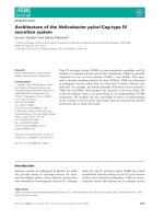
Fig. 2. In vitro self-ubiquitination of TRIM5a. (A) UbcH5B-depen-
dent self-ubiquitination of TRIM5a. MBP-fused TRIM5a was puri-
fied using amylose resin beads and incubated with the reaction
mixture containing RH-tagged ubiquitin, recombinant E1 enzyme,
and various poly-His-tagged recombinant E2 enzymes (UbcH2,
UbcH5B, UbcH7, UbcH10, and hCDC34). After this reaction, MBP–
TRIM5a immobilized on the beads was washed to remove the
reaction mixture and solubilized in SDS treatment solution. MBP–
TRIM5a was then analyzed by western blotting, using antibody to
RH to detect ubiquitinated MBP–TRIM5a (upper panel), and anti-
body to MBP to detect both nonubiquitinated and ubiquitinated
MBP–TRIM5a (lower panel). Molecular size markers are shown on
the left in kilodaltons (kDa). (B) Minimum requirements for the
in vitro self-ubiquitination of TRIM5a. In the in vitro ubiquitination
assay, the complete reaction mixture contained RH–ubiquitin, E1
enzyme, and UbcH5B as an E2 enzyme. To determine the mini-
mum requirements for the self-ubiquitination of TRIM5a, MBP–
TRIM5a immobilized on amylose resin beads was incubated in the
incomplete reaction mixture lacking one of these components
(lanes 2–4). As a positive control, MBP–TRIM5a immobilized on
amylose resin beads was incubated in the complete mixture
(lane 5). As a negative control, amylose resin beads alone without
immobilization of MBP–TRIM5a were incubated in the complete
mixture (lane 1). After the reaction, the beads were treated in SDS-
containing solution to solubilize MBP–TRIM5a. Then, MBP–TRIM5a
was analyzed by western blotting, using antibody to RH to detect
ubiquitinated MBP–TRIM5a (upper panel), and antibody to MBP to
detect both nonubiquitinated and ubiquitinated MBP–TRIM5 a
(lower panel). The incomplete reaction mixture shown in lanes 2, 3
and 4 lacked RH–ubiquitin, E1 enzyme, and UbcH5B, respectively.
A
B
K. Yamauchi et al. Ubiquitination of TRIM5a and its role
FEBS Journal 275 (2008) 1540–1555 ª 2008 The Authors Journal compilation ª 2008 FEBS 1543
tinated (or multimonoubiquitinated) when overexpres-
sed with HA–ubiquitin in HEK293T cells (lanes 3 and
7). In contrast, the ubiquitination of the C15A mutant
was extremely weak, even when overexpressed with
HA–ubiquitin in HEK293T cells (Fig. 4, lanes 4 and
8). This faint ubiquitination of TRIM5a(C15A) might
have been catalyzed by the wild-type TRIM5a or other
E3 ubiquitin ligases that are endogenously expressed in
HEK293T cells. These results indicate that TRIM5a
ubiquitinates itself through the function of its RING-
finger domain in HEK293T cells.
Ro52 strongly ubiquitinates itself and TRIM5a
in HEK293T cells
TRIM5a functions as an E3 ubiquitin ligase, because
it ubiquitinates itself both in vitro (Figs 2 and 3) and
in vivo (Fig. 4), as does Ro52 [7]. Because TRIM5a is
structurally similar to Ro52 (Fig. 1C), we wondered
whether TRIM5a and Ro52 cross-ubiquitinate (or
trans-ubiquitinate) each other in addition to undergo-
ing self-ubiquitination. In other words, we wondered
whether Ro52 ubiquitinates TRIM5a and whether
TRIM5a ubiquitinates Ro52. To test the first possibil-
ity, we performed the in vivo ubiquitination assay,
using a wild-type Ro52 as an E3 ubiquitin ligase. As
a substrate, we used a RING mutant of Ro52
(positive control) or of TRIM5a to avoid the self-
ubiquitination. Specifically, RH-tagged Ro52(C16A) or
TRIM5a(C15A) was expressed with HA-tagged ubiqu-
itin and FLAG-tagged Ro52 (wild-type or its mutant
C16A) in HEK293T cells. The cells were then
harvested and lysed under denaturing conditions.
Ro52(C16A)–RH or TRIM5a(C15A)–RH in the lysate
was precipitated with cobalt-coated TALON beads,
solubilized in SDS solution, and then analyzed by wes-
tern blotting, using antibody to RH to detect both
nonubiquitinated and ubiquitinated forms, and anti-
body to HA to detect ubiquitinated forms. As shown
in Fig. 5A, both Ro52(C16A)–RH (upper panel) and
TRIM5a(C15A)–RH (lower panel) were strongly
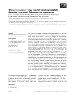
Fig. 4. E3 activity of wild-type TRIM5a and its RING mutant C15A
in HEK293T cells. RH-tagged wild-type TRIM5a or its RING mutant
C15A was expressed with or without HA-tagged ubiquitin in
HEK293T cells by plasmid transfection. Twenty hours after trans-
fection, the cells were harvested and lysed under denaturing
conditions. TRIM5a–RH (wild-type or C15A) in the lysate was pre-
cipitated with cobalt-coated TALON beads and solubilized in 2%
SDS solution. The solubilized TRIM5a–RH was then analyzed by
western blotting, using antibody to RH to detect both nonubiquiti-
nated and ubiquitinated TRIM5a–RH (lanes 1–4), and antibody to
HA to detect ubiquitinated TRIM5a–RH (lanes 5–8).
AB
Fig. 3. E3 activity of wild-type TRIM5a and its RING mutant in vitro. (A) Schematic representation of the RING-finger domain of TRIM5a.
The amino acid sequence and structure of the RING-finger domain are shown. Asterisks indicate conserved Cys and His residues in the
RING-finger domain. Arrows indicate Cys15, which was replaced by Ala to generate the TRIM5a(C15A). (B) In vitro ubiquitination assay using
wild-type TRIM5a and its RING mutant C15A. MBP–TRIM5a (wild-type) or MBP–TRIM5a(C15A) was purified with amylose resin beads from
bacterial lysate and incubated with the reaction mixture containing RH–ubiquitin, E1 enzyme, and UbcH5B. After the reaction, MBP–TRIM5a
immobilized on the beads was solubilized and analyzed by western blotting, using antibody to MBP to detect both nonubiquitinated and ubi-
quitinated MBP–TRIM5a (lanes 1 and 2), and antibody to RH to detect ubiquitinated MBP–TRIM5a (lanes 3 and 4).
Ubiquitination of TRIM5a and its role K. Yamauchi et al.
1544 FEBS Journal 275 (2008) 1540–1555 ª 2008 The Authors Journal compilation ª 2008 FEBS
monoubiquitinated and polyubiquitinated when coex-
pressed with wild-type FLAG–Ro52 in HEK293T cells
(lanes 4 and 9). In contrast, the ubiquitination of
Ro52(C16A)–RH and TRIM5a(C15A)–RH was extre-
mely weak when wild-type FLAG–Ro52 was not coex-
pressed in HEK293T cells (Fig. 5A, lanes 3 and 8) and
when FLAG-tagged inactive Ro52(C16A) was coex-
pressed in HEK293T cells (Fig. 5A, lanes 5 and 10).
This faint ubiquitination of Ro52(C16A) and
TRIM5a(C15A) might have been catalyzed by the
wild-type Ro52, TRIM5a, or other E3 ubiquitin ligases
that are endogenously expressed in HEK293T cells.
These results indicate that Ro52 ubiquitinates both
itself and TRIM5a in HEK293T cells.
TRIM5a ubiquitinates itself, but not Ro52,
in HEK293T cells
Next, we examined whether TRIM5a ubiquitinates
Ro52 in HEK293T cells, using a wild-type TRIM5a as
an E3 ubiquitin ligase. As a substrate, we used a
RING mutant of Ro52 or of TRIM5a (positive con-
trol) to avoid self-ubiquitination. Specifically, RH-
tagged Ro52(C16A) or TRIM5a(C15A) was expressed
with HA–ubiquitin and FLAG–TRIM5a (wild-type or
its mutant C15A) in HEK293T cells. The cells were
then harvested and lysed under denaturing conditions.
Ro52(C16A)–RH or TRIM5a(C15A)–RH in the lysate
was precipitated with cobalt-coated TALON beads,
solubilized in SDS solution, and then analyzed by wes-
tern blotting, using antibody to RH and antibody to
HA. As shown in the upper panel of Fig. 5B, the
Ro52(C16A)–RH was weakly monoubiquitinated and
polyubiquitinated in HEK293T cells when wild-type
FLAG–TRIM5a was not coexpressed (lanes 3 and 8)
A
B
C
Fig. 5. In vivo assay of self-ubiquitination and cross-ubiquitination
between Ro52 and TRIM5a. (A) In vivo ubiquitination by Ro52 E3
ubiquitin ligase. To examine the ubiquitination of RH-tagged
Ro52(C16A) and TRIM5a(C15A) by FLAG–Ro52, Ro52(C16A)–RH or
TRIM5a(C15A)–RH was expressed with HA–ubiquitin and FLAG–
Ro52 (wild-type or C16A) in HEK293T cells by plasmid transfection.
Twenty hours after transfection, the cells were harvested and lysed
under denaturing conditions. Ro52(C16A)–RH or TRIM5a(C15A)–RH
in the lysate was precipitated with TALON beads and solubilized in
2% SDS solution. The solubilized Ro52(C16A)–RH (upper panel) or
TRIM5a(C15A)–RH (lower panel) was then analyzed by western
blotting, using antibody to RH to detect both nonubiquitinated and
ubiquitinated forms (lanes 1–5), and antibody to HA to detect the
ubiquitinated form (lanes 6–10). (B) In vivo ubiquitination by TRIM5a
E3 ubiquitin ligase. To examine the ubiquitination of RH-tagged
Ro52(C16A) or TRIM5a(C15A) by FLAG–TRIM5a, Ro52(C16A)–RH
or TRIM5a(C15A)–RH was expressed with HA–ubiquitin and FLAG–
TRIM5a (wild-type or C15A) in HEK293T cells by plasmid transfec-
tion. Twenty hours after transfection, the cells were harvested
and lysed under denaturing conditions. Ro52(C16A)–RH or
TRIM5a(C15A)–RH in the lysate was precipitated with cobalt-
coated TALON beads and solubilized in 2% SDS solution. The solu-
bilized Ro52(C16A)–RH (upper panel) or TRIM5a(C15A)–RH (lower
panel) was then analyzed by western blotting, using antibody to RH
to detect both nonubiquitinated and ubiquitinated forms (lanes 1–5),
and antibody to HA to detect the ubiquitinated form (lanes 6–10). A
nonspecific band is indicated by an asterisk. (C) Schematic sum-
mary of self-ubiquitination and cross-ubiquitination between Ro52
and TRIM5a.
K. Yamauchi et al. Ubiquitination of TRIM5a and its role
FEBS Journal 275 (2008) 1540–1555 ª 2008 The Authors Journal compilation ª 2008 FEBS 1545
and when FLAG-tagged inactive TRIM5a(C15A) was
coexpressed (lanes 5 and 10). Importantly, the level of
the ubiquitination of Ro52(C16A) was not changed
when wild-type FLAG–TRIM5a was coexpressed
(Fig. 5B, lanes 4 and 9), suggesting that Ro52 is not
ubiquitinated by TRIM5a in HEK293T cells. The faint
ubiquitination of Ro52(C16A) seen in Fig. 5B
(lanes 3–5 and 8–10) seemed to be catalyzed by the
wild-type Ro52 or other E3 ubiquitin ligases that were
endogenously expressed in HEK293T cells. In contrast,
TRIM5a(C15A)–RH was more strongly ubiquitinated
by wild-type FLAG–TRIM5a (Fig. 5B, lower panel,
lanes 4 and 9).
TRIM5a is ubiquitinated by Ro52 more strongly
than TRIM5a in HEK293T cells
As summarized in Fig. 5C, we showed two things using
the in vivo ubiquitination assay. First, TRIM5a is ubiq-
uitinated by itself and Ro52. Second, Ro52 is ubiquiti-
nated by itself, but not by TRIM5a. These results
raised a question: which E3 ligase predominantly ubiq-
uitinates TRIM5a? In other words, is TRIM5a ubiqui-
tinated more strongly by itself or by Ro52? To address
this question, we performed an in vivo ubiquitination
assay (Fig. 6). FLAG-tagged wild-type TRIM5a and
wild-type Ro52 were used as E3 ubiquitin ligases, and
TRIM5a(C15A)–RH was used as a substrate. In brief,
FLAG-tagged wild-type TRIM5a and Ro52 were
expressed with HA–ubiquitin and TRIM5a(C15A)–RH
in HEK293T cells, by plasmid transfection. The cells
were then harvested. Some of the cells were lysed in the
SDS treatment solution, and FLAG-tagged proteins
were analyzed by western blotting, using antibody to
FLAG. As shown in the upper panel of Fig. 6, the
expression levels of FLAG–TRIM5a and Ro52 were
almost equal (lane 3 versus lane 4). The rest of the cells
were also lysed under denaturing conditions to precipi-
tate TRIM5a(C15A)–RH with TALON beads.
TRIM5a(C15A)–RH was then solubilized in 2% SDS
solution and analyzed by western blotting, using anti-
body to HA and antibody to RH. As shown in the
middle panel of Fig. 6, TRIM5a(C15A)–RH is ubiqui-
tinated by FLAG–Ro52 (wild-type) more strongly than
by FLAG–TRIM5a (wild-type) in HEK293T cells
(lane 4 versus lane 3), suggesting that Ro52 has higher
E3 ligase activity for this ubiquitination.
UnpEL
⁄
Usp4 deubiquitinates Ro52, but not
TRIM5a, in HEK293T cells
Recently, we showed that UnpEL is an isopeptidase
used to deubiquitinate Ro52 [10]. Because TRIM5a
and Ro52 are phylogenetically and structurally similar,
as described above, we hypothesized that TRIM5a is
also deubiquitinated by UnpEL. To test this hypothe-
sis, we performed an in vivo deubiquitination assay
using UnpEL as described previously [10,21] (Fig. 7).
As a control, we used a deubiquitinating enzyme,
YopJ (Fig. 8) (see below). Specifically, TRIM5a and
ubiquitin were expressed in HEK293T cells along with
empty vector, wild-type UnpEL, or UnpEL(C311A),
Fig. 6. Ubiquitination of TRIM5a by TRIM5a and Ro52: a compara-
tive study. To compare the ligase activities of TRIM5a and Ro52
for the ubiquitination of TRIM5a,anin vivo ubiquitination assay
was performed. FLAG-tagged TRIM5a (wild-type) and Ro52 (wild
type) were expressed with HA–ubiquitin and TRIM5a(C15A)–RH as
a substrate in HEK293T cells. Twenty hours after transfection, the
cells were harvested. Some of the cells were lysed and analyzed
by western blotting, using antibody to FLAG to show the expres-
sion level of FLAG–TRIM5a (wild-type) and Ro52 (wild-type) (upper
panel). The rest of the cells were also lysed under denaturing
conditions to precipitate TRIM5a(C15A)–RH with TALON beads.
TRIM5a(C15A)–RH was then solubilized in 2% SDS solution and
analyzed by western blotting, using antibody to HA (middle panel)
and antibody to RH (lower panel).
Ubiquitination of TRIM5a and its role K. Yamauchi et al.
1546 FEBS Journal 275 (2008) 1540–1555 ª 2008 The Authors Journal compilation ª 2008 FEBS
in which Ala was substituted for the active site
Cys311. TRIM5 a was then precipitated using TALON
beads, after which it was solubilized and then analyzed
by western blotting to detect ubiquitinated TRIM5a
(Fig. 7, lanes 1–3). To demonstrate the isopeptidase
activity of UnpEL [10], Ro52 was also used as a posi-
tive control for the substrate (Fig. 7, lanes 4–6). As
shown in the upper and lower panels of Fig. 7, there
was strong ubiquitination of Ro52 when Ro52 and
ubiquitin were coexpressed with empty vector (lane 4).
Importantly, however, their coexpression with wild-
type UnpEL greatly reduced the level of ubiquitinated
Ro52, because of UnpEL’s isopeptidase activity
(Fig. 7, lane 5). In contrast, the coexpression of Ro52
and ubiquitin with UnpEL(C311A) did not affect the
ubiquitination of Ro52 (Fig. 7, lane 6), because of the
substitution of Ala for the active site Cys311 in
UnpEL(C311A). Thus, we clearly detected the isopep-
tidase activity of UnpEL when Ro52 was the substrate
but not when TRIM5a was the substrate (Fig. 7,
lanes 1–3). Specifically, we detected ubiquitination of
TRIM5a when TRIM5a and ubiquitin were coex-
pressed with empty vector (Fig. 7, lane 1). Unexpect-
edly, however, their coexpression with wild-type
UnpEL did not reduce the level of ubiquitinated
TRIM5a (Fig. 7, lane 2), indicating that UnpEL does
not deubiquitinate TRIM5a in HEK293T cells.
YopJ deubiquitinates both Ro52 and TRIM5a
in HEK293T cells
YopJ is one of the Yersinia outer proteins encoded by
pathogenic Yersinia species. In particular, YopJ is a
cysteine protease that is thought to remove ubiquitin
or a ubiquitin-like modification from target proteins in
host cells [22]. As described above, we chose YopJ as a
control against UnpEL because we initially expected
that UnpEL would deubiquitinate both Ro52 and
TRIM5a, but YopJ would not. To test the possibility
that YopJ would not deubiquitinate either Ro52 or
TRIM5a, we performed an in vivo deubiquitination
assay. First, we used Ro52 as a substrate. Specifically,
Ro52 and ubiquitin were expressed in HEK293T cells
along with empty vector, wild-type YopJ, or
YopJ(C172S), in which Ser was substituted for the
active site Cys172. Ro52 was then precipitated, solubi-
lized, and analyzed by western blotting to detect ubi-
quitinated Ro52 (Fig. 8A). As shown in the upper and
lower panels of Fig. 8A, we detected strong ubiquitina-
tion of Ro52 when Ro52 and ubiquitin were co-
expressed with empty vector (lane 2). Surprisingly, in
contrast, their coexpression with wild-type YopJ
greatly reduced the level of ubiquitinated Ro52, due to
its isopeptidase activity (Fig. 8A, lane 4). The coex-
pression of Ro52 and ubiquitin with YopJ (C172S),
however, did not affect the ubiquitination of Ro52
(Fig. 8A, lane 6), because of substitution of Ser for the
active site Cys172 in this mutant YopJ. Thus, the
detection of isopeptidase activity of YopJ when Ro52
was used as a substrate was unexpected.
Because TRIM5a and Ro52 are phylogenetically
and structurally similar, as described above, we then
hypothesized that TRIM5a is also deubiquitinated by
YopJ. To test this hypothesis, we performed the same
in vivo deubiquitination assay as described above. As
shown in Fig. 8B, we strongly detected the ubiquitina-
Fig. 7. In vivo deubiquitination by isopeptidase activity of human
UnpEL. HA-tagged ubiquitin was coexpressed with RH-tagged
TRIM5a (lanes 1–3) or Ro52 (lanes 4–6) in HEK293T cells. In addi-
tion, empty vector (lanes 1 and 4), FLAG–UnpEL (wild-type) (lanes 2
and 5) or FLAG-tagged UnpEL mutant with a single substitution
(C311A) (lanes 3 and 6) was also coexpressed. The cells were
lysed in 6
M guanidine hydrochloride. TRIM5a–RH or Ro52–RH in
the lysate was then precipitated with cobalt-coated TALON beads
and analyzed by western blotting, using antibody to HA to detect
ubiquitinated TRIM5a–RH or Ro52–RH (upper panel), and antibody
to RH to detect all derivatives of TRIM5a–RH or Ro52–RH (lower
panel).
K. Yamauchi et al. Ubiquitination of TRIM5a and its role
FEBS Journal 275 (2008) 1540–1555 ª 2008 The Authors Journal compilation ª 2008 FEBS 1547
tion of TRIM5a when TRIM5a and ubiquitin were co-
expressed with empty vector (lane 2). In contrast, their
coexpression with wild-type YopJ greatly reduced the
level of ubiquitinated TRIM5a, because of its isopepti-
dase activity (Fig. 8B, lane 4). The coexpression of
TRIM5a and ubiquitin with YopJ(C172S), however,
did not affect the ubiquitination of TRIM5a (Fig. 8B,
lane 6). This is because an active site Cys172 was
replaced by Ser in YopJ(C172S). Thus, we found by
chance that YopJ deubiquitinates both Ro52 and
TRIM5a in HEK293T cells.
Finally, we examined the enzymatic specificity of
YopJ, using hypoxia-inducible factor (HIF)1a(DC) as
a negative control for the substrate. HIF1a(DC), an
N-terminal fragment (amino acids 1–330) of HIF1a,
was previously shown to be polyubiquitinated [7,23].
To confirm that YopJ does not deubiquitinate
HIF1a(DC), HIF1a(DC) and ubiquitin were expressed
with empty vector, wild-type YopJ or YopJ(C172S) in
HEK293T cells. HIF1a(DC) was then precipitated,
solubilized, and analyzed by western blotting to detect
ubiquitinated HIF1a(DC) (Fig. 8C). As shown in the
upper panel of Fig. 8C, we detected strong ubiquitina-
tion of HIF1a(DC) when HIF1a(DC) and ubiquitin
were coexpressed with empty vector (lane 2) or
YopJ(C172S) (lane 6). As expected, the wild-type YopJ
did not affect this ubiquitination (Fig. 8C, lane 4),
indicating that YopJ does not deubiquitinate
HIF1a(DC).
Ubiquitinated Ro52 and TRIM5a are not stabilized
by proteasome inhibitors
In the sections above, we demonstrated the ubiquitina-
tion of TRIM5a in vitro and in vivo. Because we previ-
ously showed that self-ubiquitination of Ro52 does not
target it to the proteasome for degradation [7], we
hypothesized that ubiquitination of TRIM5a does not
lead to proteasomal degradation either. To test this
hypothesis, we performed an in vivo ubiquitination
assay (Fig. 9). Specifically, using the proteasome inhib-
itor MG115 (Fig. 9A) or MG132 (Fig. 9B), we inhib-
ited the proteasomal degradation in HeLa cells to
determine whether the ubiquitinated TRIM5a was
accumulated. Briefly, TRIM5a–RH was coexpressed
with HA–ubiquitin in HeLa cells in the presence or
absence of proteasome inhibitor MG115 or MG132.
The cells were then harvested and lysed under denatur-
ing conditions. Afterwards, TRIM5a–RH in the lysate
was precipitated with TALON beads, solubilized, and
then analyzed by western blotting using antibody
to HA to detect ubiquitinated TRIM5a–RH, and
AB C
Fig. 8. In vivo deubiquitination by isopeptidase activity of YopJ. (A) In vivo deubiquitination assay of YopJ using Ro52 as a substrate.
(B) In vivo deubiquitination assay of YopJ using TRIM5a as a substrate. (C) In vivo deubiquitination assay of YopJ using HIF1a N-terminal
fragment (DC) as a substrate. RH-tagged Ro52, TRIM5a or HIF1a(DC) was expressed without HA–ubiquitin (lanes 1, 3, and 5) or with
HA–ubiquitin (lanes 2, 4, and 6) in HEK293T cells. In addition, empty vector (lanes 1 and 2), FLAG–YopJ (wild-type) (lanes 3 and 4) or
FLAG-tagged YopJ mutant with a single substitution (C172S) (lanes 5 and 6) was also coexpressed. The cells were lysed in 6
M guanidine
hydrochloride. RH-tagged substrate, such as Ro52, TRIM5a, or HIF1a(DC), in the lysate was then precipitated with cobalt-coated TALON
beads and analyzed by western blotting, using antibody to HA to detect ubiquitinated substrate (upper panel), and antibody to RH to detect
all derivatives of the substrate (lower panel). A nonspecific band is indicated by an asterisk.
Ubiquitination of TRIM5a and its role K. Yamauchi et al.
1548 FEBS Journal 275 (2008) 1540–1555 ª 2008 The Authors Journal compilation ª 2008 FEBS
antibody to RH to detect both nonubiquitinated and
ubiquitinated TRIM5a–RH. As a positive control for
the effect of proteasome inhibitor on proteasomal
degradation of ubiquitinated proteins, we used
HIF1a(DC), because we had previously detected a
clear effect of MG132 on the proteasomal degradation
of HIF1a(DC) [7,23]. As a negative control for the
effect of proteasome inhibitor, we used Ro52 [7]. As
shown in the lower panel of Fig. 9, the treatment with
proteasome inhibitors (MG115 and MG132) did not
increase the expression of either ubiquitinated
TRIM5a–RH (lane 9 versus lane 12) or unubiquitinat-
ed TRIM5a–RH (lane 3 versus lane 6). These results
suggested that the ubiquitination of TRIM5a does not
lead to its proteasomal degradation in HeLa cells. As
expected, a negative control Ro52 was not stabilized
by the treatment with MG115 and MG132 either
(Fig. 9, middle panel). In contrast, a positive control
HIF1a(DC) was stabilized by the treatment with
MG115 and MG132. As shown in the upper panel of
Fig. 9, the treatment with MG115 and MG132
increased the expression of HIF1 a(DC)–RH (lane 3
versus lane 6). Furthermore, the treatment increased
the amount of ubiquitinated HIF1a(DC)–RH (Fig. 9,
lane 9 versus lane 12), because MG115 and MG132
inhibited the proteasomal degradation of the ubiquiti-
nated HIF1a(DC)–RH, resulting in its accumulation.
These results suggested that the ubiquitination of
HIF1a(DC) targets it to proteasomal degradation.
Monoubiquitin-fusion of Ro52 and TRIM5a causes
their translocation from cytoplasmic bodies to
cytoplasm in human cells
As described above, the ubiquitination of Ro52 and
TRIM5a did not cause their proteasomal degradation.
This raises the following question: what is the biologi-
cal relevance of the ubiquitination of Ro52 and
TRIM5a? Because monoubiquitination appeared to be
dominant in their ubiquitination (see Discussion), we
investigated the biological relevance of their mono-
ubiquitination, using monoubiquitin-fused Ro52 and
TRIM5a. Specifically, we examined whether the direct
fusion of a monoubiquitin to Ro52 or TRIM5 a causes
its translocation. In the molecule of monoubiquitin-
fused protein, however, the monoubiquitin links to the
N-terminal Met residue of the protein with an a-pep-
tide bond. This linkage is artificial, not being found
naturally in cells. In the molecule of naturally mono-
ubiquitinated protein in cells, the monoubiquitin links
to the target Lys residue of the protein with an isopep-
tide bond. Thus, the monoubiquitin-fusion product is
different from the natural monoubiquitination product,
A
B
Fig. 9. Effects of proteasome inhibitors on the expression of
HIF1a, Ro52 and TRIM5a in HeLa cells. (A) Expression of RH-
tagged HIF1a(DC), Ro52 and TRIM5a in MG115-treated cells. (B)
Expression of RH-tagged HIF1a(DC), Ro52 and TRIM5a in MG132-
treated cells. HIF1a(DC)–RH, Ro52–RH or TRIM5a –RH was coex-
pressed in HeLa cells with empty vector (lanes 2, 5, 8, and 11) or
HA-tagged ubiquitin (lanes 3, 6, 9, and 12). The HeLa cells were
cultured for 6 h in the absence (lanes 1–3 and 7–9) or presence
(lanes 4–6 and 10–12) of a proteasome inhibitor, either MG115 or
MG132. After incubation, the cells were harvested and lysed under
denaturing conditions. RH-tagged proteins in the lysate were pre-
cipitated with cobalt-coated TALON beads and solubilized in 2%
SDS solution. The solubilized RH-tagged proteins were then ana-
lyzed by western blotting in which both nonubiquitinated and ubi-
quitinated forms were detected by antibody to RH (lanes 1–6),
and the ubiquitinated form was detected by antibody to HA
(lanes 7–12).
K. Yamauchi et al. Ubiquitination of TRIM5a and its role
FEBS Journal 275 (2008) 1540–1555 ª 2008 The Authors Journal compilation ª 2008 FEBS 1549
but they are structurally similar. Therefore, using the
direct fusion of a monoubiquitin to target proteins, we
are able to investigate their monoubiquitination [24].
To investigate the subcellular translocation of
monoubiquitin-fused Ro52 and TRIM5a, the cDNA
of monoubiquitin (UbG) was first fused to that of
Ro52–enhanced green fluorescent protein (EGFP) or
TRIM5a–EGFP. Then, the UbG–Ro52–EGFP or
UbG–TRIM5a–EGFP was expressed in HEK293
cells. As controls, EGFP alone, Ro52–EGFP and
TRIM5a–EGFP were also expressed in HEK293 cells.
The cells were then examined with a fluorescence
microscope.
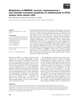
As shown in Fig. 10C,D, Ro52–EGFP mainly local-
ized to the rod- or spindle-like cytoplasmic bodies, a
finding that we and other groups have previously
reported [5,8,9,25]. In addition, although the signal
was weak, Ro52–EGFP was diffusely located in the
cytoplasm. However, Ro52 was hardly detectable in
the nucleus. As shown in Fig. 10G,H, TRIM5a–EGFP
strongly localized to the dot-like cytoplasmic bodies.
Interestingly, however, the shape of the TRIM5a(+)
cytoplasmic bodies differed from that of the Ro52(+)
cytoplasmic bodies. Furthermore, unlike Ro52–EGFP,
TRIM5a–EGFP was clearly located in the cytoplasm.
However, like Ro52–EGFP, TRIM5a–EGFP was
hardly detectable in the nucleus. Thus, in spite of small
differences, both Ro52–EGFP and TRIM5a–EGFP
predominantly localized to cytoplasmic bodies.
In contrast to Ro52–EGFP and TRIM5a–EGFP,
monoubiquitin-fused Ro52–EGFP (Fig. 10E,F) and
TRIM5a-EGFP (Fig. 10I,J) diffusely located to the
cytoplasm in HEK293 cells, suggesting that monoubiq-
uitination is a signal for Ro52 and TRIM5a to trans-
locate from cytoplasmic bodies to the cytoplasm.
Discussion
TRIM5a is a RING-finger protein that belongs to the
TRIM family [4]. Many RING-finger proteins have
been reported to recruit E2 ubiquitin-conjugating
enzymes and act as E3 ubiquitin ligases [13,14]. Fur-
thermore, some groups recently reported that TRIM
family members, such as ARD1 [26], TRIM37 [27],
and Ro52 [7], function as E3 ubiquitin ligases. In addi-
tion, TRIM5d, an alternative splicing product of the
trim5 gene, was shown to have E3 ubiquitin ligase
activity in vitro [15]. On the basis of these reports, we
hypothesized that TRIM5a is also an E3 ubiquitin
ligase. In the study presented here, we tested this
hypothesis using both in vitro and in vivo ubiquitina-
tion assays. As expected, we observed that TRIM5a
ubiquitinated itself in the presence of E1 enzyme and
E2 enzyme UbcH5B, indicating that TRIM5a func-
tions as an E3 enzyme for its self-ubiquitination, thus
proving our hypothesis. These findings, however,
raised two questions: what role does the self-ubiquiti-
nation play in the function of TRIM5 a, and what are
the substrates, other than the TRIM5a itself, in the
TRIM5a-mediated ubiquitination?
Recently, Diaz-Griffero et al. reported that TRIM5a
is polyubiquitinated, resulting in its rapid degradation
by the 26S proteasome [28]. This observation seems to
partially answer our first question, because the self-
ubiquitination of TRIM5a might lead to its rapid
degradation. In other words, the activity of TRIM5a
might be negatively regulated by its feedback mecha-
nism. Indeed, an E3 ubiquitin ligase Nrdp1 is regu-
lated by a similar mechanism. Namely, Nrdp1
polyubiquitinates itself, resulting in its proteasomal
degradation [29]. On the basis of these previous obser-
vations, we initially thought that the self-ubiquitination
of TRIM5a leads to its proteasomal degradation. To
confirm this, we performed an in vivo ubiquitination
assay using HeLa cells treated or not treated with pro-
teasome inhibitors. Although our assay was similar to
the assay performed by Diaz-Griffero et al. [28], the
results, contrary to our expectations, showed that
proteasome inhibitors do not increase either the ubi-
quitinated or nonubiquitinated forms of TRIM5a, sug-
gesting that the self-ubiquitination of TRIM5a does
not cause its proteasomal degradation. Given this, we
then asked, what is the biological relevance of ubiquiti-
nation of TRIM5a?
Importantly, when we performed the in vivo ubiqui-
tination assay of TRIM5a, we clearly detected the
monoubiquitination of TRIM5a in addition to its
polyubiquitination. Strangely, the monoubiquitination
was even stronger than the polyubiquitination when
TRIM5a was detected by western blotting using the
antibody to RH (Fig. 4, lane 3; Fig. 5, lane 4; Fig. 9,
lane 3). On the other hand, however, the polyubiquiti-
nation was strongly detected when antibody to HA
was used for western blotting (Fig. 4, lane 7; Fig. 5,
lane 9; Fig. 9, lane 9). Why did these two antibodies
produce such discrepant results in terms of degree of
polyubiquitination? We believe that a polyubiquitin
chain on TRIM5a–RH consists of multiple molecules
of HA–ubiquitin. Because each of these molecules
reacts with an antibody to HA, the polyubiquitin chain
is labeled with multiple molecules of antibody to HA,
which causes the antibody to HA to detect a much
higher level of polyubiquitin chain expression than is
actually the case. In contrast, detection of the poly-
ubiquitinated TRIM5a–RH by antibody to RH reflects
the actual level of the expression, because the antibody
Ubiquitination of TRIM5a and its role K. Yamauchi et al.
1550 FEBS Journal 275 (2008) 1540–1555 ª 2008 The Authors Journal compilation ª 2008 FEBS
to RH reacts only with a single RH-epitope of the
polyubiquitinated TRIM5a–RH. On the basis of this
explanation of our findings, the monoubiquitinated
form clearly exists in the actual population of ubiquiti-
nated TRIM5a. Because monoubiquitination plays
roles in proteasome-unrelated events, such as protein
trafficking and interaction with other proteins [30–33],
the monoubiquitination of TRIM5a might be involved
Ro52-EGFP Ro52-EGFP
TRIM5α-EGFP TRIM5α-EGFP
EGFP
+ DAPI
EGFP
AB
CD
UbG-Ro52-EGFPUbG-Ro52-EGFP
UbG-TRIM5α-EGFP
UbG-TRIM5α-EGFP
EF
GH
IJ
Fig. 10. Subcellular location of Ro52 and TRIM5a and their monoubiquitin-fused forms in HEK293 cells. EGFP alone, Ro52–EGFP, UbG-fused
Ro52–EGFP, TRIM5a–EGFP or UbG-fused TRIM5a–EGFP was expressed in HEK293 cells by plasmid transfection. After 20 h, the cells were
fixed and then treated with 4¢,6-diamidino-2-phenylindole (DAPI) to stain the nucleus. Afterwards, the cells were analyzed under a fluores-
cence microscope. The localization of EGFP alone (A, B), Ro52–EGFP (C, D), UbG-fused Ro52–EGFP (E, F), TRIM5a–EGFP (G, H) and
UbG-fused TRIM5a–EGFP (I, J) was shown by the green fluorescence of EGFP. The nuclear counterstaining was shown by the blue fluores-
cence of DAPI (B, D, F, H, J).
K. Yamauchi et al. Ubiquitination of TRIM5a and its role
FEBS Journal 275 (2008) 1540–1555 ª 2008 The Authors Journal compilation ª 2008 FEBS 1551
in these events. To test this hypothesis, we directly
fused monoubiquitin to the N-terminal Met residue
of TRIM5a and expressed it in HEK293 cells. The
monoubiquitin-fused TRIM5a diffusely located to the
cytoplasm in the cells, whereas TRIM5a without
monoubiquitin fusion mainly localized to cytoplasmic
bodies. Although the assay system is artificial, these
results suggest that monoubiquitination is a signal
for TRIM5a to translocate from cytoplasmic bodies to
the cytoplasm, supporting our hypothesis described
above.
In this study, we further investigated ‘cross-talk’ or
‘cross-ubiquitination’ between TRIM5a and Ro52,
because TRIM5a is phylogenetically and structurally
similar to Ro52. Our in vivo ubiquitination assay
revealed that TRIM5a does not ubiquitinate Ro52 but
is strongly ubiquitinated by Ro52. Importantly, the
ubiquitination of TRIM5a by Ro52 is stronger than
its self-ubiquitination, suggesting that Ro52 regulates
TRIM5a, but TRIM5a does not regulate Ro52, by
ubiquitination in cells. In addition to investigating
the ubiquitination of TRIM5a, we investigated its
deubiquitination. We found that the ubiquitination
of TRIM5a is downregulated by a deubiquitinating
enzyme, YopJ. Interestingly, both TRIM5a and YopJ
are closely involved in infectious diseases. TRIM5a is
a key player in host cells in the species-specific restric-
tion of HIV infection [3,4], whereas YopJ is one of the
pathogenic proteins derived from Yersinia species [22].
So far, there has been no report of a relationship
between HIV infection and Yersinia infection. How-
ever, our finding that the ubiquitination of TRIM5a is
clearly controlled by YopJ suggests that the species-
specific restriction of HIV infection might be altered
by Yersinia infection.
Although we identified TRIM5a as an E3 ubiquitin
ligase, we could not determine its substrates other than
itself. Therefore, we were not able to answer the sec-
ond question above. Nevertheless, some proteins have
been reported to interact or colocalize with TRIM5a
or its isoform TRIM5d. Specifically, TRIM5a interacts
with retroviral capsids [34,35], and TRIM5d colocalizes
with the novel topoisomerase I-interacting proteins
BTBD1 and BTBD2 in the cytoplasmic bodies [15].
These TRIM5-associating proteins derived from retro-
viruses and host cells are therefore thought to be can-
didates for the substrates ubiquitinated by TRIM5a.
To thoroughly elucidate TRIM5a-mediated biological
functions, such as the species-specific restriction of
HIV infection, it will be important to identify the sub-
strates in TRIM5a-mediated ubiquitination. This is
because TRIM5a seems to modify the function or sta-
bility of its substrates through ubiquitination, and this
modification might result in the TRIM5 a-mediated
biological events.
Experimental procedures
Cell culture
HEK293 cells, HEK293T cells and HeLa cells were
obtained from the American Type Culture Collection
(Manassas, VA, USA) and were maintained in DMEM
supplemented with 10% fetal bovine serum and antibiotics.
Antibodies
Mouse antibody to HA (16B12) was purchased from
Covance (Richmond, CA, USA). Mouse antibody to RH
(specific for the amino acid sequences RGSHHHH and
GGSHHHH) was purchased from Qiagen (Santa Clara,
CA, USA). Mouse antibody to FLAG (M2) was purchased
from Sigma (St Louis, MO, USA). Rabbit antibody to
MBP was purchased from New England Biolabs (Beverly,
MA, USA).
Preparation of cDNAs
The cDNAs of ubiquitin, Ro52 [36], TRIM5a (GenBank
accession number: DQ288685), UnpEL [10] and E2 ubiqu-
itin-conjugating enzymes, such as UbcH2, UbcH5B, UbcH7,
UbcH10 and hCDC34 [7] were amplified by PCR, using
appropriate primers from a human testis, heart or brain
cDNA library (Invitrogen, Carlsbad, CA, USA). The cDNA
of YopJ was kindly provided by K. Orth (University of
Texas Southwestern Medical Center, Dallas, TX, USA).
Site-directed mutagenesis
To abolish the ligase activity of TRIM5a, a Cys fi Ala sub-
stitution was generated at Cys15 in its RING-finger domain.
To abolish the deubiquitinating enzyme activity of YopJ, a
Cys fi Ser substitution was generated at Cys172 in its Cys-
box domain. For this purpose, the cDNA of wild-type
TRIM5a or YopJ was mutated by PCR-based site-directed
mutagenesis, as described previously [37]. The mutated
cDNA was subcloned into pMAL-c2 or pcDNA3 ⁄ RH-C
(see below). Ro52 with a Cys fi Ala substitution at Cys16
in its RING-finger domain had been previously generated
[7]. UnpEL with a Cys fi Ala substitution at Cys311 in its
Cys-box domain had been previously generated [10].
Plasmid construction for direct fusion of
monoubiquitin to Ro52 and TRIM5a
For the monoubiquitin fusion, we used truncated ubiquitin
(termed UbG), in which the last Gly residue at the 76th
Ubiquitination of TRIM5a and its role K. Yamauchi et al.
1552 FEBS Journal 275 (2008) 1540–1555 ª 2008 The Authors Journal compilation ª 2008 FEBS
amino acid of wild-type ubiquitin was deleted. Because of
this truncation, UbG-fused proteins are not hydrolyzed by
ubiquitin C-terminal hydrolases. To amplify a cDNA
encoding UbG, PCR was used. The cDNA of UbG was
then inserted between the HindIII site and the KpnI site of
pEGFP-N1 (Clontech, Palo Alto, CA, USA) to generate
plasmid pUbG–EGFP. Finally, a cDNA of Ro52 or
TRIM5a was inserted into pUbG–EGFP at the KpnI site
between UbG cDNA and EGFP cDNA to generate
pUbG–Ro52–EGFP or pUbG–TRIM5a–EGFP.
In vitro ubiquitination assay
For the in vitro ubiquitination assay, we first expressed
several recombinant proteins in bacteria using the eukaryo-
tic expression vectors pMAL-c2 (New England Biolabs)
and pTrcHisB (Invitrogen), as described previously [7,37].
These proteins included MBP-fused TRIM5a (MBP–
TRIM5a), RH-tagged ubiquitin (RH–Ub), and poly-His-
tagged E2 ubiquitin-conjugating enzymes. Next, amylose
resin bead-immobilized MBP–TRIM5a was incubated with
RH–ubiquitin, an E1 ubiquitin-activating enzyme (Boston
Biochem, Cambridge, MA, USA), and a poly-His-tagged
E2 ubiquitin-conjugating enzyme in reaction buffer (50 m m
Tris ⁄ HCl, pH 7.5,, 2 mm ATP, 4 mm MgCl
2
,2mm dith-
iothreitol) for 30 min at 37 °C. After this reaction, the
beads were washed with washing buffer (25 mm Tris ⁄ HCl,
pH 7.5, 100 mm NaCl, 0.5% NP-40) and treated for 1 h at
50 °C in sample treatment solution containing 2% SDS
and 5% b-mercaptoethanol. Finally, the solubilized MBP–
TRIM5a was analyzed by western blotting, using antibody
to RH to detect ubiquitinated TRIM5a and antibody to
MBP to detect both nonubiquitinated and ubiquitinated
TRIM5a.
Expression in human cells by transfection
To express Ro52 or TRIM5a tagged with an RH-epitope
(RGSHHHHHH) at the C-terminus in human cells, the
cDNA was ligated into pcDNA3 ⁄ RH-C [38]. To express
ubiquitin tagged with an HA-epitope at the N-terminus, the
cDNA was inserted into pcDNA3 ⁄ HA-N [39]. To express
UnpEL, YopJ, Ro52 (wild-type or C16A) or TRIM5a
(wild-type or C15A) tagged with a FLAG-epitope at the
N-terminus, the cDNA was inserted into pcDNA3 ⁄ FLAG-N
[10]. To coexpress RH-tagged HIF1a(DC), which is an
N-terminal fragment (amino acids 1–330) of HIF1a,we
used pcDNA3 ⁄ HIF1a(DC)–RH [7]. The plasmids were
transfected into HEK293 cells, HEK293T cells and HeLa
cells using FuGENE6 (Roche Applied Science, Indiana-
polis, IN, USA) or Lipofectamine 2000 (Invitrogen). The
transfected cells were harvested for TALON-bead precipita-
tion 20 h after transfection. To express Ro52 or TRIM5a
fused with EGFP at the C-terminus in HEK293 cells, the
cDNA was subcloned into pEGFP-N1 (Clontech).
In vivo ubiquitination assay and TALON-bead
precipitation
To investigate the ubiquitination of TRIM5a and Ro52,
HA-tagged ubiquitin was coexpressed with RH-tagged
TRIM5a and Ro52 in HEK293T cells by the cotransfection
method. As the sequence of the RH tag is RGSHHHHHH,
RH-tagged protein can be purified by cobalt-immobilized
resin beads (TALON beads; Clontech) [40]. The total cell
lysate of the transfectants expressing each RH-tagged pro-
tein and HA–ubiquitin was prepared in lysis buffer (20 mm
Tris ⁄ HCl, pH 8.0, 6 m guanidine hydrochloride, 100 mm
NaCl). DNA in the sample was sheared with a 22-gauge
needle, and then the lysate was centrifuged at 100 000 g for
30 min at 15 °C. The supernatant was incubated with
TALON beads for 1 h at room temperature. The beads
were washed once with lysis buffer, and then twice with
washing buffer (20 mm Tris ⁄ HCl, pH 7.0, 15 mm imidazole,
8 m urea, 100 mm NaCl). Finally, the beads were washed
twice with NaCl ⁄ P
i
and treated for 1 h at 50 °C in sample-
treating solution containing 2% SDS and 5% b-mercapto-
ethanol. The solubilized RH-tagged protein was then
analyzed by western blotting, using antibody to RH to
detect all derivatives of the RH-tagged protein, and anti-
body to HA to detect the RH-tagged protein conjugated
with HA–ubiquitin.
In vivo deubiquitination assay
To determine whether UnpEL or YopJ deubiquitinates
TRIM5a and Ro52 by their isopeptidase activity, we per-
formed an in vivo deubiquitination assay as described previ-
ously [10,21]. Briefly, FLAG-tagged UnpEL or YopJ was
expressed in HEK293T cells with HA-tagged ubiquitin and
an RH-tagged substrate, such as Ro52 (positive control) or
TRIM5a, by using plasmid cotransfection. Twenty hours
after transfection, cells were lysed in the lysis buffer con-
taining 6 m guanidine hydrochloride (see above). The
RH-tagged substrate in the lysate was then precipitated
with cobalt-coated TALON beads, washed, and solubilized
in sample-treating solution containing 2% SDS and 5%
b-mercaptoethanol (see above). Finally, the RH-tagged
substrate was analyzed by western blotting, using antibody
to RH to detect all derivatives of the substrate, and anti-
body to HA to detect the substrate conjugated with
HA–ubiquitin.
Treatment with proteasome inhibitor
MG115 and MG132 were purchased from Boston Biochem
to treat cells as described previously [28,41]. In brief,
1 · 10
6
HeLa cells were transfected by Lipofectamine 2000.
After overnight culture, the culture medium was replaced
with fresh medium containing proteasome inhibitor MG115
K. Yamauchi et al. Ubiquitination of TRIM5a and its role
FEBS Journal 275 (2008) 1540–1555 ª 2008 The Authors Journal compilation ª 2008 FEBS 1553
(50 lm) or MG132 (20 lm). The cells were further cultured
at 37 °C with the proteasome inhibitor for 6 h. The cells
were then harvested, and the total cell lysates were prepared
for TALON-bead precipitation.
Western blotting
Protein samples were treated for 1 h at 50 °Cina
sample-treating solution containing 2% SDS and 5%
b-mercaptoethanol. After SDS ⁄ PAGE, western blotting
was performed according to the protocol provided with
the ECL detection system (Amersham Pharmacia Biotech,
Piscataway, NJ, USA). As a secondary antibody, horse-
radish peroxidase-conjugated anti-mouse IgG or anti-
rabbit IgG (Santa Cruz Biotechnology, Santa Cruz, CA,
USA) was used.
Fluorescence microscopy
To investigate the subcellular location of Ro52 and
TRIM5a in cultured cells, we performed fluorescence
microscopy studies. For these studies, HEK293 cells were
cultured on a coverslip in a 3.5 cm dish and then trans-
fected with 2 lg of pEGFP-N1 (control), pEGFP-
N1 ⁄ Ro52, pEGFP-N1 ⁄ TRIM5a, pUbG–Ro52–EGFP, or
pUbG–TRIM5a–EGFP. After 20 h, the cells were fixed
with a 4% paraformaldehyde solution (pH 7.5) for 20 min
at room temperature. The cells were then counter-
stained with DAPI (5 lgÆmL
)1
NaCl ⁄ P
i
) for 5 min and
analyzed under a BX60 fluorescence microscope (Olympus,
Center Valley, PA, USA). The localization of EGFP,
Ro52–EGFP, TRIM5a–EGFP, UbG–Ro52–EGFP or UbG–
TRIM5a–EGFP was shown by the green fluorescence of
EGFP.
Computational analysis of TRIM family members
The chromosomal localization of genes encoding human
TRIM family members was analyzed by the map viewer
on the NCBI website. For phylogenetic analysis, amino
acid sequences of TRIM family members encoded by genes
on human chromosome 11p15 were aligned using clus-
tal w. The alignment was then used to build phylogenetic
trees in mega3.1 using the neighbor-joining method. The
following amino acid sequences of human TRIM family
members were retrieved from the GenBank database on the
NCBI website: TRIM3 (accession number: AAG53474),
TRIM5a (ABB90543), TRIM6 (AAG53484), Ro52 ⁄
TRIM21 (NP_003132), TRIM22 (CAA57684), TRIM34
(AAG53516), and TRIM68 (NP_060543). To investigate
domains in TRIM5a and Ro52, we referred to the existing
literature [3,7]. Only the coiled-coil domain of TRIM5a,
however, was determined using the smart program on the
website.
Acknowledgements
We thank Dr Kim Orth (University of Texas South-
western Medical Center) for providing the plasmid
with the insert of YopJ cDNA. This work was sup-
ported in part by National Institutes of Health Grants
R01DK56298 and R01AG024497 (to T. Kamitani).
References
1 Hofmann W, Schubert D, LaBonte J, Munson L, Gibson
S, Scammell J, Ferrigno P & Sodroski J (1999) Species-
specific, postentry barriers to primate immunodeficiency
virus infection. J Virol 73, 10020–10028.
2 LaBonte JA, Babcock GJ, Patel T & Sodroski J (2002)
Blockade of HIV-1 infection of New World monkey
cells occurs primarily at the stage of virus entry. J Exp
Med 196, 431–445.
3 Stremlau M, Owens CM, Perron MJ, Kiessling M,
Autissier P & Sodroski J (2004) The cytoplasmic body
component TRIM5a restricts HIV-1 infection in Old
World monkeys. Nature 427, 848–853.
4 Torok M & Etkin LD (2001) Two B or not two B?
Overview of the rapidly expanding B-box family of pro-
teins. Differentiation 67, 63–71.
5 Reymond A, Meroni G, Fantozzi A, Merla G, Cairo S,
Luzi L, Riganelli D, Zanaria E, Messali S, Cainarca S
et al. (2001) The tripartite motif family identifies cell
compartments. EMBO J 20, 2140–2151.
6 Fukuda-Kamitani T & Kamitani T (2002) Ubiquitina-
tion of Ro52 autoantigen. Biochem Biophys Res Com-
mun 295, 774–778.
7 Wada K & Kamitani T (2006) Autoantigen Ro52 is an
E3 ubiquitin ligase. Biochem Biophys Res Commun 339,
415–421.
8 Wada K, Tanji K & Kamitani T (2006) Function and
subcellular location of Ro52b. Biochem Biophys Res
Commun 340, 872–878.
9 Wada K & Kamitani T (2006) UnpEL ⁄ Usp4 is ubiqui-
tinated by Ro52 and deubiquitinated by itself. Biochem
Biophys Res Commun 342, 253–258.
10 Wada K, Tanji K & Kamitani T (2006) Oncogenic
protein UnpEL ⁄ Usp4 deubiquitinates Ro52 by its
isopeptidase activity. Biochem Biophys Res Commun
339, 731–736.
11 Yeh ETH, Gong L & Kamitani T (2000) Ubiquitin-like
proteins: new wines in new bottles. Gene 248, 1–14.
12 Amerik AY & Hochstrasser M (2004) Mechanism and
function of deubiquitinating enzymes. Biochim Biophys
Acta 1695, 189–207.
13 Jackson PK, Eldridge AG, Freed E, Furstenthal L, Hsu
JY, Kaiser BK & Reimann JD (2000) The lore of the
RINGs: substrate recognition and catalysis by ubiquitin
ligases. Trends Cell Biol 10, 429–439.
Ubiquitination of TRIM5a and its role K. Yamauchi et al.
1554 FEBS Journal 275 (2008) 1540–1555 ª 2008 The Authors Journal compilation ª 2008 FEBS
14 Liu YC (2004) Ubiquitin ligases and the immune
response. Annu Rev Immunol 22, 81–127.
15 Xu L, Yang L, Moitra PK, Hashimoto K, Rallabhandi
P, Kaul S, Meroni G, Jensen JP, Weissman AM &
D’Arpa P (2003) BTBD1 and BTBD2 colocalize to
cytoplasmic bodies with the RBCC ⁄ tripartite motif pro-
tein, TRIM5delta. Exp Cell Res 288, 84–93.
16 Asaoka K, Ikeda K, Hishinuma T, Horie-Inoue K,
Takeda S & Inoue S (2005) A retrovirus restriction fac-
tor TRIM5alpha is transcriptionally regulated by inter-
ferons. Biochem Biophys Res Commun 338, 1950–1956.
17 Saurin AJ, Borden KL, Boddy MN & Freemont PS
(1996) Does this have a familiar RING? Trends Bio-
chem Sci 21, 208–214.
18 Fang S, Lorick KL, Jensen JP & Weissman AM (2003)
RING finger ubiquitin protein ligases: implications for
tumorigenesis, metastasis and for molecular targets in
cancer. Semin Cancer Biol 13, 5–14.
19 Deng L, Wang C, Spencer E, Yang L, Braun A, You J,
Slaughter C, Pickart C & Chen ZJ (2000) Activation of
the IkappaB kinase complex by TRAF6 requires a
dimeric ubiquitin-conjugating enzyme complex and a
unique polyubiquitin chain. Cell 103, 351–361.
20 Uchida C, Miwa S, Kitagawa K, Hattori T, Isobe T,
Otani S, Oda T, Sugimura H, Kamijo T, Ookawa K
et al. (2005) Enhanced Mdm2 activity inhibits pRB
function via ubiquitin-dependent degradation. EMBO J
24, 160–169.
21 Gong L, Kamitani T, Millas S & Yeh ETH (2000) Iden-
tification of a novel isopeptidase with dual specificity
for ubiquitin and NEDD8 conjugated proteins. J Biol
Chem 275, 14212–14216.
22 Viboud GI & Bliska JB (2005) Yersinia outer proteins:
role in modulation of host cell signaling responses and
pathogenesis. Annu Rev Microbiol 59, 69–89.
23 Kanaya K & Kamitani T (2003) pVHL-independent
ubiquitination of HIF1alpha and its stabilization by
cobalt ion. Biochem Biophys Res Commun 306, 750–755.
24 Wu ZH, Shi Y, Tibbetts RS & Miyamoto S (2006)
Molecular linkage between the kinase ATM and NF-
kappaB signaling in response to genotoxic stimuli.
Science 311, 1141–1146.
25 Rhodes DA, Ihrke G, Reinicke AT, Malcherek G,
Towey M, Isenberg DA & Trowsdale J (2002) The
52 000 MW Ro ⁄ SS-A autoantigen in Sjogren’s syn-
drome ⁄ systemic lupus erythematosus (Ro52) is an inter-
feron-gamma inducible tripartite motif protein
associated with membrane proximal structures. Immu-
nology 106, 246–256.
26 Vichi A, Payne DM, Pacheco-Rodriguez G, Moss J &
Vaughan M (2005) E3 ubiquitin ligase activity of the
trifunctional ARD1 (ADP-ribosylation factor domain
protein 1). Proc Natl Acad Sci USA 102, 1945–1950.
27 Kallijarvi J, Lahtinen U, Hamalainen R, Lipsanen-
Nyman M, Palvimo JJ & Lehesjoki AE (2005) TRIM37
defective in mulibrey nanism is a novel RING finger
ubiquitin E3 ligase. Exp Cell Res 308, 146–155.
28 Diaz-Griffero F, Li X, Javanbakht H, Song B, Welikala
S, Stremlau M & Sodroski J (2006) Rapid turnover and
polyubiquitylation of the retroviral restriction factor
TRIM5. Virology 349, 300–315.
29 Wu X, Yen L, Irwin L, Sweeney C & Carraway KL III
(2004) Stabilization of the E3 ubiquitin ligase Nrdp1 by
the deubiquitinating enzyme USP8. Mol Cell Biol 24,
7748–7757.
30 Polo S, Confalonieri S, Salcini AE & Di Fiore PP (2003)
EH and UIM: endocytosis and more. Sci STKE,
doi: 10.1126/stke.2132003re17
31 Haglund K, Di Fiore PP & Dikic I (2003) Distinct
monoubiquitin signals in receptor endocytosis. Trends
Biochem Sci 28, 598–603.
32 Di Fiore PP, Polo S & Hofmann K (2003) When ubiqu-
itin meets ubiquitin receptors: a signalling connection.
Nat Rev Mol Cell Biol 4, 491–497.
33 Miyase S, Tateishi S, Watanabe K, Tomita K, Suzuki
K, Inoue H & Yamaizumi M (2005) Differential regula-
tion of Rad18 through Rad6-dependent mono- and
polyubiquitination. J Biol Chem 280, 515–524.
34 Sebastian S & Luban J (2005) TRIM5alpha selectively
binds a restriction-sensitive retroviral capsid. Retrovirol-
ogy 2, 40–42.
35 Stremlau M, Perron M, Lee M, Li Y, Song B, Javanbakht
H, Diaz-Griffero F, Anderson DJ, Sundquist WI &
Sodroski J (2006) Specific recognition and accelerated
uncoating of retroviral capsids by the TRIM5-alpha
restriction factor. Proc Natl Acad Sci USA 103, 5514–5519.
36 Itoh K, Itoh Y & Frank MB (1991) Protein heterogene-
ity in the human Ro ⁄ SSA ribonucleoproteins. The 52-
and 60-kD Ro ⁄ SSA autoantigens are encoded by sepa-
rate genes. J Clin Invest 87, 177–186.
37 Kamitani T, Nguyen HP & Yeh ETH (1997) Activa-
tion-induced aggregation and processing of the human
Fas antigen – detection with cytoplasmic domain spe-
cific antibodies. J Biol Chem 272, 22307–22314.
38 Kamitani T, Nguyen HP, Kito K, Fukuda-Kamitani T
& Yeh ETH (1998) Covalent modification of PML by
the sentrin family of ubiquitin-like proteins. J Biol
Chem 273, 3117–3120.
39 Kamitani T, Kito K, Nguyen HP & Yeh ETH (1997)
Characterization of NEDD8, a developmentally down-
regulated ubiquitin-like molecule. J Biol Chem 272,
28557–28562.
40 Wada H, Yeh ETH & Kamitani T (2000) A dominant
negative Ubc12 mutant sequesters NEDD8 and inhibits
NEDD8-conjugation in vivo. J Biol Chem 275, 17008–
17015.
41 Kamitani T, Kito K, Fukuda-Kamitani T & Yeh ETH
(2001) Targeting of NEDD8 and its conjugates for pro-
teasomal degradation by NUB1. J Biol Chem 276,
46655–46660.
K. Yamauchi et al. Ubiquitination of TRIM5a and its role
FEBS Journal 275 (2008) 1540–1555 ª 2008 The Authors Journal compilation ª 2008 FEBS 1555