Báo cáo khoa học: Arginine-induced conformational change in the c-ring ⁄a-subunit interface of ATP synthase ppt
Bạn đang xem bản rút gọn của tài liệu. Xem và tải ngay bản đầy đủ của tài liệu tại đây (799.63 KB, 14 trang )
Arginine-induced conformational change in the
c-ring
⁄
a-subunit interface of ATP synthase
Thomas Vorburger
1
, Judith Zingg Ebneter
1
, Alexander Wiedenmann
1
, Damien Morger
1
,
Gerald Weber
1
, Kay Diederichs
2
, Peter Dimroth
1
and Christoph von Ballmoos
1
1 Institut fu
¨
r Mikrobiologie, ETH Zu
¨
rich Ho
¨
nggerberg, Switzerland
2 Fachbereich Biologie, Universita
¨
t Konstanz M656, Germany
F
1
F
0
ATP synthases are responsible for production of
the majority of ATP, the universal energy currency in
every living organism. These enzymes synthesize ATP
from ADP and inorganic phosphate by a rotary mech-
anism, utilizing the electrochemical gradient provided
by oxidative phosphorylation, decarboxylation phos-
phorylation or photophosphorylation. The vast major-
ity of F-ATPases use protons as their coupling ions,
but those of some anaerobic bacteria use Na
+
ions
instead. The enzyme can be divided into two domains,
each capable of acting as an independent motor.
In bacterial systems, the catalytic F
1
domain, consist-
ing of subunits a
3
b
3
cde, is connected to the mem-
brane-embedded F
0
domain via two stalks. The F
0
domain consists of one a subunit, two b subunits and
10–15 c subunits, depending on the organism [1]. Dur-
ing ATP synthesis, the flux of H
+
or Na
+
through F
0
following the electrochemical potential is used to drive
rotation of the c-ring relative to the stator subunits
ab
2
da
3
b
3
. This rotational torque applied to the central
Keywords
a ⁄ c interface; ATP synthase; c-ring; cysteine
cross-linking; ion-binding pocket
Correspondence
C. von Ballmoos, Institut fu
¨
r Mikrobiologie,
ETH Zu
¨
rich Ho
¨
nggerberg, Wolfgang-Pauli-
Str. 10, CH-8093 Zu
¨
rich, Switzerland
Fax: +41 44 6321378
Tel: +41 44 6323830
E-mail:
(Received 23 January 2008, revised 29
February 2008, accepted 3 March 2008)
doi:10.1111/j.1742-4658.2008.06368.x
The rotational mechanism of ATP synthases requires a unique interface
between the stator a subunit and the rotating c-ring to accommodate sta-
bility and smooth rotation simultaneously. The recently published c-ring
crystal structure of the ATP synthase of Ilyobacter tartaricus represents the
conformation in the absence of subunit a. However, in order to understand
the dynamic structural processes during ion translocation, studies in the
presence of subunit a are required. Here, by intersubunit Cys–Cys cross-
linking, the relative topography of the interacting helical faces of subunits a
and c from the I. tartaricus ATP synthase has been mapped. According to
these data, the essential stator arginine (aR226) is located between the
c-ring binding pocket and the cytoplasm. Furthermore, the spatially vicinal
residues cT67C and cG68C in the isolated c-ring structure yielded largely
asymmetric cross-linking products with aN230C of subunit a, suggesting a
small, but significant conformational change of binding-site residues upon
contact with subunit a. The conformational change was dependent on the
positive charge of the stator arginine or the aR226H substitution. Energy-
minimization calculations revealed possible modes for the interaction
between the stator arginine and the c-ring. These biochemical results and
structural restraints support a model in which the stator arginine operates
as a pendulum, moving in and out of the binding pocket as the c-ring
rotates along the interface with subunit a. This mechanism allows efficient
interaction between subunit a and the c-ring and simultaneously allows
almost frictionless movement against each other.
Abbreviations
CuP, copper-(1,10-phenanthroline)
2
SO
4
; EIPA, ethyl isopropyl amiloride; NEM, N-ethylmaleimide.
FEBS Journal 275 (2008) 2137–2150 ª 2008 The Authors Journal compilation ª 2008 FEBS 2137
stalk, consisting of subunits c and e, drives the confor-
mational changes in the catalytic F
1
part, enabling
ATP synthesis [2,3].
During ATP synthesis, it is envisaged that coupling
ions enter the F
0
part from the periplasm through an
aqueous pathway located within subunit a, and are
bound to the appropriately positioned binding sites on
the rotating c-ring. From there, they are released into
the cytoplasmic reservoir through a poorly understood
pathway [3]. Although subunits a and c most likely pro-
vide exclusively the required features for the ion path-
way, Na
+
or H
+
translocation across the membrane is
only observed in the presence of subunit b [4,5]. The
high-resolution structures of the isolated Na
+
-binding
c-ring from Ilyobacter tartaricus and the K-ring from
Enterococcus hirae revealed precisely how the Na
+
ion
is stably coordinated within binding sites outside the
a ⁄ c interface [6,7]. However, ion loading and unloading
of these binding sites from or towards either reservoir
requires the presence of subunit a [8,9]. It is therefore
important to investigate the dynamic structural changes
in the c subunits that are in contact with subunit a.
Efforts to understand the interaction between sub-
unit a and the c-ring were made several years ago by
Fillingame et al. They presented an elaborate study on
the interacting helical faces of subunits a and c of
Escherichia coli ATPase using disulfide cross-linking
[10]. Based on NMR structures of the monomeric
c subunit in organic solvent mixtures at various pH
values, a mechanism for ion translocation in F
0
was
proposed, which involves swiveling of the outer helix
of subunit c by 180° to be congruent with both bio-
chemical and structural data [11,12]. The recently pub-
lished crystal structure of the I. tartaricus c-ring and an
E. coli c-ring homology model revealed that such a large
conformational change is unlikely, as all residues on the
c-ring, which were found to form disulfide bridges with
subunit a, are facing outwards [6]. Large conforma-
tional changes were not found in NMR studies of the
c-monomer of the H
+
-translocating ATP synthase of
Bacillus PS3 in organic solvents over a broad pH range
(pH 2–8) [13]. Very recently, Fillingame et al. retreated
from their swiveling model. They propose that such a
twinned conformation of the c-subunit is indeed found
in membranes, but does not necessarily contribute to the
mechanism of ion translocation [14].
In the present study, we engineered various cysteine
mutants within subunits a and c of I. tartaricus ATP
synthase, and quantified the formation of ac complexes
by disulfide cross-linking. We provide experimental
evidence for a small but significant conformational
change within the structure of the ion-binding site
upon contact with subunit a. This conformational
change is dependent on the presence of the conserved
arginine in the stator. These results are supported by
energy-minimization calculations of the interaction
between the stator arginine and the c-ring, and suggest
a general molecular model for rotation of subunit c
against subunit a.
Throughout the paper, the cytoplasmic and periplas-
mic reservoirs are denoted as N-side and P-side,
respectively.
Results
Based on suppressor mutations, helix 4 of subunit a,
containing the universally conserved arginine, was pro-
posed to interact closely with the c-ring [15]. This find-
ing was corroborated by a detailed study of Cys–Cys
cross-link formation between residues of helix 4 from
subunit a and those of helix 2 from subunit c [10].
In the present study, we investigate by similar means
the interaction between interfacial helices of subunits a
and c in the I. tartaricus enzyme, and reconcile this
data with newly available structural and functional
knowledge of the c-ring.
Characterization of the a ⁄ c interface by cysteine
cross-linking experiments
Cell membranes, containing combined cysteine substi-
tutions in helices 4 and 2 of subunits a and c, respec-
tively, were isolated under reducing conditions and
subjected to copper phenanthroline-mediated oxida-
tion as described in Experimental procedures. Due to
the low expression levels of the recombinant Na
+
-
translocating ATP synthases, we enriched hydro-
phobic proteins, including subunit a and c and their
cross-linking products, by organic extraction under
acidic conditions as described in Experimental proce-
dures. This process is highly reproducible and did not
increase the variance in our experiments. The forma-
tion of cross-linking products was analyzed by SDS–
PAGE and immunoblotting using antibodies against
subunits a and c. Cross-linking products containing
subunits a and c were identified by reaction with both
antibodies (Fig. 1A). Immunoblots against subunit a
were routinely used for quantification as indicated in
Fig. 1B. Immunoblots against subunit c produced
similar results, but their quantification was less accu-
rate due to the large excess of subunit c monomer
compared with ac cross-linking products. Appropriate
control experiments were performed. If the reaction
was stopped using N-ethylmaleimide (NEM) and
EDTA prior to incubation with copper-(1,10-phenan-
throline)
2
SO
4
(CuP), no formation of cross-linking
Conformations of the ATPase ion-binding pocket T. Vorburger et al.
2138 FEBS Journal 275 (2008) 2137–2150 ª 2008 The Authors Journal compilation ª 2008 FEBS
products was observed (data not shown). Likewise,
SDS–PAGE under reducing conditions to break disul-
fide bonds indicated that no cross-linking products
were formed (data not shown).
In a first series of experiments, 16 cysteine pairs
were constructed and the amount of intersubunit
cross-link formation was quantified (Table 1). Overall,
we found cross-linking yields of up to 50%, compara-
ble to the study by Jiang and Fillingame [10]. Ten
pairs yielded substantial amounts of ac cross-linking
products (> 18%), whereas the remaining mutants
yielded only little or no cross-linking products.
Table 2 shows the separation of these mutants into
five categories with respect to their ac cross-linking
yields. When these data were compared with
cross-linking data for the E. coli enzyme, six of the
corresponding Cys pairs produced ac cross-linking
products to a comparable extent. For four of the
mutant pairs, the tendency to form ac cross-links
deviated significantly between the I. tartaricus enzyme
and the E. coli enzyme. Finally, for three I. tartaricus
Cys–Cys double mutants, no data was available
regarding the E. coli homologues. As would have
been predicted from the crystal structure for the
I. tartaricus c-ring and the homology model for the
E. coli c-ring [6], the strongest cross-linking yields
were obtained with residues facing towards the out-
side in the c-ring structures, reinforcing the notion
that no major conformational change takes place in
the c-ring structure upon entry into the a ⁄ c interface.
Taken together, overall similar ac cross-linking pat-
terns are found in the enzymes of I. tartaricus and
E. coli (Fig. 2A,B), albeit with significantly different
yields between some of the corresponding pairs. These
differences imply that a direct comparison of c-ring
structures based on their primary amino acid
sequences is difficult. It is likely that the majority of
the c-ring residues are involved in overall organization
and stability of the c -ring to provide a scaffold for a
few functionally important residues.
Replacement of the conserved aR226 by
uncharged residues changes the cross-linking
pattern
In the crystal structure of the c-ring, the spatial
localization of residues cT67 and cG68 from two
adjacent helices of the binding pocket is very similar,
and, when substituted by cysteine, their distances to
aN230C are likely to be almost identical (Fig. 2C,D).
In the absence of any driving force, the ATP syn-
thase is in its idling mode, performing back-and-
forth rotations within a narrow angle, which allows
Na
+
exchange across the membrane [16,17]. These
movements ensure that residues cT67C and cG68C are
accessible for cross-link formation by aN230C from
any angle. This scenario predicts that cT67C and
cG68C form similar amounts of cross-linking
products with aN230C. Experimentally, however,
about 25% cross-linking product formation was
found in the cT67C mutant, whereas only very low
amounts of cross-linking product (< 5%) were
observed with the cG68C mutant (Fig. 1A, lanes 1
and 3), suggesting a distinct spatial arrangement of
these residues in the a⁄ c interface compared to the
crystal structure.
The different spatial orientation of these two c-ring
residues within and outside of the interface with sub-
unit a might be elicited by electrostatic interactions
between the binding site and the stator arginine.
Therefore, in subsequent experiments, the stator aR226
was replaced by either A, H, Q or S to yield the
triple mutants aR226X ⁄ aN230C ⁄ cT67C and aR226X ⁄
aN230C ⁄ cG68C (X = A, H, Q or S, respectively). The
B
A
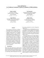
Fig. 1. (A) Identification of ac cross-linking products by western
blot analysis and antibody detection. Membranes were oxidized
using CuP for 1 h at room temperature and subunits a and c were
extracted using chloroform ⁄ methanol. After electrophoresis under
non-reducing conditions, proteins were transferred to nitrocellulose
membranes and visualized by immunoblotting. Antibodies against
subunit a (left panel) and subunit c (right panel) were utilized to
identify the ac cross-linking products. Bands marked Af* are arti-
facts from DK8 that are not related to the ATP synthase. Shown is
a representative analysis of cT67C ⁄ aN230C (lane 1), cT67C (lane 2)
and cG68C ⁄ aN230C mutants (lane 3). (B) Quantification of ac
cross-link formation in subunit a immunoblots. Immunoblots were
scanned and the bands corresponding to subunit a and to the
cross-linking product ac were quantified and expressed as volumes
(Vol
a
and Vol
ac
) using QUANTITY ONE software. For every blot, a back-
ground volume (Vol
Bg
) was calculated from three individual squares.
The amount of cross-link formation was then calculated according
to the equation shown.
T. Vorburger et al. Conformations of the ATPase ion-binding pocket
FEBS Journal 275 (2008) 2137–2150 ª 2008 The Authors Journal compilation ª 2008 FEBS 2139
AB
CD
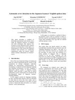
Fig. 2. (A) Location of cross-links in the
I. tartaricus a ⁄ c interface found in this study.
Green (good yield), yellow (medium yield),
red (minor or no yield). (B) Location of
cross-links in the E. coli a ⁄ c interface [10].
Blue (good yield), yellow (medium yield), red
(minor or no yield). (C) Top view into the
binding pocket of the I. tartaricus c-ring.
Residues 67 and 68 are mutated to cyste-
ines to illustrate their almost identical loca-
tion within the binding site. (D) Side view
into the binding pocket of the I. tartaricus
c-ring. Residues 67 and 68 are mutated to
cysteines to illustrate their almost identical
position within the membrane bilayer. All
images were prepared using
PYMOL (DeLano
Scientific).
Table 1. Relative yield of ac cross-linking products between cyste-
ines introduced in subunits a and c at the positions indicated. The
developed immunoblots were scanned and bands corresponding to
ac and a were quantified. The relative yield of ac cross-linking prod-
ucts was calculated as shown in Fig 1B, and 100% cross-linking
would therefore correspond to the presence of the entire subunit a
in the form of ac cross-linking products. At least three individual
measurements (new protein expression) were performed to deter-
mine product formation.
Cys pair
Relative yield of ac
cross-linking product (%)
aI223C ⁄ cV58C 46.9 ± 4.6
aI223C ⁄ cL59C 37.4 ± 4.5
aN230C ⁄ cS66C 37.7 ± 6.3
aN230C ⁄ cT67C 25.4 ± 6.7
aN230C ⁄ cG68C 4.8 ± 1.8
aN230C ⁄ cI69C 23.9 ± 5.9
aN230C ⁄ cY70C 30.3 ± 7.1
aA233C ⁄ cI69C 8.6 ± 2.6
aA233C ⁄ cY70C 36.4 ± 2.4
aI237C ⁄ cV73C 23.6 ± 6.6
aG239C ⁄ cL76C 7.1 ± 3.7
aG239C ⁄ cI77C 2.3 ± 2.1
aL240C ⁄ cL76C 18.2 ± 4.4
aL240C ⁄ cI77C 4.1 ± 3.2
aL241C ⁄
cL76C 21.4 ± 2.3
aL241C ⁄ cI77C 6.0 ± 1.3
Table 2. Comparison between ac cross-link formation using cyste-
ine mutants in the a ⁄ c interface of the E. coli and I. tartaricus ATP
synthases. Corresponding cross-linking products are shown in the
same row and relative cross-linking yields have been characterized
as follows: ±, < 5%; +, 6–10%; ++, 11–20%; +++, 21–40%;
++++, > 40%. ND, not determined.
I. tartaricus ATPase E. coli ATPase [10]
Cys pair (I. t. numbering) Cys pair (E. c. numbering)
aI223C ⁄ cV58C ++++ aL207C ⁄ cF54C +
aI223C ⁄ cL59C +++ aL207C ⁄ cI55C ++
aN230C ⁄ cS66C +++ aN214C ⁄ cA62C +++
aN230C ⁄ cT67C +++ aN214C ⁄ cI63C ND
aN230C ⁄ cG68C ± aN214C ⁄ cP64C ND
aN230C ⁄ cI69C +++ aN214C ⁄ cM65C +++
aN230C ⁄ cY70C +++ aN214C ⁄ cI66C +
aA233C ⁄ cI69C +
aA217C ⁄ cM65C ±
aA233C ⁄ cY70C +++ aA217C ⁄ cI66C ±
aI237C ⁄ cV73C +++ aI221C ⁄ cG69C +++
aG239C ⁄ cL76C + aI223C ⁄ cL72C +++
aG239C ⁄ cI77C ± aI223C ⁄ cY73C ND
aL240C ⁄ cL76C ++ aL224C ⁄ cL72C +
aL240C ⁄ cI77C ± aL224C ⁄ cY73C ++++
aL241C ⁄ cL76C +++ aI225C ⁄ cL72C +
aL241C ⁄ cI77C + aI225C ⁄ cY73C +++
Conformations of the ATPase ion-binding pocket T. Vorburger et al.
2140 FEBS Journal 275 (2008) 2137–2150 ª 2008 The Authors Journal compilation ª 2008 FEBS
results for relative cross-linking product formation
(compared to X = R) for these triple mutants are
shown in Fig. 3A. For the aN230C ⁄ cG68C cysteine
pair, the yield of cross-linking products for all aR226X
substitutions was significantly increased (up to 20%)
compared to the wild-type background. On the other
hand, the aR226X substitution did not significantly
affect cross-link formation by the aN230C ⁄ cT67C
cysteine pair.
To further investigate the influence of the stator
arginine on the conformational changes of the c sub-
unit, the amounts of cross-link formation between
aN230C and cysteine mutants of subunit c around the
binding site (residues 66–70) in the wild-type and
aR226H background were compared. The results in
Fig. 3B,C indicate that the aR226H substitution
decreased the amount of cross-link formation by the
pair aN230C ⁄ cS66C to about 70% of that of the wild-
type, while that for the aN230C ⁄ cG68C pair increased
about 280%, and that for the pairs aN230C ⁄ cT67C,
aN230C ⁄ cI69C and aN230C ⁄ cY70C was not signifi-
cantly affected.
Cross-linking product formation by aN230C
⁄
cG68C is influenced by the protonation state of
histidine in aR226H
To elucidate whether the altered side chains themselves
or the presence or absence of a positive charge within
the a ⁄ c interface is responsible for the amount of ac
cross-link formation, we took advantage of the fact
that the protonation state of a histidine residue can be
changed in the near-neutral range [pK
a
(His) = 6.0].
The experiments described above were repeated at
pH 5 and 6 in order to protonate the histidine in
aR226H. To control the influence of the pH on the
formation of Cys–Cys cross-linking products, we
included control experiments at both acidic pH values
in which the arginine at position 226 was not changed.
The results of these measurements (Fig. 4A) show
the amounts of cross-link formation at the various pH
values normalized to the amounts at pH 5. In the con-
trol reactions in the presence of aR226, labeling at
pH 6 and 8 was increased approximately 2.5-fold and
4-fold, respectively, compared to pH 5, reflecting the
A
C
B
Fig. 3. (A) Effect of aR226X mutations on formation of Cys–Cys cross-linking products between aN230C and cT67C or cG68C, respectively.
The values shown are the ratios of cross-linking product formation between aN230C and cT67C or cG68C, respectively, in the R226X back-
ground versus those in the wild-type background. Details are given in Fig. 1 and Experimental procedures. CuP-catalyzed air oxidation of the
membranes was carried out at pH 8. The numbers below the figure are the average (mean) yields of ac cross-link formation (as a percentage
of the total amount of a subunit). (B) Formation of ac cross-linking products between aN230C and mutants cS66C, cT67C, cG68C, cI69C
and cY70C in the presence or absence of the aR226H replacement. The values shown are the ratios between the triple and the double
mutants. The absolute cross-link formation yields (mean) are shown below. (C) Western blot analysis using antibodies against subunit a for
the experiment described in (B).
T. Vorburger et al. Conformations of the ATPase ion-binding pocket
FEBS Journal 275 (2008) 2137–2150 ª 2008 The Authors Journal compilation ª 2008 FEBS 2141
pH dependence of the disulfide formation reaction.
In the aR226H ⁄ aN230C ⁄ cT67C mutant, comparable
values were obtained. In the aR226H ⁄ aN230C ⁄ cG68C
mutant, however, the same measurements resulted in a
4-fold (pH 6) and 17-fold (pH 8) increased cross-link
formation. These results show that formation of the
aN230C ⁄ cG68C cross-linking products is severely
diminished in presence of a positively charged amino
acid at position 226 of the a subunit, i.e. either the
wild-type (aR226) or the protonated form of the
aR226H mutant.
Effect of the cG25I mutation on cross-link
formation between aN230C and cT67C or cG68C,
respectively
The various amounts of cross-link formation in the
presence or absence of a positive charge might result
from a partial helical rotation due to electrostatic
interactions between the stator charge and the abutting
rotor site. Likewise, several side chains from the bind-
ing site might be significantly rearranged upon contact
with the stator charge on subunit a (see Discussion).
Both kinds of structural changes are preferred as the
helix packing between inner and outer helices is not
tight in this region due to the absent side chain of
cG25 on the inner helices. Although residue cG25 is
conserved in Na
+
-translocating ATP synthases, it does
not belong to the G-X-G-X-G-X-G motif responsible
for the tight packing between the inner helices [18].
Replacement of the small glycine by a bulky isoleucine
residue might occupy the space needed for the confor-
mational changes envisaged above. We therefore deter-
mined the yield of aN230C ⁄ cT67C and aN230C ⁄
cG68C cross-linking products in the presence and
absence of the cG25I substitution. Importantly, the
cG25I mutation did not disturb the assembly of an
oligomeric c-ring as judged by SDS–PAGE after purifi-
cation of the enzyme (data not shown). As shown in
Fig. 4B, the cG25I replacement had only little effect
on the formation of cross-linking products by the
aN230C ⁄ cT67C cysteine pair but increased that of the
aN230C ⁄ cG68C pair about 3-fold over the wild-type
(cG25) control.
ATP synthesis measurements with single
mutants cG25I, cT67C and cG68C
We wished to determine whether the effect of the
cG25I mutation on cross-link formation is reflected
by functional enzyme studies. For this reason,
mutants cG25I, cT67C, cG68C and the recombinant
wild-type enzyme were purified, reconstituted into pro-
teoliposomes and tested for ATP synthesis activity
AB
Fig. 4. (A) pH dependence of cross-link formation between aN230C and cT67C or cG68C, respectively, in the wild-type or aR226H back-
ground. Membranes containing the mutant proteins were exposed to CuP at pH 5, 6 and 8, and the relative yields of ac cross-linking prod-
ucts were determined. The values shown are the ratios of cross-link yields at the pH indicated to the yields at pH 5, to illustrate the
influence of pH on cross-link formation. The absolute cross-link formation yields (means) are displayed below the figure. If three or more
experiments were performed, error bars are indicated. (B) Influence of cG25I on formation of cross-linking products. Yields of ac cross-link-
ing products for the two Cys–Cys pairs aN230C ⁄ cT67C and aN230C ⁄ cG68C in the presence or absence of the cG25I mutation at pH 8 are
shown. The corresponding western blot analysis using antibodies against subunit a is shown below.
Conformations of the ATPase ion-binding pocket T. Vorburger et al.
2142 FEBS Journal 275 (2008) 2137–2150 ª 2008 The Authors Journal compilation ª 2008 FEBS
after energization by a K
+
⁄ valinomycin-induced diffu-
sion potential (positive inside). Maximal enzyme activ-
ity was observed in the wild-type enzyme, but mutant
cG68C also showed a substantial synthesis rate (about
30% of wild-type) (Fig. 5). No significant ATP synthe-
sis was observed in the cG25I mutant, emphasizing the
functional importance of the small glycine residue.
Likewise, we were not able to detect any activity in the
cT67C mutant, indicating the physiological importance
of threonine at position 67.
Energy-minimization calculations for interaction
of aR226 with the c-ring
To further probe critical interactions in the a ⁄ c inter-
face, energy-minimization calculations for interaction
between a seven amino acid stretch of subunit a
(aI225–aM231), containing the conserved residues
aR226 and aN230, and the c-ring crystal structure
were performed. The minimization consistently
adjusted the conformation of aI225 to aM231 such
that the plane of the guanidino group of aR226 was
placed optimally in the entrance of the binding pocket
of the c-ring. While full mobility (no harmonic
restraints) was allowed for the subunit a stretch and
the side chains of the c-ring residues, various degrees
of motional freedom were applied to the back-
bone of the c-ring helices using harmonic restraints
(10 kcalÆmol
)1
A
˚
2
). The resulting conformation of
aR226 after energy minimization was found to be
insensitive to the exact starting conformation applied,
and visually identified hydrogen-bond patterns indi-
cated a possible mode of interaction between aR226
and the binding pocket. The detailed results of these
calculations are discussed below.
Discussion
A stator charge-induced conformational change
within the binding pocket
Elucidation of the high-resolution structures of the
Na
+
-dependent rotor rings of I. tartaricus F-ATP syn-
thase and E. hirae V-ATPase represents a significant
step towards a mechanistic understanding of ion trans-
location in these enzymes [6,7]. In the I. tartaricus
structure, the ion-binding pocket is located close to the
outer surface of the c-ring, but is shielded from the
hydrophobic environment by the side chains of cE65,
cS66 and cY70. The side chain of cY70 is not directly
involved in Na
+
coordination, but forms a hydrogen
bond to the conserved cE65 that stabilizes the overall
shape of the binding pocket. In this conformation, the
aromatic side chain seems to be ideally suited to shield
the polar binding pocket from the lipid bilayer. The
significance of the phenolic group of cY70 for stability
of the binding site has been demonstrated by an about
30-fold decrease in Na
+
binding affinity in the cY70F
mutant [19].
Electrostatic interactions between the binding site
and the stator arginine have been proposed to dis-
charge the ion in the subunit a ⁄ c interface, and this
hypothesis has been experimentally verified [5]. In this
study, we wished to determine whether a conforma-
tional change within the binding pocket, induced by
the positive stator charge, provides a molecular ratio-
nale for dislodging of the ion, and probed the dis-
tances between c-ring residues near the binding site
and helix 4 of subunit a by Cys–Cys cross-linking
experiments. Notably, the aN230C residue, which is
located one helical turn towards the P-side of the sta-
tor arginine, formed substantially fewer cross-linking
products with cG68C than with cT67C, although
both side chains adopt a very similar position in the
structure of the isolated c-ring. These data indicate
Time (s)
0 102030405060
mol ATP/ mol enzyme
0
200
400
600
800
wt
cG25I
cG68C
cT67C
Fig. 5. ATP synthase activities in the wild-type I. tartaricus ATP
synthase and c subunit mutants. The purified enzymes were recon-
stituted into proteoliposomes and the synthesis of ATP was
followed after application of a K
+
⁄ valinomycin diffusion potential.
In control experiments, the membrane potential was dissipated by
addition of the K
+
⁄ H
+
exchanger nigericin, and the values obtained
by these measurements were subtracted. The luminescence time
traces of representative experiments for the wild-type and indicated
mutant enzymes are shown. The rates of ATP synthesis were
calculated under the assumption that 100% of ATP synthase
molecules were incorporated into the liposomes during the recon-
stitution process.
T. Vorburger et al. Conformations of the ATPase ion-binding pocket
FEBS Journal 275 (2008) 2137–2150 ª 2008 The Authors Journal compilation ª 2008 FEBS 2143
that cG68 is shielded or displaced from helix 4 of
subunit a in the subunit a ⁄ c interface. Factors elicit-
ing the corresponding conformational change at the
ion-binding site could thus be monitored by compar-
ing cross-linking yields between aN230C and cT67C
or cG68C. Importantly, upon replacement of the sta-
tor arginine by electroneutral amino acids, formation
of cross-linking products between aN230C and
cG68C was specifically augmented, while those with
cT67C, cI69C or cY70C were not affected. Hence,
the stator arginine appears to elicit a distinct confor-
mational change in the c subunit binding site without
affecting the global conformation of the c-ring. These
conclusions were corroborated by comparing cross-
link formation in the aR226H background under var-
ious protonation states of the histidine. At low pH,
when the histidine is protonated, the cross-linking
pattern resembles that in the presence of arginine.
At higher pH, however, when the histidine is expected
to be neutral, the pattern resembled that in the
aR226A or aR226S mutants. A similar effect of pH
to that observed in cross-linking experiments with the
aR226H mutant was also found in ATP-driven Na
+
transport and Na
+
exchange experiments with this
mutant [5].
Is it possible to envisage molecular details of this
conformational change on the basis of the c-ring struc-
ture? Swiveling of part of the outer helix of subunit c
(containing cE65 and cG68) would be one possibility
for bringing the cT67C and cG68C residues into
unequal positions with respect to aN230C. It is also
conceivable that side-chain movements of several resi-
dues in the presence of the stator charge would induce
a new energetically favorable conformation that blocks
access to the cG68C residue. Previously, the stator
charge was thought to interact electrostatically with
the acidic side chain of the ion binding glutamate, ini-
tiating a large side-chain movement (opposite to the
direction of rotation) that opens the binding site [6,7].
In this scenario, residue cG68C (which is on the same
helix as the rotated cE65) would become further
exposed and not shielded from contact with subunit a
as observed in our present experiments. Upon helical
rotation in the opposite direction as proposed above,
however, cG68C would be disconnected from the inter-
face, and cross-link formation would be impeded. We
reasoned that the rotating part of the helix is most
likely distal to cV63, where the helix is broken because
the backbone carbonyl of cV63 is involved in Na
+
coordination. It is interesting to note that cG68 is
positioned opposite another glycine (cG25) on the
inner helix. The space provided by the absence of side
chains would allow a helical segment around cG68 to
rotate towards the inner helices (Fig. 6A). A similar
cavity is formed by glycines 27 and 66 in the K-ring of
E. hirae [7]. If this hypothesis is valid, the conforma-
tional change should be obstructed by replacement of
the glycine on the inner helix by a more bulky residue.
Indeed, in the cG25I mutant, a significantly increased
amount of cross-link formation with cG68C was
observed, indicating that the bulky side chain pre-
vented the conformational change in the rotor ⁄ stator
interface. The functional importance of cG25 is under-
lined by ATP synthesis measurements – no detectable
ATP formation was observed in the cG25I mutant.
Instead of helical rotation, it is also feasible that inter-
action with the stator charge pushes part of the helix
containing cG68 and cE65 towards the center of the
c-ring. Likewise, the cavity formed by glycines cG68
and cG25 might accommodate this helical motion.
Energy-minimization calculations support the
proposed conformational change
The data reported in this study allowed us to produce
a model of the interacting helical faces of subunit a
and the c-ring. As significant cross-link formation with
aN230C was found with residues 66–70 of the c-ring,
it was assumed that the position of the aN230C resi-
due is directly opposite the binding site. This sugges-
tion was corroborated by strong cross-link formation
between aA233C and cY70C, but only weak cross-link
formation between aA233C and cI69C. This positions
the relative height of cY70 between residues aN230
and aA233. These considerations indicate that the sta-
tor arginine is clearly shifted towards the N-side with
respect to the binding site. Consequently, the long side
chain from aR226 reaches the binding site from the
N-side by perfectly fitting the curved surface of the
hourglass shape of the c-ring. Such an interaction of
the arginine with the binding site allows close contact
of the two subunits and should also serve as an effi-
cient seal to prohibit ions arriving from the periplasm
from escaping to the cytoplasm.
In order to gain insight into the interaction of the
stator arginine with the binding site, we modeled a
stretch of seven amino acids of helix 4 of subunit a
into the c-ring structure and computationally mini-
mized the energy of this assembly. Depending on the
applied parameters, two possible coordinations of the
arginine within the binding pocket were obtained. The
binding of the arginine is stabilized by a number of
hydrogen bonds to the Na
+
-binding ligands (oxygen
atoms of cE65, cV63 and cQ32). These hydrogen
bonds minimize the polarity of the arginine in the
hydrophobic environment of the a ⁄ c interface within
Conformations of the ATPase ion-binding pocket T. Vorburger et al.
2144 FEBS Journal 275 (2008) 2137–2150 ª 2008 The Authors Journal compilation ª 2008 FEBS
the membrane. In all calculations, a hydrogen bond
was formed between the cNH group and the backbone
oxygen of cV63, guiding the arginine side chain down-
wards into the binding pocket. In Fig. 6C,D, two con-
formations of arginine coordination are depicted.
In Fig. 6C, movement of the backbone was restricted
within harmonic restraints, and therefore only side-
chain movements are observed. As expected, the argi-
nine is able to form four hydrogen bonds with cQ32,
cV63 and cE65. Another hydrogen bond is formed
with aN230 of subunit a. In Fig. 6D, where no restric-
tions were imposed on the backbone of the outer rings
of helices, a different coordination of the arginine was
obtained. Again, cQ32, cV63, cE65 and aN230 formed
hydrogen bonds with the arginine. However, unlike in
the calculation above, only one oxygen atom of cE65
was involved in arginine coordination, and the other
oxygen formed a hydrogen bond with cT67. To allow
for this interaction, the side chain of cT67 was reori-
ented, which simultaneously enabled it to form a
hydrogen bond with the NH
2
group of arginine aR226
that reacted with the second oxygen of the glutamate
in the first model.
In both calculations, the interaction with the argi-
nine forces the glutamate to move away from its origi-
nal position towards the cavity formed by cG25 ⁄ cG68,
as suggested above. Most interestingly, this movement
releases the hydrogen bond between cE65 and cY70,
indicating that the polar arginine uses both oxygens of
the glutamate to form hydrogen bonds. Loss of the
hydrogen bond between cE65 and cY70 allows the side
chain of cY70 to accommodate to a new environment,
which could be an important step in the ion-transloca-
tion mechanism, e.g. by enabling the contact of the
periplasmic access pathway with the binding site.
Only a very minor rotation of a helical strip
(although in the proposed direction) as suggested
above was observed in the calculations; instead there
was a shift towards the inner ring of helices, as pro-
posed alternatively. It is not possible, however, to
draw direct conclusions from these observations, as
important parameters of the native a ⁄ c interaction
were neglected in the energy-minimization calculation
(e.g. influence of membrane potential, influence of the
peripheral stalk, etc). Nevertheless, the calculation
indicates some structural flexibility within the helical
strip between the helix break at cV63 and the unstruc-
tured region around cY80. Such flexibility might per-
mit an efficient c-ring rotation when in contact with
subunit a and accommodate transient structural
AB
CD
Fig. 6. (A) Perspective view of the surface
of the c-ring of I. tartaricus. The atom
boundaries are displayed as surfaces to
visualize the cavity at the P-side of the ion-
binding site. The residues of the ion-binding
site and the glycine residues cG25 and
cG68 around the cavity are also shown.
(B) Side-chain movements observed after
energy-minimization calculations for the
c-ring and a heptapeptide of helix 4 of sub-
unit a. The calculated positions of the bind-
ing-site residues in the presence (light blue)
or absence (light pink) of harmonic back-
bone restraints of the outer helices are
shown with respect to the crystal structure
(green) used as the starting point for the cal-
culations. Red, oxygen; blue, nitrogen. (C,D)
Coordination of the stator arginine after
energy-minimization calculations for the
c-ring and a heptapeptide of helix 4 of sub-
unit a. The calculated positions and possible
hydrogen bonds of the binding-site residues
on the c-ring and the stator arginine in the
presence (C) or absence (D) of harmonic
backbone restraints of the outer helices are
shown. Putative hydrogen bond lengths are
marked in A
˚
. All images were prepared
using
PYMOL (DeLano Scientific).
T. Vorburger et al. Conformations of the ATPase ion-binding pocket
FEBS Journal 275 (2008) 2137–2150 ª 2008 The Authors Journal compilation ª 2008 FEBS 2145
changes during loading of Na
+
onto the binding site.
Additionally, we performed a simulation in which
aR226 was replaced by a histidine. The binding-site
residues adopted similar positions as in the calculation
with arginine (cE65 pushed towards the cavity, hydro-
gen bond to c70Y lost), reinforcing our findings from
the cross-linking studies (data not shown).
A similar localization of the stator arginine, i.e.
slightly shifted towards the N-side with respect to the
conserved acidic residue in the c-ring, was also pro-
posed for E. coli ATP synthase [10]. It might be that
the described interaction of subunits a and c in the
I. tartaricus enzyme is a general feature of all ATP
synthases.
Implications for the ion-translocation mechanism
The Na
+
⁄ H
+
antiporter inhibitor ethyl isopropyl amil-
oride (EIPA) is also known to block Na
+
-dependent
ATP hydrolysis of the I. tartaricus enzyme in a
Na
+
-dependent manner [20], indicating that EIPA and
Na
+
compete for the same binding site (Fig. 7). As the
structure of the amiloride derivative mimics that of the
stator arginine by combining a positively charged guani-
dino group with a hydrophobic environment, EIPA is
suggested to block the enzyme by occupying the binding
site. It is of interest that the H
+
-translocating enzyme
of E. coli is not inhibited by EIPA and that this
enzyme lacks residues equivalent to cQ32 and cT67,
which might act as coordination sites for the arginine.
Whether a free backbone carbonyl (cV63 for I. tartari-
cus) for formation of a hydrogen bond to the cNH
group is also present in the E. coli enzyme is unclear,
but this has been speculated recently [19]. Based on
these considerations, interaction of the arginine with
the proton-binding site is expected to be weaker than
with the Na
+
ion-binding site. A strong interaction
between the binding site and the arginine is not favor-
able for high turnover rates, and hence the different
affinities of the two enzymes for the stator arginine
might explain the different translocation rates within
F
0
(1000 Na
+
⁄ s versus 8000 H
+
⁄ s) [21,22]. Therefore,
the incoming Na
+
ion is thought to weaken the rather
strong interaction between the arginine and the bind-
ing site and to promote its loading onto the binding
site, aided by the membrane potential as described pre-
viously [3]. Such a scenario is supported by the
requirement of Na
+
ions for rotation, even under
ATP-hydrolyzing conditions [5]. The repelled arginine
is then attracted by the next incoming rotor site and
displaces the Na
+
ion to form the intermediate
described above. Such a concerted mechanism ensures
that only small energy barriers have to be overcome
during rotation in order to guarantee smooth enzyme
function. According to our data, the side chain of the
glutamate is not pulled towards subunit a, but is
pressed inwards, which makes a large back-flipping of
the acidic side chain obsolete. Such a model would
also explain the earlier and so far unexplained finding
that, in the E. coli ATP synthase, the essential cD61
on the outer helix of the c-ring can be transferred to
position 24 on the inner helix with retention of activity
[23]. Taking the envisaged side-chain drift of aR226
towards the P-side into account, it is tempting to spec-
ulate that, during rotation, the long side chain of
aR226 oscillates like a pendulum between the binding
sites of the c-ring and subunit a. Such a mechanism is
favored by the highly conserved aG229, which might
provide space for back pressure during rotation
between two binding sites. A functional aspect of this
glycine residue is anticipated but so far unexplained, as
rotation during ATP hydrolysis is severely impeded
(> 90% inhibition) in the corresponding mutant of
the E. coli ATP synthase (aG213C) [9].
Possible roles for cG25 and cT67
The deficiency of the cG25I mutant in ATP synthesis
demonstrates the functional importance of this
EIPA (µ
M
)
0.1 1 10 100 1000
%ATP hydrolysis activity
0
20
40
60
80
100
120
0.2 mM Na
+
2 mM Na
+
Fig. 7. Inhibition of ATP hydrolysis activity by EIPA. Purified ATP
synthase from I. tartaricus in the presence of either 0.2 m
M NaCl
(filled circles) or 2 m
M NaCl (open circles) was incubated with vari-
ous concentrations of EIPA, and ATP hydrolysis rates were deter-
mined using the coupled enzyme assay as described previously
[30]. Logarithmic scaling of the x axis and exponential decay fitting
were applied to illustrate the competition of EIPA and Na
+
for the
same binding site. Inset: chemical structure of EIPA.
Conformations of the ATPase ion-binding pocket T. Vorburger et al.
2146 FEBS Journal 275 (2008) 2137–2150 ª 2008 The Authors Journal compilation ª 2008 FEBS
residue. To account for this, two major scenarios are
possible. In the first, the cY70 side chain, which is no
longer hydrogen-bonded to cE65, could rotate into
this cavity, as proposed previously [1]. This could
open the binding site and an incoming Na
+
ion could
displace the bound arginine. In the second scenario,
the cavity formed by the glycines might act as vesti-
bule for the incoming Na
+
ion. Free access of the
cavity to the binding site would perfectly suit the
requirement to allow displacement of the stator
charge through the Na
+
ion. Again, the uncoordi-
nated side chain of the cY70 might be displaced (not
into the cavity, however) and act as gate to the vesti-
bule.
Surprisingly, the cT67C mutant was also unable to
synthesize ATP under the conditions used. However,
unlike the cG25I mutant, no steric reasons are
assumed for this observation. One of the minimization
calculations (Fig. 6D) suggests a possible role for cT67
as a hydrogen-accepting group for arginine (and donor
for cE65), which would not be possible in the cT67C
mutant. However, whether such an intermediate con-
tribution of cT67 occurs during catalysis cannot be
confirmed by the present data and requires further
investigation.
Experimental procedures
Materials
Unless otherwise stated, chemicals were purchased from
Fluka (Buchs, Switzerland).
Construction of mutants
Plasmid pItTr5His carries the whole atp operon
(atpIBEFHAGDC)ofI. tartaricus [24] with the following
modifications: the start codons of atpF and atpA were
changed from TTG to ATG, a Bsu15I single site was intro-
duced between atpE and atpF, and a His
10
tag was fused to
the N-terminus of subunit b. The endogenous cysteine at
position aC76 of subunit a was then changed to alanine,
resulting in plasmid pItTr6His which encodes the entire
I. tartaricus ATP synthase with a Cys-less F
0
part. In this
study, cysteine and other substitutions were introduced into
subunits a and c on plasmid pItTr6His. E. coli DH5a
served as host for cloning and was cultivated in LB medium
supplemented with 200 lgÆmL
)1
ampicillin. Amino acid
substitutions were introduced by performing a two-step
PCR procedure using two oligonucleotide pairs. One pair
contained the codon for the desired mutation, the sequence
of the other was derived from the wild-type. The presence
of the mutant codons was confirmed by automated
sequencing of the cloned DNA at Microsynth AG
(Balgach, Switzerland).
Membrane preparation
Plasmids coding for cysteine-substituted I. tartaricus ATP
synthases were expressed in the E. coli atp operon deletion
strain DK8 [24a]. The cells were collected, washed with a
buffer containing 10 mm Tris ⁄ HCl pH 8.0 and 10 mm dith-
iothreitol, and, if necessary, stored at )80 °C.
The cell pellet was resuspended (5 mLÆg
)1
cells, wet
weight) in French press buffer I (50 mm Tris ⁄ HCl pH 8.0,
5mm MgCl
2
,2mm NaCl, 10% glycerol, 10 mm dithiothrei-
tol, 0.1 mm diisopropylfluorophosphate, 50 lg DNase I) and
disrupted by three passages through a French pressure cell.
Unbroken cells and large cell debris were removed by centri-
fugation (8000 g,4°C, 15 min). The membranes were pel-
leted by ultracentrifugation (200 000 g, 45 min, 4 °C), and
washed with 20 mL of French press buffer I containing
1mm dithiothreitol. After centrifugation, the washed mem-
branes were resuspended in 1 mL of assay buffer (50 mm
Tris ⁄ HCl pH 8.0, 5 mm MgCl
2
,2mm NaCl, 10% glycerol)
for standard cross-linking assays. To determine the pH
dependency of formation of ac cross-linking products, mem-
brane samples were resuspended in assay buffer containing
1mm instead of 50 mm Tris ⁄ HCl, pH 8.0. All steps were car-
ried out at 4 °C or on ice.
Copper phenanthroline-catalyzed air oxidation
of membranes
Unless otherwise noted, copper cross-linking was per-
formed by mixing a 100 lL aliquot of membranes in assay
buffer with 100 lL of CuP-solution which consisted of
10 mm o-phenanthroline and 3 mm CuSO
4
in assay buffer.
To measure the influence of varying proton concentrations
on the formation of ac cross-linking products, the pH of
a75lL aliquot of membranes was adjusted by the addi-
tion of 25 lL MMT buffer (100 mm Mes, 100 mm Mops,
100 mm Tricine, adjusted to the desired pH with 5 m
KOH). To stop the oxidation reaction, EDTA and NEM
(stock solution in dimethylsulfoxide) were added to final
concentrations of 15 mm each, followed by incubation for
another 10 min at room temperature. In control experi-
ments, in which NEM and EDTA were added 10 min
prior to CuP, no formation of ac cross-linking products
was observed.
Extraction of subunits a and c from oxidized
membrane samples by organic solvents
The extraction of subunits a and c from oxidized mem-
branes was performed as described previously [25]. An
aliquot of 20 lL 5% acetic acid was added to 80 lLof
T. Vorburger et al. Conformations of the ATPase ion-binding pocket
FEBS Journal 275 (2008) 2137–2150 ª 2008 The Authors Journal compilation ª 2008 FEBS 2147
oxidized membranes. After addition of 1 mL of chloro-
form ⁄ methanol (1 : 1, v ⁄ v) and vigorous shaking, insoluble
proteins were removed by centrifugation at 15 800 g for
5 min. The supernatant, transferred to a new test tube, was
mixed with 200 lL of 5% acetic acid. Mixing and a subse-
quent centrifugation step (15 800 g, 1 min) induced a phase
separation. The lower organic phase, containing subunits a
and c, was mixed with 20 lL of a 1% SDS solution, dried
under vacuum and solubilized in 60 lL non-reducing
1· SDS sample buffer (50 mm Tris ⁄ HCl pH 6.8, 1% SDS,
10% glycerol, 0.1 mgÆ mL
)1
bromophenol blue).
SDS–PAGE and immunoblotting
SDS–PAGE was performed under non-reducing conditions
as described previously [26]. The proteins were transferred
to nitrocellulose sheets (Protran nitrocellulose transfer
membrane; Schleicher & Schuell BioScience GmbH, Das-
sel, Germany) using a semi-dry western blotting procedure
as described by the manufacturer of the blotting apparatus
(GE Healthcare, Glattbrugg, Switzerland). After blocking
of the nitrocellulose membrane overnight with blocking
buffer [1% blocking reagent (Boehringer Mannheim
GmbH, Mannheim, Germany) in TTBS (20 mm Tris ⁄ HCl
pH 7.5, 500 mm NaCl and 0.05% Tween-20)], it was
washed twice with TTBS. The membranes were then incu-
bated with rabbit anti-a or anti-c serum for 3 h. The anti-
a antibody was custom-made by Eurogentec SA (Seraing,
Belgium), and recognizes the I. tartaricus subunit a seg-
ment from amino acid positions 90–103, and the anti-c
antibody was raised against the highly similar c subunit of
P. modestum. Both sera were diluted 1 : 6000 in TTBS
supplemented with 3% BSA. The membrane was rinsed
twice with TTBS and incubated for 2 h with a 1 : 3000
dilution of alkaline phosphatase-conjugated goat anti-rab-
bit IgG (Bio-Rad, Hercules, CA, USA) in TTBS. Subse-
quently, the blots were washed first with TTBS for
2 · 5 min and then with Tris ⁄ HCl for 5 min. The alkaline
phosphatase conjugate was visualized by performing a
color development reaction using 5-bromo-4-chloro-3-
indoyl phosphate p-toluidine salt and p-nitroblue tetrazo-
lium chloride.
Quantification of ac cross-linking product
formation
The developed immunoblots were scanned and the soft-
ware quantity one (Bio-Rad, Hercules, CA, USA) was
applied for quantitative analysis of bands detected on the
western blots as indicated in Fig. 1B. A linear depen-
dence between the protein amount and the western blot
signal was observed in the applied concentration range,
as verified in a control experiment with a serial dilution
of a sample.
Purification and reconstitution of recombinant
ATP synthase
The protocol for His-tagged E. coli ATP synthase purifica-
tion was used with modifications [27]. Briefly, about 5 g (wet
weight) of E. coli DK8 cells containing heterologously
expressed I. tartaricus ATPase were resuspended in 25 mL
French press buffer II (200 mm Tris ⁄ HCl pH 7.8, 100 mm
KCl, 5 mm MgCl
2
, 0.1 mm EDTA, 2.5% glycerol, 0.1 mm
diisopropylfluorophosphate, 50 lg DNase I) and disrupted
in a French pressure cell. Unbroken cells and large cell deb-
ris were removed by centrifugation (8000 g, 10 min, 4 °C).
Membranes were collected by ultracentrifugation (200 000 g,
45 min, 4 °C) and solubilized for 1 h at 4 °Cin20mL
extraction buffer (50 mm Tris ⁄ HCl pH 7.5, 100 mm KCl,
250 mm sucrose, 40 mm aminocaproic acid, 15 mm
p-aminobenzamidine, 5 mm MgCl
2
, 0.1 mm EDTA,
0.2 mm dithiothreitol, 0.8% soybean phosphatidyl choline,
1.5% octyl glucoside, 0.5% sodium cholate, 0.5% sodium
deoxycholate, 2.5% glycerol and 30 mm imidazole). After
centrifugation (200 000 g, 1 h, 4 °C), the supernatant was
sterile-filtered and loaded onto a HisTrap HP 1 mL column
(GE Healthcare). The column was washed with 15 mL of
washing buffer (1 : 1 dilution of extraction buffer and
30 mm imidazole), and the ATPase was eluted using elution
buffer (washing buffer but with 400 mm imidazole).
Enzyme-containing fractions were pooled and stored in
liquid nitrogen.
For reconstitution, 60 mg soybean phosphatidylcholine
were homogenized in 2 mL liposome buffer (10 mm
Hepes ⁄ KOH pH 6.5, 100 mm NaCl, 5 mm MgCl
2
, 0.1 mm
EDTA, 0.2 mm dithiothreitol). The suspension was soni-
cated on ice for 1 min using a tip-type sonicator (MSE Soni-
prep 150, Labtec AG, Wohlen, Switzerland). To the
liposome suspension, sodium cholate (1.5% final concentra-
tion) and purified ATP synthase in a lipid : protein ratio
200 : 1 (w ⁄ w) were added. The mixture was kept for 30 min
at 4 °C, and then a 500 lL sample was loaded on a PD-10
gel filtration column (GE Healthcare) equilibrated with lipo-
some buffer. Turbid fractions were pooled and proteolipo-
somes collected by ultracentrifugation (200 000 g, 30 min,
4 °C) and suspended in 100 lL of liposome buffer.
ATP synthesis measurements
An aliquot of 250 lL sample buffer (10 mm Tris ⁄ HCl, pH
6.5, 100 mm KCl, 5 mm MgCl
2
) was mixed with 50 lLof
luciferase reagent (ATP bioluminescence assay kit CLS II,
Roche Diagnostics, Rotkreuz, Switzerland) and 5 lL of lipo-
somes. The ATP synthesis reaction was started by injection
of 250 lL injector buffer (10 mm Tris ⁄ HCl pH 6.5, 10 mm
potassium phosphate buffer pH 6.5, 100 mm KCl, 5 mm
MgCl
2
,1mm ADP, 2 lm valinomycin), and the lumines-
cence was followed using a luminometer (Glomax, Promega,
Conformations of the ATPase ion-binding pocket T. Vorburger et al.
2148 FEBS Journal 275 (2008) 2137–2150 ª 2008 The Authors Journal compilation ª 2008 FEBS
Du
¨
bendorf, Switzerland). Control experiments were carried
out as above, but in the presence of 1 lm nigericin.
Energy-minimization calculations
Energy calculations were performed using cns 1.2 [28] on a
Linux workstation using a force field with explicit hydro-
gens, corresponding to the files protein-allhdg.top and pro-
tein-allhdg.param. Minimization utilized the limited-memory
Broyden–Fletcher–Goldfarb–Shanno method available in
cns, which was run until convergence; this usually required
less than 4000 iterations. The starting structure used was
the 2.4 A
˚
X-ray structure of the I. tartaricus c-ring [6] with
crystallographic waters removed, and residues aI225–
aM231 (helix 4 of subunit a) in an ideal helical conforma-
tion placed parallel to the outer helix of the c-ring at a dis-
tance between helix axes of 12 A
˚
. The longitudinal rotation
of the model helix was such that the side chain of aR226
pointed towards and into the binding pocket of the
I. tartaricus c-ring. Harmonic restraints were placed on the
backbone atoms of the c-ring, with the intention that only
the residues of the a subunit and the side chains of the
c-ring should be free to move during minimization. To
avoid trapping in local minima, several starting arrange-
ments differing by minor reorientations were tried, as well
as removal of the harmonic restraints on the backbone
atoms around the binding pocket. Manual placement of
aI225–aM231, as well as visualization of the results of
energy minimization, was performed using coot [29] and
pymol (DeLano Scientific, Palo Alto, CA, USA).
Acknowledgements
We thank Benjamin Oberfeld (ETH Zurich, Switzer-
land) for beneficial discussions. Gregory M. Cook
University of Otago, Dunedin, New Zealand is
acknowledged for critical reading of the manuscript.
This work was supported by the Swiss National
Science Foundation.
References
1 Meier T, Morgner N, Matthies D, Pogoryelov D, Keis
S, Cook GM, Dimroth P & Brutschy B (2007) A tri-
decameric c ring of the adenosine triphosphate (ATP)
synthase from the thermoalkaliphilic Bacillus sp. strain
TA2.A1 facilitates ATP synthesis at low electrochemi-
cal proton potential. Mol Microbiol 65, 1181–1192.
2 Capaldi RA & Aggeler R (2002) Mechanism of the
F
1
F
0
-type ATP synthase, a biological rotary motor.
Trends Biochem Sci 27, 154–160.
3 Dimroth P, von Ballmoos C & Meier T (2006) Catalytic
and mechanical cycles in F-ATP synthases. EMBO Rep
7, 276–282.
4 Greie JC, Heitkamp T & Altendorf K (2004) The trans-
membrane domain of subunit b of the Escherichia coli
F
1
F
0
ATP synthase is sufficient for H
+
-translocating
activity together with subunits a and c. Eur J Biochem
271, 3036–3042.
5 Wehrle F, Kaim G & Dimroth P (2002) Molecular mech-
anism of the ATP synthase’s F
0
motor probed by muta-
tional analyses of subunit a. J Mol Biol 322, 369–381.
6 Meier T, Polzer P, Diederichs K, Welte W & Dimroth
P (2005) Structure of the rotor ring of F-type Na
+
-AT-
Pase from Ilyobacter tartaricus. Science 308, 659–662.
7 Murata T, Yamato I, Kakinuma Y, Leslie AG & Walker
JE (2005) Structure of the rotor of the V-type Na
+
-
ATPase from Enterococcus hirae. Science 308, 654–659.
8 Angevine CM, Herold KA & Fillingame RH (2003)
Aqueous access pathways in subunit a of rotary ATP
synthase extend to both sides of the membrane. Proc
Natl Acad Sci USA 100, 13179–13183.
9 Angevine CM, Herold KA, Vincent OD & Fillingame
RH (2007) Aqueous access pathways in ATP synthase
subunit a: reactivity of cysteine substituted into trans-
membrane helices 1, 3 and 5. J Biol Chem 282, 9001–
9007.
10 Jiang W & Fillingame RH (1998) Interacting helical
faces of subunits a and c in the F
1
F
0
ATP synthase of
Escherichia coli defined by disulfide cross-linking. Proc
Natl Acad Sci USA 95, 6607–6612.
11 Fillingame RH, Angevine CM & Dmitriev OY (2003)
Mechanics of coupling proton movements to c-ring
rotation in ATP synthase. FEBS Lett 555, 29–34.
12 Rastogi VK & Girvin ME (1999) Structural changes
linked to proton translocation by subunit c of the ATP
synthase. Nature 402, 263–268.
13 Nakano T, Ikegami T, Suzuki T, Yoshida M & Akutsu
H (2006) A new solution structure of ATP synthase
subunit c from thermophilic Bacillus PS3, suggesting a
local conformational change for H
+
-translocation.
J Mol Biol 358 , 132–144.
14 Vincent OD, Schwem BE, Steed PR, Jiang W & Fillin-
game RH (2007) Fluidity of structure and swiveling of
helices in the subunit c ring of Escherichia coli ATP
synthase as revealed by cysteine–cysteine cross-linking.
J Biol Chem 282, 33788–33794.
15 Fraga D, Hermolin J & Fillingame RH (1994) Trans-
membrane helix–helix interactions in F
0
suggested by
suppressor mutations to Ala24 fi Asp ⁄ Asp61 fi Gly
mutant of ATP synthase subunit. J Biol Chem 269,
2562–2567.
16 Kaim G & Dimroth P (1998) Voltage-generated torque
drives the motor of the ATP synthase. EMBO J 17,
5887–5895.
17 Dimroth P, von Ballmoos C, Meier T & Kaim G (2003)
Electrical power fuels rotary ATP synthase. Structure
11, 1469–1473.
T. Vorburger et al. Conformations of the ATPase ion-binding pocket
FEBS Journal 275 (2008) 2137–2150 ª 2008 The Authors Journal compilation ª 2008 FEBS 2149
18 Vonck J, Krug von Nidda T, Meier T, Matthey U,
Mills DJ, Ku
¨
hlbrandt W & Dimroth P (2002) Molecu-
lar architecture of the undecameric rotor of a bacterial
Na
+
-ATP synthase. J Mol Biol 321, 307–316.
19 von Ballmoos C & Dimroth P (2007) Two distinct pro-
ton binding sites in the ATP synthase family. Biochemis-
try 46, 11800–11809.
20 Kluge C & Dimroth P (1993) Kinetics of inactivation of
the F
1
F
0
ATPase of Propionigenium modestum by dic-
yclohexylcarbodiimide in relationship to H
+
and Na
+
concentration: probing the binding site for the coupling
ions. Biochemistry 32, 10378–10386.
21 Feniouk BA, Kozlova MA, Knorre DA, Cherepanov
DA, Mulkidjanian AY & Junge W (2004) The proton-
driven rotor of ATP synthase: ohmic conductance
(10 fS), and absence of voltage gating. Biophys J 86,
4094–4109.
22 Kluge C & Dimroth P (1992) Studies on Na
+
and H
+
translocation through the F
0
part of the Na
+
-translo-
cating F
1
F
0
ATPase from Propionigenium modestum:
discovery of a membrane potential dependent step.
Biochemistry 31, 12665–12672.
23 Miller MJ, Oldenburg M & Fillingame RH (1990)
The essential carboxyl group in subunit c of the F
1
F
0
ATP synthase can be moved and H
+
-translocating
function retained. Proc Natl Acad Sci USA 87,
4900–4904.
24 Oberfeld B (2006) F
1
F
0
ATP synthase: identification of a
plug within the c-ring and heterologous expression of a
sodium-translocating enzyme. PhD Thesis, ETH Zu
¨
rich,
Switzerland.
24a Kilonsky DJ, Brusilow WS & Simoni RD (1984) In vivo
evidence for the role of the e subunit as an inhibitor of
the proton-translocating ATPase of Escherichia coli.
J Bacteriol 160, 1055–1060.
25 von Ballmoos C (2005) Molecular interactions along the
ion pathway of the F
1
F
0
ATP synthase. PhD Thesis,
ETH Zu
¨
rich, Switzerland.
26 Scha
¨
gger H & von Jagow G (1987) Tricine–sodium
dodecyl sulfate–polyacrylamide gel electrophoresis for
the separation of proteins in the range from 1 to
100 kDa. Anal Biochem 166, 368–379.
27 Ishmukhametov RR, Galkin MA & Vik SB (2005)
Ultrafast purification and reconstitution of His-tagged
cysteine-less Escherichia coli F
1
F
0
ATP synthase.
Biochim Biophys Acta 1706, 110–116.
28 Brunger AT, Adams PD, Clore GM, DeLano WL,
Gros P, Grosse-Kunstleve RW, Jiang JS, Kuszewski J,
Nilges M, Pannu NS et al. (1998) Crystallography &
NMR system: a new software suite for macromolecular
structure determination. Acta Crystallogr D 54,
905–921.
29 Emsley P & Cowtan K (2004) Coot: model-building
tools for molecular graphics. Acta Crystallogr D 60,
2126–2132.
30 Laubinger W & Dimroth P (1988) Characterization of
the ATP synthase of Propionigenium modestum as a pri-
mary sodium pump. Biochemistry 27, 7531–7537.
Conformations of the ATPase ion-binding pocket T. Vorburger et al.
2150 FEBS Journal 275 (2008) 2137–2150 ª 2008 The Authors Journal compilation ª 2008 FEBS