Báo cáo khoa học: A novel mechanism of TGFb-induced actin reorganization mediated by Smad proteins and Rho GTPases docx
Bạn đang xem bản rút gọn của tài liệu. Xem và tải ngay bản đầy đủ của tài liệu tại đây (797.22 KB, 14 trang )
A novel mechanism of TGFb-induced actin reorganization
mediated by Smad proteins and Rho GTPases
Lina Vardouli
1
, Eleftheria Vasilaki
1,2
, Elsa Papadimitriou
1
, Dimitris Kardassis
1,2
and Christos Stournaras
1
1 Department of Biochemistry, School of Medicine, University of Crete, Heraklion, Greece
2 Institute of Molecular Biology and Biotechnology, Foundation for Research & Technology-Hellas, Heraklion, Greece
It is well established that reorganization of the actin
cytoskeleton is one of the earliest cellular responses to
various extracellular stimuli [1–5]. Binding of ligands
to the appropriate receptors triggers specific signaling
cascades, which may generate rapid and long-term
modifications of actin polymerization dynamics and
microfilament organization [6–9].
Transforming growth factor b (TGFb) is a pleiotropic
cytokine that regulates homeostasis in various cell types
such as epithelial and endothelial cells, and regulates
other cell functions such as growth, differentiation and
apoptosis [10–12]. In addition, TGFb influences epithe-
lial–mesenchymal transitions, events critical for normal
embryogenesis and also for tumorigenesis, tumor-cell
invasiveness and metastasis [13–15]. The classical TGFb
signaling apparatus consists of a plasma membrane
complex of type I (TbRI) and type II (TbRII) receptors
and downstream Smad signaling effectors [16]. Activa-
tion of TbRI by TGFb leads to phosphorylation of the
receptor-regulated Smad proteins (R-Smad proteins)
Smad2 and Smad3, which in turn oligomerize with the
common partner Smad4 and rapidly translocate to the
Keywords
actin; Rho GTPases; Smad; TGFb; a-SMA
Correspondence
C. Stournaras, Department of Biochemistry,
School of Medicine, University of Crete,
GR-71110 Heraklion, Greece
Fax: +30 2810 394530
Tel: +30 2810 394563
E-mail:
(Received 22 April 2008, revised 1 June
2008, accepted 12 June 2008)
doi:10.1111/j.1742-4658.2008.06549.x
In previous studies, we have demonstrated that RhoA ⁄ B-dependent signal-
ing regulates TGFb-induced rapid actin reorganization in Swiss 3T3 fibro-
blasts. Here we report that TGFb regulates long-term actin remodeling by
increasing the steady-state mRNA levels of the RhoB gene in mouse Swiss
3T3 fibroblasts and human hepatoma HepG2 cells. We show that this regu-
lation is specific for the RhoB gene and is facilitated by enhanced activity
of the RhoB promoter. Adenovirus-mediated gene transfer of Smad2 and
Smad3 in Swiss 3T3 fibroblasts induced transcription of the endogenous
RhoB gene but not the RhoA gene. Interestingly, in JEG-3 choriocarcinoma
cells that lack endogenous Smad3, TGFb-induced transcriptional up-regu-
lation of the RhoB gene was not observed, but it was restored by adeno-
viral Smad3 overexpression. In addition, Smad2 and Smad3 triggered
activation of RhoA and RhoB GTPases and long-term actin reorganization
in Swiss 3T3 fibroblasts. Finally, Smad3, and to a lesser extent Smad2,
induced transcription of the a-smooth muscle actin (a-SMA) gene, and
enhanced the incorporation of a-SMA into microfilaments in Swiss 3T3
fibroblasts. These data reveal a novel mechanism of cross-talk between
the classical TGFb ⁄ Smad pathway and Rho GTPases, regulating the rapid
and the long-term actin reorganization that may control the fibroblast–
myofibroblast differentiation program.
Abbreviations
ALK5, activin like kinase 5; ca, constitutively active; FITC, fluorescein isothiocyanate; GAPDH, glyceraldehyde-3-phosphate dehydrogenase;
GEF, guanine nucleotide exchange factor; GST, glutathione S-transferase; luc, luciferase; RBD, Rho binding domain; RT-PCR, reverse
transcription-polymerase chain reaction; SBE, Smad binding element; a-SMA, a-smooth muscle actin; TbRI, TGFb receptor type I; TGFb,
transforming growth factor b; TI, Triton X-100 insoluble; TS, Triton X-100 soluble.
4074 FEBS Journal 275 (2008) 4074–4087 ª 2008 The Authors Journal compilation ª 2008 FEBS
nucleus where they bind to promoters and regulate
the expression of various target genes in a positive or a
negative manner [16]. This pathway is negatively regu-
lated by multiple signaling inputs. The best understood
is a feedback loop that involves Smad7, an inhibitory
Smad, which blocks R-Smad phosphorylation by TbRI
and directs lysosomal degradation of the receptor, thus
ensuring termination of the pathway [17,18]. Further-
more, TGFb was shown to modulate cell morphology
and growth in a concerted manner via mechanisms that
control the actin cytoskeleton in a variety of cell types
[19–22]. In Swiss 3T3 fibroblasts, TGF b has been
shown to induce rapid actin polymerization and
formation of stress fibers via a non-genomic RhoA ⁄ B ⁄
ROCK ⁄ Limk2 ⁄ cofilin signaling cascade downstream of
TbRI [23]. Similarly, in other studies, it has been
reported that RhoA signaling is activated early follow-
ing TGFb treatment in smooth muscle cells [24], while
RhoA and Rac1 are regulated by TGFb during aortic
endothelial morphogenesis [25]. However, the mecha-
nisms controlling the cross-talk between the classical
TGFb ⁄ Smad pathways, regulation of Rho GTPases and
long-term actin cytoskeleton reorganization have not
been addressed so far.
In the present work, we focus on the regulatory role
of the TGFb ⁄ Smad pathway in the activation and ⁄ or
transcriptional regulation of the Rho GTPases and
long-term actin cytoskeleton restructuring. Using vari-
ous cell models, we analyzed the TGFb-induced tran-
scriptional regulation of the RhoA ⁄ B GTPases. Using
adenoviral overexpression of Smad2 ⁄ 3, we studied the
role of Smad proteins in regulation of the RhoA ⁄ B
genes, activation of these small GTPases and the
induction of actin reorganization. Finally, to address
the biological significance of the TGFb-induced actin
reorganization, we assessed the Smad-induced tran-
scriptional regulation of a-SMA, and its expression
and incorporation into the microfilamentous network
of fibroblasts. Our results provide evidence for a novel
signaling mechanism in TGFb-induced actin cytoskele-
ton reorganization mediated by Smad proteins and
Rho GTPases that may regulate fibroblast–myofibro-
blast differentiation.
Results
TGFb1 induces transcription of the gene coding
for the small GTPase RhoB in Swiss 3T3
fibroblasts and hepatoma HepG2 cells
We have shown previously that TGFb induces rapid
and sustained activation of the small GTPases RhoA
and RhoB, and that this activation is essential for
TGFb-induced actin cytoskeleton reorganization in
fibroblasts [23]. In the present study, we sought to
examine whether TGFb, in addition to inducing Rho
protein activation, regulates the expression of these
two Rho GTPases.
First, we investigated the requirement for active gene
transcription in TGFb-induced actin cytoskeleton poly-
merization. For this purpose, Swiss 3T3 fibroblasts were
treated with TGFb for 24 h in the absence or presence
of the general inhibitor of transcription actinomycin D,
and changes in the actin cytoskeleton were monitored
by fluorescence using rhodamine phalloidin staining. As
shown in Fig. 1A, TGFb caused potent actin cytoskele-
ton reorganization as evidenced by the formation of
stress fibers. Importantly, long-term TGFb-induced
cytoskeleton reorganization was abolished in the pres-
ence of actinomycin D, suggesting that, in addition to
short-term activation events, TGFb elicits long-term
actin cytoskeleton regulation requiring transcriptional
induction of TGFb target genes (Fig. 1A).
We then examined the effect of the TGFb signaling
pathway in transcriptional regulation of the genes
coding for the human Rho GTPases A and B by two
different approaches: measuring the mRNA levels and
measuring the activity of the promoters of the two
genes in response to TGFb stimulation. First, we
determined the effect of TGFb on the mRNA levels of
the RhoA and RhoB genes in Swiss 3T3 fibroblasts by
RT-PCR experiments. For this purpose, Swiss 3T3
cells were serum-starved for 24 h and then stimulated
with 5 ngÆmL
)1
TGFb1 for various time periods (from
30 min up to a maximum of 24 h). As shown in
Fig. 1B, treatment of Swiss 3T3 cells with TGFb1
resulted in a rapid increase (1.6-2.2-fold) in the steady-
state mRNA levels of the RhoB gene, which was
initiated at 30 min and persisted for a period of 24 h
post-induction (top panel). In contrast, transcriptional
activation of the RhoA gene by TGFb1 was not
observed (Fig. 1B, middle panel). As expected, TGFb
did not affect the mRNA levels of the glyceraldehyde-
3-phosphate dehydrogenase (GAPDH) gene used as a
control (Fig. 1B, bottom panel).
In a similar manner, we showed that TGFb induces
transcription of the RhoB gene but not the RhoA gene in
human hepatoma HepG2 cells. In this experiment,
HepG2 cells were serum-starved for 24 h, treated with
TGFb1(5ngÆmL
)1
) for various time periods (from 1 h
up to a maximum of 24 h), and then subjected to
RT-PCR analysis for determination of the RhoB and
RhoA mRNA levels. As shown in Fig. 1C (top panel),
treatment of HepG2 cells with TGFb1 resulted in rapid
transcriptional activation of the RhoB gene. The induc-
tion of transcription started at 1 h of treatment with
L. Vardouli et al. Rho GTPases ⁄ Smad proteins in TGFb-induced actin reorganization
FEBS Journal 275 (2008) 4074–4087 ª 2008 The Authors Journal compilation ª 2008 FEBS 4075
TGFb1, reached a maximum (2.6-fold) at 2 h, and
declined thereafter (1.4-fold activation at 24 h). In
agreement with the findings in Swiss 3T3 fibroblasts, no
transcriptional activation of the RhoA gene by TGFb1
was observed in HepG2 cells (Fig. 1C, middle panel).
Overexpression of the Smad3 protein via
adenovirus-mediated gene transfer induced
transcription of the human RhoB gene
By using recombinant adenoviruses expressing Smad2
and Smad3 proteins, we investigated the role of Smad
proteins in TGFb-induced transcriptional activation of
the RhoA and RhoB genes. For this purpose, Swiss
3T3 cells were infected with recombinant adenoviruses
expressing Smad2 (ad-Smad), Smad3 (ad-Smad3) or a
control adenovirus expressing the b-galactosidase gene
(ad-LacZ), and were treated with TGFb1(5ngÆmL
)1
)
for 2 h or left untreated. RT-PCR analysis revealed
that overexpression of Smad3 caused a significant
increase in RhoB mRNA levels (2.1-fold) even in the
absence of TGFb stimulation (Fig. 2A, top panel,
lane 5), and treatment with TGFb1 slightly enhanced
this effect (2.3-fold) (Fig. 2A, top panel, lane 6). In
contrast, adenovirus-mediated overexpression of the
Smad2 protein did not significantly upregulate expres-
sion of the RhoB gene either in the absence (1.3-fold)
or presence of added TGFb (1.9-fold) (Fig. 2A, top
panel, lanes 3 and 4). In line with our findings in
Fig. 1, transcription of the RhoA gene was not affected
by overexpression of Smad proteins in Swiss 3T3 cells
(Fig. 2A, middle panel).
Smad3 is required for TGFb1-induced
transcriptional activation of the RhoB gene
The results presented in Fig. 2A indicate that Smad3,
and to a much lesser extent Smad2, are transcriptional
activators of the human RhoB gene. To further evalu-
ate these findings, we used the JEG-3 human chorio-
carcinoma cell line that does not express endogenous
Smad3 protein [26]. We first determined mRNA levels
for the RhoA and RhoB genes following treatment of
JEG-3 cells with TGFb1 in the absence or presence of
exogenous Smad3 expressed via adenovirus-mediated
gene transfer. For this purpose, JEG-3 cells were
adenovirally infected with ad-Smad3 or ad-LacZ as a
negative control, serum-starved for 24 h and stimulated
or not with 5 ngÆmL
)1
TGFb1 for 24 h. Cells were then
lysed and processed for total RNA extraction and
RT-PCR. As shown in Fig. 2B (top panel), treatment
of JEG-3 cells with TGFb1 resulted in minor (1.6-fold)
transcriptional activation of the endogenous RhoB
gene. Importantly, TGFb caused a potent (4.5-fold)
induction of RhoB gene expression when JEG-3 cells
were infected with an adenovirus expressing Smad3,
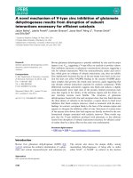
Fig. 1. TGFb1 induces rapid and sustained transcriptional upregula-
tion of the RhoB gene but not the RhoA gene in Swiss 3T3 and
HepG2 cells. (A) Active transcription is required for TGFb-induced
actin cytoskeleton reorganization. Swiss 3T3 fibroblasts were trea-
ted with 5 ngÆmL
)1
TGFb for 24 h in the absence or presence of
actinomycin D (5 lgÆmL
)1
). Changes in actin cytoskeleton were
monitored using rhodamine phalloidin staining. (B,C) TGFb induces
the transcription of the RhoB gene but not of the RhoA gene.
Swiss 3T3 (B) or HepG2 (C) cells were serum-starved for 24 h and
stimulated with TGFb 1 for the indicated time points or left
untreated. RT-PCR analysis was performed with primers specific
for the mRNA of the RhoB or RhoA genes. The bottom panels rep-
resent the mRNA levels of the housekeeping gene GAPDH at the
same time points. The RhoB and RhoA mRNA levels were normal-
ized to the intensity of the corresponding GAPDH mRNA of each
sample. Data are representative of two independent experiments.
The fold increase in mRNA levels is shown below each PCR image.
Rho GTPases ⁄ Smad proteins in TGFb-induced actin reorganization L. Vardouli et al.
4076 FEBS Journal 275 (2008) 4074–4087 ª 2008 The Authors Journal compilation ª 2008 FEBS
indicating that Smad3 is required for TGFb1-induced
transcriptional upregulation of the RhoB gene. In
agreement with the mRNA data, levels of RhoB pro-
tein were also potently increased by TGFb1 only in the
presence of adenovirally expressed Smad3 (Fig. 2C, top
panel). In line with our initial findings in Swiss 3T3 and
HepG2 cells, the transcription levels of the RhoA gene
were not affected by Smad3 overexpression in JEG-3
cells (Fig. 2B, middle panel).
TGFb and Smad proteins activate the promoter
of the RhoB gene
To further characterize the mechanism of transcrip-
tional upregulation of the RhoB gene by TGFb1, we
cloned the promoters of the human RhoB and RhoA
genes in fusion with the firefly luciferase gene (Fig. 3A)
and used them in transactivation experiments in order
to determine their responsiveness to the TGFb ⁄ Smad
signaling cascade. For this purpose, HepG2 cells were
transiently transfected with the reporter plasmids
)726 ⁄ +86 RhoB-Luc and )799 ⁄ +166 RhoA-Luc in
the absence or the presence of expression vectors for
the constitutively active form of the TGFb receptor I
(ALK5ca) and the TGFb signaling mediators Smad2,
Smad3 and Smad4. As shown in Fig. 3B, the constitu-
tively active form of the TGFb receptor type I
(ALK5ca) enhanced the activity of the )726 ⁄ +86
RhoB promoter 2.2-fold. The activity of this promoter
was also enhanced by overexpression of Smad3 and
Smad4 proteins (3.4-fold) and to a lesser extent by
overexpression of Smad2 and Smad4 proteins
(2.3-fold), and this activity was stimulated further by
simultaneous expression of the ALK5ca receptor (3.6-
and 2.6-fold, respectively). In contrast, ALK5ca,
Smad2 ⁄ Smad4 and Smad3 ⁄ Smad4 proteins had a
minor effect (1.2-1.6-fold) on the activity of the human
)799 ⁄ +166 RhoA promoter (Fig. 3C).
In conclusion, the combined data in Figs 2 and 3
indicate that transcriptional activation of the RhoB
gene by TGFb is mediated, at least in part, by TGFb-
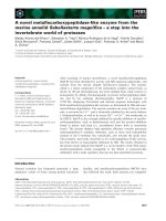
Fig. 2. Adenovirus-mediated overexpression of Smad proteins in
Swiss 3T3 cells and JEG-3 choriocarcinoma cells that lack endoge-
nous Smad3 confirmed the important role of Smad3 in transcrip-
tional upregulation of the RhoB gene. (A) RT-PCR analysis of Swiss
3T3 cells infected with ad-LacZ as control (lanes 1 and 2),
ad-Smad2 (lanes 3 and 4) or ad-Smad3 (lanes 5 and 6). Cells were
serum-starved for 24 h and stimulated with 5 ngÆmL
)1
TGFb1
(lanes 2, 4 and 6) for 2 h. The bottom panel represents the mRNA
levels of the housekeeping gene GAPDH at the same time points.
The RhoB and RhoA mRNA levels were normalized to the intensity
of the corresponding GAPDH mRNA of each sample. Data are rep-
resentative of two independent experiments. The fold increase in
mRNA levels is shown below each PCR image. (B) RT-PCR analysis
of JEG-3 (Smad3) ⁄ )) cells infected with ad-LacZ as control (lanes
1 and 2) or ad-Smad3 (lanes 3 and 4). Cells were serum-starved for
24 h and stimulated with 5 ngÆmL
)1
TGFb1 (lanes 2 and 4) for 24 h.
RT-PCR analysis was performed with primers specific for the RhoB
or RhoA mRNA. The bottom panel represents the mRNA levels of
the housekeeping gene GAPDH. The RhoB and RhoA mRNA levels
were normalized to the intensity of the corresponding GAPDH
mRNA of each sample. Data are representative of two independent
experiments. (C) Immunoblotting analysis of RhoB and adenovirally
overexpressed Smad2 and Smad3 proteins in JEG-3 cells. Follow-
ing adenovirus infection, cells were serum-starved for 24 h and
stimulated with 5 ngÆmL
)1
TGFb1 for 24 h (+) or left unstimulated
()). Total extracts from the infected cells were analyzed by SDS–
PAGE and Western blotting using antibodies against RhoB and
against total or phosphorylated forms of Smad2 and Smad3.
L. Vardouli et al. Rho GTPases ⁄ Smad proteins in TGFb-induced actin reorganization
FEBS Journal 275 (2008) 4074–4087 ª 2008 The Authors Journal compilation ª 2008 FEBS 4077
regulated Smad proteins, which act as transcriptional
regulators of the activity of the RhoB promoter.
Smad2 and Smad3 proteins activate RhoA and
RhoB GTPases and induce actin polymerization
and microfilament reorganization in Swiss 3T3
fibroblasts
To examine whether Smad proteins, in addition to
serving as transcriptional regulators of RhoB gene
expression in response to TGFb, also play a role in the
activation of Rho proteins as GTPases by this cyto-
kine, we performed affinity precipitation assays using
the rhotekin Rho-binding domain fused to glutathione
S-transferase (GST–RBD). For this purpose, cells
were infected with Smad2- or Smad3-expressing adeno-
viruses or with a LacZ-expressing adenovirus as a neg-
ative control. Following serum deprivation for 24 h,
cells were treated with 5 ngÆmL
)1
TGFb1 for 10 min,
and GST–RBD was used to isolate GTP-loaded RhoA
and RhoB from cell lysates. Both proteins were moni-
tored by immunoblotting using anti-RhoA and anti-
RhoB specific serum, and the protein band intensities
were normalized relative to the total RhoA or RhoB
content of non-adsorbed cell lysates. As shown in
Fig. 4A (top panel), RhoA was activated by TGFb
(3.2-fold) or by overexpression of Smad2 or Smad3
(3.9-fold) in Swiss 3T3 fibroblasts. In contrast, RhoB
was activated only slightly by Smad2 (1.2-fold) or
Smad3 (1.9-fold) compared to the activation of RhoA
(Fig. 4B, top panel).
The activation of GTPase activity of RhoA and
RhoB proteins and the transcriptional induction of the
RhoB gene by Smad proteins suggest that the TGFb–
Smad pathway may contribute to the long-term actin
reorganization induced by TGFb in Swiss 3T3 fibro-
blasts. To test this hypothesis, we analyzed actin archi-
tecture in Swiss 3T3 cells infected with adenoviruses
expressing Smad2 and Smad3. In these double staining
experiments, the actin cytoskeleton organization was
assessed by direct fluorescence using rhodamine phal-
loidin, and indirect immunofluorescence against the
Flag epitope of Smad2 and Smad3 revealed R-Smad
staining. Cells were serum-starved for 24 h, and then
stimulated, or not, with 5 ngÆmL
)1
TGFb1 for 24 h.
As shown in Fig. 5, control starved cells expressing the
LacZ gene exhibited typical morphology, i.e. their
actin cytoskeleton was restricted to cortical actin and
the main cell body was devoid of stress fibers
(Fig. 5B). Treatment with TGF b1 resulted in cell
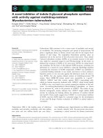
Fig. 3. TGFb1 increases the activity of the promoter of the RhoB gene in human hepatoma HepG2 cells. (A) Schematic representation of
the reporter plasmids )726 ⁄ +86 RhoB-Luc and )799 ⁄ +166 RhoA-Luc that were used in the transactivation experiments shown in (B) and
(C). Numbers refer to the transcription start site of each gene (+1). (B,C) HepG2 cells were transiently transfected with the )726 ⁄ +86
RhoB-Luc or the )799 ⁄ +166 RhoA-Luc plasmid (1 lg) together with the expression vector for the constitutively active form of TGFb recep-
tor I (ALK5ca) independently or in combination with expression vectors for Smad2, Smad3 and Smad4 (1 lg each) as indicated beneath each
histogram. The CMV-b-gal plasmid expressing b-galactosidase (1 lg) was included in each sample for normalization of transfection variability.
Luciferase activity was determined in cell lysates at 48 h after transfection, and the mean values and SEM from at least two independent
experiments performed in duplicate are shown as percentage relative luciferase activity.
Rho GTPases ⁄ Smad proteins in TGFb-induced actin reorganization L. Vardouli et al.
4078 FEBS Journal 275 (2008) 4074–4087 ª 2008 The Authors Journal compilation ª 2008 FEBS
flattening and scattering, supported by changes in the
organization of the actin cytoskeleton (Fig. 5D). In
contrast to control adenovirus (LacZ), ectopic expres-
sion of Smad2 in serum-starved cells resulted in
increased actin polymerization, even in the absence of
TGFb1 stimulation (Fig. 5F). Likewise, adenoviral
overexpression of Smad3 resulted in even more pro-
found changes in the organization of the actin cyto-
skeleton, resulting in cell flattening and cell shape
change. Cells appeared elongated or spindle-shaped,
with a parallel arrangement of actin bundles (Fig. 5J).
Addition of TGFb did not cause any additional
changes in actin cytoskeleton organization in the
ad-Smad3-infected cells (Fig. 5, panels J and L), but
enhanced the formation of stress fibers in the
ad-Smad2-infected cells (Fig. 5, panels F and H).
These morphological findings were further corrobo-
rated by quantitative immunoblot analysis of the Tri-
ton X-100-soluble (TS) and -insoluble (TI) actin
cytoskeleton fractions of cells overexpressing the
R-Smad proteins (Table 1). As calculated from the rel-
ative band intensities, the G ⁄ total actin ratio of Swiss
3T3 fibroblasts treated with TGFb 1 for 24 h was
clearly and reproducibly decreased in comparison with
control cells serum-starved for 24 h. Furthermore, cells
ectopically expressing Smad2 or Smad3 revealed a
greater decrease in the G ⁄ total actin ratio, correspond-
ing to a clear shift of the dynamic equilibrium towards
polymerized actin. Thus, quantitative analysis of actin
dynamics revealed that Smad2 and Smad3 overexpres-
sion in Swiss 3T3 fibroblasts induces actin polymeriza-
tion and microfilament reorganization.
To address the contribution of Smad3 protein to the
actin reorganization induced by TGFb, we infected
JEG-3 cells with adenoviruses expressing Smad3 (or
LacZ as a negative control), and assessed the polymeri-
zation dynamics of the actin cytoskeleton by quantita-
Fig. 4. Overexpression of Smad2 and Smad3 proteins via adeno-
virus-mediated gene transfer induced activation of Rho GTPases in
Swiss 3T3 fibroblasts. Rho–GTP loading assay with Swiss 3T3 cells
infected with ad-LacZ as control (lanes 1 and 2), ad-Smad2 (lanes 3
and 4) or ad-Smad3 (lanes 5 and 6). Cells were serum-starved for
24 h and stimulated with 5 ngÆmL
)1
TGFb1 (lanes 2, 4 and 6) for
15 min. Immunoblots of the GST–RBD pulldown (RhoA–GTP or
RhoB–GTP), or total cell extracts with RhoA antibody (A) or RhoB
antibody (B) and Flag-M5 antibody (Smad2 and Smad3) are shown.
Densitometric analysis of the RhoA–GTP and RhoB–GTP immuno-
blots was performed, and the fold increase of the active ⁄ total ratio
values of each condition is shown. These data are representative of
two independent experiments.
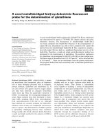
Fig. 5. Smad2 and Smad3 induce actin reor-
ganization in Swiss 3T3 fibroblasts. Swiss
3T3 cells infected with the adenoviruses
ad-LacZ, ad-Smad2 or ad-Smad3 were sub-
sequently serum-starved for 24 h and stimu-
lated (+) or not ()) with 5 ngÆmL
)1
TGFb1
for 24 h (+). Indirect immunofluorescence
against the Flag epitope of Smad2 and
Smad3 is shown in the left panels (FITC,
fluorescein isothiocyanate) and direct
fluorescence of actin is shown in the right
panels. The bar represents 10 lm.
L. Vardouli et al. Rho GTPases ⁄ Smad proteins in TGFb-induced actin reorganization
FEBS Journal 275 (2008) 4074–4087 ª 2008 The Authors Journal compilation ª 2008 FEBS 4079
tive immunoblot analysis of the Triton X-100-soluble
(TS) and -insoluble (TI) actin cytoskeleton fractions of
cells (Table 2). The G ⁄ total actin ratio of JEG-3 cells
treated with TGFb1 for 24 h was decreased compared
to control serum-starved cells, indicating slight actin
polymerization. Furthermore, the G ⁄ total actin ratio
decreased in cells overexpressing Smad3, and this ratio
decreased further following treatment with TGFb1,
corresponding to a clear shift of the dynamic equilib-
rium toward actin polymerization. These results indi-
cate that Smad3 is crucial for the TGFb1-induced
actin reorganization mediated by Rho GTPases.
Smad3 (and to a lesser extent Smad2) induces
the expression of a-smooth muscle actin
TGFb stimulation of various cell types, including
Swiss 3T3 fibroblasts, resulted in increased a-smooth
muscle actin (a-SMA) expression [24,27–29 and
unpublished data]. Having established that overexpres-
sion of R-Smad proteins results in long-term actin
polymerization, we investigated the involvement of
Smad2 and Smad3 in TGFb1-induced a-SMA gene
regulation and protein incorporation into the actin
network of Swiss 3T3 fibroblasts. For this purpose, we
transiently overexpressed Smad2 and Smad3 using
adenoviral cell infections. Swiss 3T3 fibroblasts
expressing Smad2, Smad3 or LacZ (as a negative con-
trol) were serum-starved for 24 h, then stimulated with
5ngÆmL
)1
TGFb1 for 24 h, and the levels of expres-
sion of a-SMA were analyzed. As shown in Fig. 6A,
control cells infected with the LacZ adenovirus and
stimulated with TGFb1 for 24 h showed a 5.4-fold
upregulation of a-SMA gene expression. In agreement
with previous findings, Smad2 overexpression resulted
in a moderate (1.5-fold) increase in a-SMA mRNA
levels, whereas stimulation with TGFb1 led to a robust
4.6-fold upregulation of a-SMA mRNA (Fig. 6A).
Interestingly, cells infected with ad-Smad3 showed a
stronger upregulation of a-SMA mRNA (2.4-fold),
which was even more prominent with TGFb1 treat-
ment (5.4-fold). Analysis of a-SMA protein levels
under the same experimental conditions as described in
Fig. 6A provided very similar results (Fig. 6B). These
data indicated that Smad3 rather that Smad2 is
involved in a-SMA transcriptional upregulation in
Swiss 3T3 fibroblasts.
To further evaluate incorporation of the newly
synthesized a-SMA to the actin cytoskeleton, we
performed immunoblot analysis of the Triton X-100-
soluble (TS) and -insoluble (TI) preparations after sub-
cellular fractionation of Swiss 3T3 fibroblasts treated
or not with TGFb and overexpressing Smad2, Smad3
or LacZ (as a negative control). Smad3 overexpression
led to incorporation of a-SMA into the insoluble (fila-
mentous) part (Fig. 6D, compare lanes 1 ⁄ 2 and 5 ⁄ 6).
In line with the weaker induction of a-SMA protein
expression in Smad2-transfected cells, a-SMA incorpo-
ration into the insoluble cytoskeleton fraction was very
low (Fig. 6C, compare lanes 1 ⁄ 2 and 5 ⁄ 6). These con-
clusions were fully supported by the morphological
analysis shown in Fig. 6E. Indirect immunostainning
of Swiss 3T3 fibroblasts with an antibody against
a-SMA, and subsequent analysis by fluorescence
microscopy revealed increased a-SMA structures after
Smad3 (but not Smad2) overexpression, similar to the
control cells stimulated with TGFb1 for 24 h (Fig. 6E).
These results indicate that ectopic expression of
Smad3 (and to a lesser extent Smad2) leads to
increased a-SMA expression in Swiss 3T3 fibroblasts.
Interestingly, however, the newly synthesized a-SMA
Table 1. Effect of the ectopic expression of Smad2 and Smad3 on
the polymerization state of actin in Swiss 3T3 fibroblasts. Swiss
3T3 fibroblasts were infected with the adenoviruses indicated,
serum-starved for 24 h, and then stimulated with 5 ngÆ mL
)1
TGFb1
for 24 h. Triton-soluble (TS) and Triton-insoluble (TI) actin cytoskele-
ton fractions were prepared as described in Experimental proce-
dures, and their actin content was analyzed by immunoblotting.
Data presented correspond to the G ⁄ total actin ratio in each condi-
tion. These data are representative of three independent experi-
ments.
Sample G ⁄ total actin ratio Standard error
ad-LacZ 0.42 0.01
ad-LacZ + TGFb1 0.38* 0.01
ad-Smad2 0.35* 0.00
ad-Smad2 + TGFb1 0.34* 0.01
ad-Smad3 0.35* 0.00
ad-Smad3 + TGFb1 0.34* 0.02
* Statistically different from ad-LacZ (control) at P < 0.05.
Table 2. Effect of the ectopic expression of Smad3 on the poly-
merization state of actin in JEG-3 cells. JEG-3 (Smad3) ⁄ )) cells
were infected with the adenoviruses indicated, serum-starved for
24 h, and then stimulated with 5 ngÆmL
)1
TGFb1 for 24 h. Triton-
soluble (TS) and Triton-insoluble (TI) actin cytoskeleton fractions
were prepared as described in Experimental procedures, and their
actin content was analyzed by immunoblotting. Data presented
correspond to the G ⁄ total actin ratio in each condition. These data
are representative of five independent experiments.
Sample G ⁄ total actin Standard error
ad-LacZ 0.45 0.01
ad-LacZ + TGFb1 0.37* 0.05
ad-Smad3 0.40* 0.03
ad-Smad3 + TGFb1 0.34* 0.05
* Statistically different from ad-LacZ (control) at P < 0.05.
Rho GTPases ⁄ Smad proteins in TGFb-induced actin reorganization L. Vardouli et al.
4080 FEBS Journal 275 (2008) 4074–4087 ª 2008 The Authors Journal compilation ª 2008 FEBS
can be incorporated into the newly formed stress fibers
only when Smad3 is overexpressed, even in the absence
of TGFb.
Discussion
In the present study, we provide evidence for the
essential role of Smad proteins and Rho GTPases in
long-term TGFb-induced actin cytoskeleton reorgani-
zation in various cell models. We also show that acti-
vation of Smad and Rho proteins by TGFb in
fibroblasts is correlated with potent induction of
a-SMA gene expression and subsequent incorporation
of a-SMA into microfilamentous structures. As a-SMA
expression is indicative of a myofibroblast phenotype
[30], our findings suggest that these proteins control a
TGFb-induced fibroblast to myofibroblast differentia-
tion program. Fibroblast to myofibroblast conversion
is a pathophysiological feature of various fibrotic dis-
eases such as idiopathic pulmonary fibrosis, asthma
and chronic obstructive pulmonary diseases (COPD)
[31–33]. Given that enhanced TGFb concentrations
have been detected in patients with various fibrotic dis-
eases including idiopathic pulmonary fibrosis [34,35],
sarcoidosis [36] and cystic fibrosis [37], in-depth under-
standing of the mechanism that underlies this TGFb-
induced conversion program, identification of the
molecules involved and elucidation of their role or their
Fig. 6. Smad3 (and to a lesser extent Smad2) induce the expression of a-smooth muscle actin (a-SMA). (A) RT-PCR of Swiss 3T3 cells
infected with control (LacZ) adenovirus or adenoviruses expressing Smad2 and Smad3. Cells were serum-starved for 24 h and stimulated
with 5 ngÆmL
)1
TGFb1 for 3 h. RT-PCR analysis was performed with primers specific for the mRNA of the a-SMA gene. The bottom panel
represents the mRNA levels of the housekeeping gene GAPDH. The a-SMA mRNA level was normalized to the intensity of the correspond-
ing GAPDH mRNA for each sample. Data are representative of two independent experiments. The fold increase in a-SMA mRNA levels is
shown below each image. (B) Immunoblot analysis of total cell extracts of Swiss 3T3 cells infected with control (LacZ) adenovirus or adeno-
viruses expressing Smad2 and Smad3. Cells were starved for 24 h and stimulated (+) or not ()) with 5 ngÆmL
)1
TGFb1 for 24 h. Immuno-
blotting of the corresponding total extracts with the a-SMA antibody and control b-tubulin antibody is shown. The fold increase in a-SMA
protein levels is shown below each image. (C,D) Immunoblot analysis of the Triton-soluble (TS) and Triton-insoluble (TI) actin cytoskeleton
fractions of Swiss 3T3 cells infected with adenoviruses expressing LacZ (lanes 1-4, both panels), Smad2 (lanes 5-8, C) or Smad3 (lanes 5-8,
D). Following adenovirus infection, cells were starved for 24 h and stimulated (+) or not ()) with 5 ngÆmL
)1
TGFb1 for 24 h (+). Immunoblot-
ting of the corresponding total extracts with the a-SMA antibody is shown. (E) Indirect fluorescence microscopy of Swiss 3T3 cells infected
with the indicated adenoviruses using an antibody against a-SMA. Following adenovirus infection, cells were serum-starved for 24 h and
stimulated (+) or not ()) with 5 ngÆmL
)1
TGFb1 for 24 h. Bars correspond to 10 lm.
L. Vardouli et al. Rho GTPases ⁄ Smad proteins in TGFb-induced actin reorganization
FEBS Journal 275 (2008) 4074–4087 ª 2008 The Authors Journal compilation ª 2008 FEBS 4081
modes of regulation may lead to the development of
novel therapeutic approaches for these diseases.
We show here that TGFb stimulation of fibroblasts
or hepatic cells causes differential regulation of expres-
sion of the genes encoding the two small GTPases
RhoA and RhoB (Fig. 1). Thus, treatment of these
cells with TGFb1 resulted in a rapid increase in
steady-state mRNA levels of the RhoB gene that was
initiated at 30 min (Swiss 3T3) or 1 h (HepG2) and
persisted for a period of 24 h post-induction, whereas
no transcriptional activation of the RhoA gene by
TGFb1 was observed (Fig. 1). These findings confirm
previous experimental evidence which suggested that
RhoB is the only member of the Rho-related subfamily
of small GTPases that is regulated at the transcrip-
tional level, and this regulation may be important for
its function due to the short half-life of this protein in
the cell [38,39]. Of interest was the periodic pattern of
induction of RhoB gene transcription by TGFb.As
shown in Fig. 1B, TGFb induced RhoB gene expres-
sion 2.6-fold at 2 h of treatment in HepG2 cells, and
this induction declined to 1.8-fold at 4 h, increased
again to 2.2-fold at 8 h and dropped back to 1.4-fold
at 24 h after TGFb addition. A similar periodic pat-
tern of RhoB gene transcriptional induction was
observed in Swiss 3T3 cells (Fig. 1A). These findings
could account for our previously reported observations
that regulation of the RhoB GTPase activity by
TGFb in Swiss 3T3 cells follows a periodic pattern
[23]. The periodic decrease in expression of the RhoB
gene during TGFb stimulation strongly suggests that
RhoB is subject to an auto-inhibitory loop. In agree-
ment with this hypothesis, we have shown that over-
expression of RhoB causes a potent reduction in the
activity of its own promoter (E. Vasilaki, unpublished
observations).
We speculated that the differential effect of TGFb
on expression of these two genes reflects a difference in
the mechanism by which the TGFb-regulated Smad
proteins (Smad2 and Smad3) regulate the activity of
the two promoters. To address this hypothesis, we
cloned the promoters of the human RhoA and RhoB
genes in front of the firefly luciferase gene, and, using
transient transfection experiments and luciferase
assays, did indeed show that the TGFb–Smad pathway
specifically targets the RhoB gene (Fig. 3). A mechanis-
tic explanation for this RhoB-specific transcriptional
response to the TGFb–Smad pathway could be that
the promoter of the human RhoA gene lacks Smad
binding elements that could serve as sites of Smad
recruitment in response to TGFb stimulation. A search
for transcription factor binding sites in the two
promoters revealed that the human RhoB promoter
contains three putative Smad binding elements
(sequence 5¢-CAGAC-3¢) [40] in the proximal
)726 ⁄ +86 region that was used in the transactivation
experiments (at positions )294 ⁄ )290, )278 ⁄ )274 and
+20 ⁄ +24), whereas the human RhoA
promoter, which
is not homologous to the human RhoB promoter, does
not contain any of these putative Smad binding ele-
ments. We are in the process of characterizing these
sites further and studying their role and participation
in the transcriptional upregulation of the RhoB
promoter in response to TGFb stimulation or Smad
overexpression. Our preliminary experiments have
shown that regulation of the RhoB promoter by the
TGFb–Smad pathway is more complex than initially
suspected, and that elements additional to the Smad
binding elements are required for this regulation
(E. Vasilaki, E. Papadimitriou, C. Stournaras &
D. Kardassis, unpublished data).
An interesting finding during this work was that
Smad proteins, in addition to serving as specific tran-
scriptional activators of the RhoB gene, can also act as
activators of Rho GTPase function. This is in line with
previous studies that had provided indications of the
involvement of the Smad pathway in the activation of
Rho protein activity, and specifically that of RhoA, by
TGFb [23,24], We have shown recently that overex-
pression of Smad7, a known and potent inhibitor of
the TGFb signaling pathway and of Smad function
that operates in the context of a feedback inhibitory
loop [41], was able to block both the activation of
RhoA GTPase activity and reorganization of the actin
cytoskeleton by TGFb1 in Swiss 3T3 fibroblasts, sug-
gesting cross-talk between Smad signaling and RhoA
activation [23]. In line with these observations, it was
recently reported that dominant-negative RhoA inhib-
its the nuclear translocation of Smad2 and Smad3 dur-
ing the smooth muscle cell differentiation induced by
TGFb, indicating that RhoA is a modulator of Smad
activation [24]. Moreover, by studying the signaling
properties of a type I TGFb receptor with a mutation
in the L45 loop that contains the Smad docking site
[16], it was demonstrated that interaction of the recep-
tor with Smad proteins is required for signaling to
Rho GTPases and the actin cytoskeleton [23]. These
data suggest that Smad proteins, in addition to their
role in long-term RhoB transcriptional regulation,
might be directly involved in activation of Rho GTP-
ases and the regulation of actin dynamics. The results
presented in this study clearly support this assumption.
Indeed, adenovirus-mediated Smad2 and Smad3 over-
expression triggered activation of RhoA, and to a
lesser extent RhoB, and caused potent long-term actin
reorganization (Figs 4 and 5). Activation of Rho
Rho GTPases ⁄ Smad proteins in TGFb-induced actin reorganization L. Vardouli et al.
4082 FEBS Journal 275 (2008) 4074–4087 ª 2008 The Authors Journal compilation ª 2008 FEBS
proteins by Smad proteins could be facilitated by dif-
ferent, not necessarily mutually exclusive mechanisms,
as discussed below.
Another interesting finding arising from this work
was the differential involvement of the two TGFb-reg-
ulated Smad proteins (Smad2 and Smad3) in TGFb-
induced Rho gene regulation and actin restructuring.
While adenovirus-mediated overexpression of Smad3
largely mimicked the TGFb effects on transcriptional
upregulation of the RhoB and a-SMA genes, as well as
on RhoA and RhoB activation and actin reorgani-
zation, Smad2 was consistently less effective in all of
these processes (Figs 2–6). This may reflect a specific
requirement for Smad3 in transcriptional regulation of
both RhoB and a-SMA genes by TGFb. This hypothe-
sis is supported by experiments in a cellular model that
lacks endogenous Smad3 expression (JEG-3 choriocar-
cinoma cells). We found that TGFb treatment of this
cell line had no effect on transcriptional upregulation
of the RhoB gene, but RhoB gene expression was
rescued after adenovirus-mediated ectopic expression
of Smad3 (Fig. 2B). Taken together, these findings
support a key role for Smad3 in mediating Rho ⁄ actin
regulation by TGFb.
The combined data from this as well as previous
studies suggest a model of short- and long-term
TGFb-induced actin cytoskeleton reorganization in
fibroblasts and other cell types. This model is shown
schematically in Fig. 7, and can be summarized as
follows. In the short-term activation process, TGFb
receptor activation by its ligand induces rapid activa-
tion of RhoA and RhoB GTPases (1), which is fol-
lowed by activation of the ROCK ⁄ LIMK ⁄ cofilin
pathway (2–4) and actin cytoskeleton restructuring (5),
as shown previously [23]. The mechanism by which
Rho activation is linked to activation of the type I
TGFb receptor (TbRI) is currently unknown. It may
be that the phosphorylated TbRI activates very
rapidly, by phosphorylation, a specific guanine nucleo-
tide exchange factor (GEF), which in turn activates
Rho proteins. In a previous study, we showed that
dominant-negative forms of either RhoA or RhoB are
equally effective in blocking TGFb-induced actin cyto-
skeleton reorganization in Swiss 3T3 cells, suggesting
that these two GTPases are in the same pathway and
activation of the one may precede activation of the
other [23]. Gene silencing experiments may shed some
light into the details of this activation process and the
specific role of each Rho protein, but it seems reason-
able to suggest, based on the available data, that
RhoA protein plays a more critical role in this short-
term activation event.
The long-term actin cytoskeleton response to TGFb
stimulation involves the Smad pathway and transcrip-
tional activation events. Thus, TGFb rapidly induces
the phosphorylation of R-Smad proteins (a), the for-
mation of R-Smad(P)–Smad4 complexes (b), transloca-
tion of these complexes to the nucleus (c), and their
Fig. 7. Mechanisms of short-term and long-
term actin cytoskeleton reorganization
induced by TGFb in fibroblasts. Schematic
representation of the proposed mechanisms
regulating actin cytoskeleton reorganization
in fibroblasts following TGFb stimulation.
Numbers in parentheses (1–5) indicate the
successive steps that lead to short-term
actin reorganization immediately after TGFb
stimulation. Letters in parentheses (a–g)
indicate events that contribute to long-term
actin cytoskeleton reorganization. Solid
arrows indicate events that have been
experimentally proven in this or previous
studies. Dashed arrows indicate hypotheti-
cal events.
L. Vardouli et al. Rho GTPases ⁄ Smad proteins in TGFb-induced actin reorganization
FEBS Journal 275 (2008) 4074–4087 ª 2008 The Authors Journal compilation ª 2008 FEBS 4083
recruitment to various TGFb-responsive genes (d).
Two genes that were shown in this study to be acti-
vated by this pathway are the RhoB and a-SMA genes.
The preferential transcriptional activation of the RhoB
gene over the RhoA gene strongly suggests an exclusive
role of RhoB GTPase in long-term actin cytoskeleton
reorganization by TGFb (f). Smad proteins may con-
tribute to long-term Rho GTPase activity by various
mechanisms, such as transcriptional activation of a
gene coding for a Rho GEF (e), physical interactions
with an existing GEF, or finally by acting as GEFs
themselves. Finally, in fibroblasts, the TGFb–Smad
pathway causes transcriptional activation of the
a-SMA gene and its incorporation into the cytoskele-
ton (g), a process that is characteristic of a fibroblast
to myofibroblast differentiation program [30].
In conclusion, in the present work, we have eluci-
dated a novel regulatory mechanism for the long-term
actin restructuring controlled by TGFb and mediated
by Smad proteins. We show for the first time that
the RhoB gene (but not the RhoA gene) is a direct
transcriptional target of the TGFb–Smad signal trans-
duction pathway, and that expression of the RhoB
gene as well as the activity of the RhoB promoter can
be induced by TGFb-regulated Smad proteins. Fur-
thermore, we provide direct evidence of a key role for
Smad proteins, and more specifically Smad3, in medi-
ating Rho activation and actin regulation by TGFb.
Finally, we demonstrate that, in fibroblasts, activation
of RhoB gene expression by TGFb is associated with
the stimulation of a-SMA expression and microfila-
ment incorporation, indicative of a fibroblast to myo-
fibroblast differentiation program.
Experimental procedures
Materials
Dulbecco’s modified Eagle’s medium (DMEM), penicil-
lin ⁄ streptomycin for cell culture, Trizol reagent for RNA
extraction and Superscript RNAse H reverse transcriptase
were purchased from Invitrogen ⁄ Life Technologies (Carls-
bad, CA, USA). Fetal bovine serum (FBS) was purchased
from BioChrom Labs (Terre Haute, IN, USA). Recombi-
nant mature human TGF-b1 was purchased from R&D Sys-
tems Inc. (Minneapolis, MN, USA). Restriction enzymes
and modifying enzymes (T4 DNA ligase and alkaline phos-
phatase) were purchased from Minotech (Heraklion,
Greece) or New England Biolabs (Beverly, MA, USA). Go-
Taq DNA polymerase, dNTPs, the luciferase assay system,
and the Wizard SV gel and PCR cleanup system were pur-
chased from Promega (Madison, WI, USA). The Super Sig-
nal West Pico chemiluminescent substrate was purchased
from Pierce (Rockford, IL, USA). Anti-Smad2 (SC-6200)
and anti-Smad3 (SC-8332), mouse monoclonal anti-RhoA
(SC-26C4) and rabbit polyclonal anti-RhoB (SC-119) were
purchased from Santa Cruz Biotechnology Inc. (Santa Cruz,
CA, USA). Anti-P-Smad2 (3101S) was purchased from Cell
Signalling Technology (Danvers, MA, USA). Anti-P-Smad3
(9514L) was a generous gift from A. Moustakas (Ludwig
Institute for Cancer Research, Uppsala, Sweden). Secondary
anti-mouse IgG and anti-rabbit IgG coupled to horseradish
peroxidase were from Chemicon International Inc. (Temecu-
la, CA, USA). GST–RBD was purchased from Millipore
(Billerica, MA, USA). Mouse anti-a-SMA (1A4), mouse
anti-Flag (M5), mouse anti-b-tubulin (T8535) and anti-a-
smooth muscle actin serum were purchased from Sigma-
Aldrich (St Louis, MI, USA). Enhanced chemiluminescence
detection systems were purchased from Santa Cruz Biotech-
nology Inc. or Amersham Biosciences (Piscataway, NJ,
USA). Rhodamine phalloidin and the Slow Fade detection
kit were purchased from Invitrogen ⁄ Molecular Probes Inc
(Carlsbad, CA, USA).
Cell culture, adenoviruses, transient transfections
and luciferase assays
Mouse Swiss 3T3 fibroblasts, human embryonic kidney
(HEK) 293 cells (used only for titration of adenoviruses),
human hepatoma HepG2 and JEG-3 choriocarcinoma cells
were obtained from the American Type Culture Collection
(Manassas, VA, USA). All cell lines were cultured in
DMEM supplemented with 10% FBS, glutamine and peni-
cillin ⁄ streptomycin at 37 °C. Adenoviruses expressing LacZ
and N-terminally Flag-tagged Smad2 and Smad3 were
provided by K. Miyazono (University of Tokyo, Japan)
[42]; all adenoviruses were amplified and titrated in
HEK293 cells as described previously [22]. Adenoviral
transient infections of cells using multiplicity of infection
leading to a maximal rate of infection efficiency and
protein expression, without obvious cytopathic effects, were
performed as previously described [43]. Transient trans-
fections in HepG2 cells were performed in six-well plates
by the Ca
3
(PO
4
)
2
co-precipitation method using 6 lgof
DNA per well. Luciferase assays were performed using
the luciferase assay kit from Promega, according to the
manufacturer’s instructions. Normalization for transfection
efficiency was performed by b-galactosidase assays.
Plasmids
The mammalian vectors expressing Smad2, Smad3 and the
constitutively active form of ALK5 were a kind gift from
A. Moustakas (Ludwig Institute for Cancer Research, Upp-
sala, Sweden). The promoter plasmids )726 ⁄ +86 RhoB-
Luc and )799 ⁄ +166 RhoA-Luc were generated by PCR
amplification of the corresponding fragments of human
Rho GTPases ⁄ Smad proteins in TGFb-induced actin reorganization L. Vardouli et al.
4084 FEBS Journal 275 (2008) 4074–4087 ª 2008 The Authors Journal compilation ª 2008 FEBS
RhoB and RhoA promoters and subsequent cloning into
the pGL3basic vector at the HindIII (RhoB-Luc) and
KpnI–HindIII (RhoA-Luc) sites. The oligonucleotides used
as primers in PCR amplification were synthesized at the
microchemical facility of the Institute of Molecular Biology
and Biotechnology (IMBB) (Heraklion, Greece), and their
sequences are shown in Table 3.
RT-PCR
Swiss 3T3 fibroblasts, JEG-3 or HepG2 cells infected or not
with the adenoviruses expressing LacZ, Smad2 or Smad3
proteins, after 24 h of serum starvation and stimulation with
5ngÆlL
)1
TGFb1 for various durations (30 min, 1, 2, 4, 8
and 24) were lysed and processed for total RNA extraction
using Trizol reagent according to the manufacturer’s instruc-
tions. The first cDNA strand was synthesized by Superscript
reverse transcriptase. The sequences of the primers used for
the PCR amplification of RhoB and RhoA cDNA are shown
in Table 3. For normalization of the samples, the cDNA of
the housekeeping glyceraldehyde-3-phosphate dehydro-
genase (GAPDH) gene was also amplified by PCR. Quantifi-
cation of the results was performed by measuring the
intensity of the bands using tina version 2 software (Raytest,
Straubenhardt, Germany).
Immunoblot analysis
Total protein extracts were analyzed by SDS–PAGE ⁄
Western blotting as described previously [44]. Protein band
intensity was quantified using tina version 2 (Raytest).
Immunofluorescence analysis and fluorescence
microscopy
For immunofluorescence or direct fluorescence of the actin
cytoskeleton, cell monolayers were serum-starved for 24 h
and then treated with TGFb1; alternatively cells were first
infected with adenoviruses, then starved for 24 h and finally
treated with TGFb1 as indicated in the figure legends. Cells
were prepared as previously described [21,43]. Staining was
performed using rhodamine phalloidin at 1 : 100 dilution
or mouse monoclonal anti-Flag M5 at 1 : 100 dilution. All
secondary antibodies were used at 1 : 100 dilution. Slides
were mounted using the Slow Fade kit. Photomicrographs
were obtained using a Leica DMLB microscope (Leica
Microsystems Imaging Solutions Ltd., Cambridge, UK)
equipped with epifluorescence illumination, and photo-
graphed using a Leica DC 300F digital camera, with a
Zeiss Plan-neofluar 40·⁄0.75 objective lens at ambient tem-
perature in the absence of immersion oil. Images were
acquired using the camera’s qed imaging software, and
image memory content was reduced and brightness ⁄
contrast were adjusted using AdobeÒ photoshopÒ soft-
ware (Adobe Systems Inc., Boston, MA, USA).
Determination of GTP-loaded Rho levels
For affinity precipitation with GST–RBD, cells were
transiently infected with LacZ or Smad2 or Smad3 adeno-
viruses 16 h prior to serum starvation and stimulation
with TGFb1. The cells were then washed with ice-cold
Tris–buffered saline and lysed with RIPA buffer (50 mm
Tris pH 7.2, 1% Triton X-100, 0.5% sodium deoxycholate,
0.1% SDS, 500 mm NaCl, 10 mm MgCl
2
, supplemented
with protease inhibitors). Equal amounts of cleared cell
lysates were incubated with 30 lg GST–RBD attached to
glutathione–Sepharose at 4 °C for 45 min and analyzed by
SDS–PAGE. GTP-bound Rho was detected by immuno-
blotting using the appropriate antibody. Protein band
intensity was quantified using PC-based image analysis
(Image Analysis Inc.). The data were then normalized using
the ratio of active ⁄ total Rho protein in each sample.
Thus the total protein levels in these experiments cannot be
compared.
Triton X-100 fractionation
The Triton X-100-soluble and -insoluble fractions of cells
transiently infected with adenoviruses in the absence or
presence of TGFb were prepared as previously described
[45]. Briefly, cells were incubated in 500 lL of Triton
extraction buffer (0.3% Triton X-100, 5 mm Tris ⁄ HCl
pH 7.4, 2 mm EGTA, 300 mm sucrose, 2 lm phalloidin,
1mm phenylmethanesulfonyl fluoride, 10 lgÆmL
)1
leupeptin and 50 mm NaF) for 5 min on ice. The Triton-
soluble proteins were precipitated with equal volumes of
6% perchloric acid. The Triton-insoluble fractions were
precipitated with 1 mL of 3% perchloric acid. Equal
volumes of each fraction were subjected to SDS–PAGE
and Western blotting using the anti-a-smooth muscle actin
serum.
Table 3. Primers used in RT-PCR experiments and for the cloning
of the RhoA and RhoB promoter regions.
Name of
primer Sequence (5¢-to3¢)
hRhoB -825 GGGATCAGAGTTCATAGTGAAAAGAG
hRhoB +86 GCG
AAGCTTCGGCCTAGCTCTCTCCCGGGTCTC
hRhoA )799 GCG
GGTACCAATGTGATGGGTGGACTGGT
hRhoA +166 GCG
AAGCTTACCAGACCGTGGACTAACGA
hRhoB sense CCCACCGTCTTCGAGAACTA
hRhoB
antisense
CTTCCTTGGTCTTGGCAGAG
hRhoA sense CCAGACTAGATGTAGTATTTTTTG
hRhoA
antisense
ATTAGAGCCAGATGCTTAAGTCC
GAPDH-F ACCACAGTCCATGCCATCAC
GAPDH-R TCCACCACCCTGTTGCTGTA
L. Vardouli et al. Rho GTPases ⁄ Smad proteins in TGFb-induced actin reorganization
FEBS Journal 275 (2008) 4074–4087 ª 2008 The Authors Journal compilation ª 2008 FEBS 4085
Acknowledgements
We would like thank Dr Aris Moustakas (Ludwig
Institute for Cancer Research, Uppsala, Sweden) and
Dr Theodore Fotsis (University of Ioannina Medical
School, Ioannina, Greece) for useful discussions and
for their comments on this manuscript. This research
was supported by a grant from the Greek General Sec-
retariat for Research and Technology (PENED-2003).
References
1 Pollard TD & Borisy GG (2003) Cellular motility
driven by assembly and disassembly of actin filaments.
Cell 112, 453–465.
2 Raftopoulou M & Hall A (2004) Cell migration: Rho
GTPases lead the way. Dev Biol 265, 23–32.
3 Gourlay CW & Ayscough KR (2005) Identification of
an upstream regulatory pathway controlling actin-medi-
ated apoptosis in yeast. J Cell Sci 118, 2119–2132.
4 Yamazaki D, Kurisu S & Takenawa T (2005) Regula-
tion of cancer cell motility through actin reorganization.
Cancer Sci 96, 379–386.
5 Thomas SG, Huang S, Li S, Staiger CJ & Franklin-
Tong VE (2006) Actin depolymerization is sufficient to
induce programmed cell death in self-incompatible
pollen. J Cell Biol 174, 221–229.
6 Theriot JA (1994) Regulation of the actin cytoskeleton
in living cells. Semin Cell Biol 5, 193–199.
7 Papakonstanti EA & Stournaras C (2002) Association
of PI-3 kinase with PAK1 leads to actin phosphoryla-
tion and cytoskeletal reorganization. Mol Biol Cell 13,
2946–2962.
8 Papakonstanti EA & Stournaras C (2004) Tumor necro-
sis factor-alpha promotes survival of opossum kidney
cells via Cdc42-induced phospholipase C-gamma1 acti-
vation and actin filament redistribution. Mol Biol Cell
15, 1273–1286.
9 Rivera GM, Antoku S, Gelkop S, Shin NY, Hanks SK,
Pawson T & Mayer BJ (2006) Requirement of Nck
adaptors for actin dynamics and cell migration stimu-
lated by platelet-derived growth factor B. Proc Natl
Acad Sci USA 103, 9536–9541.
10 Massague
´
J (1998) TGFb signal transduction. Annu Rev
Biochem 67, 753–791.
11 Roberts A & Sporn M (1992) Transforming growth fac-
tor-beta: recent progress and new challenges. J Cell Biol
119, 1017–1021.
12 Roberts AB (1998) Molecular and cell biology of TGF-
beta. Miner Electrolyte Metab 24, R111–R119.
13 Dumont N & Arteaga CL (2003) Targeting the TGF
beta signaling network in human neoplasia. Cancer Cell
3, 531–536.
14 Grunert S, Jechlinger M & Beug H (2003) Diverse cellu-
lar and molecular mechanisms contribute to epithelial
plasticity and metastasis. Nat Rev Mol Cell Biol 4, 657–
665.
15 Roberts AB & Wakefield LM (2003) The two faces of
transforming growth factor beta in carcinogenesis. Proc
Natl Acad Sci USA 100, 8621–8623.
16 Shi Y & Massague
´
J (2003) Mechanisms of TGF-beta
signaling from cell membrane to the nucleus. Cell 113,
685–700.
17 Moustakas A, Souchelnytskyi S & Heldin CH (2001)
Smad regulation in TGF-beta signal transduction.
J Cell Sci 114, 4359–4369.
18 Di Guglielmo GM, Le Roy C, Goodfellow AF &
Wrana JL (2003) Distinct endocytic pathways regulate
TGF-beta receptor signalling and turnover. Nat Cell
Biol 5, 410–421.
19 Bhowmick NA, Ghiassi M, Bakin A, Aakre M, Lund-
quist CA, Engel ME, Arteaga CL & Moses HL (2001)
Transforming growth factor-beta1 mediates epithelial to
mesenchymal transdifferentiation through a RhoA-
dependent mechanism. Mol Biol Cell 12, 27–36.
20 Edlund S, Landstrom M, Heldin CH & Aspenstrom P
(2002) Transforming growth factor-beta-induced mobili-
zation of actin cytoskeleton requires signaling by small
GTPases Cdc42 and RhoA. Mol Biol Cell 13, 902–914.
21 Moustakas A & Stournaras C (1999) Regulation of
actin organisation by TGF-beta in H-ras-transformed
fibroblasts. J Cell Sci 112, 1169–1179.
22 Piek E, Moustakas A, Kurisaki A, Heldin CH & ten
Dijke P (1999) TGF-(beta) type I receptor ⁄ ALK-5 and
Smad proteins mediate epithelial to mesenchymal trans-
differentiation in NMuMG breast epithelial cells. J Cell
Sci 112, 4557–4568.
23 Vardouli L, Moustakas A & Stournaras C (2005)
LIM-kinase 2 and cofilin phosphorylation mediate
actin cytoskeleton reorganization induced by
transforming growth factor-beta. J Biol Chem 280,
11448–11457.
24 Chen S, Crawford M, Day RM, Briones VR, Leader
JE, Jose PA & Lechleider RJ (2006) RhoA modulates
Smad signaling during transforming growth factor-beta-
induced smooth muscle differentiation. J Biol Chem
281, 1765–1770.
25 Varon C, Basoni C, Reuzeau E, Moreau V, Kramer IJ
&Ge
´
not E (2006) TGFbeta1-induced aortic endothelial
morphogenesis requires signaling by small GTPases
Rac1 and RhoA. Exp Cell Res 312, 3604–3619.
26 Xu G, Chakraborty C & Lala PK (2001) Expression of
TGF-beta signaling genes in the normal, premalignant,
and malignant human trophoblast: loss of smad3 in
choriocarcinoma cells. Biochem Biophys Res Commun
287, 47–55.
27 Kennard S, Liu H & Lilly B (2008) Transforming
growth factor-beta (TGFb1) down-regulates Notch3 in
fibroblasts to promote smooth muscle gene expression.
J Biol Chem 283, 1324–1333.
Rho GTPases ⁄ Smad proteins in TGFb-induced actin reorganization L. Vardouli et al.
4086 FEBS Journal 275 (2008) 4074–4087 ª 2008 The Authors Journal compilation ª 2008 FEBS
28 Qiu P, Ritchie RP, Fu Z, Cao D, Cumming J, Miano
JM, Wang DZ, Li HJ & Li L (2005) Myocardin
enhances Smad3-mediated transforming growth factor-
beta1 signaling in a CArG box-independent manner:
Smad-binding element is an important cis element for
SM22alpha transcription in vivo. Circ Res 97, 983–
991.
29 Subramanian SV, Polikandriotis JA, Kelm RJ Jr, David
JJ, Orosz CG & Strauch AR (2004) Induction of vascu-
lar smooth muscle alpha-actin gene transcription in
transforming growth factor beta1-activated myofibro-
blasts mediated by dynamic interplay between the Pur
repressor proteins and Sp1 ⁄ Smad coactivators. Mol Biol
Cell 15, 4532–4543.
30 Adler KB, Low RB, Leslie KO, Mitchell J & Evans JN
(1989) Contractile cells in normal and fibrotic lung. Lab
Invest 60, 473–485.
31 Pardo A & Selman M (2002) Idiopathic pulmonary
fibrosis: new insights in its pathogenesis. Int J Biochem
Cell Biol 34, 1534–1538.
32 Scotton CJ & Chambers RC (2007) Molecular targets
in pulmonary fibrosis: the myofibroblast in focus. Chest
132, 1311–1321.
33 Darby IA & Hewitson TD (2007) Fibroblast differentia-
tion in wound healing and fibrosis. Int Rev Cytol 257,
143–179.
34 Khalil N, O’Connor RN, Unruh HW, Warren PW,
Flanders KC, Kemp A, Bereznay OH & Greenberg AH
(1991) Increased production and immunohistochemical
localization of transforming growth factor-beta in idio-
pathic pulmonary fibrosis. Am J Respir Cell Mol Biol 5,
155–162.
35 Broekelmann TJ, Limper AH, Colby TV & McDonald
JA (1991) Transforming growth factor beta 1 is present
at sites of extracellular matrix gene expression in human
pulmonary fibrosis. Proc Natl Acad Sci USA 88, 6642–
6646.
36 Salez F, Gosset P, Copin MC, Lamblin-Degros C,
Tonnel AB & Wallaert B (1998) Transforming
growth factor-beta1 in sarcoidosis. Eur Respir J 12,
913–919.
37 Wojnarowski C, Frischer T, Hofbauer E, Grabner C,
Mosgoeller W, Eichler I & Ziesche R (1999) Cytokine
expression in bronchial biopsies of cystic fibrosis
patients with and without acute exacerbation. Eur
Respir J 14, 1136–1144.
38 Prendergast GC (2001) Actin’ up: RhoB in cancer and
apoptosis. Nat Rev Cancer 1, 162–168.
39 Zalcman G, Closson V, Linare
`
s-Cruz G, Lerebours F,
Honore
´
N, Tavitian A & Olofsson B (1995) Regulation
of Ras-related RhoB protein expression during the cell
cycle. Oncogene 10, 1935–1945.
40 Shi Y, Wang YF, Jayaraman L, Yang H, Massague
´
J
& Pavletich NP (1998) Crystal structure of a Smad
MH1 domain bound to DNA: insights on DNA bind-
ing in TGF-beta signalling. Cell 94, 585–594.
41 ten Dijke P & Hill CS (2004) New insights into TGF-
beta–Smad signalling. Trends Biochem Sci 29, 265–273.
42 Fujii M, Takeda K, Imamura T, Aoki H, Sampath TK,
Enomoto S, Kawabata M, Kato M, Ichijo H & Miyaz-
ono K (1999) Roles of bone morphogenetic protein
type I receptors and Smad proteins in osteoblast and
chondroblast differentiation. Mol Biol Cell 10, 3801–
3813.
43 Kurisaki K, Kurisaki A, Valcourt U, Terentiev AA,
Pardali K, ten Dijke P, Heldin CH, Ericsson J & Mou-
stakas A (2003) Nuclear factor YY1 inhibits transform-
ing growth factor beta- and bone morphogenetic
protein-induced cell differentiation. Mol Cell Biol 23,
4494–4510.
44 Pardali K, Kurisaki A, Moren A, ten Dijke P, Kardas-
sis D & Moustakas A (2000) Role of Smad proteins
and transcription factor Sp1 in p21(Waf1 ⁄ Cip1) regula-
tion by transforming growth factor-beta. J Biol Chem
275, 29244–29256.
45 Papakonstanti EA & Stournaras C (2007) Actin cyto-
skeleton architecture and signaling in osmosensing.
Methods Enzymol 428, 227–240.
L. Vardouli et al. Rho GTPases ⁄ Smad proteins in TGFb-induced actin reorganization
FEBS Journal 275 (2008) 4074–4087 ª 2008 The Authors Journal compilation ª 2008 FEBS 4087