Báo cáo khoa học: Hemitoxin, the first potassium channel toxin from the venom of the Iranian scorpion Hemiscorpius lepturus ppt
Bạn đang xem bản rút gọn của tài liệu. Xem và tải ngay bản đầy đủ của tài liệu tại đây (522.67 KB, 10 trang )
Hemitoxin, the first potassium channel toxin from the
venom of the Iranian scorpion Hemiscorpius lepturus
Najet Srairi-Abid
1,
*, Delavar Shahbazzadeh
1,2,
*, Imen Chatti
1
, Saoussen Mlayah-Bellalouna
1
,
Hafedh Mejdoub
3
, Lamia Borchani
1
, Rym Benkhalifa
1
, Abolfazl Akbari
4
and Mohamed El Ayeb
1
1 Laboratoire des Venins et Toxines, Institut Pasteur de Tunis, Tunisia
2 Biotechnology Department, Institute Pasteur of Iran, Tehran, Iran
3 USCR Se
´
quenceur de Prote
´
ines, Faculte
´
des Sciences de Sfax, Tunisia
4 Razi Vaccine & Serum Research Institute, Karaj, Iran
Hemiscorpius lepturus is the most dangerous scorpion
of Khuzestan, the south-west, hot and humid province
of Iran. In addition to inducing typical symptoms of
necrosis and ulceration of the skin and hemolysis
of blood cells, H. lepturus venom exerts its most
toxic effects on the central nervous system and cardio-
vascular system [1].
It is known that scorpion peptides possessing
neurotoxic activity in mice are generally able to
modulate Na
+
,K
+
or Ca
2+
channels [2]. In previ-
ous work, we showed that the neurotoxic fraction of
H. lepturus venom contains a peptide active on Ca
2+
channels that we named hemicalcin [3]. Herein, we
identified from the toxic fraction of H. lepturus
venom a peptide that is able to displace [
125
I]a-den-
drotoxin (aDTX) from its site on rat brain synapto-
somes.
Several scorpion K
+
channel inhibitors have been
previously characterized. These inhibitors target pri-
marily the Shaker-related subfamily of voltage-gated
Keywords
Hemiscorpius lepturus; hemitoxin; K
+
channel; scorpion toxin; structure–function
relationships
Correspondence
N. Srairi-Abid, Laboratoire des Venins et
Toxines, Institut Pasteur de Tunis, 13, Place
Pasteur, Tunis BP-74 1002, Tunisia
Fax: +216 71 791 833
Tel: +216 71 783 022
E-mail:
*These authors contributed equally to this
work
(Received 18 April 2008, revised 28 June
2008, accepted 22 July 2008)
doi:10.1111/j.1742-4658.2008.06607.x
Hemitoxin (HTX) is a new K
+
channel blocker isolated from the venom
of the Iranian scorpion Hemiscorpius lepturus. It represents only 0.1% of
the venom proteins, and displaces [
125
I]a-dendrotoxin from its site on rat
brain synaptosomes with an IC
50
value of 16 nm. The amino acid sequence
of HTX shows that it is a 35-mer basic peptide with eight cysteine residues,
sharing 29–69% sequence identity with other K
+
channel toxins, especially
with those of the aKTX6 family. A homology-based molecular model gen-
erated for HTX shows the characteristic a ⁄ b-scaffold of scorpion toxins.
The pairing of its disulfide bridges, deduced from MS of trypsin-digested
peptide, is similar to that of classical four disulfide bridged scorpion toxins
(Cys1–Cys5, Cys2–Cys6, Cys3–Cys7 and Cys4–Cys8). Although it shows
the highest sequence similarity with maurotoxin, HTX displays different
affinities for Kv1 channel subtypes. It blocks rat Kv1.1, Kv1.2 and Kv1.3
channels expressed in Xenopus oocytes with IC
50
values of 13, 16 and
2nm, respectively. As previous studies have shown the critical role played
by the b-sheet in Kv1.3 blockers, we suggest that Arg231 is also important
for Kv1.3 versus Kv1.2 HTX positive discrimination. This article gives
information on the structure–function relationships of Kv1.2 and Kv1.3
inhibitors targeting developing peptidic inhibitors for the rational design of
new toxins targeting given K
+
channels with high selectivity.
Abbreviations
HgeTx1, Hadrurus gertschi scorpion toxin 1; HsTx1, Heterometrus spinnifer scorpion toxin 1; HTX, hemitoxin; ICV, intracerebroventricular;
IsTx, Ischnuridae toxin; MTX, maurotoxin; OcKTx1–5, K
+
channel Opistophthalmus carinatus scorpion toxins 1–5; Pi1, Pi4 and Pi7,
Pandinus imperator scorpion toxins 1, 4 and 7, respectively; aDTX, a-dendrotoxin.
FEBS Journal 275 (2008) 4641–4650 ª 2008 The Authors Journal compilation ª 2008 FEBS 4641
K
+
channels and ⁄ or the Ca
2+
-dependent K
+
channels.
Those toxins can be either long-chain or short-chain
peptides. Long-chain toxins are composed of 58–64
amino acids, with only six cysteines [4–6]. Short-chain
toxins usually contain 30–40 amino acids, with three
or four disulfide bridges [7–10]. It has been shown that
these toxins have in common a structural core made of
an a-helix linked by three covalent bridges to an
antiparallel two-stranded b-sheet [11], despite the high
variability in their sequence, which is thought to be
responsible for their differential affinities for each
subtype of K
+
channel.
The structure–activity relationships of some
scorpion K
+
channel inhibitors have been studied
extensively, by using mainly monosubstituted mutants
[12–16] or synthetic chimeras of already known toxins
[17]. Nevertheless, such mutational studies have been
confined to small variations of the original structure
under investigation. To extend the exploration of the
structure–activity relationships of this toxin family,
more substantial variations in structure have to be
investigated. The discovery of new natural toxins
might give access to active structures displaying multi-
point mutations as compared with already known
toxins.
We have purified one polypeptide from H. lepturus
scorpion venom, hemitoxin (HTX), that showed
70% identity ( 80% similarity) with mauroto-
xin (MTX), a potent Kv1.2 channel blocker (IC
50
=
0.8 nm) [10]. Despite the important sequence identity
with MTX, HTX is 25 times less active on Kv1.2
channels (IC
50
=16nm) and 90 times more potent on
Kv1.3 channels (2 nm) than MTX. Here, we aimed to
determine the amino acids involved in HTX discrimi-
nation between Kv1.2 and Kv1.3 channels.
Results
Purification of HTX
The toxic fraction (18.3 mg) of H. lepturus venom
(obtained as described by Shahbazzedah et al. [3]) was
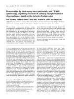
purified by HPLC on a C8 column (Fig. 1). Only
0.37 mg was loaded per HPLC run. HTX was eluted
at 16 min. An analytical HPLC run of HTX showed a
single symmetric peak. HTX represents about 0.1% of
the H. lepturus venom.
Toxicity of HTX
Intracerebroventricular (ICV) injection of HTX causes
neurotoxic symptoms in mice. The LD
50
of HTX was
determined to be 0.3 lg per 20 g body weight.
[
125
I]aDTX displacement by HTX
Figure 2 shows the results of binding experiments in
which increasing concentrations of aDTX or HTX
were added to a fixed concentration of rat brain
membranes (50 lg in 500 lL of synaptosome buffer)
in the presence of [
125
I]aDTX (30 000 c.p.m.). Specific
binding is defined as the difference between total and
0 20 40 60
Elution time
(
min
)
HTX
HCa
0
.
3
0.2
0.1
100
50
A
280
%B
Fig. 1. Purification of HTX. HTX was purified from the neurotoxic
fraction of H. lepturus venom [3]. Twenty microliters containing
0.37 mg was loaded per HPLC run on a C8 column using a gradient
of buffer B (0.1% trifluoroacetic acid in acetonitrile) as described in
Experimental procedures and represented in the figure by dotted
line. HTX was collected at 16 min. HCa, hemicalcin.
Fig. 2. Inhibition of binding of [
125
I]aDTx to rat brain synaptosomal
membranes with HTX (
) and aDTX ( ). As described in Experi-
mental procedures, nonspecific binding (NS) was determined in the
presence of 1 l
M unlabeled aDTX and was subtracted from all
data points. Total binding (B
0
) was determined in the absence
of ligand. Specific binding (%) was obtained by calculating
100 · (B – NS) ⁄ B
0
. The data were analyzed using the computer
program
PRISM.
Hemitoxin – a new a-Ktx6 toxin N. Srairi-Abid et al.
4642 FEBS Journal 275 (2008) 4641–4650 ª 2008 The Authors Journal compilation ª 2008 FEBS
nonspecific binding. HTX inhibits binding of
[
125
I]aDTX to rat brain membranes with an apparent
IC
50
value of 16 nm. This value is nearly 10
times higher than that obtained with aDTX (IC
50
=
1.5 nm), and suggests that HTX is a novel peptide
directed against voltage-gated K
+
channels.
Electrophysiological characterization
The inhibitory effect of HTX was studied on Xeno-
pus laevis oocytes expressing Kv1.1 and Kv1.2 chan-
nels, which are mostly present in the central nervous
system, and also on Kv1.3 channels, which are known
to be essential for lymphocyte proliferation. HTX
(50 nm) caused high and reversible current inhibition
(with a mean value of 81.3%) on Kv1.3 channels. The
Kv1.1 and Kv1.2 channel currents were, respectively,
blocked by 71.8% and 66% with 50 nm HTX (Fig. 3).
At each concentration (0.5, 1, 5, 10, 20, 50 and
100 nm) and in the )40 to +40 mV range, the block-
ing potency of HTX slightly increased with the pulse
level. The blocking potency of the toxin was assessed
by measuring the current remaining after stepwise
increases in HTX concentration. Fitting of the dose–
response data to hyperbolic curves gave IC
50
values of
13 ± 0.1, 16 ± 0.1 and 2 ± 0.1 nm for Kv1.1, Kv1.2
and Kv1.3 channels, respectively (data not shown).
Data were obtained at least three times for each
concentration.
Sequence determination and comparison with
other K
+
channel scorpion toxins
Edman degradation of 2 nmol of S-pyridyl-ethylated
peptides led to the identification of the complete
amino acid sequence of HTX (Fig. 4). HTX is 35 resi-
dues long and contains eight cysteines. The experi-
mental masses of native HTX (3899.24 ± 0.67 Da)
250 ms
10
000
5000
0
–5000
0 100 200 300
Control
50 n
M HTX
Control
50 n
M HTX
Control
50 n
M HTX
Imemb (nA)
Time (ms)
Kv 1.1 Kv 1.2 Kv 1.3
10
000
5000
0
–5000 –2000
0
2000
4000
0 100 200 300
Imemb (nA)
Imemb (nA)
Time (ms)
0 100 200 300
Time (ms)
+20 mV
–80 mV
Fig. 3. Effect of HTX on Kv1.1, Kv1.2 and Kv1.3 channels. Blockade of Kv1.1, Kv1.2 and Kv1.3 currents, using 50 nM HTX. Results are
expressed as the relative current persisting in the presence of the toxin. The control panel corresponds to channel currents in the absence
of HTX. Depolarization was with +20 mV amplitude with 250 ms duration from a holding potential of )80 mV. Data were obtained at least
three times for each concentration.
Fig. 4. Amino acid sequence of HTX, and comparison with the other a-KTx-6 scorpion toxins. Cysteine residues are shown in bold, dots indi-
cate completely conserved residues, and gaps (–) have been introduced to enhance similarity. MTX was from S. maurus [10]; Pi1, Pi4 and
Pi7 were purified from P. imperator [9,21]; HsTx1 [22] and spinoxin (Protein Data Bank code Iv56A [25]) was from He. spinnifer; OcKTx1–5
were from O. carinatus [23]; IsTx was from Op. madagascarensis [24]; anuroctoxin was from A. phaiodactylus [26]; and HgeTx1 was from
Ha. gertschi [27].
N. Srairi-Abid et al. Hemitoxin – a new a-Ktx6 toxin
FEBS Journal 275 (2008) 4641–4650 ª 2008 The Authors Journal compilation ª 2008 FEBS 4643
obtained by MALDI ionization are effectively identi-
cal to the average masses calculated from the corre-
sponding sequence for the fully oxidized form:
3898 Da for HTX. Moreover, only a monomeric form
was observed in the mass spectra, suggesting that no
intermolecular disulfide bridge is formed. These results
indicate that the eight cysteines of HTX are engaged
in four intramolecular disulfide bridges. The sequence
of HTX showed a high level of identity with K
+
channel blockers of the a-KTx6 family, especially with
maurotoxin ( 70 %) from Scorpio maurus [10]
(Fig. 4). The protein sequence data for HTX will
appear in the UniProt database under the accession
number P85528.
Disulfide bridge pairing of HTX
MS of trypsin-digested HTX gave a major peak at
3922 Da, corresponding to the native peptide, thus
showing that the trypsin had a weak effect on native
HTX. This is known to be the case for scorpion
toxins, which are difficult to digest in their native
structure (without reducing their disulfide bridges)
because of their compact structure. Careful examina-
tion of the spectra revealed minor peaks at 2364.91,
1182.95 and 788.97 Da (data not shown) correspond-
ing to the three out of four disulfide bridge-linked pep-
tides (DCYSPCK + NCK + ETGCPR + CYGCS)
with +1, +2 and +3 charged masses, respectively. A
mass of 527.2 Da was obtained, corresponding to the
CTLSK + CINR peptide. These results are in favor
of a conventional HTX disulfide bridge pairing (Cys3–
Cys24, Cys9–Cys29, Cys13–Cys31 and Cys19–Cys37).
Molecular model of HTX
The 3D model of HTX obtained using solution struc-
tures of MTX (Protein Data Bank code 1TXM) [18]
and Pandinus imperator scorpion toxin 1 (Pi4) (Protein
Data Bank code 1N8M) [19] as templates showed the
same unique disulfide pattern of MTX (HTX mod1,
Fig. 5). Taking into account experimental data on
disulfide bridge pairing of HTX, we restarted its
molecular modeling using only Pi4 coordinates as tem-
plate. The model obtained (HTX mod2) is shown in
Fig. 5. As expected, the folds of HTX mod1 and HTX
mod2 appear to be very similar to the folding of the
a-KTx6 toxin experimental structures (Fig. 5). They
showed the basic characteristics of the a ⁄ b-fold of
scorpion toxins. The main elements of the regular
secondary structure are a double-stranded antiparallel
b-sheet comprising residues 21–25 and 28–32, and a
long a-helix composed of residues 7–17.
HTX mod1 presents a disulfide bridge pairing simi-
lar to that of MTX, whereas HTX mod2 presents the
conventional pairing of four disulfide-bridged scorpion
toxins.
Discussion
We have described the isolation and characterization
of a new toxin from H. lepturus scorpion venom
named HTX. HTX displaced [
125
I]aDTX from rat
brain synaptosomes, indicating that it is a K
+
channel
blocker. Comparison of its sequence, composed of 35
amino acids including eight cysteine residues, with the
others in the literature shows that it belongs to
the aKTx6 family, according to the criteria defined by
Tytgat et al. [20] (Fig. 4). HTX could be considered as
the 15th member of the a-KTx6 subfamily (systematic
number: a-KTx6.15). As shown in Fig. 4, the other 14
peptides in the a-KTx6 subfamily, including P. impera-
tor scorpion toxin 1 (Pi1), Pi4 and P. imperator scor-
pion toxin 7 (Pi7), were obtained from P. imperator
[9,21], Heterometrus spinnifer scorpion toxin 1 (HsTx1)
was obtained from He. spinnifer [22], MTX was
obtained from S. maurus [10], K
+
channel Opistoph-
R14
R14
Q16
K30
K30
E16
R33
K15
K30K30
R21
R21
K15
K14
K14
E16
E16
MTX
HsTx1
HTX mod1 HTX mod2
K15
K15
Fig. 5. Homology model of HTX. Backbone ribbon representation
of the models of HTX: HTX mod1 was obtained using atomic coor-
dinates of both MTX and Pi4 as templates. HTX mod2 was
obtained using atomic coordinates of only Pi4. Both models of HTX
were compared to structures of MTX (Protein Data Bank
code 1TXM [18]), the most potent Kv1.2 channel, and HsTx1 (Pro-
tein Data Bank code 1QUZ [22]), the most potent Kv1.3 channel
scorpion toxin. Disulfide bridges are in yellow stick representation.
Hemitoxin – a new a-Ktx6 toxin N. Srairi-Abid et al.
4644 FEBS Journal 275 (2008) 4641–4650 ª 2008 The Authors Journal compilation ª 2008 FEBS
thalmus carinatus scorpion toxins 1–5 (OcKTx1–5)
were obtained from O. carinatus [23], Ischnuridae toxin
(IsTx) was obtained from Opisthacanthus madagascar-
ensis [24], spinoxin was obtained from He. spinnifer
[25] (Protein Data Bank code 1v56A), anuroctoxin was
obtained from Anuroctonus phaidodactylus [26], and
Hadrurus gertschi scorpion toxin 1 (HgeTx1) was
obtained from Ha. gertschi [27]. The a-KTx6 subfamily
has not yet been found in the venom from Buthidae
scorpions. Pi1, Pi4, Pi7, HsTx1, OcKTx1–5, MTX and
spinoxin have been isolated from the venom of the
Scorpionidae, IsTx from the Ischnuridae, HgeTx1 from
the Caraboctonidae, and anuroctoxin from the Chacti-
dae. Therefore, HTX was the first example of such a
peptide from the recently defined family of the Lioche-
lidae [28].
HTX has the highest sequence similarity (80%) with
MTX, a K
+
channel inhibitor scorpion toxin of 34
amino acids, and has a unique pairing of its four disul-
fide bridges [10,29,30]. Despite its high sequence simi-
larity with MTX, HTX shows different affinities for
Kv1 channel subtypes. HTX reversibly blocked Kv1.1,
Kv1.2 and Kv1.3 voltage-gated K
+
channel currents
expressed in Xenopus oocytes with IC
50
values of 13,
16 and 2 nm, respectively. In comparison to the other
a-KTx6 peptides, HTX is an intermediate voltage-
gated K
+
channel blocker peptide. HsTx1 potently
blocks voltage-gated Kv1.3 channels with an IC
50
of
approximately 12 pm [31]. MTX is a potent and selec-
tive inhibitor of the intermediate-conductance Ca
2+
-
activated K
+
channels and the voltage-gated Kv1.2
channel. It blocks the Kv1.1, Kv1.2 and Kv1.3 channel
currents with IC
50
values of 45, 0.8 and 180 nm,
respectively [30,32]. HTX appears to be 90 times more
potent on Kv1.3 channels and 20 times less potent on
Kv1.2 channels than MTX.
It will be very interesting to determine which amino
acids or structure elements are responsible for these
differences in affinity for Kv1.2 and Kv1.3 K
+
channel
subtypes.
It was possible to obtain a 3D structure model of
HTX from Pi4 atomic coordinates and from a combi-
nation of MTX and Pi4 coordinates. Both models
showed the same general folding, but HTX mod1,
which was obtained using both MTX and Pi4 experi-
mental structures as templates, showed a disulfide
bridge pairing similar to that of MTX. This pattern
may be more favorable in terms of energy. Neverthe-
less, experimental data showed that HTX presents the
conventional disulfide pattern, although it conserves
the three amino acids (Agr14 ⁄ Lys14, Lys15 and
Gly33) that were described as being responsible for the
nonconventional pairing of MTX disulfide bridges [32].
It is necessary to mention that even MTX was never
shown to contain the claimed pattern of disulfide
bridges by direct experimental analysis of native pep-
tide. It was determined after chemical synthesis; that
is, the disulfide pairs published were obtained from
in vitro oxidation of synthetic MTX, but its in vivo
pairing may be different.
In protein–protein interactions, at least six para-
meters, i.e. solvation potential, residue interface pro-
pensity, hydrophobicity, planarity, protrusion, and
accessible surface area, are important determinants for
binding [33]. Positively charged (lysine or arginine) and
aromatic (tyrosine or phenylalanine) residues were
described as being critical for the binding to the volt-
age-gated K
+
channels in a number of toxins
[16,31,34,35].
It was established that Kv channel toxins exerted
their activity through the solvent-exposed face of their
b-sheet or helix, depending on the target type of K
+
channel. In their study, Fajloun et al. [32] and Visan
et al. [36] demonstrated that Arg14 and Lys15 of the
MTX helix were very important in its interaction with
Kv1.2 channels. Their substitution causes a drastic
decrease in Kv1.2 channel affinity. Visan et al. [36]
suggested that the change in the 3D structure of
MTX-R14Q or MTX-K15Q might place Lys23, which
is important in the toxin–channel interaction [37], in a
different position, and thus might alter an important
electrostatic contact.
The observed differences in affinity for Kv1.2 chan-
nels between HTX and MTX may be related to the
charges of their respective helixes. The tripeptides KKE
and RKQ, located respectively on the HTX helix and
the MTX helix (residues 14–16), may account for the
difference in affinity (20-fold) for Kv1.2 channels. In
particular, substitution of the acidic residue Glu16 by
Gln16 is thought to be involved by preventing, with its
negative charge, the interaction of Lys14 and Glu355
with Kv1.2 channels, as described for MTX [36].
HTX has 57% similarity with HsTx1, the most
potent Kv1.3 channel a-KTx6 toxin, and it is 6000
times less active on Kv1.3 channels. When comparing
the net global charges of HTX, HsTx1 and MTX, both
HTX and HsTx1 have +6 and MTX has +5. The
charges of the b-sheets, often involved in interactions
with Kv1.3 channels, are +3 for MTX, +4 for HTX
and +5 for HsTx1. This suggested that the positive
charge of these b-sheet toxin regions should favor the
Kv1.3 channel interaction. Docking calculations con-
firm that Lys23 and Met25 of HsTX1 interact with the
GYGDH motif of Kv1.3, and Arg33 can contact
Asp386. Arg33 was thus reported to be important for
the activity of the four disulfide-bridged toxins [38].
N. Srairi-Abid et al. Hemitoxin – a new a-Ktx6 toxin
FEBS Journal 275 (2008) 4641–4650 ª 2008 The Authors Journal compilation ª 2008 FEBS 4645
Arg21 of HTX is situated on the same potential sur-
face and parallel to Arg33 of HsTx1, and thus these
residues could be equivalent (Fig. 5). Arg21 may be
involved in the interaction of HTX with Kv1.3 chan-
nels, instead of Arg33. In addition, HsTx1 contains
Lys28, which is thought to be important, through its
positive charge, in the interaction with Kv1.3 channels.
Lys28 may play the role of the additional positively
charged residue of HsTx1. The simultaneous presence
of Arg33 and Lys28 may explain, at least in part, why
HsTx1 displays more affinity for Kv1.3 channels than
does HTX.
MTX, HsTx1 and HTX may constitute interesting
Kv1 channel-interacting natural toxins, and can be
used as a basis for structure–activity relationship stud-
ies aimed at determining the structural elements modu-
lating selectivity, specificity and affinity for Kv1.2 and
Kv1.3 channels.
The structure–activity study of HTX allowed us to
obtain more precise information regarding the role of
the charged residues 14–16 of the a-helix in Kv1.2
channel interactions. On the other hand, it demon-
strates the importance of positive charge of b-sheet in
the interaction of four disulfide-bridged toxins with
Kv1.3 channels. Work is in progress to design and syn-
thesize a chimeric peptide containing Arg21, Lys28
and Arg33 that should have a more positive charge
on its b-sheet, as compared to the known a-KTx6
peptides, probably accompanied by a higher activity
on Kv1.3 channels.
Experimental procedures
Scorpion venom
Venom of H. lepturus scorpions from Khuzestan (Iran) was
collected by the veterinarian service of RAZI Vaccine
Development and Serum Research Institute of Iran and
kept frozen at )20 °C in its crude form until use.
Purification of HTX
Crude venom was dissolved in water and loaded onto
Sephadex G-50 gel filtration chromatography columns
(2 · K26 ⁄ 50) to isolate the neurotoxic fraction. Columns
were equilibrated with 20 mm ammonium acetate (pH 4.7).
The neurotoxic fraction was identified by injection into
mice by the ICV route. After lyophilization, the neurotoxic
fraction was fractionated by HPLC using a C8 reversed-
phase HPLC column (5 lm, 4.6 · 250 mm, Beckman,
Fullerton, CA, USA) equipped with a Beckman Series 125
pump and a Beckman diode array detector set at 214 and
280 nm. Elution was controlled using gold software.
Proteins were eluted from the column at a rate of 0.8 mLÆ-
min
)1
using a linear gradient (45 min) from 12% to 40% of
buffer B (0.1% trifluoroacetic acid in acetonitrile) in buf-
fer A (0.1% trifluoroacetic acid in water). The protein con-
centration was measured by the Bradford method [39].
In vivo toxicity tests and LD
50
determination
HTX was tested for in vivo toxicity on 20 ± 2 g male
C57 ⁄ BL6 mice, by ICV injection of 5 lL of 0.1% (w ⁄ v)
BSA solution containing increasing amounts of HTX. Six
mice were used for each dose; control mice were injected
with only 0.1% BSA in water to ensure that symptoms
were not due to experimental conditions. ICV administra-
tion was performed under ether anesthesia, according to
the method described by Galeotti et al. [40].
[
125
I]aDTX displacement by HTX
Preparation of [
125
I]aDTX
Synthetic aDTX (10 lg) was incubated at room tempera-
ture in a micro test tube (Eppendorf, Paris, France) coated
with 1 lg of iodogen (Pierce, Rockford, IL, USA) with
2 mCi of
125
I (Amerham Pharmcia Biotech, Little Chalfont,
UK) in 200 lL of 0.1 m sodium phosphate. After 15 min,
20 lL of 0.1 m sodium thiosulfate was added, and the reac-
tion mixture was injected onto a C18 column (Beckman).
After washing of the column with 25% solvent B (0.1%
trifluoroacetic acid, 50% acetonitrile) in solvent A (0.1%
trifluoroacetic acid), separation was achieved using a
40 min gradient of 25–60% solvent B in solvent A at a rate
of 1 mLÆmin
)1
. The fraction containing pure mono-iodin-
ated aDTX (2000 CiÆmmol
)1
) was kept at 4 °C after the
addition of BSA (1 mgÆ mL
)1
).
Preparation of rat brain synaptosomal membranes
Synaptosomal membranes were prepared as previously
described [41]. Membranes contained 0.92 lgÆlL
)1
of pro-
teins as determined by the Bradford method.
Binding assays
All binding experiments were performed at room tempera-
ture. Tubes were set up in duplicate. The total volume per
tube was 500 lL (containing 50 lg of synaptosomal mem-
branes). The buffer used was 0.1% BSA (> 99% pure) in
synaptosome buffer (130 mm NaCl, 3 mm KCl, 2 mm
CaCl
2
.2H
2
O, 2 mm MgCl
2
.6H
2
O, 20 mm Tris ⁄ HCl, pH 7.4)
(BSA ⁄ SB). Total binding was determined in a tube conta-
ining [
125
I]aDTX, 100 lgÆmL
)1
protein and synaptosome
buffer. Nonspecific binding was determined by displacing
[
125
I]aDTX with 1 lm dendrotoxin. HTX was added to the
test tubes at various concentrations. Tubes were incubated
Hemitoxin – a new a-Ktx6 toxin N. Srairi-Abid et al.
4646 FEBS Journal 275 (2008) 4641–4650 ª 2008 The Authors Journal compilation ª 2008 FEBS
at room temperature (19–21 °C) for 30 min while being
rotated on the mixer. The synaptosomal pellet was
recovered by centrifuging at 13 000 g for 3 min in a micro-
centrifuge. The supernatant, which contains unbound
[
125
I]aDTX, was discarded. The pellet was washed with
50 lL of BSA ⁄ SB to remove any excess unbound
[
125
I]aDTX. The radioactivity was estimated using a
Gamma LKB counter. Curves and IC
50
were determined
using prism graph pad software [42].
Electrophysiological characterization
Mature female Xenopus laevis were anesthetized by immer-
sion in a 0.17% solution of tricaine (ethyl m -aminobenzo-
ate). The ovarian lobes were surgically isolated and rinsed
in standard modified Barth’s saline (MBS) of the following
composition: 88 mm NaCl, 1 mm KCl, 2.4 mm CaCl
2
,
0.82 mm MgSO
4
, 2.4 mm NaHCO
3
, 0.41 mm MgCl
2
,
0.33 mm Ca(NO
3
)
2
, and 10 mm Hepes (pH 7.4).
Stage V–VI oocytes were defolliculated by collagenase
treatment (type A and type B; Roche, Boehringer,
Germany; 2 mgÆmL
)1
in Ca
2+
-free MBS), and then
mechanically by using two thin forceps. Rat Kv1.1 Kv1.2
and Kv1.3 cRNAs [generous gift from M. Crest, Departe-
ment de Signalisation Neuronale, Centre de Recherche de
Neurobiologie–Neurophysiologie de Marseille (CRN2M),
France] were stored at 1 lgÆmL
)1
in diethylpyrocarbonate-
treated water and injected at a concentration of 4 ng per
oocyte using an automatic injector (Drummond Nanoject,
Broomall, PA, USA). Oocytes were incubated at 16–18 °C
in sterile MBS supplemented with 0.1 mm gentamicin
(Sigma-Aldrich, Lyon, France).
Ionic currents through the Kv1.1, Kv1.2 and Kv1.3 chan-
nels were recorded during the week following RNA injec-
tion with the two-electrode voltage-clamp method using a
Gene Clamp 500 amplifier (Axon Instruments, Foster City,
CA, USA).
Oocytes were immersed in Ca
2+
-free saline and impaled
with two glass intracellular electrodes filled with 3 m KCl.
The resistance of the pulled electrodes (P-97 puller; Sutter
Instruments, Novato, CA, USA) was 1–2 MW . The holding
potential was set at )80 mV. The perfusion system was
controlled by a Manifold Solution Changer (MSC-200;
Bio-Logic, Grenoble, France). Data acquisition and analy-
sis were performed using clampex and clampfit from
pclamp8 software (Molecular Devices, Sunnyvale, CA,
USA). Leak and capacitive currents were subtracted during
analysis using a P ⁄ 4orP⁄ 8 protocol [43].
Determination of amino acid sequence of HTX
Reduction of HTX with dithiothreitol, and alkylation with
4-vinylpyridine, were performed as previously described [44].
The sequence of the reduced ⁄ carboxymethylated toxin
was determined using an automatic liquid-phase protein
sequencer (model 476A; Applied Biosystems, Foster City,
CA, USA) using standard Edman protein degradation [45].
HTX was deposited onto Biobrene-precycled glass-fiber
disks.
MS
The molecular mass of native HTX was determined with a
Voyager-DE PRO MALDI-TOF Workstation mass spec-
trometer (Perseptive Biosystems, Inc., Framingham, MA,
USA). The peptide was dissolved in acetonitrile ⁄ H
2
O
(30 : 70) with 0.3% trifluoroacetic acid to obtain a concen-
tration of 1–10 pmolÆlL
)1
. The matrix was prepared as
follows. a-Cyanohydroxycinnamic acid was dissolved in
50% acetonitrile in 0.3% trifluoroacetic acid ⁄ H
2
O to obtain
a saturated solution of 10 lgÆlL
)1
; 0.5 lL of peptide solu-
tion was then mixed with 0.5 lL of matrix and placed on
the sample plate. This mixture was allowed to dry. Mass
spectra were recorded in reflectron mode, externally cali-
brated with suitable standards, and analyzed using the
grams ⁄ 386 software of Galactic Industries Corporation
and the Savitzky–Golay algorithm [46].
MS was used also to determine disulfide bridges. After
overnight digestion of 10 lg of HTX with 0.2 lg of trypsin
(in Tris ⁄ HCl, 100 mm, pH 8.5), the sample was infused at
a rate of 3 lLÆmin
)1
on an ESI MicroTofQ mass spectro-
meter (Bruker Daltonic GmbH, Bremen, Germany).
Sequence comparison
Peptides showing sequence similarity with HTX were identi-
fied with blast2 [47] on the nonredundant database.
Sequences with E-values less than 10
)3
were a-KTx6 K
+
channel blockers. These were aligned, and sequence similar-
ities between these toxins were calculated manually.
Molecular modeling
A 3D structure model of HTX was generated by homology
modeling with the program modeller 9v2 [48]. Homolo-
gous polypeptides with known structures were identified by
a blast2 [26] search of the Protein Data Bank [49] (RCSB
organization) using the sequence of HTX as entry. The
solution structures of MTX (Protein Data Bank
code 1TXM) [18] and Pi4 (Protein Data Bank code 1N8M)
[19] were first used as templates. Also, another molecular
modeling was performed using only Pi4 coordinates. Disul-
fide bridges were not introduced as constraints in molecular
modeling. Two sets of 20 models were generated. All their
Protein Data Bank files were analyzed for their energetic
and geometric characteristics. In each case, only one model
combining the best Ramachandran plot (for geometric con-
formity) ( />[50] and good scores for the objective function values [48],
N. Srairi-Abid et al. Hemitoxin – a new a-Ktx6 toxin
FEBS Journal 275 (2008) 4641–4650 ª 2008 The Authors Journal compilation ª 2008 FEBS 4647
and the VICTOR ⁄ FRST energy function proposed by
Tosatto ( [51], was consid-
ered. The best models were then visualized with the
viewerlite50 program ( />dstudio/).
Acknowledgements
This research was supported by MRST and the Inter-
national Network of the Pasteur Institutes. We are
indebted to P. Mansuelle (IFR Jean Roche, Marseille)
for MS of native HTX, to C. Villard and D. Lafitte
(Plateforme Proteomique IFR 125 Site Timone) for
HTX disulfide bridge pairing determination, and to
Professor H. Louzir, head of the Pasteur Institute of
Tunisia, and Professor A. R. Najafabadi, head of the
Pasteur Institute of Iran, for their helpful advice.
References
1 Radmanesh M (1990) Clinical study of Hemiscorpion
lepturus in Iran. J Trop Med Hyg 93, 327–332.
2 Loret EP & Hammock BD (2001) Structure and neuro-
toxicity of venoms. In Scorpion Biology and Research
(Brownell PH & Polis GA, eds), pp. 204–233. Oxford
University Press, New York, NY.
3 Shahbazzadeh D, Srairi-Abid N, Feng W, Ram N, Bor-
chani L, Ronjat M, Akbari A, Pessah IN, De Waard M
& El Ayeb M (2007) Hemicalcin, a new toxin from the
Iranian scorpion Hemiscorpius lepturus which is active
on ryanodine-sensitive Ca
2+
channels. Biochem J 404,
89–96.
4 Rogowski RS, Krueger BK, Collins JH & Blaustein MP
(1994) Tityustoxin Ka blocks voltage-gated noninacti-
vating K
+
channels and unblocks inactivating K
+
channels blocked by a-dendrotoxin in synaptosomes.
Proc Natl Acad Sci USA 91, 1475–1479.
5 Legros C, Ceard B, Bougis PE & Martin-Eauclaire MF
(1998) Evidence for a new class of scorpion
toxins active against K
+
channels. FEBS Lett 431,
375–380.
6 Srairi-Abid N, Guijarro JI, Benkhalifa R, Mantegazza
M, Cheikh A, Ben-Aissa M, Haumont PY, Delepierre
M & El Ayeb M (2005) A new type of scorpion
Na
+
-channel-toxin-like polypeptide active on K
+
channels. Biochem J 388, 455–464.
7 Miller C (1995) The charybdotoxin family of K
+
channel-blocking peptides. Neuron 15, 5–10.
8 Romi-Lebrun R, Lebrun B, Martin-Eauclaire MF,
Ishiguro M, Escoubas P, Wu FQ, Hisada M, Pongs O
& Nakajima T (1997) Purification, characterization,
and synthesis of three novel toxins from the Chinese
scorpion Buthus martensi, which act on K
+
channels.
Biochemistry 36, 13473–13482.
9 Olamendi-Portugal T, Gomez-Lagunas F, Gurola GB
& Possani LD (1996) A novel structural class of K
+
-
channel blocking toxin from the scorpion Pandinus
imperator. Biochem J 315, 977–981.
10 Kharrat R, Mabrouk K, Crest M, Darbon H, Oughi-
deni R, Martin-Eauclaire MF, Jacquet G, el Ayeb M,
Van Rietschoten J, Rochat H et al. (1996) Chemical
synthesis and characterization of maurotoxin, a short
scorpion toxin with four disulfide bridges that acts on
K+ channels. Eur J Biochem 242, 491–498.
11 Bontems F, Roumestand C, Gilquin B, Menez A &
Toma F (1991) Refined structure of charybdotoxin:
common motifs in scorpion toxins and insect defensins.
Science 254, 1521–1523.
12 Aiyar J, Rizzi JP, Gutman GA & Chandy KG (1996)
The signature sequence of voltage-gated potassium
channels projects into the external vestibule. J Biol
Chem 271, 31013–31016.
13 Aiyar J, Withka JM, Rizzi JP, Singleton DH, Andrews
GC, Lin W, Boyd J, Hanson DC, Simon M, Dethlefs B
et al. (1995) Topology of the pore-region of a K
+
chan-
nel revealed by the NMR-derived structures of scorpion
toxins. Neuron 15, 1169–1181.
14 Stampe P, Kolmakova-Partensky L & Miller C (1994)
Intimations of K
+
channel structure from a complete
functional map of the molecular surface of charybdo-
toxin. Biochemistry 33, 443–450.
15 Park CS & Miller C (1992) Mapping function to struc-
ture in a channel-blocking peptide: electrostatic mutants
of charybdotoxin. Biochemistry 31, 7749–7755.
16 Ranganathan R, Lewis JH & MacKinnon R (1996)
Spatial localization of the K
+
channel selectivity filter
by mutant cycle-based structure analysis. Neuron 16,
131–139.
17 Giangiacomo KM, Sugg EE, Garcia-Calvo M, Leonard
RJ, McManus OB, Kaczorowski GJ & Garcia ML
(1993) Synthetic charybdotoxin–iberiotoxin chimeric
peptides define toxin binding sites on calcium-activated
and voltage-dependent potassium channels. Biochemis-
try 32, 2363–2370.
18 Blanc E, Sabatier JM, Kharrat R, Meunier S, el Ayeb
M, Van Rietschoten J & Darbon H (1997) Solution
structure of maurotoxin, a scorpion toxin from Scorpio
maurus, with high affinity for voltage-gated potassium
channels. Proteins 29, 321–333.
19 Guijarro JI, M’Barek S, Go
´
mez-Lagunas F, Garnier D,
Rochat H, Sabatier JM, Possani L & Delepierre M
(2003) Solution structure of Pi4, a short four-disulfide-
bridged scorpion toxin specific of potassium channels.
Protein Sci 12, 1844–1854.
20 Tytgat J, Chandy G, Garcia ML, Gutman GA,
Martin-Eauclaire MF, van der Walt JJ & Possani LD
(1999) A unified nomenclature for short-chain
peptides isolated from scorpion venoms:
Hemitoxin – a new a-Ktx6 toxin N. Srairi-Abid et al.
4648 FEBS Journal 275 (2008) 4641–4650 ª 2008 The Authors Journal compilation ª 2008 FEBS
alpha-KTx molecular subfamilies. Trends Physiol Sci
20, 444–447.
21 Olamendi-Portugal T, Gomez-Langunas F, Gurola GB
& Possani LD (1998) Two similar peptides from the
venoms of the scorpion Pandinus imperator, one highly
effective blocker and the other inactive on K
+
channels.
Toxicon 36, 759–770.
22 Lebrun B, Romi-Lebrun R, Martin-Eauclaire MF,
Yasuda A, Ishiguro M, Oyama Y, Pongs O &
Nakajima T (1997) A four-disulfide-bridged toxin, with
high affinity towards voltage-gated K+ channels,
isolated from Heterometrus spinnifer (Scorpionidae)
venom. Biochem J 328, 321–327.
23 Zhu S, Huy I, Dayson K, Verdonck F & Tytgat J
(2004) Evolutionary trace analysis of scorpion toxins
specific for K-channels. Proteins 54 , 361–370.
24 Yamaji N, Dai L, Sugase K, Andriantsiferana M, Nak-
ajima T & Iwashita T (2004) Solution structure of IsTx.
A male scorpion toxin from Opisthacanthus madagascar-
iensis (Ischnuridae). Eur J Biochem 271, 3855–3864.
25 Sugahara Y, Nirthanan S, Huys I, Kobayashi K,
Kohno T, Tytgat J, Gopalakrishnakone P & Sato K
(2004) Synthesis and characterization of spinoxin, a
novel peptide toxin from the Malaysian black
scorpion. In Peptide Science – Proceedings of the 40th
Symposium on Japanese Peptide Symposium (Ueki M,
ed), pp. 283–284. Protein Research Foundation,
Osaka.
26 Bagda
´
ny M, Batista CV, Valdez-Cruz NA, Somodi S,
Rodriguez de la Vega RC, Licea AF, Varga Z,
Ga
´
spa
´
r R, Possani LD & Panyi G (2005) Anuroctox-
in, a new scorpion toxin of the alpha-KTx 6 subfam-
ily, is highly selective for Kv1.3 over IKCa1 ion
channels of human T lymphocytes. Mol Pharmacol
67, 1034–1044.
27 Schwartz EF, Schwartz CA, Go
´
mez-Lagunas F,
Zamudio FZ & Possani LD (2006) HgeTx1, the first
K
+
-channel specific toxin characterized from the venom
of the scorpion Hadrurus gertschi Soleglad. Toxicon 48,
1046–1053.
28 Soleglad ME & Fet V (2003) High-level systematics and
phylogeny of the extant scorpions (Scorpiones: Orthos-
terni). Euscorpius 11, 1–175.
29 Kharrat R, Mansuelle P, Sampieri F, Crest M,
Oughideni R, Van Rietschoten J, Martin-Eauclaire MF,
Rochat H & El Ayeb M (1997) Maurotoxin, a four
disulfide bridged toxin from Scorpio maurus venom:
purification, structure and action on potassium
channels. FEBS Lett 406, 284–290.
30 Castle NA, London DO, Creech C, Fajloun Z, Stocker
JW & Sabatier JM (2003) Maurotoxin: a potent
inhibitor of intermediate conductance Ca
2+
-activated
potassium channels. Mol Pharmacol 63, 409–418.
31 Dauplais M, Lecoq A, Song J, Cotton J, Jamin N,
Gilquin B, Roumestand C, Vita C, de Medeiros CL,
Rowan EG et al. (1997) On the convergent
evolution of animal toxins. J Biol Chem 272,
4302–4309.
32 Fajloun Z, Mosbah A, Carlier E, Mansuelle P, Sandoz
G, Fathallah M, Di Luccio E, Devaux C, Rochat H,
Darbon H et al. (2000) Maurotoxin versus Pi1 ⁄ HsTx1
scorpion toxins. J Biol Chem 275, 39394–39402.
33 Jones S & Thornton J (1997) Analysis of protein–pro-
tein interaction sites using surface patches. J Mol Biol
272, 121–132.
34 Goldstein S, Pheasant DJ & Miller C (1994) The char-
ybdotoxin receptor of a Shaker Kv1 channel: peptide
and channel residues mediating recognition. Neuron 12,
1377–1388.
35 Bednarek MA, Johnson BA, Stevens SP, Bugianesi
RM, Felix JP, Leonard RJ, Slaughter RS, Kaczorowski
GJ & Williamson JM (1996) SAR of margatoxin, a
potent and selective inhibitor of voltage-activated potas-
sium channel Kv1.3 in human T-cell lymphocytes. In
Peptides: Chemistry, Structure and Biology (Pravin TP,
Kaumaya X & Hodges RS, eds), Mayflower Scientific
Ltd., Kingswinford.
36 Visan V, Fajloun Z, Sabatier JM & Grissmer S (2004)
Mapping of maurotoxin binding sites on hKv1.2,
hKv1.3, and hIKCa1 channels. Mol Pharmacol 66,
1103–1112.
37 Fu W, Cui M, Briggs JM, Huang X, Xiong B, Zhang
Y, Luo X, Shen J, Ji R & Chen K (2002) Brownian
dynamics simulations of the recognition of the scorpion
toxin maurotoxin with the voltage-gated potassium ion
channels. Biophys J 83, 2370–2385.
38 Savarin P, Romi-Lebrun R, Zinn-Justin S, Lebrun B,
Nakajima T, Gilquin B & Menez A (1999) Structural
and functional consequences of the presence of a fourth
disulfide bridge in the scorpion short toxins: solution
structure of the potassium channel inhibitor HsTx1.
Protein Sci 8, 2672–2685.
39 Bradford MM (1976) A rapid and sensitive method
for the quantitation of microgram quantities of protein
utilizing the principle of protein-dye binding. Anal
Biochem 72, 248–254.
40 Galeotti N, Bartolini A & Ghelardini C (2003) Diphen-
hydramine-induced amnesia is mediated by Gi-protein
activation. Neuroscience 122, 471–478.
41 Gray EG & Whittaker VP (1962) The isolation of nerve
endings from brain: an electron microscopic study of
cell fragments derived by homogenization centrifuga-
tion. J Anat 96, 79–88.
42 Swift ML (1997) GRAPHPAD PRISM, data analysis,
and scientific graphing. J Chem Inf Comput Sci 37,
411–412.
43 Soto E, Salceda E, Cruz R, Ortega A & Vega R (2000)
Microcomputer program for automated action potential
waveform analysis. Comput Methods Programs Biomed
62, 141–144.
N. Srairi-Abid et al. Hemitoxin – a new a-Ktx6 toxin
FEBS Journal 275 (2008) 4641–4650 ª 2008 The Authors Journal compilation ª 2008 FEBS 4649
44 Srairi-Abid N, Mansuelle P, Mejri T, Karoui H,
Rochat H, Sampieri F & El Ayeb M (2000)
Purification, characterization and molecular modelling
of two toxin-like proteins from the Androctonus
australis Hector venom. Eur J Biochem 267,
5614–5620.
45 Edman P & Begg G (1967) A protein sequenator. Eur J
Biochem, 1, 80–91.
46 Savitzky A & Golay MJE (1964) Smoothing and differ-
entiation of data by simplified least squares procedures.
Anal Chem 36, 1627–1639.
47 Altschul SF, Madden TL, Schaffer AA, Zhang J, Zhang
Z, Miller W & Lipman DJ (1997) Gapped BLAST and
PSI-BLAST: a new generation of protein database
search programs. Nucleic Acids Res 25, 3389–3402.
48 Sali A & Blundell T (1993) Comparative protein model-
ing by satisfaction of spatial restraints. J Mol Biol 234,
779–815.
49 Abola EE, Manning NO, Prilusky J, Stampf DR &
Sussman JL (1996) The protein data bank: current
status and future challenges. J Res Natl Inst Stand
Technol 101, 231–241.
50 Kleywegt GJ & Jones TA (1996) Phi ⁄ psi-chology:
Ramachandran revisited. Structure 4, 1395–1400.
51 Tosatto SC (2005) The victor ⁄ FRST function for model
quality estimation. J Comput Biol 12, 1316–1327.
Hemitoxin – a new a-Ktx6 toxin N. Srairi-Abid et al.
4650 FEBS Journal 275 (2008) 4641–4650 ª 2008 The Authors Journal compilation ª 2008 FEBS