Báo cáo khoa học: Distinct but critical roles for integrin aIIbb3 in platelet lamellipodia formation on fibrinogen, collagen-related peptide and thrombin pot
Bạn đang xem bản rút gọn của tài liệu. Xem và tải ngay bản đầy đủ của tài liệu tại đây (564.8 KB, 12 trang )
Distinct but critical roles for integrin a
IIb
b
3
in platelet
lamellipodia formation on fibrinogen, collagen-related
peptide and thrombin
Kelly Thornber
1
, Owen J. T. McCarty
2,3
, Steve P. Watson
2
and Catherine J. Pears
1
1 Department of Biochemistry, University of Oxford, UK
2 Centre for Cardiovascular Sciences, Institute of Biomedical Research, University of Birmingham, UK
3 Department of Biomedical Engineering, Oregon Health & Science University, Portland, OR, USA
Platelets play an essential role in the formation of a
haemostatic plug at the site of vascular injury. This
process requires adhesion of the platelet to the
exposed subendothelial matrix, followed by powerful
intracellular signalling events that lead to platelet–
platelet interactions and thrombus formation. One
critical feature in this scheme is the dramatic alter-
ation in platelet morphology in response to activa-
tion. Thus, the resting, discoid platelet undergoes a
series of morphological changes that include round-
ing, generation of filopodia and lamellipodia, and for-
mation of actin stress fibres [1]. These events serve to
stabilize the thrombus, thereby enabling it to with-
stand the high shear forces found in arteries and arte-
rioles.
Stable adhesion of platelets to the subendothelial
matrix is dependent upon sustained activation of
integrins [2]. Integrins are glycoprotein heterodimers
Keywords
a
IIb
b
3
; adhesion; integrins; lamellipodia;
platelets
Correspondence
C. Pears, Department of Biochemistry,
South Parks Road, University of Oxford,
Oxford, OX1 3QU, UK
Fax: +44 1865 275259
Tel: +44 1865 275737
E-mail:
Website:
(Received 11 July 2006, revised 22 August
2006, accepted 12 September 2006)
doi:10.1111/j.1742-4658.2006.05500.x
Integrins are the major receptor type known to facilitate cell adhesion and
lamellipodia formation on extracellular matrix proteins. However, collagen-
related peptide and thrombin have recently been shown to mediate platelet
lamellipodia formation when presented as immobilized surfaces. The aims
of this study were to establish if there exists a role for the platelet integrin
a
IIb
b
3
in this response; and if so, whether signalling from the integrin is
required for lamellipodia formation on these surfaces. Real-time analysis
was used to compare platelet morphological changes on surfaces of fibrino-
gen, collagen-related peptide or thrombin in the presence of various
pharmacological inhibitors and platelets from ‘knockout’ mice. We demon-
strate that collagen-related peptide and thrombin stimulate distinct patterns
of platelet lamellipodia formation and elevation of intracellular Ca
2+
to
that induced by the integrin a
IIb
b
3
ligand, fibrinogen. Nevertheless, lamelli-
podia formation on collagen-related peptide and thrombin is dependent
upon engagement of a
IIb
b
3
, consistent with release of a
IIb
b
3
ligand(s) from
platelet granules. However, the requirement for signalling by the integrin
on fibrinogen can be bypassed by the addition of thrombin to the solution.
These observations reveal a critical role for a
IIb
b
3
in forming lamellipodia
on collagen-related peptide and thrombin which is dependent on its ability
to function as an adhesive receptor but not necessarily on its ability to sig-
nal. These results suggest that integrins may play an important role in
lamellipodia formation triggered by nonintegrin ligands in platelets and
possibly in other cell types.
Abbreviations
CRP, collagen-related peptide.
5032 FEBS Journal 273 (2006) 5032–5043 ª 2006 The Authors Journal compilation ª 2006 FEBS
composed of a-subunits and b-subunits that exist in an
inactive or low-affinity conformation in nonactivated
cells. Intracellular signals within the platelet (known as
‘inside-out’ signalling) promote a conformational
change in the extracellular domain, leading to an
increase in affinity, thereby promoting integrin–ligand
interactions [2]. In turn, clustering of integrins gene-
rates a series of intracellular signals (‘outside-in’ signal-
ling) that serve to reinforce platelet activation [3].
The major platelet integrin a
IIb
b
3
is a receptor for
fibrinogen, von Willebrand factor, vitronectin, CD40
ligand and fibronectin. Integrin a
IIb
b
3
plays a vital role
in supporting platelet adhesion to the extracellular
matrix and promoting platelet–platelet interaction
(aggregation). In addition, integrin a
IIb
b
3
generates
outside-in signals that mediate platelet activation. It is
now established that engagement of a
IIb
b
3
activates
Src family kinases, leading to activation of Syk [4],
SLP-76 [5], Vav1 ⁄ 3 [4] and phospholipase Cc2 (PLCc2)
[6], and thereby to activation of several second messen-
ger pathways, including protein kinase C [7] and Ca
2+
[8]. These signalling events promote actin assembly,
leading to formation of filopodia, lamellipodia and
stress fibres [9].
In addition to integrin ligands, formation of filo-
podia and lamellipodia has been described on a
monolayer of collagen-related peptide (CRP), which
selectively activates the immunoglobulin receptor glyco-
protein VI (GPVI) [10,11], and on thrombin that has
been immobilized by fibrin. In platelets, thrombin binds
to and signals via GPIba and the G protein-coupled
protease-activated receptor (PAR) receptors (PAR 1
and 4) [12,13]. Immobilized thrombin that has become
trapped by fibrin is able to promote platelet adhesion
and aggregate formation at intermediate rates of flow,
leading to the speculation that it may function as an
adhesion ligand in vivo [13]. In epithelial cells, it has
been suggested that immobilized thrombin can bind to
integrins through an RGD site, raising the possibility
that is may bind directly to integrins in platelets [14].
The present study was undertaken to investigate the
mechanism by which CRP and thrombin are able to
support platelet lamellipodia formation in comparison
to that induced by fibrinogen. It was of particular
interest to discern whether thrombin and CRP stimu-
late lamellipodia formation directly, or whether they
require a
IIb
b
3
. The results demonstrate a critical role
for a
IIb
b
3
in promoting platelet lamellipodia formation
on CRP, thrombin and on fibrinogen, but that out-
side-in signalling by the integrin is not required for
lamellipodia formation on fibrinogen in the presence
of thrombin. These results further emphasize the
importance of integrin engagement in lamellipodia
formation but demonstrate that signalling by a
IIb
b
3
is
not essential for this response.
Results
Morphological changes of human platelets on
fibrinogen, CRP and thrombin
The three ligands fibrinogen, CRP and thrombin sup-
port platelet adhesion and lamellipodia formation when
presented as a monolayer, even though they bind to
distinct classes of surface receptor. This raises the ques-
tion of the molecular basis of adhesion to these ligands
and whether they induce distinct patterns of change in
morphology. To address this, real-time imaging of
platelets adhering to each surface was undertaken using
a coating of ligands that induces maximal platelet adhe-
sion. These experiments were performed in the presence
of concentrations of apyrase and indomethacin shown
to fully block the effect of the feedback agonists ADP
and thromboxane A
2
, respectively, in order to directly
monitor the ability of each ligand to support adhesion
and lamellipodia formation.
Consistent with previous reports, our results dem-
onstrate that platelets exposed to immobilized fibrin-
ogen go through sequential formation of filopodia
and lamellipodia over a period of 30 min (Fig. 1 and
supplementary Video S1). Both structures were stable
and did not retract once formed, although discrete
movements could still be seen around the periphery
of the cell. Fluorescent labelling of the actin cytoske-
leton revealed that stress fibres were formed within
platelets that had undergone full lamellipodia forma-
tion (Fig. 1B). In contrast, a distinct pattern of
platelet morphological changes was observed on the
GPVI-specific agonist CRP (Fig. 1A and supplement-
ary Video S2). Limited small filopodia were seen,
with wave-like lamellipodia appearing before filopo-
dia formation was complete, in contrast to the
spherically synchronized growth on fibrinogen. Full
lamellipodia formation was reached within 5–9 min,
three times more rapidly than on fibrinogen
(Fig. 1C). Strikingly, even after platelets had reached
90% of their final surface area, the lamellipodia were
very dynamic (supplementary Video S2), even though
they were accompanied by formation of stress fibres
(Fig. 1B). A similar pattern of rapid yet unstable
lamellipodia formation and stress fibre formation
was observed for platelets adhering to immobilized
thrombin (Fig. 1A,B and supplementary Video S3).
Significantly, lamellipodia formation on thrombin
was not altered in the presence of the fibrin poly-
merization inhibitor Gly-Pro-Arg-Pro (GPRP) (data
K. Thornber et al. Platelet lamellipodia formation via a
IIb
b
3
FEBS Journal 273 (2006) 5032–5043 ª 2006 The Authors Journal compilation ª 2006 FEBS 5033
not shown), demonstrating that this response was
not dependent upon fibrin formation.
These results demonstrate that a distinct pattern of
morphological change is induced by platelet adhesion to
CRP and thrombin, compared to that seen on fibrin-
ogen.
Different Ca
2+
mobilization patterns in platelets
exposed to fibrinogen, CRP and thrombin
Experiments were undertaken to investigate whether
the distinct pattern of lamellipodia formation on
the three ligands is associated with differences in the
A
B
0
20
40
60
80
100
120
0 200 400 600 800 1000 1200 1400 1600
Time (sec)
% of maximum surface area
FG
CRP
THR
C
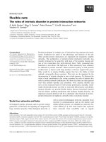
Fig. 1. Distinct morphological changes in human washed platelets exposed to fibrinogen, collagen-related peptide (CRP) and thrombin. (A)
Human washed platelets were exposed to surfaces of fibrinogen (FG), CRP or thrombin (THR). Representative morphology of a single plate-
let on each surface at the time points shown (s). (B) Rhodamine–phalloidin staining to show actin stress fibres of platelets after 30 min of
exposure to fibrinogen, CRP and thrombin. (C) Mean surface area of platelets at indicated time points, quantified using
IMAGEJ. Values are
mean ± SEM of the percentage of maximum surface area of five platelets from three independent experiments.
Platelet lamellipodia formation via a
IIb
b
3
K. Thornber et al.
5034 FEBS Journal 273 (2006) 5032–5043 ª 2006 The Authors Journal compilation ª 2006 FEBS
elevation of intracellular Ca
2+
, in view of the pivotal
role that the divalent cation plays in the regulation of
actin rearrangements in platelets through pathways
such as activation of the capping protein gelsolin [10]
and myosin light chain kinase. Intracellular Ca
2+
was
measured by loading with the calcium reporter dye
Oregon Green-BAPTA 1-AM and monitoring fluore-
scent fluctuations for a minimum of 5 min after adhe-
sion to each surface. The functional role of Ca
2+
was
investigated by pretreatment with the Ca
2+
chelator
1,2-bis-(o-aminophenoxy)-ethane-N,N,N¢,N¢-tetraace tic
acid (BAPTA-AM).
Our results demonstrate that platelet adhesion to
fibrinogen was followed by a characteristic lag phase
of 1–3 min before a series of rhythmic spikes of Ca
2+
.
These were seen at intervals of approximately 30 s,
each lasting 5–15 s. Generally, the magnitude of spikes
declined slightly over time (Fig. 2A). In contrast, adhe-
sion to CRP generated an immediate increase in intra-
cellular Ca
2+
, which subsequently declined over a
period of 3–10 min. Minimal oscillations were
observed during the sustained elevation in Ca
2+
.
A distinct pattern of intracellular Ca
2+
signalling was
observed in platelets on thrombin. An initial rapid ele-
vation in intracellular Ca
2+
was followed by a sus-
tained phase of increased Ca
2+
levels superimposed by
a series of small Ca
2+
oscillations (Fig. 2A).
Maximal concentrations of the Ca
2+
chelator BAPTA-
AM inhibited intracellular Ca
2+
elevation on all three
surfaces by over 90% (Fig. 2A). This was accompanied
by a complete inhibition of lamellipodia on fibrinogen,
whereas only a partial inhibition of lamellipodia was
observed on CRP and thrombin (Fig. 2B, Table 1).
Platelet adhesion to thrombin was significantly reduced
in the presence of BAPTA-AM, whereas the degree of
adhesion to CRP and fibrinogen remained unchanged
(Table 1). Filopodia formation was observed on all
three surfaces in the presence of BAPTA-AM, indica-
ting that their formation is independent of Ca
2+
mobilization.
Taken together, these results demonstrate a critical
role for Ca
2+
in lamellipodia formation on all three
surfaces and suggest that distinct patterns of Ca
2+
signalling could contribute to the different patterns of
lamellipodia formation induced by each ligand.
a
IIb
b
3
is required for platelet lamellipodia
formation on fibrinogen, CRP and thrombin
Although the above results demonstrate marked differ-
ences between the three ligands in the pattern of Ca
2+
mobilization, which presumably reflect the differing
signalling strengths of their receptors, it is possible that
adhesion and lamellipodia formation are mediated
through integrin activation, in view of the critical role
of integrins in mediating adhesion and cell spreading.
To address this, experiments were designed to compare
the role of the major platelet integrin a
IIb
b
3
, in sup-
porting spreading on the three surfaces, using the
a
IIb
b
3
antagonist lotrafiban [15,16].
As expected, lotrafiban abrogated platelet adhesion
to fibrinogen (Fig. 2B), but, importantly, dramatically
reduced lamellipodia formation on CRP and thrombin
surfaces, although it also increased the level of adhe-
sion (Table 1). These findings demonstrate that despite
the distinct morphological changes on CRP and
thrombin in comparison to those on fibrinogen, the
generation of lamellipodia is dependent upon engage-
ment of a
IIb
b
3
in all cases. This is most likely explained
by release of fibrinogen, von Willebrand factor and
other a
IIb
b
3
ligands from platelet a-granules. Consistent
with this, secretion of platelet a-granules in platelets
that had adhered to CRP and thrombin was confirmed
by immunofluorescence staining for the a-granule
marker P-selectin, on the surface of adhered platelets
(Fig. 3).
These results demonstrate that formation of lamelli-
podia on CRP, thrombin and fibrinogen is dependent
on engagement of integrin a
IIb
b
3
as a consequence of
release of a
IIb
b
3
ligands from platelet a-granules.
Thrombin can bypass a
IIb
b
3
outside-in signalling
in mediating lamellipodia formation
The above observations demonstrate a critical role for
a
IIb
b
3
in lamellipodia formation on CRP and throm-
bin. This could be due to a role of a
IIb
b
3
in supporting
adhesive events or in generating intracellular signals
that drive formation of lamellipodia. It is difficult to
distinguish between these two possibilities in the case
of CRP, because of the similarity in the signalling
pathways used by GPVI and integrin a
IIb
b
3
, both of
which are mediated by sequential activation of Src and
Syk family kinases and subsequent activation of
PLCc2 [6]. In contrast, thrombin signals through a
G protein-dependent pathway and induces a full
repertoire of platelet responses in the presence of the
Src family kinase inhibitor 4-amino-5-(4-chlorophenyl)-
7-(t-butyl)pyrazolo[3,4-d]pyrimidine (PP2). Experi-
ments were therefore designed to investigate the effect
of PP2 on the ability of thrombin to promote lamelli-
podia formation during platelet adhesion.
As previously reported, the Src kinase inhibitor PP2
blocked formation of lamellipodia on fibrinogen [17]
(Fig. 4), with average platelet surface area being signi-
ficantly reduced from 26.3 ± 0.9 to 17.6 ± 0.7 lm
2
.
K. Thornber et al. Platelet lamellipodia formation via a
IIb
b
3
FEBS Journal 273 (2006) 5032–5043 ª 2006 The Authors Journal compilation ª 2006 FEBS 5035
A
B
Fig. 2. Different Ca
2+
signalling patterns on each surface. Washed human platelets were exposed to surfaces of fibrinogen (FG), collagen-
related peptide (CRP) or thrombin (THR). (A) Platelets were loaded with Oregon Green-BAPTA 1-AM calcium dye and exposed to each sur-
face for 10 min. The fluorescent Ca
2+
fluctuations of three single platelets on each surface are shown in the presence (+) or absence (–) of
BAPTA-AM (10 l
M, added 10 min prior to surface exposure). Representative traces from three individual platelets are shown. (B) Platelets
were exposed to each surface for 45 min in the absence or presence of BAPTA-AM (10 l
M, added 10 min prior to surface exposure) or the
a
IIb
b
3
antagonist lotrafiban (10 lM,10 min). Images shown are representative of at least three independent experiments. Adhesion and sur-
face area data from these experiments are shown in Table 1.
Platelet lamellipodia formation via a
IIb
b
3
K. Thornber et al.
5036 FEBS Journal 273 (2006) 5032–5043 ª 2006 The Authors Journal compilation ª 2006 FEBS
In contrast, adhesion was not significantly altered.
Importantly, the inhibitory effect of PP2 on lamelli-
podia formation could be completely overcome by
addition of thrombin (Fig. 4), which induced a larger
increase in surface area to that induced by fibrinogen
(37.5 ± 1.5 lm
2
), most likely reflecting the increased
signalling strength of the platelet thrombin receptors
PAR1 and PAR4. A similar effect was seen with the
PAR1-specific peptide, thrombin receptor activating
peptide [TRAP (data not shown)].
Fig. 3. P-Selectin exposure on collagen-rela-
ted peptide (CRP) and thrombin. Washed
human platelets were exposed to surfaces
of CRP or thrombin (THR) for 45 min before
fixing and staining with fluorescein isothio-
cyanate (FITC)-conjugated anti-P-selectin
serum. Cells were imaged using differential
interference contrast (DIC) or fluorescence
(P-selectin). Images shown are representa-
tive of at least three independent experi-
ments.
Table 1. Human platelet data. See Fig. 2 for experimental details. Values are reported as follows: adherent platelets ¼ mean ± SEM of three
experiments; platelet surface area ¼ mean ± SEM of at least 100 cells. On a control surface of BSA, adhesion and surface area data are
1.5 ± 0.1 · 10
2
mm
)2
and 7.3 ± 0.2 lm
2
, respectively.
Treatment
Fibrinogen CRP Thrombin
Adherent platelets ⁄
mm
2
(· 10
2
)
Platelet surface
area (lm
2
)
Adherent platelets ⁄
mm
2
(· 10
2
)
Platelet surface
area (lm
2
)
Adherent platelets ⁄
mm
2
(· 10
2
)
Platelet surface
area (lm
2
)
None 57.6 ± 0.9 26.3 ± 0.9 65.7 ± 0.9 35.0 ± 1.1 67.9 ± 1.9 32.2 ± 1.1
BAPTA-AM 57.2 ± 0.6 17.7 ± 0.6
a
50.5 ± 0.6 22.3 ± 1.2
a
36.3 ± 1.0
a
25.8 ± 0.9
a
Lotrafiban 3.3 ± 0.4
a
9.3 ± 0.4
a
95.1 ± 1.3
a
16.7 ± 0.5
a
93.0 ± 1.4
a
12.8 ± 0.4
a
a
P < 0.05 with respect to untreated samples for each surface.
Fig. 4. Thrombin can overcome Src kinase inhibition of lamellipodia on fibrinogen. Washed human platelets were exposed to a surface of
fibrinogen for 45 min in the absence or presence of the Src kinase inhibitor PP2 (20 l
M, added 5 min prior to surface exposure) and ⁄ or
thrombin (1 UÆmL
)1
, 1 min) in suspension (+ THR). Images shown are representative of at least three independent experiments.
K. Thornber et al. Platelet lamellipodia formation via a
IIb
b
3
FEBS Journal 273 (2006) 5032–5043 ª 2006 The Authors Journal compilation ª 2006 FEBS 5037
These results demonstrate that thrombin is able to
stimulate formation of lamellipodia on fibrinogen in
the absence of signalling downstream of the integrin.
Under these circumstances, therefore, the integrin is
functioning solely as an adhesive receptor.
Role of a
IIb
b
3
and PLCc2 in adhesion and
lamellipodia formation of murine platelets
Experiments were undertaken in mouse platelets to
confirm the role of integrin a
IIb
b
3
in mediating lamelli-
podia formation on fibrinogen, CRP and thrombin,
and to establish the importance of PLCc2 activation in
this response. These experiments served to extend the
observations made in human to mice platelets and also
to provide an alternative line of evidence to support
the results obtained above through the use of platelets
deficient in the a
IIb
integrin subunit and PLCc2.
As previously documented [9,18,19], and in contrast
to human platelets, mouse platelets generate only filo-
podia and limited lamellipodia on fibrinogen in the
presence of apyrase and indomethacin (Fig. 4A). In
contrast, robust lamellipodia formation in mouse
platelets is observed on CRP or thrombin surfaces.
Adhesion and lamellipodia formation on fibrinogen
were abrogated in the presence of the a
IIb
b
3
antagonist
lotrafiban (Fig. 5A, Table 2) and in mice lacking the
a
IIb
gene (Fig. 5A, Table 2). Similarly, adhesion of
mouse platelets to CRP was fully blocked in the pres-
ence of lotrafiban or in the absence of a
IIb
(Fig. 5A,
Table 2), demonstrating that adhesion on CRP is crit-
ically dependent on functional a
IIb
b
3
, presumably as a
consequence of secretion of fibrinogen and other a
IIb
b
3
ligands. In contrast, a
IIb
b
3
-blocked and a
À=À
IIb
mouse
platelets retained the ability to adhere to immobilized
thrombin, although lamellipodia formation was elimin-
ated (Fig. 5A, Table 2).
The role of Src kinases and Ca
2+
mobilization in
lamellipodia formation in mouse platelets on fibrino-
gen and CRP was investigated using platelets deficient
in the major isoform of PLCc in platelets, PLCc2
[19,20]. Blockade of Src kinases with PP2 (data not
shown) or the absence of PLCc2 led to inhibition of
lamellipodia formation on fibrinogen, but had no
effect on adhesion (Fig. 5B, Table 2), in agreement
with previous observations [18,19]. The addition of
thrombin in suspension could overcome this inhibitory
effect, leading to extensive lamellipodia formation
(Fig. 5, Table 2). In contrast, the absence of PLCc2
abrogated adhesion of mouse platelets to CRP
(Fig. 5B), although adhesion and lamellipodia forma-
tion could be restored by addition of thrombin (Fig. 5,
Table 2).
These experiments extend the observations on
human platelets to mouse platelets, namely that inte-
grin a
IIb
b
3
is necessary for platelet lamellipodia forma-
tion on CRP, thrombin and fibrinogen, but that
thrombin is able to mediate lamellipodia formation in
the absence of outside-in signalling from the integrin.
The roles of a
IIb
b
3
and a
2
b
1
in platelet
lamellipodia formation on collagen
Experiments were designed to investigate whether the
critical role of a
IIb
b
3
in mediating lamellipodia forma-
tion in human and mouse platelets on fibrinogen, CRP
and thrombin also extends to a further platelet ligand,
collagen, which binds directly to a second platelet
integrin, a
2
b
1
as well as GPVI. This question was
addressed by monitoring lamellipodia formation on
collagen in human platelets in the presence of the
a
IIb
b
3
antagonist lotrafiban and in mouse platelets defi-
cient in the integrin subunit a
IIb
.
Human and mouse platelets generate filopodia and
lamellipodia on fibrillar collagen (Fig. 6), and this is
reduced by approximately 55% in human platelets in
the presence of lotrafiban (28.7 ± 1.1 to 17.8 ±
0.5 lm
2
, compared to 26.3 ± 0.9 to 9.3 ± 0.4 lm
2
on
fibrinogen) and in a
IIb
-deficient mouse platelets (from
18.4 ± 0.6 to 11.3 ± 0.4 lm
2
in wild-type and a
IIb
-
deficient platelets, respectively, compared with 11.1 ±
0.3 to 5.5 ± 0.5 lm
2
on fibrinogen). The degree of
platelet adhesion, however, was not significantly
altered by loss of a
IIb
b
3
function in either human or
mouse platelets (not shown), presumably because it is
mediated through integrin a
2
b
1
.
These results demonstrate that a
IIb
b
3
contributes to
lamellipodia formation on an integrin-binding ligand,
namely collagen, but that, in its absence, limited
lamellipodia formation is mediated by integrin a
2
b
1
.
Discussion
It is well established that integrins play a critical role
in lamellipodia formation in a wide variety of cell
types, although it is unclear whether lamellipodia for-
mation can also be induced by engagement of noninte-
grin receptors. Nevertheless, it is well established that
other cell surface receptors may facilitate cell adhesion
and actin remodelling through integrin activation and
stimulation of actin polymerization. An example of
this is Syndecan-1, which has been shown to mediate
lamellipodia formation in Raji lymphoblastoid cells
independently of integrins [21], although other synde-
cans are thought to operate through interaction with
neighbouring integrins [22]. More recently, two studies
Platelet lamellipodia formation via a
IIb
b
3
K. Thornber et al.
5038 FEBS Journal 273 (2006) 5032–5043 ª 2006 The Authors Journal compilation ª 2006 FEBS
have described lamellipodia formation in platelets
upon adhesion to thrombin and the synthetic collagen
CRP [10,12]. The present study investigated the
molecular basis of lamellipodia formation in platelets
on these two surfaces, alongside studies on fibrinogen,
which mediates lamellipodia formation through inte-
grin a
IIb
b
3
. The results demonstrate a critical role for
integrin a
IIb
b
3
in mediating lamellipodia formation on
A
B
Fig. 5. Murine washed platelet adhesion and lamellipodia formation. (A) Wild-type (WT) and a
IIb
-deficient (a
À=À
IIb
) platelets were exposed to
surfaces of fibrinogen (FG), collagen-related peptide (CRP) or thrombin (THR) for 45 min (B) WT and PLCc2-deficient (PLCc2
– ⁄ –
) platelets
were exposed to surfaces of fibrinogen (FG) or CRP for 45 min in the absence or presence of thrombin (1 UÆmL
)1
) in suspension (+ THR).
Images shown are representative of at least three separate experiments. Adhesion and surface area data from these experiments are
shown in Table 2.
K. Thornber et al. Platelet lamellipodia formation via a
IIb
b
3
FEBS Journal 273 (2006) 5032–5043 ª 2006 The Authors Journal compilation ª 2006 FEBS 5039
all three surfaces, although each surface induces a dis-
tinct pattern of formation of filopodia and lamellipo-
dia. A distinct pattern of lamellipodia formation on
CRP was also reported by the Hartwig group [10].
The critical role of a
IIb
b
3
in mediating lamellipodia
formation on CRP and thrombin is likely to be medi-
ated by release of a
IIb
b
3
ligands from platelet a-gran-
ules, which become immobilized on the surface,
enabling them to support lamellipodia formation.
Moreover, secreted fibrinogen has been shown to be
prebound to the platelet surface, which would there-
fore put it in the right place to support lamellipodia
formation [23]. Thrombin may also directly support
adhesion through an RDG motif that becomes
exposed upon immobilization [14], although it must
also bind to other receptors, as it cannot generate
lamellipodia in the absence of functional a
IIb
b
3
. Inter-
estingly, blockade of a
IIb
b
3
caused an increase in plate-
let adhesion to CRP and thrombin. This can be
explained by the reduction in lamellipodia formation
and therefore the corresponding increase in available
matrix area, and by the observations of Patel et al.
that adhered platelets cause the lateral movement of
depositing platelets away from themselves and on to
the matrix below [24].
The above discussion indicates that the difference in
the pattern of lamellipodia between fibrinogen, throm-
bin and CRP is likely to be due to their different signal
Table 2. Murine platelet data. See Fig. 5 for experimental details. Values are reported as follows: adherent platelets ¼ mean ± SEM of three
experiments; platelet surface area ¼ mean ± SEM of at least 100 cells. On a control surface of BSA, adhesion and surface area data are
0.9 ± 0.1 · 10
2
mm
)2
and 4.6 ± 0.6 lm
2
, respectively. THR, thombin added in suspension; WT, wild-type; LOT, lotrafiban.
Genotype ⁄
treatment
Fibrinogen CRP Thrombin
Adherent platelets ⁄
mm
2
(· 10
2
)
Platelet surface
area (lm
2
)
Adherent platelets ⁄
mm
2
(· 10
2
)
Platelet surface
area (lm
2
)
Adherent platelets ⁄
mm
2
(· 10
2
)
Platelet surface
area (lm
2
)
WT ⁄ None 63.5 ± 0.9 11.1 ± 0.3 52.8 ± 0.9 17.9 ± 0.4 30.3 ± 0.3 15.7 ± 0.5
WT ⁄ LOT 1.1 ± 0.1
a
5.5 ± 0.5
a
5.7 ± 0.4
a
6.5 ± 0.3
a
15.4 ± 0.3
a
5.5 ± 0.4
a
a
À=À
IIb
⁄ None 2.4 ± 0.1
a
4.4 ± 0.2
a
4.3 ± 0.3
a
3.7 ± 0.3
a
22.6 ± 0.9
a
5.5 ± 0.3
a
PLCc2
– ⁄ –
⁄ None 62.0 ± 0.5 10.0 ± 0.4
a
6.1 ± 0.3
a
9.0 ± 0.5
a
PLCc2
– ⁄ –
⁄ THR 51.7 ± 0.3
b
18.1 ± 0.5
b
36.8 ± 0.7
b
16.6 ± 0.7
a,b
a
P < 0.05 with respect to untreated wild-type samples for each surface.
b
P < 0.05 for samples treated with thrombin in suspension
compared to the equivalent sample without thrombin.
Fig. 6. Lamellipodia formation on collagen in
the absence of a
IIb
b
3
. Human washed plate-
lets in the absence or presence of the a
IIb
b
3
antagonist lotrafiban (10 lM, added 10 min
prior to surface exposure), and murine plate-
lets deficient in a
IIb
(a
À=À
IIb
) or wild-type (WT)
littermate controls were exposed to immobi-
lized collagen for 45 min. Images shown are
representative of at least three independent
experiments.
Platelet lamellipodia formation via a
IIb
b
3
K. Thornber et al.
5040 FEBS Journal 273 (2006) 5032–5043 ª 2006 The Authors Journal compilation ª 2006 FEBS
strengths rather than the nature of the ligand medi-
ating lamellipodia formation. As shown in the present
study, immobilized CRP and thrombin induced a
much greater increase in intracellular Ca
2+
relative to
fibrinogen, and this presumably contributes to the gen-
eration of wave-like structures that are absent on
fibrinogen.
The ability of integrin a
IIb
b
3
to activate Src kinase-
dependent signalling cascades that lead to lamellipodia
formation is widely recognized [3]. This study shows,
however, that under certain conditions, the ability of
the integrin to activate Src kinases is not essential for
lamellipodia formation, as these structures can be
induced by thrombin or the PAR1-specific agonist
TRAP. Nevertheless, there is evidence that outside-in
signalling by a
IIb
b
3
is important in supporting throm-
bus formation in vivo. For example, increased rebleed-
ing is seen following removal of a small portion of the
tail in mice with a knock-in mutation of the integrin b
3
subunit in which the two conserved tyrosine residues
have been replaced by phenylalanine residues, thereby
impairing a
IIb
b
3
outside-in signalling [25]. Similarly,
thrombus formation in a heat injury model is reduced
in mice deficient in the protein tyrosine phosphatase
PTP1b, which plays a critical role in a
IIb
b
3
-mediated
outside-in signalling [26]. Alternatively, impaired
thrombus formation in these studies could be a conse-
quence of reduced clot retraction, which also depends
on outside-in signalling.
In summary, the present study has shown that
immobilized fibrinogen, CRP and thrombin stimulate
distinct patterns of morphological change and Ca
2+
signalling during platelet adhesion. However, despite
these differences, lamellipodia formation on all surfaces
is critically dependent upon integrin a
IIb
b
3
, as a conse-
quence of release of a
IIb
b
3
ligands from platelet a-gran-
ules. The distinct pattern of lamellipodia may therefore
be explained by the differing levels of elevation of
intracellular Ca
2+
and other intracellular signals. The
present study also shows that outside-in signalling
from integrin a
IIb
b
3
is not required for lamellipodia
formation on fibrinogen in the presence of thrombin.
Thus, under these conditions, the essential role of inte-
grin a
IIb
b
3
in supporting lamellipodia formation can be
attributed to its ability to function as an adhesive
receptor.
Experimental procedures
Reagents
Fibrinogen depleted of plasminogen, von Willebrand factor
and fibronectin were obtained from Kordia Laboratory
Supplies, Leiden, NL. Oregon Green bis-(o-aminophen-
oxy)ethane-N,N,N¢,N¢-tetraacetic acid (BAPTA 1-AM) and
rhodamine–phalloidin were purchased from Molecular
Probes (Cambridge Bioscience, Cambridge, UK), and fluo-
rescein isothiocyanate-conjugated anti-P-selectin serum was
obtained from BD Pharmingen (Erembodegem, Belgium).
All other reagents were obtained as described in McCarty
et al. [16].
Preparation of washed platelets
Human studies were carried out with ethical approval from
the Central Oxford Research Committee (Ref: C00:203)
and with the understanding and written consent of each
subject. Platelets were prepared as previously described [9],
and resuspended at 2 · 10
7
ml
)1
in modified Hepes ⁄
Tyrodes buffer (129 mm NaCl, 0.34 mm Na
2
HPO
4
, 2.9 mm
KCl, 12 mm NaHCO
3
,20mm Hepes, 5 mm glucose, 1 mm
MgCl
2
; pH 7.3) containing 0.1 lgÆmL
)1
prostacyclin. In
selected experiments, platelet suspensions were treated with
10 lm lotrafiban, 10 lm BAPTA-AM and 20 lm PP2 for
10 min, or with 1 UÆmL
)1
thrombin for 1 min, before use.
Concentrations of inhibitors were used that are maximally
effective. These concentrations were identified in previous
studies from the Watson group and others [27–30]. The
generation of mice disrupted in the genes encoding a
IIb
(a
À=À
IIb
) or PLCc2 (PLCc2
– ⁄ –
) was as described [24,31].
Wild-type littermates were used as controls. Washed murine
platelets were isolated and resuspended in modified
Hepes ⁄ Tyrodes buffer as previously described [9]. All
experiments were performed in the presence of 2.5 UÆmL
)1
apyrase and 10 lm indomethacin, and in the absence of
exogenously added Ca
2+
. Animals were bred, and blood
was removed under an approved Home Office Project
licence.
Adhesion assays
Coverslips were incubated with a suspension of fibrinogen
(100 lgÆmL
)1
), CRP (1 lgÆmL
)1
), thrombin (1 UÆmL
)1
)or
collagen (100 lgÆmL
)1
) for 1 h at room temperature before
washing with phosphate buffered saline and blocking with
denatured BSA (5 mgÆmL
)1
) for 1 h. Platelets were exposed
to coverslips for 45 min, before fixing, staining where neces-
sary, and mounting as described in McCarty et al. [9].
Adherent platelets were imaged using Ko
¨
hler illuminated
Nomarski differential interference contrast optics with a
Zeiss (Carl Zeiss Ltd., Welwyn Garden City, UK) 63· oil
immersion 1.40 NA plan-apochromat lens on a Zeiss
Axiovert ZOOM microscope (Zeiss). Time-lapse events
were captured by a Hamamatsu Orca 285 cooled digital
camera (Cairn Research, Faversham, UK) using slidebook
4.0 (Intelligent Imaging Innovations, Inc., Denver, CO). To
compute the surface area of platelets, time-lapse images
were manually outlined and quantitated by determining the
K. Thornber et al. Platelet lamellipodia formation via a
IIb
b
3
FEBS Journal 273 (2006) 5032–5043 ª 2006 The Authors Journal compilation ª 2006 FEBS 5041
number of pixels within each outline using a Java plugin
for the image j software package (NIH, Bethesda, MD,
USA). Imaging a graticule under the same conditions
allowed the conversion of pixel size to micrometres. Adhe-
sion data in each experiment were obtained by counting the
number of platelets on five random images of each cover-
slip, with each image encompassing an area of 15 400 lm
2
.
For immunofluorescence studies, coverslips were blocked in
0.1% BSA following fixation and stained for 1 h at room
temperature before washing, mounting and imaging as
described above.
Single-platelet Ca
2+
measurement
Washed human platelets (2 · 10
8
mL
)1
) were incubated
with the Ca
2+
-sensitive dye Oregon Green BAPTA 1-AM
(15 lm) for 1 h at 30 °C. Platelets were subsequently
washed, resuspended at 2 · 10
8
ml
)1
and left for a mini-
mum of 30 min before experimentation. Platelets
(1 · 10
7
mL
)1
) were allowed to sediment onto fibrinogen-,
CRP- or thrombin-coated coverslips over a period of
10 min. Fluorescence changes in single platelets were meas-
ured using a Zeiss Axiovert 200M microscope fitted with an
Optoscan Monochromator System (Cairn Research). A
Hamamatsu Orca 285 camera and slidebook software were
used for image capture and subsequent analysis.
Analysis of data
Experiments were carried out on at least three occasions,
and images shown are representative data from one
experiment. Unless stated otherwise, results are shown
as mean ± SEM. Statistical significance of differences
between means was determined by one-way anova.If
means were shown to be significantly different, multiple
comparisons by pairs were performed by the Tukey test.
Probability values of P < 0.05 were selected to be statisti-
cally significant.
Acknowledgements
We thank the British Heart Foundation and the Med-
ical Research Foundation for funding this work, and
Mark Larson, Simon Calaminus and Andrew Pearce
for their help and advice. KT and SPW hold a British
Heart Foundation studentship and chair, respectively.
References
1 Hartwig JH (1992) Mechanisms of actin rearrangements
mediating platelet activation. J Cell Biol 118, 1421–
1442.
2 Hynes RO (1992) Integrins: versatility, modulation, and
signaling in cell adhesion. Cell 69, 11–25.
3 Shattil SJ & Newman PJ (2004) Integrins: dynamic scaf-
folds for adhesion and signaling in platelets. Blood 104,
1606–1615.
4 Miranti CK, Leng L, Maschberger P, Brugge JS &
Shattil SJ (1998) Identification of a novel integrin
signaling pathway involving the kinase Syk and the
guanine nucleotide exchange factor Vav1. Curr Biol 8,
1289–1299.
5 Judd BA, Myung PS, Leng L, Obergfell A, Pear WS,
Shattil SJ & Koretzky GA (2000) Hematopoietic
reconstitution of SLP-76 corrects hemostasis and
platelet signaling through alpha IIb beta 3 and collagen
receptors. Proc Natl Acad Sci USA 97, 12056–12061.
6 Watson SP, Auger JM, McCarty OJ & Pearce AC
(2005) GPVI and integrin alphaIIb beta3 signaling in
platelets. J Thromb Haemost 3, 1752–1762.
7 Giuliano S, Nesbitt WS, Rooney M & Jackson SP
(2003) Bidirectional integrin alphaIIbbeta3 signalling
regulating platelet adhesion under flow: contribution of
protein kinase C. Biochem J 372, 163–172.
8 Pelletier AJ, Bodary SC & Levinson AD (1992) Signal
transduction by the platelet integrin alpha IIb beta 3:
induction of calcium oscillations required for protein-
tyrosine phosphorylation and ligand-induced spreading
of stably transfected cells. Mol Biol Cell 3, 989–998.
9 McCarty OJ, Larson MK, Auger JM, Kalia N,
Atkinson BT, Pearce AC, Ruf S, Henderson RB,
Tybulewicz VL, Machesky LM et al. (2005) Rac1 is
essential for platelet lamellipodia formation and
aggregate stability under flow. J Biol Chem 280, 39474–
39484.
10 Falet H, Barkalow KL, Pivniouk VI, Barnes MJ, Geha
RS & Hartwig JH (2000) Roles of SLP-76, phosphoino-
sitide 3-kinase, and gelsolin in the platelet shape changes
initiated by the collagen receptor GPVI ⁄ FcR gamma-
chain complex. Blood 96, 3786–3792.
11 Falet H, Hoffmeister KM, Neujahr R & Hartwig JH
(2002) Normal Arp2 ⁄ 3 complex activation in platelets
lacking WASp. Blood 100, 2113–2122.
12 Adam F, Guillin MC & Jandrot-Perrus M (2003)
Glycoprotein Ib-mediated platelet activation. A signal-
ling pathway triggered by thrombin. Eur J Biochem 270,
2959–2970.
13 Weeterings C, Adelmeijer J, Myles T, de Groot PG &
Lisman T (2006) Glycoprotein Ibalpha-mediated platelet
adhesion and aggregation to immobilized thrombin
under conditions of flow. Arterioscler Thromb Vasc Biol
26, 670–675.
14 Papaconstantinou ME, Carrell CJ, Pineda AO, Bobof-
chak KM, Mathews FS, Flordellis CS, Maragoudakis
ME, Tsopanoglon NE, Di Cera E et al. (2005)
Thrombin functions through its RGD sequence in a
non-canonical conformation. J Biol Chem 280, 29393–
29396.
Platelet lamellipodia formation via a
IIb
b
3
K. Thornber et al.
5042 FEBS Journal 273 (2006) 5032–5043 ª 2006 The Authors Journal compilation ª 2006 FEBS
15 Liu F, Craft RM, Morris SA & Carroll RC (2000) Lotra-
fiban: an oral platelet glycoprotein IIb⁄ IIIa blocker.
Expert Opin Invest Drugs 9, 2673–2687.
16 McCarty OJ, Zhao Y, Andrew N, Machesky LM,
Staunton D, Frampton J & Watson SP (2004) Evalua-
tion of the role of platelet integrins in fibronectin-depen-
dent spreading and adhesion. J Thromb Haemost 2,
1823–1833.
17 Goncalves I, Hughan SC, Schoenwaelder SM, Yap
CL, Yuan Y & Jackson SP (2003) Integrin alpha IIb
beta 3-dependent calcium signals regulate platelet–
fibrinogen interactions under flow. Involvement of
phospholipase C gamma 2. J Biol Chem 278, 34812–
34822.
18 Obergfell A, Eto K, Mocsai A, Buensuceso C, Moores
SL, Brugge JS, Lowell CA & Shattil SJ (2002) Coordi-
nate interactions of Csk Src, and Syk kinases with
[alpha]IIb[beta]3 initiate integrin signaling to the cyto-
skeleton. J Cell Biol 157, 265–275.
19 Wonerow P, Pearce AC, Vaux DJ & Watson SP (2003)
A critical role for phospholipase Cgamma2 in alphaIIb-
beta3-mediated platelet spreading. J Biol Chem 278,
37520–37529.
20 Suzuki-Inoue K, Inoue O, Frampton J & Watson SP
(2003) Murine GPVI stimulates weak integrin activation
in PLCgamma2– ⁄ – platelets: involvement of PLCgam-
ma1 and PI3-kinase. Blood 102, 1367–1373.
21 Lebakken CS, McQuade KJ & Rapraeger AC (2000)
Syndecan-1 signals independently of beta1 integrins dur-
ing Raji cell spreading. Exp Cell Res 259, 315–325.
22 Beauvais DM & Rapraeger AC (2004) Syndecans in
tumor cell adhesion and signaling. Reprod Biol Endo-
crinol 2,3.
23 Legrand C, Dubernard V & Nurden AT (1989) Studies
on the mechanism of expression of secreted fibrinogen
on the surface of activated human platelets. Blood 73,
1226–1234.
24 Patel D, Vaananen H, Jirouskova M, Hoffmann T,
Bodian C & Coller BS (2003) Dynamics of GPII-
b ⁄ IIIa-mediated platelet–platelet interactions in
platelet adhesion ⁄ thrombus formation on collagen in
vitro as revealed by videomicroscopy. Blood 101,
929–936.
25 Law DA, DeGuzman FR, Heiser P, Ministri-Madrid K,
Killeen N & Phillips DR (1999) Integrin cytoplasmic
tyrosine motif is required for outside-in alphaIIbbeta3
signalling and platelet function. Nature 401, 808–811.
26 Arias-Salgado EG, Haj F, Dubois C, Moran B,
Kasirer-Friede A, Furie BC, Neel BG, Shattil SJ (2005)
PTP-1B is an essential positive regulator of platelet
integrin signaling. J Cell Biol 170, 837–845.
27 Erhardt JA, Ohlstein EH, Toomey JR, Gabriel MA,
Willette RN, Yue TL, Barone FC & Parsons AA (2001)
Activation of caspase-3 ⁄ caspase-3-like activity in rat
cardiomyocytes by an RGD peptide, but not the GPII-
b ⁄ IIIa antagonist lotrafiban. Thromb Res 103, 143–148.
28 Inoue O, Suzuki-Inoue K, Dean WL, Frampton J &
Watson SP (2003) Integrin alpha2beta1 mediates out-
side-in regulation of platelet spreading on collagen
through activation of Src kinases and PLCgamma2.
J Cell Biol 160, 769–780.
29 Jung SM, Ohnuma M, Watanabe N, Sonoda M, Handa
M & Moroi M (2005) Analyzing the mechanism of
Rap1 activation in platelets: Rap1 activation is related
to the release reaction mediated through the collagen
receptor GPVI. Thromb Res 118, 509–521.
30 Redondo PC, Ben-Amor N, Salido GM, Bartegi A,
Pariente JA & Rosado JA (2005) Ca2+-independent
activation of Bruton’s tyrosine kinase is required for
store-mediated Ca2+ entry in human platelets. Cell
Signal 17, 1011–1021.
31 Wang D, Feng J, Wen R, Marine JC, Sangster MY,
Paraganes E, Hoffmeyer A, Jackson CW, Cleveland JL,
Murray PJ et al. (2000) Phospholipase Cgamma2 is
essential in the functions of B cell and several Fc recep-
tors. Immunity 13, 25–35.
Supplementary material
The following supplementary material is available
online:
Video S1. Real-time analysis of platelet on fibrinogen.
Video S2. Real-time analysis of platelet on collagen-
related peptide (CRP).
Video S3. Real-time analysis of platelet on thrombin.
This material is available as part of the online article
from
K. Thornber et al. Platelet lamellipodia formation via a
IIb
b
3
FEBS Journal 273 (2006) 5032–5043 ª 2006 The Authors Journal compilation ª 2006 FEBS 5043