Báo cáo khoa học: Characterization of a membrane-bound aminopeptidase purified from Acyrthosiphon pisum midgut cells A major binding site for toxic mannose lectins pptx
Bạn đang xem bản rút gọn của tài liệu. Xem và tải ngay bản đầy đủ của tài liệu tại đây (2.28 MB, 15 trang )
Characterization of a membrane-bound aminopeptidase
purified from Acyrthosiphon pisum midgut cells
A major binding site for toxic mannose lectins
Plinio T. Cristofoletti
1
, Flavia A. Mendonc¸a de Sousa
2
, Yvan Rahbe
´
2
and Walter R. Terra
1
1 Departamento de Bioquı
´
mica, Instituto de Quı
´
mica, Universidade de Sa˜o Paulo, Brazil
2 UMR INRA-INSA de Lyon BF2I, Biologie Fonctionnelle Insectes & Interactions, Villeurbanne, France
Aminopeptidase N (APN) is an exopeptidase that cata-
lyzes the sequential release of N-terminal amino acids
of peptides (EC 3.4.11.2). It is found in the midgut of
insect larvae either as soluble enzyme or associated
with the microvillar membrane. Properties of APN
preparations from midgut tissue have been described
for at least six orders of insects [1,2].
APNs are the major proteins in some insect midgut
microvillar membranes. Probably linked to its abun-
dance, APN is one of the targets of the insecticidal
Bacillus thuringiensis d-endotoxins [3–6]. These toxins,
after binding to receptors such as APNs, form chan-
nels through which midgut cell contents leak, finally
leading to insect death [7]. Also, in humans, APN is
the binding site for coronavirus infection [8].
Such findings raised interest in this enzyme, leading
to APN cloning from target insects in the Lepidoptera
family [3,6,9–20]. All these APNs are inserted into
the midgut microvillar membrane by a C-terminal
glycosyl-phosphatidylinositol (GPI) anchor. Sequence
comparisons with vertebrate and fungal aminopepti-
dases showed that their most striking similarities
were in the zinc-binding motif, including residues
His142, His146, and Glu166 (putative zinc ligands,
Keywords
aminopeptidase N; Aphididae; glycosyl-
phosphatidylinositol (GPI) anchor; mannose
lectin receptor; substrate specificity
Correspondence
Y. Rahbe
´
, UMR INRA-INSA de Lyon BF2I,
Biologie Fonctionnelle Insectes &
Interactions, Bat. Louis-Pasteur, F-69621
Villeurbanne cedex, France
Fax: +33 4 72 43 85 34
Tel: +33 4 72 43 83 56
E-mail:
Database
The sequence described here has been
deposited in the GenBank database with
the accession number DQ440823
(Received 25 August 2006, revised 13
October 2006, accepted 19 October 2006)
doi:10.1111/j.1742-4658.2006.05547.x
A single membrane-bound aminopeptidase N (APN) occurs in the pea
aphid (Acyrthosiphon pisum Harris) midgut, with a pH optimum of 7.0, pI
of 8.1 and molecular mass of 130 kDa. This enzyme accounts for more
than 15.6% of the total gut proteins. After being solubilized in detergent,
APN was purified to homogeneity. The enzyme is a glycoprotein rich in
mannose residues, which binds the entomotoxic lectins of the concanavalin
family. The internal sequence of APN is homologous with a conservative
domain in APNs, and degenerated primers of highly conserved APN motifs
were used to screen a gut cDNA library. The complete sequence of APN
has standard residues involved in zinc co-ordination and catalysis and a
glycosyl-phosphatidylinositol anchor, as in APNs from Lepidoptera. APN
has a broad specificity towards N-terminal amino acids, but does not
hydrolyze acidic aminoacyl-peptides, thus resembling the mammalian
enzyme (EC 3.4.11.2). The k
cat
⁄ K
m
ratios for different di-, tri-, tetra-, and
penta-peptides suggest a preference for tripeptides, and that subsites S
1
,S
2
¢
and S
3
¢ are pockets able to bind bulky aminoacyl residues. Bestatin and
amastatin bound APN in a rapidly reversible mode, with K
i
values of
1.8 lm and 0.6 lm, respectively. EDTA inactivates this APN (k
obs
0.14 m
)1
Æs
)1
, reaction order of 0.44) at a rate that is reduced by competitive
inhibitors. In addition to oligopeptide digestion, APN is proposed to be
associated with amino-acid-absorption processes which, in contrast with
aminopeptidase activity, may be hampered on lectin binding.
Abbreviations
APN, aminopeptidase N; ConA, concanavalin A; ConBr, concanavalin A ortholog from Canavalia brasiliensis; EST, expressed sequence tag;
GPI, glycosyl-phosphatidylinositol; LeupNA,
L-leucine-p-nitroanilide; WGA, wheat germ agglutinin.
5574 FEBS Journal 273 (2006) 5574–5588 ª 2006 The Authors Journal compilation ª 2006 FEBS
numbering according to thermolysin), and Glu143 (cat-
alytic active residue). This conserved motif classifies
the enzymes as members of the M
1
family of neutral
zinc metallopeptidases [21]. In spite of these research
efforts, there are few detailed studies on the substrate
specificity of lepidopteran microvillar APNs [1,2].
Kinetic data on a midgut APN from Coleop-
tera showed its similarity to mammalian APN, a
family showing a broad specificity towards aminoacyl
b-naphthylamides. Chemical modification experiments
revealed that a metal ion, a carboxylic group, and the
lateral chains of His, Arg and Tyr are important for
enzyme activity [22,23].
APN sequences obtained so far are restricted to the
Lepidoptera, although insect targets of B. thuringiensis
toxins now include many Coleoptera (beetles) and Dip-
tera (flies, mosquitoes). A homologue aminopeptidase
has been found in the Drosophila genome [24], and sev-
eral enzymes found in Rhynchosciara americana have
been characterized [25,26]. No membrane-bound
aminopeptidase from Hemiptera (bugs, aphids, white-
flies, scales) has been studied so far [2], nor has any
truly hemipteran-active B. thuringiensis toxin yet been
identified. In fact, the Hemipteran Dysdercus peruvi-
anus has a soluble aminopeptidase [27]. Although they
are key components of trophic and toxic interactions
involving insects, comparative structural and func-
tional data on insect aminopeptidases are lacking.
In aphids, APN occurs in the apical network of
lamellae, which in this insect replaces the usual regu-
larly arranged microvilli [28]. Furthermore, Sauvion
et al. [29] found strong interaction of the lectin conca-
navalin A (ConA) with putative glycosylated receptors
at the cell surface. In this paper, we describe the purifi-
cation to homogeneity of the midgut membrane-bound
APN from adult pea aphids Acyrthosiphon pisum
(Hemiptera: Aphididae) and the cloning of its corres-
ponding cDNA. The data show that this APN prefers
tripeptides, has broad amino-acid specificity, and is the
most important mannose-specific lectin-binding site in
midgut membranes.
Results
Solubilization of A. pisum membrane-bound
midgut APN
About 98% of APN midgut activity [l-leucine-p-nitro-
anilide (LeupNA) as substrate] was found to be mem-
brane-bound. The soluble fraction was eluted as a single
peak from a Mono Q column, with a retention time sim-
ilar to that of the solubilized enzyme (data not shown).
The soluble enzyme was disregarded in further studies.
Acyrthosiphon pisum membrane-bound APN was well
solubilized by all detergents tested (detergent concentra-
tion, % solubilization, % activity recovery): Chaps
(32.7 mm, 90 ± 6%, 97 ± 8%), deoxycholate (7.3 mm,
91 ± 7%, 81 ± 9%), Triton X-100 (9.7 mm,96±
5%, 116 ± 9%), Nonidet (9.7 mm, 91 ± 9%, 79 ±
8%), Control (8 ± 1% solubilization, 100 ± 8%
recovery). As the best yield (solubilization) and recovery
of activity were found with Triton X-100, this detergent
was chosen for preparing the starting sample.
Purification of A. pisum midgut APN
The solubilized A. pisum APN was purified to homo-
geneity by one chromatographic step using a
Mono Q column (Fig. 1A). From starting material
A
BC
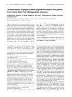
Fig. 1. Chromatographic purification of midgut aminopeptidase from
A. pisum. (A) Chromatography on Mono Q equilibrated with 20 m
M
Tris ⁄ HCl buffer (pH 7.0) ⁄ 0.1% Triton X-100. Elution was accom-
plished with a gradient of 0–600 m
M NaCl gradient in the same Tris
buffer (substrate used LeupNA). (B) SDS ⁄ PAGE of samples
obtained after the steps from A. pisum APN purification (12% poly-
acrylamide slab gels, silver staining). Lane 1, midgut homogenate;
lane 2, Triton X-100-released proteins from midgut cell membranes;
lane 3, Mono Q eluate (purified aminopeptidase). (C) Glycoprotein
detection (Dig Glycan detection kit), after western blots of proteins.
Lane 4, midgut homogenate; lane 5, purified APN; lane 6, purified
with the differentiation kit with the mannose-specific lectin Galan-
thus nivalis agglutinin.
P. T. Cristofoletti et al. Aphid midgut aminopeptidase
FEBS Journal 273 (2006) 5574–5588 ª 2006 The Authors Journal compilation ª 2006 FEBS 5575
consisting of 300 guts, with total activity 2.2 U and
343 lg protein, it was possible to recover 28 lg puri-
fied APN with specific activity 40.3 UÆmg
)1
. The
final yield was 50%, with a purification factor of
6.4. SDS ⁄ PAGE of purified APN resulted in a single
150-kDa protein band (Fig. 1B). The enzyme was
found in the midgut as a major protein band and
was preferentially solubilized by Triton X-100 (Fig. 1B,
lane 2).
SDS ⁄ PAGE of proteins in fractions eluted from a
gel-filtration column showed a correspondence between
eluted activity and band intensity in stained gels
(not shown), indicating homogeneity of the purified
enzyme. The molecular mass calculated from gel filtra-
tion was 200 ± 30 kDa, a little higher than that
obtained from SDS ⁄ PAGE.
In addition, APN can be purified using a single
chromatographic step in ConA–Br-Sepharose (data
not shown). The purified protein had the same mobil-
ity on SDS ⁄ PAGE and the same internal peptide
sequence (see below) as APN purified on a Mono Q
column.
Properties of the purified APN from A. pisum
Acyrthosiphon pisum APN is a glycoprotein (Fig. 1C)
and seems to be the major and ⁄ or most glycosylated
protein from aphid midgut extracts (Fig. 1C, lane 4).
It binds specifically to the lectin (Galanthus nivalis
agglutinin) that recognizes a mannose moiety (Fig. 1C,
lane 6). This agrees with the APN pattern of elution
from ConA–Br-Sepharose columns (see above).
The APN purified from A. pisum had a pH optimum
of 7.0 ± 0.5 (Fig. 2A) when assayed with LeupNA as
substrate. Isoelectric focusing gave a single peak of pI
8.4 ± 0.2 (Fig. 2B), and density-gradient ultracentrifu-
gation produced a single peak of molecular mass
130 ± 20 kDa (Fig. 2C).
A
CD
B
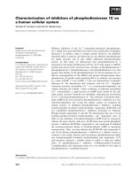
Fig. 2. Properties of purified midgut APN from A. pisum. (A) Effect of pH on enzyme activity (optimal pH 7.0 ± 0.5). Buffers used: 100 mM
sodium phosphate buffer (pH 5–7) and 100 mM Tris ⁄ HCl buffer (pH 7–9.5). (B) Isoelectric focusing (pI 8.4 ± 0.2). (C) Density-gradient centrif-
ugation. Hb, Haemoglobin; Ct, catalase. Molecular mass was calculated as 130 kDa. (D) Arrhenius plot. Activation energy was determined
as E
a
¼ 42.2 kJÆmol
)1
.
Aphid midgut aminopeptidase P. T. Cristofoletti et al.
5576 FEBS Journal 273 (2006) 5574–5588 ª 2006 The Authors Journal compilation ª 2006 FEBS
The thermodynamic parameters of activation for
A. pisum APN (Fig. 1D) were calculated by Arrhenius
plot (plot of k
cat
against 1 ⁄ T). From the slope of the
line, the activation energy (E
a
) was determined to be
42.2 kJÆmol
)1
. Other thermodynamic parameters of
activation were calculated using the relations of the
transition state theory [23]. Thus, DS
à
, DG
à
and DH
à
at 25 °C were estimated to be )65.1 JÆmol
)1
ÆK
)1
()15.5 calÆmol
)1
ÆK
)1
), 59.0 kJÆmol
)1
(14 kcalÆmol
)1
)
and 39.7 kJÆmol
)1
(9.5 kcalÆmol
)1
), respectively.
Purified APN (Mono Q column) was submitted to
MS sequencing. First, it was treated with trypsin. The
digested protein was separated by Q-ToF, and two of
the resulting peptides were submitted to MS sequencing.
The resulting sequences were (a) MDLLAIPDFR, (b)
AGAMENWGMNTYK, and (c) NDSKITIYTYK.
The same sequence as produced by peptide number 2
could be recovered from the protein purified by ConA–
Br-Sepharose, together with a series of more than 19
mass hits covering the entire sequence (25 p.p.m. preci-
sion cut-off), including matching oxidized methionines.
A Mowse score of 3.82E + 9 identified the purified and
cloned sequences unambiguously (see below).
Kinetic parameters of A. pisum APN
The purified aphid APN showed a broad specificity
towards N-terminal aminoacyl residues, although it
was unable to hydrolyze l-aspartic acid a-(b-naphthyl-
amide) (Table 1). The preferred substrates (higher
k
cat
⁄ K
m
values) were those bearing leucine or methion-
ine at the N-terminus, and the least preferable those
presenting a proline at the N-terminus (Table 1). There
was a slight preference for tripeptides (Table 1), as
judged by a comparison of k
cat
⁄ K
m
values for peptides
of the Leu-(Gly)
n
series which differ only in the num-
ber of Gly residues (Table 1).
Leucine hydroxamate is a simple intersecting linear
competitive inhibitor of APN (Fig. 3), with K
i
¼
5±1lm; the same is true for arginine hydroxamate
(K
i
¼ 34±7lm). K
i
values for aminoacyl hydroxa-
mates depend on the hydroxamate used, not on the
substrate used (l-leucine-b-naphthylamide or l-argin-
ine-b-naphthylamide) (not shown). This indicates that
l-leucine-b-naphthylamide and l-arginine-b-naphthyla-
mide are hydrolyzed at the same active site.
Acyrthosiphon pisum APN inhibition by amastatin
and bestatin are rapidly reversible by dilution (not
shown), as observed with microsomal aminopeptidase
[30]. Their pattern of inhibition is an intersecting, com-
petitive, linear type, with K
i
¼ 1.8 lm for bestatin and
K
i
¼ 0.6 lm for amastatin (not shown).
Inactivation of A. pisum APN by EDTA follows
pseudo-first-order kinetics with k
obs
¼ 0.14 m
)1
Æs
)1
,
which is virtually completely suppressed by the com-
petitive inhibitor arginine hydroxamate at a concentra-
Table 1. Kinectic parameters for purified APN from A. pisum. Relative values of k
cat
⁄ K
m
were calculated using LeupNA as reference for syn-
thetic substrate. For peptide subtrates, PheGlyGlyPhe was used as reference. The values were determined at least twice by 10 independent
determinations with different substrate concentrations. SEM values were calculated by fitting data by a weighted linear regression using
the software SigmaPlotÒ. AlabNA,
L-alanine-b-naphthylamide; AlapNA, L-alanine-p-nitroanilide; ArgpNA, L-arginine-p-nitroanilide; MetbNA,
L-methionine-b-naphthylamide; MetpNA, L-methionine-p-nitroanilide; ProbNA, L-proline-b-naphthylamide.
Substrate K
m
(mM) k
cat
(s
)1
) k
cat
⁄ K
m
(mM
)1
Æs
)1
) k
cat
⁄ K
m
(relative)
LeupNA 0.057 ± 0.007 119 ± 15 2090 ± 360 100
MetpNA 0.026 ± 0.008 45 ± 3 1720 ± 540 80.2
ArgpNA 0.19 ± 0.02 158 ± 31 832 ± 180 40.5
AlapNA 1.1 ± 0.04 222 ± 4 202 ± 9 9.9
LeubNA 0.038 ± 0.003 105 ± 2 2760 ± 220 133
ArgbNA 0.058 ± 0.06 72 ± 2 1220 ± 130 59.0
MetbNA 0.043 ± 0.005 28 ± 1 642 ± 76 30.9
AlabNA 0.28 ± 0.04 67 ± 2 240 ± 8 11.4
ProbNA 1.6 ± 0.2 6.8 ± 0.2 4.3 ± 0.5 0.2
AspbNA – < 0,19
LeuGly 0.26 ± 0.08 5.6 ± 0.4 22 ± 7 11.1
LeuGlyGly 0.28 ± 0.06 26 ± 2 92 ± 20 48.9
LeuGlyGlyGly 0.9 ± 0.1 16 ± 1 19 ± 2 10
LeuGlyGlyGlyGly 1.1 ± 0.1 17 ± 1 15 ± 1 7.8
LeuLeuLeu 0.070 ± 0.02 6.4 ± 0.3 91 ± 23 47.8
PheGly 0.60 ± 0.08 13 ± 4 22 ± 8 11.1
PheGlyGly 1.1 ± 0.3 23 ± 2 22 ± 6 11.1
PheGlyPheGly 0.71 ± 0.07 92 ± 2 130 ± 13 68.8
PheGlyGlyPhe 0.27 ± 0.03 50 ± 2 188 ± 21 100
P. T. Cristofoletti et al. Aphid midgut aminopeptidase
FEBS Journal 273 (2006) 5574–5588 ª 2006 The Authors Journal compilation ª 2006 FEBS 5577
tion corresponding to 25-fold its K
i
value (Fig. 4). The
reaction order with respect to EDTA was 0.44. As
EDTA has two metal-binding sites, the data support
the conclusion that removal of only one metal ion is
sufficient to inactivate the enzyme. In agreement with
this, the partially EDTA-inactivated enzyme has the
same K
m
and pH optimum as native aminopeptidase
(not shown).
Carbohydrate and lectin interactions with
A. pisum APN
The enzyme strongly interacts with lectins that bind
mannose-like Galanthus nivalis agglutinin and ConA, as
observed in blotting assays (Fig. 1C) and in the purifica-
tion steps (see above). The interaction with lectins was
evaluated by density-gradient ultracentrifugation
(Fig. 5). After 30 min of preincubation of APN with the
lectins, wheat germ agglutinin (WGA), which binds to
sialic acid and N-acetylglucosamine moieties, or ConA,
which binds to glucose and mannose moieties, the sam-
ples were submitted to density-gradient ultracentrifuga-
tion. APN with WGA results in a single peak (Fig. 5A),
as observed in Fig. 2C, which corresponds to the APN
without bound lectin (Fig. 5A). Mixing ConA with
APN resulted in all the activity being at the bottom of
the tube, meaning a molecular mass higher than
400 kDa (Fig. 5B), resulting from lectin binding and
agglutination. When a competitive monosaccharide
(a-methyl mannoside) was added to the incubation
mixture of ConA with APN at a concentration of
Fig. 3. Inhibition of purified A. pisum APN by leucine hydroxamate.
Lineweaver–Burk plots of LeupNA-hydrolyzing activity against differ-
ent concentrations (m
M) of leucine hydroxamate. Insert: replots of
slopes calculated from Lineweaver–Burk plots against the concen-
tration of leucine hydroxamate. K
i
¼ 5±1lM (n ¼ 4).
Fig. 4. Inactivation of A. pisum APN by EDTA at 37 °C. Reaction
mixtures contained different concentrations of EDTA in 100 m
M
Tris ⁄ HCl buffer, pH 7.0, containing 0.1% Triton X-100. After differ-
ent incubation times, the reaction was stopped by 100 times dilu-
tion. Inactivation by 50 m
M EDTA in the absence (d) or presence
(s) of 850 l
M (25 · K
i
) arginine hydroxamate, which is a competit-
ive inhibitor of aminopeptidase. Buffer used: 100 m
M Tris ⁄ HCl,
pH 7.0, containing 0.1% Triton X-100. The insert shows a plot of
the log of the observed first-order rate kinetics of inactivation con-
stant against log of EDTA concentration. n, the slope of the plot,
was calculated as 0.44 and estimates the number of molecules of
EDTA needed to inactivate each active site of the enzyme.
C
B
A
Fig. 5. Density-gradient ultracentrifugation of A. pisum APN in the
presence of the glucose ⁄ mannose-binding lectin ConA and WGA
(sialic acid ⁄ N-acetylglucosamine binding). (A) APN with WGA lectin;
(B) APN with ConA; (C) APN with ConA and 500 m
M a-methyl man-
noside, a competitive sugar. Note that the sedimentation of APN
with WGA is closer to that in Fig. 2C.
Aphid midgut aminopeptidase P. T. Cristofoletti et al.
5578 FEBS Journal 273 (2006) 5574–5588 ª 2006 The Authors Journal compilation ª 2006 FEBS
500 mm, a peak of intermediate molecular mass was
observed (Fig. 5C), corresponding to the partially
aggregated form of APN with ConA (the molecular
mass of ConA is 73 kDa under the density-gradient con-
ditions).
The activity recovered from the density gradients
was similar with and without lectins (data not shown).
The kinetics parameters of APN (k
cat
and K
m
) associ-
ated with ConA were unaffected, indicating that the
catalytic site and the mannosylated site(s) are quite far
apart on the enzyme molecule.
Sequence coding the A. pisum APN
Full-length cDNA was obtained for A. pisum APN with
3234 bp (GenBank accession number DQ440823). This
sequence codes for a protein of 973 amino acids with
residues 1–17 corresponding to the signal peptide pre-
dicted with signalp ( />SignalP) [31]. The mature protein has a putative ungly-
cosylated molecular mass of 109 011 Da and pI 5.30.
The full-length cDNA contains a short 5¢-UTR from 1
to 132 bp and 3¢-UTR from 3055 to 3234 bp.
The protein encoded by this cDNA contains the
three peptide sequences obtained from the purified
enzyme, showing identity between the purified enzyme
and the cDNA sequence, as well as MS peaks with
high Mowse score (see above) which unambiguously
identified the cloned sequence.
The coding protein has high similarity to other ami-
nopeptidases. It possesses the domain HEXXH + G,
characteristic of gluzincins, and the domain GAMEN,
found in many of these enzymes (Fig. 6). Puta-
tive N-glucosylation sites (predicted at http://
www.cbs.dtu.dk/services/NetNGlyc/) are assigned in
Fig. 6, as well as the GPI anchor site in its C-terminal
domain, as predicted by the DGPI software [32]. The
presence of the signal peptide and GPI anchor signal
are consistent with the known characteristics of insect
APNs. O-glycosylation sites were predicted to be pre-
sent in the region (close to the C-terminus) of the APN
using the NetOGlyc 3.1 server [33]. clustalw sequence
alignment with other insect aminopeptidases (Fig. 7)
showed that A. pisum APN has a weak similarity to
class 2 aminopeptidases from Lepidoptera [20].
Discussion
Occurrence, properties and sequence of
A. pisum APN
Acyrthosiphon pisum has a membrane-bound and a sol-
uble aminopeptidase corresponding to 98% and 2% of
the midgut aminopeptidase activity, respectively, when
LeupNA is used as substrate. There is a single mole-
cular species of A. pisum membrane-bound amino-
peptidase in this tissue, as judged by Mono Q
chromatography after solubilization of almost 100%
of its activity.
The membrane-bound APN from A. pisum was puri-
fied to homogeneity, with a yield of 51.9% and specific
activity of 40.3 UÆmg
)1
. Taking into account that the
specific activity of the homogenized midgut of this
insect is 6.3 UÆmg
)1
per animal and that each midgut
has 19 lg protein [27], it is possible to calculate that
there are about 3 lg APN per midgut and that APN
amounts to 15.6% of midgut protein. This is con-
firmed by SDS ⁄ PAGE of the midgut homogenate,
where it is possible to recognize APN as a major pro-
tein in the preparation.
APN is a glycosylated protein of molecular mass
130 kDa (density-gradient centrifugation) and pI
8.4. Molecular masses determined by SDS⁄ PAGE
(150 kDa) or gel filtration (200 kDa) are probably
artifacts. The molecular mass of the unglycosylated
protein is 109 kDa and pI 5.3 (predicted from the
amino-acid sequence). The data led us to conclude that
16% of the molecular mass of APN is carbohydrate.
Immunoblot for identification of glycosylated pro-
teins recognized APN as the most abundant glycopro-
tein in the midgut and thus as an important target for
lectins. Taking into account that more than one lectin
can bind a single APN molecule (Fig. 5), and the
abundance of APN in microvillar membrane, this
enzyme is potentially the most important lectin-binding
site in aphid midgut. The calculated amount of ami-
nopeptidase in microvillar membrane may explain the
capacity of each aphid to feed on a diet containing lec-
tins amounting to as much as 1 lg of ConA in 48 h
[29]. Also immunohistochemical observations of lectin
binding on the midgut demonstrated that the stomach
(ventriculus 1) cell membranes are the primary target
for ConA, followed by the intestine (remaining midgut
chambers) cell membranes [29]. Activity measurements
found APN along the midgut, and imunolocalization
with APN antibodies showed that APN is associated
with a specialized plasma membrane associated with
the apical lamellae. These consist of a complex net-
work of lamellae, linked one to another by trabecullae
to resist the osmotic pressure caused by high-sugar
phloem-sap ingestion [28]. The apical lamellae replace
the regularly arranged microvilli observed in most mid-
gut cells. APN localization data are in agreement with
the lectin-binding site found by Sauvion et al. [29].
However, as presented here, APN activity is not affec-
ted by glucose ⁄ mannose-binding lectin.
P. T. Cristofoletti et al. Aphid midgut aminopeptidase
FEBS Journal 273 (2006) 5574–5588 ª 2006 The Authors Journal compilation ª 2006 FEBS 5579
Fig. 6. cDNA coding sequence of A. pisum APN and its deduced sequence (GenBank accession number DQ440823). The predicted signal
peptide is underlined, and the C-terminal GPI cleavage signal sequence is dotted underlined. The characteristic zinc binding ⁄ gluzincin motif,
HEXXH + E, and the gluzincin aminopeptidase motif, GAMEN, are highlighted in a bold ⁄ gray box. Peptides identified by MS analysis are dou-
ble underlined. Boxed residues correspond to the MS sequenced peptides. Putative N-glycosylated asparagine residues are dark-shaded
using the NetNGlyc 1.0 server ( and putative O-glycosylated threonines residues identified by the
NetOGlyc 3.1 server ( are also shaded.
Aphid midgut aminopeptidase P. T. Cristofoletti et al.
5580 FEBS Journal 273 (2006) 5574–5588 ª 2006 The Authors Journal compilation ª 2006 FEBS
Acyrthosiphon pisum APN has a broad specificity
towards the N-terminal amino-acid residues of pep-
tides, but it does not hydrolyze acidic aminoacyl-
peptides, and, although it is by no means proven, it
appears to prefer peptides longer than dipeptides.
Thus, A. pisum APN resembles the vertebrate enzyme
(EC 3.4.11.2) [34] and the following insect midgut
enzymes: microvillar membrane APN from Teneb-
rio molitor [22], soluble Tineola bisselliela (Lepido-
ptera) aminopeptidase [35,36], soluble Attagenus
megatoma (Coleoptera) aminopeptidase [37], soluble
and microvillar R. americana (Diptera) aminopeptidas-
es [25,26], and membrane-bound Spodoptera littoralis
(Lepidoptera) aminopeptidase [38]. Another resem-
blance between A. pisum APN and the vertebrate
enzyme are both inhibited by bestatin and amastatin,
which in both cases is rapidly reversible [30]. It should
be noted, however, that A. pisum APN has a K
m
value
for peptides much smaller than those for T. molitor
APN [22].
Substrates with different N-terminal amino-acid resi-
dues are hydrolyzed at the same site of A. pisum APN,
as hydroxamate K
i
values do not depend on the ami-
noacyl b-naphthylamide used as substrate.
Substrates with a bulky aminoacyl residue in posi-
tion P
1
(numbering of Schechter & Berger [39]) are
better substrates for APN. The same is true for the P
2
¢
position (compare Leu-Gly-Gly with Leu-Leu-Leu in
Table 1), but not for the P
1
¢ position (compare amino-
acyl-naphthylamide with aminoacyl-p-nitroanilide).
This suggests that the subsites S
1
,S
2
¢ and probably S
3
¢
of the enzyme are pockets able to bind bulky amino-
acyl residues, and this hypothesis agrees with the fact
that amastatin is a better inhibitor of A. pisum APN
than bestatin. Bestatin has bulky residues putatively
able to interact with S
1
¢ and S
2
¢ of the enzyme (see
above), and amastatin with S
1
,S
1
¢ and S
2
¢ [40].
Acyrthosiphon pisum APN is the first insect digestive
aminopeptidase that does not belong to the order
Lepidoptera to be fully characterized and sequenced.
0.1
AAB70755 Pxy
AAX39863 Tni APN1
AAF08254 Hvi
AAN75693 Har APN1
AAF37558 Hpu APN1
AAC33301 Bmo
Q11001 Mse
A pisum APN
CAA66467 Pxy
AAX39864 Tni APN2
AAD31184 Ldi APN2
BAA32140 Bmo
P91885 Mse APN2
CAA10950 Pxy
BAA33715 Bmo
AAX39866 Tni APN4
AAK69605 Sli
AAF37559 Hpu APN2
AAK58066 Hvi
AAC36148 Pin
AAX39865 Tni APN3
AAF01259 Pxy APN3
Q11000 Hvi
AAN75694 Har APN2
AAF37560 Hpu APN3
AAF99701 Epo
AAD31183 Ldi APN1
AAL83943 Bmo APN3
Family 1
Family 3
Famil
y
2
Family 4
Fig. 7. Sequence tree of Lepidoptera aminopeptidases and A. pisum APN. The tree was obtained using the CLUSTALX alignment program.
Families were numbered as described by Wang et al. [20]. Sequences used: Helicoverpa armigera, Har, APN1 (HaAPN1) (GenBank acces-
sion number AAN75693) and APN2 (HaAPN2) (GenBank accession number AAN75694) [19]; Helicoverpa puntigera, Hpu, APN1 (HpAPN1)
(GenBank accession number AAF37558), APN2 (HpAPN2) (GenBank accession number AAF37559) and APN3 (H pAPN3) (GenBank acces-
sion number AAF37560) [15]; Heliothis virescens, Hvi, 110 kDa APN(HvAPN 110 kDa) (GenBank accession number AAK58066) [18], 120-kDa
APN(HvAPN 120 kDa) (GenBank accession number ACC46929) [9] and 170-kDa APN(HvAPN 170 kDa) (GenBank accession number
AAF08254) [11]; Plutella xylostella, Pxy, APNA (PxAPNA) (GenBank accession number AAB70755) [12], APN1 (PxAPN1) (GenBank accession
number CAA66467) [5], APN3 (PxAPN3) (GenBank accession number AAF01259) [17] and APN4 (PxAPN4) (GenBank accession number
CAA10950); Bombyx mori, Bmo, APN1 (BmAPN1) (GenBank accession number AAC33301) [6], APN2 (BmAPN2) (GenBank accession num-
ber BAA32140) [10], APN3 (BmAPN3) (GenBank accession number AAL83943) [17] and APN4 (BmAPN4) (GenBank accession number
BAA33715); Epiphyas postvittana, Epo, APN(EpAPN) (GenBank accession number AAF99701) [13]; Lymantria dispar, Ldi, APN1 (LdAPN1)
and APN2 (LdAPN2) (GenBank accession numbers AAD31183 and AAD31184) [64]; Plodia interpunctella, Pin, APN(PiAPN) (GenBank acces-
sion number AAC36148) [14]; Manduca sexta, Mse, APN1 (MsAPN1) (GenBank accession number CAA61452) [3] and APN2 (MsAPN2)
(GenBank accession number CAA66466) [5]; Spodoptera litura, Sli, APN(SlAPN) (GenBank accession number AAK69605) [16].
P. T. Cristofoletti et al. Aphid midgut aminopeptidase
FEBS Journal 273 (2006) 5574–5588 ª 2006 The Authors Journal compilation ª 2006 FEBS 5581
A rapid survey of the A. pisum databank (http://
urgi.infobiogen.fr/cgi-bin/annotation_form.pl?organism
¼ apisum) allows the identification of more than 25
contigs with some relation to the word ‘aminopepti-
dase’, and similarly, a high number of ‘aminopepti-
dases’ are encoded in the Drosophila genome (http://
flybase.org). A total of 29 A. pisum expressed sequence
tags (ESTs) have full complementarities with the
cloned APN in almost 60 000 ESTs. From these 29
ESTs, 22 belong to libraries from the digestive tract,
and seven from libraries from whole insect (none from
other tissue libraries), meaning that this aminopepti-
dase is potentially very specific to the aphid midgut.
The APN sequence has all identified residues essen-
tial for zinc binding and catalysis. In the sequence, it
was easy to recognize the signal peptide, several poten-
tial glycosylation sites, as well as a GPI anchor at its
C-terminus. This anchor is possibly an adaptation to a
phloem-based diet, avoiding excretion of the enzyme
into the honeydew, as the phylogenetically related
Hemipteran Dysdercus peruvianus has a soluble amino-
peptidase, in spite of the fact that, in this case, the
enzyme is trapped between the microvillar and perimic-
rovillar membranes [27,28].
Function of A. pisum APN and lectin toxicity
The role of the microvillar aminopeptidase is postula-
ted to be hydrolysis of oligopeptides formed by the
action of luminal proteinases [1,2,22]. In aphids, a
cathepsin L was found to be partially associated with
modified perimicrovillar membranes and is possibly
involved in degradation of toxic proteins found in the
phloem sap [28,41]. APN is certainly responsible for
the final digestion of peptides generated by cathep-
sin L. Another possibility is that APN is somehow
associated with putative amino-acid-binding sites at
the plasma membranes associated with the apical
lamellae (modified perimicrovillar membranes). These
are thought to increase the amino-acid concentration
(usually low in the aphid diet) [28], thus facilitating
absorption. APN may also be directly linked to
absorptive sites in apical lamellae, as has been sugges-
ted for Lepidoptera [42]. Finally, APN may serve
as the primary digestive enzyme responsible for
the assimilation of the phloem sap small peptide frac-
tion, chemical components largely unexplored at the
moment.
As B. thuringiensis is not effective in aphid control,
lectins have been used as insecticidal agents against
aphids [43]. The soluble protein, ferritin, is the snow-
drop lectin-binding protein in the planthopper Nilapar-
vata lugens [44]. The authors postulated that alteration
of iron metabolism might be related to its lectin toxic-
ity. Although ferritin was not the most abundant pro-
tein in midgut preparations, this protein was the most
specifically recognized in N. lugens. However, none of
the 2D PAGE protein spots observed in pea aphid
homogenates as binding to ConBr was identified as
corresponding to ferritin (F. A. Mendonca de Sousa &
Y. Rahbe
´
, unpublished), although the ferritin gene is
largely transcribed in A. pisum midguts [45]. It is poss-
ible that the mechanism of toxicity found in planthop-
per is different from that found in aphids.
In A. pisum, the lectin, ConA, is a potent toxin
affecting survival and growth, but WGA is relatively
ineffective [46]. These data agree with the fact that
aphids do not possess a peritrophic membrane [28].
Consequently, this toxicity must result from lectin
binding to target proteins in the apical membranes
from the midgut, although not related to the inhibition
of APN activity. One explanation of this effect is a
decrease in amino-acid absorption caused by ConA
binding to APN, with deleterious effects on the puta-
tive associated proteins thought to bind to amino acids
(see above). Indeed, ConA-intoxicated aphids have
been shown to display altered hemolymph free amino-
acid profiles and modified excretion of asparagine in
their honeydew [47]. It is still possible that a reduction
in membrane protein lateral mobility or its resist-
ance to phloem osmotic pressure is the major cause of
lectin toxicity to aphids. These possibilities need to be
evaluated.
Experimental procedures
Animals
Acyrthosiphon pisum Harris aphids (Hemiptera: Aphididae),
clone Ap-LL01, were maintained in the laboratory on
broad bean seedlings (Vicia faba ) in ventilated plexiglass
cages (21 °C; 70% relative humidity; 16 h light ⁄ 8 h dark-
ness). For the experiments, a limited number of mass-reared
adults were allowed to lay eggs for 24 h on young Vicia
plants, and the resulting apterous insects were used as
9-day-old adults.
Chemicals
Buffer salts, detergents, molecular-mass markers, protein
inhibitors, and most substrates were purchased from
Sigma-Aldrich (St Louis, MO, USA). Glycoprotein detec-
tion kits came from Boehringer-Mannheim (Mannheim,
Germany). The peptides Leu-Gly-Gly-Gly and Leu-
Gly-Gly-Gly-Gly were gifts from Dr L. Juliano (Unifesp,
Sa
˜
o Paulo, Brazil).
Aphid midgut aminopeptidase P. T. Cristofoletti et al.
5582 FEBS Journal 273 (2006) 5574–5588 ª 2006 The Authors Journal compilation ª 2006 FEBS
Preparation of samples
Adult apterous aphids were immobilized on a flat surface,
using adhesive tape, and their guts were removed under a
stereomicroscope in Yeager’s physiological solution [48].
The midguts were separated and homogenized in double-
distilled water with the aid of a Potter-Elvehjem homoge-
nizer. The homogenates were labeled crude homogenate
and stored. Crude homogenates were used to assay APN
or were centrifuged at 100 000 g for 1 h at 4 °C, resulting
in a supernatant (labeled midgut soluble fraction) and a
pellet (midgut cell membranes). Washed midgut cell mem-
branes were prepared by dispersing the midgut cell mem-
branes in water, followed by three freezing and thawing
cycles, and re-centrifugation at 100 000 g for 1 h at 4 °C.
All centrifugations were performed on a Hitachi Ultracen-
trifuge model Himac 70P-72 with an RPS 40T rotor.
Protein determination and enzymatic assays
Protein was determined as described by Bradford [49] using
ovalbumin as standard. When samples contained detergent,
protein was determined by the method of Smith et al. [50], as
modified by Morton & Evans [51], using BSA as standard.
Routine assays of APN were performed using 1 mm Leu-
pNA as substrate (initially solubilized in dimethyl sulfoxide)
in 100 mm Tris ⁄ HCl buffer, pH 7.0, at 30 °C. Unless other-
wise specified, the same conditions were used for all other
substrates. Naphthylamine liberated from aminoacyl-
b-naphthylamides, nitroaniline from aminoacyl-p-nitroani-
lides, and phenylalanine and leucine from the different
peptides were determined spectrophotometrically by the
methods of Hopsu et al. [52], Erlanger et al. [53] and
Nicholson & Kim [54], respectively. In each determination,
incubations were continued for at least four different peri-
ods of time, and the initial rates were calculated. All assays
were performed so that the measured activity was propor-
tional to protein and incubation time. Controls without
enzyme or without substrate were included. One enzyme
unit (U) is defined as the amount that hydrolyzes 1 lmol
substrateÆmin
)1
,at30°C.
Solubilization of APN by detergents
In order to evaluate the solubilizing efficiency of deter-
gents, samples of 200 lL midgut homogenate at a concen-
tration of 13 guts per mL (which contains 50 lg protein)
of midgut cell membranes were suspended in 10 mm Hepes
buffer, pH 7.4, in the presence and absence of several
detergents. After 17 h at 4 °C with shaking, the suspen-
sions were centrifuged at 100 000 g for 1 h at 4 °C, and
supernatants were assayed for APN. APN activity was
determined in the resulting supernatants and referred to the
original preparation of cell membranes (as percentage
solubilization). Recovery is the percentage of the sum of
solubilized plus nonsolubilized activity referred to the ori-
ginal preparation of cell membranes. Data are mean ±
SEM calculated from determinations carried out in three
different preparations.
For routine solubilization of APN, midgut cell mem-
branes were suspended in 10 mm Hepes buffer, pH 7.4,
containing 10 mm Triton X-100. After 1 h at 4 °C, the sus-
pension was centrifuged at 25 000 g for 30 min at 4 °C,
and the supernatant used as a source of enzyme.
Purification of detergent-solubilized APN
Cell membranes corresponding to 300 A. pisum midguts
(wet weight 6 mg) were solubilized with Triton X-100 as
described above, and applied to a Mono Q HR 5 ⁄ 5 column
(0.5 cm internal diameter · 5 cm) equilibrated with 20 mm
Tris ⁄ HCl buffer, pH 7.0, containing 0.1% Triton X-100 in
an FPLC system. Controls showed that protease inhibitors
are not necessary. Elution was carried out with a gradient
of 0–0.6 m NaCl in the same buffer. The flux was 1.0 mLÆ
min
)1
, and fractions of 0.4 mL were collected. Fractions
showing activity with LeupNA were pooled, and purifica-
tion was checked by SDS ⁄ PAGE.
Alternatively, the APN was purified on a 3-mL ConA–
Br-Sepharose column (6 · 30 mm). The column was
washed with 15 mL 20 mm acetate buffer, pH 4.2, contain-
ing 0.5 m NaCl, then with 15 mL 100 mm citrate ⁄ phos-
phate buffer, pH 6.0, containing 2 mm CaCl
2
, before being
equilibrated with 15 mL 20 mm Tris ⁄ HCl buffer, pH 7.0,
with 0.1% Triton X-100. The solubilized samples were
applied to the column and eluted with 20 mm Tris ⁄ HCl
buffer, pH 7.0, containing 0.1% Triton X-100 and 0.5 m
a-methyl mannoside. Fractions of 1 mL were collected at a
flow rate of 1 mLÆmin
)1
.
SDS/PAGE
Electrophoresis of A. pisum samples in denaturing con-
ditions (SDS ⁄ PAGE) was carried out on 7.5% (w ⁄ v)
polyacrylamide gels containing 0.1% (w ⁄ v) SDS, on a dis-
continuous pH system [55], using Mini Protean II cells
(Bio-Rad, Hercules, CA, USA). Samples were lyophilized
and suspended in sample buffer containing 60 mm
Tris ⁄ HCl buffer, pH 6.8, 2.0% (w ⁄ v) SDS, 5% (v ⁄ v)
2-mercaptoethanol, 10% glycerol and 0.2% (w ⁄ v) bromo-
phenol blue and heated for 3 min at 95 °C in a water bath
before being loaded on to the gels. Electrophoresis was car-
ried out at 200 V until the tracking dye reached the bottom
of the gel. The gel was then silver-stained [56] or stained
using 0.1% (w ⁄ v) Coomassie Blue R in 10% acetic
acid ⁄ 40% methanol for 30 min. In the last case, destaining
was achieved with several washes in a solution containing
40% methanol and 10% acetic acid.
P. T. Cristofoletti et al. Aphid midgut aminopeptidase
FEBS Journal 273 (2006) 5574–5588 ª 2006 The Authors Journal compilation ª 2006 FEBS 5583
Isoelectric focusing
Isoelectric focusing was performed as described by Terra
et al. [57], in columns of 7.5% polyacrylamide gel con-
taining 10% ampholytes pH 3–10 (Pharmalyte 3–10, Phar-
macia, Uppsala, Sweden). Samples were applied after
polymerization and prefocusing (30 min at 31 VÆcm
)1
)on
the top of the alkaline side of the gels. For samples in
detergent, 0.1% Triton X-100 was added to the gels and
fractionation buffer. Recoveries of activities applied to the
gels were 80–100%.
Density-gradient centrifugation
Samples of purified APN (100 lL) containing 1 lg puri-
fied protein with or without 100 lg lectins were layered on
the top of 10-mL glycerol gradients (10–30%, w ⁄ v) made
up in 100 mm Tris ⁄ HCl buffer, pH 7.0, in the presence or
absence of 500 mm a-methyl mannoside. Centrifugation
and collection of fractions were performed as described pre-
viously [58]. The molecular mass of APN was calculated by
the method of Martin & Ames [59], using the sedimentation
rates of hemoglobin (64.5 kDa) and bovine liver catalase
(232 kDa) as reference standards. Activity recovery was
80–100%.
Determination of molecular mass by gel filtration
Samples of 200 lL, containing 2–4 lg purified protein, were
applied to a gel-filtration column in an FPLC system (Phar-
macia-LKB Biotechnology, Uppsala, Sweden) by using a
Superose HR 10 ⁄ 30 column (1.0 cm internal diam-
eter · 30 cm) equilibrated and eluted in 100 mm Tris ⁄ HCl
buffer, pH 7.0, containing 0.1% Triton X-100. Fractions of
0.4 mL were collected at a flow rate of 0.5 mLÆmin
)1
.
Molecular masses were calculated using the following
proteins as standards: aprotinin (6.5 kDa), cytochrome
c (12.4 kDa), ovalbumin (45 kDa), BSA (65 kDa), and
b-amylase (200 kDa). Recoveries were 80–100%.
Detection of carbohydrates in purified APN
Protein samples were blotted on to nitrocellulose sheets
after SDS ⁄ PAGE [60]. Detection of carbohydrates was per-
formed with the DIG Glycan Detection kit, and identifica-
tion of carbohydrate moieties was accomplished with
lectins by using the DIG Glycan Differentiation kit. The
procedures followed the supplier’s instructions (Boehringer
Mannheim).
Kinetic studies
The effect of substrate concentration on the activity of puri-
fied APN was determined using at least 10 different sub-
strate concentrations. K
m
and k
cat
values (mean ± SEM)
were determined by a weighted linear regression using the
software SigmaPlotÒ (Jandel Scientific, Systat Software Inc.,
Richmond, CA, USA). In the inhibition studies, purified
APN was incubated with four different inhibitor concentra-
tions in each of 10 different concentrations of substrate. K
i
values were calculated as described by Segel [61].
EDTA inactivation
Purified APN was incubated with EDTA (1–50 mm) for dif-
ferent times at 40 °C in 100 mm citrate ⁄ phosphate buffer
pH 6. The EDTA-inactivation reactions were stopped by
100-fold dilution of reaction mixtures with 100 mm
Tris ⁄ HCl buffer (pH 7.0) ⁄ 0.1% Triton X-100. The remain-
ing activity was measured using LeupNA as substrate,
under the conditions described above. Protection against
inactivation by EDTA was investigated with aminoacyl
hydroxamates, which are simple linear competitive inhibitors
of A. pisum APN. The reaction order was determined by
incubation of APN with different concentrations of EDTA.
Microsequencing of purified APN
Purified APN was electroblotted on to poly(vinylidene
difluoride) membranes after SDS ⁄ PAGE [62]. The mem-
branes were stained for protein using 0.1% Coomassie Blue
R-250 in a 50% (v ⁄ v) methanol, and were destained with
50% methanol. Dried poly(vinylidene difluoride) mem-
branes were submitted to tryptic digestion, and the resulting
peptides (two peptides) were submitted to MS sequencing
at the sequencing facility of the Pasteur Institute (Paris).
Alternatively, in-gel digestion was performed for protein
identification: spots were excised from preparative gels
using pipette tips. The spots were washed with 100 lL
25 mm NH
4
HCO
3
for 30 min, twice destained for 30 min
with 100 lL25mm NH
4
HCO
3
⁄ acetonitrile (v ⁄ v), and
dehydrated in acetonitrile. Gel spots were completely dried
using a vacuum centrifuge before trypsin digestion. The
dried gel volume was evaluated, and 3 vol. trypsin (V5111;
Promega, Madison, WI, USA; 10 ngÆlL
)1
in 25 mm
NH
4
HCO
3
) was added. Digestion was performed at 37 °C
over 5 h. The gel pieces were centrifuged, and 8–12 lL
acetonitrile (depending on gel volume) was added to extrac-
ted peptides. The mixture was sonicated for 5 min and cen-
trifuged. For MALDI-TOF MS analysis, 1 lL supernatant
was loaded directly on to the MALDI target. The matrix
solution (5 mgÆmL
)1
a-cyano-4-hydroxycinnamic acid in
50% acetonitrile containing 0.1% trifluoroacetic acid) was
added immediately and allowed to dry at room tempera-
ture. A Voyager DE-Pro model MALDI-TOF mass spec-
trometer (Perseptive BioSystems, Farmingham, MA, USA)
was used in positive-ion reflector mode for peptide mass
fingerprinting. External calibration was performed with a
Aphid midgut aminopeptidase P. T. Cristofoletti et al.
5584 FEBS Journal 273 (2006) 5574–5588 ª 2006 The Authors Journal compilation ª 2006 FEBS
standard peptide solution (Proteomix; LaserBio Laborator-
ies, Sophia-Antipolis, France). Internal calibration was per-
formed using peptides resulting from autodigestion of
porcine trypsin. Monoisotopic peptide masses were assigned
and used from NCBI database searches (plus A. pisum
APN sequence) with the ‘ms-fit’ software.
Cloning of APN from A. pisum
Total RNA was extracted from midgut epithelium of
A. pisum with Trizol following the instructions of the
manufacturer, Invitrogen, which are based on those of
Chomczynski & Sacchi [63]. mRNA was purified with
Qiagen mRNA purification kit, and a cDNA library was
constructed with the kit Smart (Clontech, Mountain View,
CA, USA), following the instructions of the manufacturer.
A partial sequence of a cDNA coding for APN was ampli-
fied using degenerated primers for the APN consensus
sequence that contains the peptide AGAMENWGM identi-
fied in MS analysis (primers APN-U538: 5¢-TTYCCITGY
TIYGAYGARCC-3¢, based on peptide ‘TGLYRSS’;
APN-L1051: 5¢-RTTICCRAACCACWKRTG-5¢, based on
peptide ‘THQWFGN’). PCR was performed using Taq
DNA polymerase (Invitrogen) using the standard method.
The PCR product was cloned in pGEM-T Easy Vector
(Promega), sequenced, and the identified fragment sequence
had high identity with that of APN.
This sequenced cDNA fragment contains the two identi-
fied peptides sequenced from purified protein, and was
blasted (blastn) against the A. pisum ESTs deposited at
NCBI [45]. The recovered fragments were clustered and
blasted again against the A. pisum ESTs until the N-termi-
nus was completed with signal peptide and 5¢-UTR
sequence. Putative 3¢ reads were recovered from A. pisum
ESTs using blast (blastn) with the best full-length blast
hit as driver (Apis mellifera, access number XP 366261).
Also, ESTs corresponding to A. pisum APN can be recov-
ered along the sequence using MS peaks with Mascot
engine ( Reads were clus-
tered into two contigs covering the 3¢ region and the 5¢
region. The final gap between these contigs was recovered
using PCR with forward primers 5¢-GGCATGGTGAG
GACTAGTTGGCCG-3¢ combined with reverse primer
5¢-GCCATGCCGCCGTCTCGTTGATGG-3¢, and the
complete sequence was obtained and deposited at GenBank
with the accession number DQ440823.
Acknowledgements
This work was supported by the Brazilian research
agencies FAPESP, CAPES ⁄ COFECUB and CNPq
(PRONEX program). We are indebted to Dr L. Juli-
ano (Medical School, UNIFESP) for the synthesis of
several peptides used as substrates, to Dr C. Ferreira
for helpful discussion, and to Mrs L.Y. Nakabayashi
for technical assistance. We thank Mrs L. Duportest
and C. Deraison for their help in obtaining the
A. pisum cDNA library, and G. Duport for her invalu-
able skills in aphid dissection. FAMS, YR and PTC
were given grants for exchanges between USP, UFC
and INRA-INSA through a French–Brazilian contract
from CAPES ⁄ COFECUB (contract 261 ⁄ 98, co-ordi-
nated by Drs S. Grenier and J. R. Parra). WRT is
a research fellow of CNPq. Many thanks go to
Dr Christophe Chambon (INRA Clermont-Ferrand
Theix Proteomic Facility) for his help with MALDI-
TOF analysis of the purified enzyme.
References
1 Terra WR & Ferreira C (1994) Insect digestive enzymes:
properties, compartmentalization and function. Comp
Biochem Physiol 109B, 1–62.
2 Terra WR & Ferreira C (2005) Biochemistry of diges-
tion. In Comprehensive Molecular Insect Science (Gilbert
LI, Iatrou K & Gill SS, eds), Vol. 4, pp. 171–224. Else-
vier, Oxford.
3 Knight PJ, Knowles BH & Ellar DJ (1995) Molecular
cloning of an insect aminopeptidase N that serves as a
receptor for Bacillus thuringiensis CryIA (c) toxin. J Biol
Chem 270, 17765–17770.
4 Valaitis AP, Lee MK, Rajamohan F & Dean DH
(1995) Brush border membrane aminopeptidase-N in
the midgut of the gypsy moth serves as the receptor for
the CryIA (c) delta-endotoxin of Bacillus thuringiensis.
Insect Biochem Mol Biol 25, 1143–1151.
5 Denolf P, Hendrickx K, Vandamme J, Jansens S, Pefe-
roen M, Degheele D & VanRie J (1997) Cloning and
characterization of Manduca sexta and Plutella xylos-
tella midgut aminopeptidase N enzymes related to Bacil-
lus thuringiensis toxin-binding proteins. Eur J Biochem
248, 748–761.
6 Yaoi K, Nakanishi K, Kadotani T, Imamura M,
Koizumi N, Iwahana H & Sato R (1999) cDNA cloning
and expression of Bacillus thuringiensis Cry1Aa toxin
binding 120 kDa aminopeptidase N from Bombyx mori.
Biochim Biophys Acta 1444, 131–137.
7 Schnepf E, Crickmore N, Van Rie J, Lereclus D, Baum
J, Feitelson J, Zeigler DR & Dean DH (1998) Bacillus
thuringiensis and its pesticidal crystal proteins. Microbiol
Mol Biol Rev 62, 775–806.
8 Yeager CL, Ashmun RA, Williams RK, Cardellichio
CB, Shapiro LH, Look AT & Holmes KV (1992)
Human aminopeptidase N is a receptor for human
Coronavirus 229E. Nature 357 (6377), 420–422.
9 Gill SS, Cowles EA & Francis V (1995) Identification,
isolation and cloning of a Bacillus thuringiensis Cry1Ac
P. T. Cristofoletti et al. Aphid midgut aminopeptidase
FEBS Journal 273 (2006) 5574–5588 ª 2006 The Authors Journal compilation ª 2006 FEBS 5585
toxin-binding protein from the midgut of the lepido-
pteran insect Heliothis virescens. J Biol Chem 270,
27277–27282.
10 Hua G, Tsukamoto K & Ikezawa H (1998) Cloning and
sequence analysis of the aminopeptidase N isozyme
(APN2) from Bombyx mori midgut. Comp Biochem Phy-
siol 121B, 213–222.
11 Oltean DI, Pullikuth AK, Lee HK & Gill SS (1999)
Partial purification and characterization of Bacillus thur-
ingiensis Cry1A toxin receptor A from Heliothis vires-
cens and cloning of the corresponding cDNA. Appl
Environ Microbiol 65, 4760–4766.
12 Chang WX, Gahan LJ, Tabashnik BE & Heckel DG
(1999) A new aminopeptidase from diamondback moth
provides evidence for a gene duplication event in Lepi-
doptera. Insect Mol Biol 8, 171–177.
13 Simpson RM & Newcomb RD (2000) Binding of Bacil-
lus thuringiensis delta-endotoxins Cry1Ac and Cry1Ba
to a 120-kDa aminopeptidase-N of Epiphyas postvittana
purified from both brush border membrane vesicles and
baculovirus-infected Sf9 cells. Insect Biochem Mol Biol
30, 1069–1078.
14 Zhu YC, Kramer KJ, Oppert B & Dowdy AK (2000)
cDNAs of aminopeptidase-like protein genes from Plo-
dia interpunctella strains with different susceptibilities to
Bacillus thuringiensis toxins. Insect Biochem Mol Biol 30,
215–224.
15 Emmerling M, Chandler D & Sandeman M (2001)
Molecular cloning of three cDNAs encoding aminopep-
tidases from the midgut of Helicoverpa punctigera, the
Australian native budworm. Insect Biochem Mol Biol
31, 899–907.
16 Agrawal N, Malhotra P & Bhatnagar RK (2002)
Interaction of gene-cloned and insect cell-expressed
aminopeptidase N of Spodoptera litura with insectici-
dal crystal protein Cry1C. Appl Environ Microbiol 68,
4583–4592.
17 Nakanishi K, Yaoi K, Nagino Y, Hara H, Kitami M,
Atsumi S, Muira N & Sato R (2002) Aminopeptidase N
isoforms from the midgut of Bombyx mori and Plutella
xylostella: their classification and the factors that deter-
mine their binding specificity of Bacillus thuringiensis
Cry1Ac toxin. FEBS Lett 519, 215–220.
18 Banks DJ, Hua G & Adang MJ (2003) Cloning of a
Heliothis virescens 110 kDa aminopeptidase N and
expression in Drosophila S2 cells. Insect Biochem Mol
Biol 33, 499–508.
19 Rajagopal R, Agrawal N, Selvapandiyan A, Sivakumar
S, Ahmad S & Bhatnagar RK (2003) Recombinantly
expressed isoenzymic aminopeptidases from Helicoverpa
armigera
(American cotton bollworm) midgut display
differential interaction with closely related Bacillus thur-
ingiensis insecticidal proteins. Biochem J 370, 971–978.
20 Wang P, Zhang X & Zhang J (2005) Molecular charac-
terization of four midgut aminopeptidase N isozymes
from the cabbage looper, Trichoplusia ni. Insect Biochem
Mol Biol 35, 611–620.
21 Rawlings ND & Barrett AJ (1995) Evolutionary families
of metallopeptidases. Methods Enzymol 248, 183–228.
22 Cristofoletti PT & Terra WR (1999) Specificity, anchor-
ing, and subsites in the active center of a microvillar
aminopeptidase purified from Tenebrio molitor (Coleop-
tera) midgut cells. Insect Biochem Mol Biol 29, 807–819.
23 Cristofoletti PT & Terra WR (2000) The role of amino
acid residues in the active site of a midgut microvillar
aminopeptidase from the beetle Tenebrio molitor.
Biochim Biophys Acta 1479, 185–195.
24 Adams MD, Celniker SE, Holt RA, Evans CA,
Gocayne JD, Amanatides PG, Scherer SE, Li PW,
Hoskins RA & Galle RF (2000) The genome sequence
of Drosophila melanogaster. Science 287, 2185–2195.
25 Ferreira C & Terra WR (1984) Soluble aminopeptidases
from cytosol and luminal contents of Rhynchosciara
americana midgut caeca. Properties and phenanthroline
inhibition. Insect Biochem 14, 145–150.
26 Ferreira C & Terra WR (1986) Substrate specificity and
binding loci for inhibitors in an aminopeptidase purified
from the plasma membrane of midgut cells of an insect
(Rhynchosciara americana). Arch Biochem Biophys 244,
478–485.
27 Silva CP, Ribeiro AF & Terra WR (1996) Enzyme mar-
kers and isolation of the microvillar and perimicrovillar
membranes of Dysdercus peruvianus (Hemiptera: Pyr-
rhocoridae) midgut cells. Insect Biochem Mol Biol 26,
1011–1018.
28 Cristofoletti PT, Ribeiro AF, Deraison C, Rahbe
´
Y&
Terra WR (2003) Midgut adaptation and digestive
enzyme distribution in a phloem feeding insect, the pea
aphid Acyrthosiphon pisum. J Insect Physiol 49, 11–24.
29 Sauvion N, Nardon C, Febvay G, Gatehouse AM &
Rahbe
´
Y (2004) Binding of the insecticidal lectin Conca-
navalin A in pea aphid, Acyrthosiphon pisum (Harris)
and induced effects on the structure of midgut epithelial
cells. J Insect Physiol 50, 1137–1150.
30 Wilkes SH & Prescott J (1985) The slow, tight binding
of bestatin and amastatin to aminopeptidases. J Biol
Chem 25, 13154–13162.
31 Bendtsen JD, Nielsen H, von Heijne G & Brunak S
(2004) Improved prediction of signal peptides: SignalP
3.0. J Mol Biol 340
, 783–795.
32 Kronegg J & Buloz D (1999) Detection ⁄ prediction of
GPI cleavage site (GPI-anchor) in a protein (DGPI).
http://129.194.185.165/dgpi/.
33 Julenius K, Molgaard A, Gupta R & Brunak S (2005)
Prediction, conservation analysis and structural charac-
terization of mammalian mucin-type O-glycosylation
sites. Glycobiology 15, 153–164.
34 Nore
´
n O, Sjostrom H, Danielsen EM, Cowell GM &
Skovbjerg H (1986) The enzymes of the enterocyte
plasma membrane. In Molecular and Cellular Basis of
Aphid midgut aminopeptidase P. T. Cristofoletti et al.
5586 FEBS Journal 273 (2006) 5574–5588 ª 2006 The Authors Journal compilation ª 2006 FEBS
Digestion (Desnuelle P, Sjostrom H & Nore
´
n O, eds).
Elsevier, Amsterdam.
35 Ward CW (1975a) Aminopeptidases in webbing clothes
moth larvae. Properties and specificities of enzymes of
highest electrophoretic mobility. Aust J Biol Sci 28,
447–455.
36 Ward CW (1975b) Aminopeptidases in webbing clothes
moth larvae. Properties and specificities of the enzymes
of intermediate electrophoretic mobility. Biochim Bio-
phys Acta 410, 361–369.
37 Baker JE & Woo SM (1981) Properties and specificities
of a digestive aminopeptidase from larvae of Attagenus
megatoma (Coleoptera: Dermestidae). Comp Biochem
Physiol 69B, 189–193.
38 Lee MJ & Anstee JH (1995) Characterization of midgut
exopeptidase activity from larval Spodoptera littoralis.
Insect Biochem Mol Biol 25, 63–71.
39 Schechter I & Berger A (1967) On the size of the active
site in proteases. Biochem Biophys Res Commun 27,
157–162.
40 Rich DH, Moon BJ & Harbeson S (1984) Inhibition of
aminopeptidases by amastatin and bestatin derivatives.
Effect of inhibitor structure on slow-binding processes.
J Med Chem 27, 417–422.
41 Deraison C, Darboux I, Duportets L, Gorojankina T,
Rahbe
´
Y & Jouanin L (2004) Cloning and characteriza-
tion of a gut-specific cathepsin L from the aphid Aphis
gossypii. Insect Mol Biol 13, 165–177.
42 Parenti P, Morandi P, Mcgivan JD, Consonnic P, Leo-
nardi G & Giordana B (1997) Properties of the amino-
peptidase N from the silkworm midgut (Bombyx mori).
Insect Biochem Mol Biol 27, 397–403.
43 Gatehouse AMR & Gatehouse JA (1996) Effects of lec-
tins on insects. In Effects of Antinutrients on the Nutri-
tional Value of Legume Diets:9th World Conference of
Food Science and Technology, COST 98 Action, Buda-
pest (Bardocz S, Gelencser E & Pusztai A, eds), pp.
14–21. COST publications, Brussels.
44 Du JP, Foissac X, Carss A, Gatehouse AMR & Gate-
house JA (2000) Ferritin acts as the most abundant
binding protein for snowdrop lectin in the midgut of
rice brown planthoppers (Nilaparvata lugens). Insect
Biochem Mol Biol 30, 297–305.
45 Sabater-Munoz B, Legeai F, Rispe C, Bonhomme J,
Dearden P, Dossat C, Duclert A, Gauthier JP, Ducray
DG, Hunter W, et al. (2006) Large-scale gene discovery
in the pea aphid Acyrthosiphon pisum (Hemiptera).
Genome Biol 7, R21.
46 Rahbe
´
Y & Febvay G (1993) Protein toxicity to aphids:
an in vitro test on Acyrthosiphon pisum. Entomologia
Expis Applicata 67, 149–160.
47 Sauvion N (1995) Effects and mechanisms of toxicity of
two lectins of the glucose-mannose group towards the
pea aphid, Acyrthosiphon pisum (Harris). Potential use
of plant lectins for creating transgenic plant resistant to
aphids. PhD Thesis, INSA-Lyon: />documents/archives0/00/00/70/06/tel-00007006-00/
tel-00007006.pdf.
48 Dreux P (1963) Modification de la solution de Robert
Levy et Yeager permettant le fonctionnement du vais-
seau dorsal de la chenille de Galleria mellonella en mil-
lieu artificiel. C R Seances Soc Biol Fil 157, 1000–1005.
49 Bradford MM (1976) A rapid and sensitive method for
the quantitation of microgram quantities of protein util-
izing the principle of protein-dye binding. Anal Biochem
72, 248–254.
50 Smith PK, Krohn RI, Hermanson GT, Mallia AK,
Gartner FH, Provenzano MD, Fujimoto EK, Goeke
NM, Olson BJ & Klenk DC (1985) Measurement of
protein using bicinchoninic acid. Anal Biochem 150,
76–85.
51 Morton RE & Evans TA (1992) Modification of the
bicinchoninic acid protein assay to eliminate lipid inter-
ference in determining lipoprotein protein content. Anal
Biochem 204, 332–334.
52 Hopsu UK, Ma
¨
kinen KK & Glenner GG (1966) Purifi-
cation of a mammalian peptidase selective for N-term-
inal arginine and lysine residues: aminopeptidase B.
Arch Biochem Biophysics 114, 557–566.
53 Erlanger BF, Kokowsky N & Cohen W (1961) The
preparation and properties of two new chromogenic
substrates of trypsin. Arch Biochem Biophysics 95,
271–278.
54 Nicholson JA & Kim YS (1975) An one-step 1-amino
acid oxidase assay for intestinal peptide hydrolase activ-
ity. Anal Biochem 63, 110–117.
55 Laemmli UK (1970) Cleavage of structural proteins dur-
ing the assembly of the head of the bacteriophage T4.
Nature 227, 680–685.
56 Blum H, Beier H & Gross HJ (1987) Improved silver
staining of plant proteins, RNA and DNA in polyacryl-
amide. Electrophoresis 8, 93–99.
57 Terra WR, Ferreira C & De Bianchi AG (1978) Physi-
cal properties and Tris inhibition of an insect trehalase
and a thermodynamic approach to the nature of its
active site. Biochim Biophys Acta 524, 131–141.
58 Terra WR & Ferreira C (1983) Further evidence that
enzymes involved in the final stages of digestion by
Rhynchosciara americana do not enter the endoperi-
trophic space. Insect Biochem 13, 143–150.
59 Martin RG & Ames BN (1961) A method for determin-
ing the sedimentation behavior of enzymes: application
to protein mixtures. J Biol Chem 236, 1372–1379.
60 Towbin H, Staehelin T & Gordon J (1979) Electro-
phoretic transfer of proteins from polyacrylamide gels
to nitrocellulose sheets: procedure and some applica-
tions. Proc Natl Acad Sci USA 76, 4350–4354.
61 Segel IH (1975) Enzyme Kinetics. Behavior and Analysis
of Rapid Equilibrium and Steady-State Enzyme Systems.
John Wiley & Sons, Inc, New York, NY.
P. T. Cristofoletti et al. Aphid midgut aminopeptidase
FEBS Journal 273 (2006) 5574–5588 ª 2006 The Authors Journal compilation ª 2006 FEBS 5587
62 Matsudaira P (1987) Sequence from picomole quantities
of proteins electroblotted onto polyvinylidene difluoride
membranes. J Biol Chem 262, 10035–10038.
63 Chomczynski P & Sacchi N (1987) Single-step method
of RNA isolation by acid guanidinium triocyanate–phe-
nol–chloroform extraction. Anal Biochem 162, 156–159.
64 Garner KJ, Hiremath S, Lehtoma K & Valaitis AP
(1999) Cloning and complete sequence characterization
of two gypsy moth aminopeptidase-N cDNAs, including
the receptor for Bacillus thuringiensis Cry1Ac toxin.
Insect Biochem Mol Biol 29, 527–535.
Aphid midgut aminopeptidase P. T. Cristofoletti et al.
5588 FEBS Journal 273 (2006) 5574–5588 ª 2006 The Authors Journal compilation ª 2006 FEBS