Báo cáo khoa học: S-Layers as a basic building block in a molecular construction kit ppt
Bạn đang xem bản rút gọn của tài liệu. Xem và tải ngay bản đầy đủ của tài liệu tại đây (1.35 MB, 12 trang )
MINIREVIEW
S-Layers as a basic building block in a molecular
construction kit
Uwe B. Sleytr, Eva M. Egelseer, Nicola Ilk, Dietmar Pum and Bernhard Schuster
Center for NanoBiotechnology, University of Natural Resources and Applied Life Sciences Vienna, Austria
Introduction
Methods for organizing materials at the nanometer
level are essential for the fabrication of supramolecular
structures and devices. Thus, molecular self-assembly
systems that exploit the molecular scale manufacturing
precision of biological systems are prime candidates in
nanobiotechnology.
Crystalline bacterial cell surface layer (S-layer) pro-
teins have been optimized during billions of years of
biological evolution as building blocks of one of the
simplest self-assembly systems (Fig. 1) [1–3]. S-Layers
are now recognized as the most common outermost
cell envelope components of prokaryotic organisms
[3,4]. Most S-layers are composed of a single protein
or glycoprotein species endowed with the ability to
assemble into monomolecular arrays on the supporting
envelope layer, representing the simplest biological
membrane developed during evolution. The wealth of
information accumulated on the structure, chemistry,
morphogenesis, genetics, and function of S-layers has
led to a broad spectrum of application in nanobiotech-
nology and biomimetics [2,5]. Most importantly,
S-layers represent very versatile self-assembly systems
with unique features as the structural basis for a com-
plete supramolecular construction kit, involving all
major types of biological molecules: proteins, lipids,
glycans, nucleic acids, and combinations of these.
Keywords
biomimetics; biosensors;
nanobiotechnology; nanoparticles; S-layers
Correspondence
U. B. Sleytr, Center for NanoBiotechnology,
University of Natural Resources and Applied
Life Science Vienna, Gregor-Mendel-Strasse
33, 1180 Vienna, Austria
Fax: +43 1 4789112
Tel: +43 1 47654 2201
E-mail:
This paper is dedicated to the memory of
Margit Sa
´
ra
(Received 6 October 2006, accepted
14 November 2006)
doi:10.1111/j.1742-4658.2006.05606.x
Crystalline arrays of protein or glycoprotein subunits forming surface
layers (S-layers) are the most common outermost envelope components of
prokaryotic organisms (archaea and bacteria). The wealth of information
on the structure, chemistry, genetics, morphogenesis, and function of
S-layers has revealed a broad application potential. As S-layers are periodic
structures, they exhibit identical physicochemical properties for each
molecular unit down to the subnanometer level and possess pores of identi-
cal size and morphology. Many applications of S-layers in nanobiotechnol-
ogy depend on the ability of isolated subunits to recrystallize into
monomolecular lattices in suspension or on suitable surfaces and interfaces.
S-Layer lattices can be exploited as scaffolding and patterning elements for
generating more complex supramolecular assemblies and structures, as
required for life and nonlife science applications.
Abbreviations
Bet v1, major birch pollen allergen; EGFP, enhanced green fluorescent protein; PSA, prostate-specific antigen; SbpA, S-layer protein of
Bacillus sphaericus CCM177; SbsB, S-layer protein of Geobacillus stearothermophilus PV72 ⁄ p2; SbsC, S-layer proteins of Geobacillus
stearothermophilus ATCC 12980; SCWP, secondary cell wall polymer; SgsE, S-layer protein of Geobacillus stearothermophilus NRS 2004 ⁄ 3a
variant 1; S-layer, crystalline bacterial cell surface layer; SLH, S-layer-homology; S-liposomes, S-layer coated liposomes; SPR, surface
plasmon resonance; SUM, S-layer ultrafiltration membrane; ZZ, two copies of the synthetic analog of the IgG-binding domain of protein A
from Staphylococcus aureus.
FEBS Journal 274 (2007) 323–334 ª 2006 The Authors Journal compilation ª 2006 FEBS 323
S-Layers, as periodic structures, exhibit identical
physicochemical properties on each molecular unit
down to the subnanometer level and possess pores of
identical size and morphology. Moreover, functional
groups are aligned on the surface and within the pores
of the lattice in well-defined positions and orientation.
The possibility to change the natural properties of
S-layer proteins by genetic engineering and incorporate
single-functional or multifunctional domains into
S-layer lattices has opened up new strategies for the fine-
tuning of their structural and functional features [6–8].
Major areas of application of S-layers include: (a)
production of isoporous ultrafiltration membranes; (b)
supporting structures for defined immobilization or
incorporation of functional molecules (e.g. antigens,
antibodies, ligands, enzymes); (c) matrix for the devel-
opment of biosensors including solid-phase immuno-
assays and label-free detection systems; (d) support
and stabilizing matrices for functional lipid mem-
branes, liposomes, and emulsomes; (e) adjuvants for
weakly immunogenic antigens and haptens; (f) matrix
for controlled biomineralization and structure for for-
mation of ordered arrays of metal clusters or nano-
particles as required for molecular electronics and
nonlinear optics or catalysts [2,3,5,6,9–11].
General aspects of S-layers
S-Layer proteins are widely distributed in the major
lineages of archaea and in Gram-positive and Gram-
negative bacteria [2–4,7]. In S-layer-carrying organ-
isms, up to 20% of the total protein synthesis effort
may be devoted to the production of S-layer proteins.
S-Layers represent a fascinating model system for
studying the dynamic process of assembly of a supra-
molecular structure during cell growth. An intact
closed S-layer on an average-sized, rod-shaped prok-
aryotic cell consists of 500 000 monomers. Thus, to
maintain a complete S-layer on the surface of a cell
growing with a generation time of 20–30 min, at least
500 copies of a single polypeptide species have to be
synthesized, translocated to the cell surface, and incor-
porated into the existing lattice per second. Under
laboratory cultivation conditions, the yield of S-layer
protein is strain-specific, ranging between 0.5 and 2.0 g
wet weight per litre growth medium.
Most S-layers are composed of a single homogen-
eous protein or glycoprotein species with a molecular
mass of 40–200 kDa. The degree of glycosylation of
S-layer proteins can vary between 2% and 10% (w ⁄ w)
[2,4]. Bacterial S-layer lattices are generally 5–20 nm
thick, whereas S-layer lattices of archaea are up to
70 nm thick. Transmission electron microscopic
studies on the mass distribution of S-layers (Fig. 1A)
and subsequent 2D and 3D analysis, including compu-
ter image enhancement, have produced structural
information down to 0.35–1.5 nm [2]. High-resolution
images of the surface topography of S-layers under
biological conditions have been obtained by scanning
force microscopy [2,12]. A common feature of S-layers
is, with respect to the orientation on the cell, their
smooth outer surface and more corrugated inner
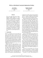
A B
Fig. 1. (A) Electron micrograph of a freeze-etched and Pt ⁄ C-shadowed preparation of a Gram-positive organism exhibiting a square (p4)
S-layer lattice. The bar corresponds to 100 nm. (B) Schematic drawing illustrating the various S-layer lattice types. In the oblique lattice, one
morphological unit (red) consists of one (p1) or two (p2) identical subunits. Four subunits constitute one morphological unit in the square
(p4) lattice type, whereas the hexagonal lattice type is either composed of three (p3) or six (p6) subunits. Modified from Sleytr et al [2] with
permission from Wiley-VCH.
Molecular construction kit based on S-layers U. B. Sleytr et al.
324 FEBS Journal 274 (2007) 323–334 ª 2006 The Authors Journal compilation ª 2006 FEBS
surface. The proteinaceous subunits of S-layers are
aligned either in lattices with oblique (p1, p2), square
(p4), or hexagonal (p3, p6) symmetry (Fig. 1B) with a
center-to-center spacing of the morphological units of
3–35 nm [2,8]. Hexagonal lattice symmetry is pre-
dominant among archaea [3]. S-Layers are highly por-
ous protein lattices with a surface porosity of 30–70%.
As S-layers are mostly composed of identical species of
subunits, they exhibit pores of identical size and mor-
phology [2,3,5]. However, in many protein lattices, two
or more distinct classes of pores with diameters in the
range 2–8 nm have been identified.
Results of amino-acid analysis and estimation of the
secondary structure by comparison of protein sequence
data and CD measurement of various S-layer proteins
can be summarized as follows. There are large amounts
of glutamic acid and aspartic acid ( 15 mol%), a
high lysine content ( 10 mol%), and large amounts
of hydrophobic amino acids ( 40–60 mol%). The
hydrophilic and hydrophobic amino acids do not form
extended clusters, and, in most S-layer proteins,
20% of the amino acids are organized as a-helix and
40% occur as b-sheets. The aperiodic folding and
b-turn content may vary between 5% and 45%. In
general, most S-layer subunits are weakly acidic
proteins with isoelectric points in the range 4–6, except
the S-layer proteins of lactobacilli and that of
Methanothermus fervidus. Post-translational modifica-
tions include cleavage of N-terminal or C-terminal
fragments, phosphorylation, and glycosylation of
amino-acid residues [13]. The latter is a remarkable
characteristic of many archaeal and some bacterial
S-layer proteins. The glycan chains and linkages differ
significantly from those of eukaryotes [14].
Self-assembly of S-layer proteins
One of the most fascinating properties of isolated
native and recombinant S-layer proteins is their ability
to form free-floating self-assembly products in solution
(flat sheets, cylinders or spheres), to recrystallize into
extended monomolecular layers on solid supports, at
the air ⁄ water interface and on lipid films, and to cover
liposomes and nanocapsules completely (Fig. 2) [5,8].
The reassembly occurs after removal of the disrupting
agent used in the dissolution and isolation procedure.
In general, complete disintegration of S-layer lattices
into their constituent protein subunits on bacterial
cells can be achieved using high concentrations of cha-
otropic agents (e.g. guanidine hydrochloride, urea), by
lowering or raising the pH, or by applying metal-
chelating agents or cation substitution [5]. The forma-
tion of self-assembled arrays is only determined by
the amino-acid sequence of the polypeptide chains
and consequently the tertiary structure of the S-layer
protein species. In various S-layer proteins from Bacil-
lacaea it has been shown that significant portions of
the C-terminal or N-terminal part can be deleted with-
out loss of the capability of the subunits for lattice
formation [15]. Further, for the S-layer protein of
Bacillus sphaericus CCM 2177 (SbpA), truncation of
the amino-acid sequence led to a change in the S-layer
lattice type from square (p4) to oblique (p1) lattice
symmetry [16].
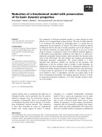
Fig. 2. Schematic drawing of the isolation of
native S-layer proteins from bacterial cells
and the reassembly of native and recombin-
ant S-layer proteins into crystalline arrays in
suspension, on a solid support, at the air ⁄
water interface and on a planar lipid film,
and on liposomes or nanocapsules. An
example of S-layer proteins reassembling
with hexagonal (p6) lattice symmetry is
shown here. Modified from Pum et al. [8],
with permission from Springer.
U. B. Sleytr et al. Molecular construction kit based on S-layers
FEBS Journal 274 (2007) 323–334 ª 2006 The Authors Journal compilation ª 2006 FEBS 325
In suspension, the formation of S-layer self-assembly
products starts by rapid nucleation of small oligomeric
precursors, which subsequently form extended
aggregates by a much slower self-assembly process.
Depending on the S-layer protein species used and on
the environmental conditions, double layers in back-to-
back orientation may be formed. On solid supports,
lattice formation is started after the attachment of
nucleation sites, which may be oligomeric S-layer pro-
tein or small self-assembly products from solution, and
is continued by lateral growth in all directions until the
proceeding front lines of the growing domains meet
[12]. In this way, a closed mosaic of monocrystalline
domains is formed. Depending on the S-layer proteins
species used, on the environmental conditions and, in
particular, on the surface properties of the support
(e.g. hydrophobicity, surface charge), the size of the
individual domains may be up to 20 lm in diameter.
Genetics of S-layer proteins and their
interaction with secondary cell wall
polymers
Several nanobiotechnological applications and functio-
nalization of S-layer lattices depend on the recrystalli-
zation of S-layer lattices in defined orientation with
respect to their inner and outer surface. In this con-
text, detailed information on the genetics of S-layer
proteins and mechanisms determining in vivo S-layer
assembly is important.
As S-layer proteins represent an important class of
secreted proteins, numerous S-layer genes from bac-
teria and archaea have been sequenced and cloned
[4,13]. For S-layer proteins of Gram-positive bacteria
at least, common structural organization principles
have been identified. A cell-wall-targeting domain was
found at the N-terminal region of many S-layer pro-
teins, which mediates binding to a specific hetero-
polysaccharide, termed secondary cell wall polymer
(SCWP), by a lectin-type binding. For Gram-positive
bacteria, at least two types of binding mechanism
between the N-terminal region of S-layer proteins and
SCWPs have been described [16–18]. With respect to
the first binding mechanism, so-called S-layer-homol-
ogy (SLH) motifs, each comprising about 55 amino
acids, recognize a distinct type of pyruvylated SCWP
as the correct anchoring structure. The second binding
mechanism has been described for Geobacillus stearo-
thermophilus wild-type strains and is characterized by
the interaction of a nonpyruvylated SCWP containing
the negatively charged 2,3-dideoxydiacetamidomanno-
samine uronic acid with a highly conserved N-terminal
region lacking an SLH domain. However, the cell-
wall-targeting domain is not necessarily located in the
N-terminal region of the S-layer protein. Well-docu-
mented examples of C-terminal anchoring are the
S-layer proteins of Lactobacillus acidophilus ATCC
4556 and Lactobacillus crispatus [7].
To elucidate the structure–function relationship of
distinct segments of S-layer proteins and to determine
which amino-acid positions are surface-located and
accessible, different strategies were pursued. In a first
approach, N-terminally and C-terminally truncated
forms were produced, and their self-assembly and
recrystallization properties investigated [16,19,20].
The S-layer protein of Geoacillus stearothermophilus
PV72 ⁄ p2 (SbsB) could be characterized by its two
functionally and structurally separated parts, namely
the specific SCWP-binding domain defined by the three
consecutive SLH motifs and the larger C-terminal part
responsible for formation of the crystalline array [21].
Interestingly, the deletion of even fewer than 15 C-ter-
minal amino acids resulted in completely water-soluble
forms of SbsB [15,19]. In contrast with SbsB, the
S-layer proteins, SbsC of Geobacillus stearothermophi-
lus ATCC 12980 and SbpA, turned out to be highly
tolerant to deletions, as up to 179 and 237 amino
acids, respectively, could be deleted at the C-terminus
without interfering with lattice formation [16,20].
Another attempt to find which amino-acid positions in
the primary sequence are located on the outer surface
of the subunits, inside the pores, or at the subunit to
subunit interface was seen in a cysteine scanning muta-
genesis study with screening of the accessibility of the
introduced cysteine residue in soluble, self-assembled
and recrystallized S-layer proteins [19].
As, to date, no structural model at atomic resolution
of any S-layer protein is available, elucidation of the
3D structure of S-layer proteins by X-ray crystallogra-
phy is also being pursued. This can be explained by (a)
the molecular mass of the S-layer subunits being too
large for NMR analysis, (b) their high tendency to
form 2D lattices preventing the formation of isotropic
3D crystals required for X-ray crystallography, and (c)
the very low solubility of isolated subunits. First, 3D
crystallization studies were carried out with water-
soluble N-terminally or C-terminally truncated forms
of SbsC. For the C-terminally truncated form, recom-
binant SbsC
31-844
, crystals that diffracted to a resolu-
tion of 3 A
˚
using synchrotron radiation could be
obtained [22]. Native and heavy atom derivative data
confirmed the results of the secondary-structure predic-
tion, which indicated that the N-terminal region com-
prising the first 257 amino acids is mainly organized as
a-helices, whereas the middle and C-terminal parts of
SbsC consist of loops and b-sheets. Information on the
Molecular construction kit based on S-layers U. B. Sleytr et al.
326 FEBS Journal 274 (2007) 323–334 ª 2006 The Authors Journal compilation ª 2006 FEBS
3D structure of S-layer proteins would open up the
possibility of rationally designing S-layer fusion pro-
teins incorporating functional domains, for example
within the pore areas of the protein lattice.
As cell surface components can generally be consid-
ered to be nonconservative structures that determine
the interaction between the living cell and its environ-
ment, the observation of phenotypic S-layer variation
was not surprising. S-Layer variation has been repor-
ted to occur in pathogens as well as in nonpathogens
and leads to the synthesis of alternate S-layer proteins,
either by the expression of complete (silent) S-layer
genes or by recombination of partial coding sequences
[7]. In pathogens, altered cell surface properties prob-
ably protect the cells from the lytic activity of the
immune system. In Campylobacter fetus, only the
C-terminal part of the S-layer protein is exchanged,
and the N-terminal region and the S-layer-specific lipo-
polysaccharides remain conserved. In nonpathogens,
S-layer variation is often induced in response to envi-
ronmental stress factors such as increased oxygen
supply [23]. In the strain variants, expression of a com-
pletely new S-layer protein is accompanied by synthesis
of a different type of SCWP, and S-layer variation can
also lead to a change in the lattice type. At the
molecular level, S-layer variation in G. stearothermo-
philus PV72 was found to be based on DNA rear-
rangements between the chromosome and a naturally
occurring megaplasmid. Regarding the development of
S-layer-deficient phenotypes, the importance of inser-
tion sequence elements has been demonstrated for at
least three different organisms [7].
S-Layer fusion proteins and
applications
For the production of nanoscale building blocks for
the bottom-up fabrication of bio-inspired materials
with designed properties, genetic approaches are cur-
rently used for the construction of functional S-layer
fusion proteins [6,7]. S-Layer fusion proteins based on
the S-layer proteins, SbsB, SbsC and SbpA, incorpor-
ate an accessible N-terminal SCWP-binding domain,
the self-assembly domain, as well as a biologically act-
ive sequence fused to the C-terminus. After heterolo-
gous expression of the genes encoding chimeric S-layer
proteins in Escherichia coli, it could be shown that the
self-assembling properties of the S-layer protein moiety
as well as the functionality of the fused sequences were
retained in all S-layer proteins (Fig. 3).
In order to build up functional monomolecular
S-layer protein lattices on artificial solid supports such
as gold, silicon, glass, indium tin oxide or polymers
(Fig. 3A,B), the surface has to be functionalized with
covalently attached chemically modified SCWP, to
which the S-layer fusion proteins bind with their N-ter-
minal part, leaving the C-terminal part with the fused
functional sequence exposed to the environment [6].
Such chimeric S-layers recrystallized on solid supports
in defined orientation (Fig. 3C) should find application
in diagnostics and biochip technology (laboratory-
on-a-chip), as well as for the development of specific
cell targeting and delivery systems [2,4,6–8,24].
In a first approach, S-layer fusion proteins compri-
sing the C-terminally truncated form, recombinant
(r)SbpA
31-1068
, and the hypervariable region of heavy
chain camel antibodies recognizing lysozyme or a pros-
tate-specific antigen (PSA) were constructed [25]. PSA
is a useful marker for screening for potential prostate
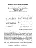
A
B
C
Self-assenbling part of
(truncated) S-layer protein
Functionality of
S-layer fusion
protein
Fig. 3. Digital image reconstructions of transmission electron micro-
graphs of negatively stained preparations of (A) the native S-layer
protein, SbsB, from Geobacillus stearothermophilus PV72 ⁄ p2 and
(B) the streptavidin fusion protein. In the lattice of the fusion pro-
tein (B), the streptavidin heterotetramers show up as additional
mass (arrows). Bars correspond to 10 nm. (C) Schematic illustration
of the self-assembling parts of S-layer fusion proteins and their
well-oriented functional domains. Such arrays theoretically provide
the highest possible order (spatial control, orientation and position)
of functional domains at the nanometer level. The knights (grey)
reassemble the functional domains (antigens, enzymes, antibodies,
ligands, etc.) and the cut squares (yellow) represent the S-layer.
Modified from Pum et al. [8], with permission from Springer.
U. B. Sleytr et al. Molecular construction kit based on S-layers
FEBS Journal 274 (2007) 323–334 ª 2006 The Authors Journal compilation ª 2006 FEBS 327
cancer patients. This fusion protein specific for PSA
was recrystallized as a monolayer on SCWP-precoated
gold chips and used as sensing layer in biochips
for surface plasmon resonance (SPR) spectroscopy
(Fig. 4). It turned out that at least three of four poss-
ible PSA molecules were bound per morphological unit
of the square S-layer lattice [25]. To summarize,
S-layer fusion proteins incorporating camel antibody
sequences can be considered key elements for the
development of sensing layers for label-free detection
systems such as SPR, surface acoustic wave or quartz
crystal microbalance, in which the binding event can
be measured directly by mass increase without the
need for any labeled molecule.
The genes encoding the chimeric S-layer proteins,
rSbsC
31-920
⁄ Bet v1 and rSbpA
31-1068
⁄ Bet v1, carrying
the major birch pollen allergen Bet v1 at the C-terminus
maintained the ability to self-assemble as well as the
functionality of the fused allergen to bind the Bet
v1-specific monoclonal mouse antibody [26]. In a recent
study, rSbsC
31-920
⁄ Bet v1 was shown to contain all rele-
vant B and T cell epitopes of Bet v1. Compared with
free Bet v1, in cells of birch pollen-allergic individuals,
the histamine-releasing capacity caused by the fusion
protein was significantly reduced, and no Th2-like
immune response was observed like after stimulation
with free Bet v1 [27]. Owing to its immunomodulating
capacity, this fusion protein is generally considered to
be a novel approach to specific treatment of allergic
diseases (e.g. as carrier ⁄ adjuvants in the design of
vaccines for immunotherapy of type 1 allergy).
In order to generate a universal affinity matrix for
binding of any kind of biotinylated molecules, mini-
mum-sized core streptavidin (118 amino acids) was
fused to either N-terminal or C-terminal positions of
rSbsB or the C-terminus of rSbpA
31-1068
[24]. After
expression of the chimeric genes in E. coli and isolation
of the fusion proteins from the host cells, a refolding
protocol was applied to obtain heterotetramers consist-
ing of one chain of the respective fusion protein and
three chains of core streptavidin (Fig. 3A,B). Fluores-
cence quenching of tryptophan residues in the binding
pockets of streptavidin confirmed that the biotin-bind-
ing capacity of soluble heterotetramers correlated with
the molecular mass of the appropriate biotinylated pro-
teins. As a first approach, monolayers generated by
recrystallization of rSbpA
31)1068
⁄ streptavidin hetero-
tetramers on plain gold chips or on gold chips precoat-
ed with thiolated SCWP were exploited for binding of
biotinylated oligonucleotides (30-mers). SPR studies
revealed that nonspecific adsorption of fluorescently
labeled oligonucleotides (15-mers) carrying two mismat-
ches was negligible. Moreover, it could be demonstra-
ted that the hybridization reaction with complementary
fluorescently labeled oligonucleotides carrying one mis-
match followed the Langmuir isotherm. The detection
limit for hybridized oligonucleotides was found to be in
the picomolar range [24]. To conclude, hybridization
experiments with biotinylated and fluorescently labeled
oligonucleotides using SPR spectroscopy indicated that
a functional sensor surface could be generated by
recrystallization of heterotetramers on gold chips. Such
promising structures that combine self-assembly prop-
erties of an S-layer protein with the biotin-binding
properties of streptavidin should find numerous
applications in (nano)biotechnology (Fig. 3C).
The S-layer fusion protein, rSbpA
31-1068
⁄ ZZ, incor-
porates two copies of the Fc-binding domain (ZZ), a
synthetic analog of the IgG-binding domain of pro-
tein A from Staphylococcus aureus [28]. As demonstra-
ted by SPR, the amount of human IgG that could be
bound on the native rSbpA
31-1068
⁄ ZZ monolayer was
slightly higher than on the rSbpA
31-1068
⁄ ZZ monolayer
cross-linked with the bifunctional imidoester dimethylpi-
melinimidate. On average, 66% of the theoretical sat-
uration capacity of a planar surface was covered by IgG
with the Fab regions in the condensed state. Novel bio-
compatible microparticles for the microsphere-based
detoxification system used for extracorporeal blood
purification of patients suffering from autoimmune
A
B
Fig. 4. (A) Schematic drawing illustrating a biosensor based on
rSbpA
31-1068
⁄ cAb-PSA-N7 recrystallized on gold chips precoated
with thiolated SCWP. The monomolecular protein lattice was able
to specifically bind PSA on the outermost surface. (B) Sensorgram
showing association (—) and dissociation (– ) – –) between PSA
and rSbpA
31-1068
⁄ cAb-PSA-N7 recrystallized on a SCWP-coated gold
chip. The sensorgram indicates specific binding of PSA to the
S-layer fusion protein.
Molecular construction kit based on S-layers U. B. Sleytr et al.
328 FEBS Journal 274 (2007) 323–334 ª 2006 The Authors Journal compilation ª 2006 FEBS
disease could be generated by recrystallization of the
S-layer fusion protein on SCWP-coated microbeads [28].
As only a marginal loss of IgG-binding capacity has
been observed, the protein lattice was cross-linked with
dimethylpimelinimidate. Thus, regeneration of the
binding matrix under acidic pH conditions could be
performed and the affinity microparticles could
be repeatedly used for extracorporeal blood purification.
S-Layer-stabilized lipid membranes and
liposomes
The advances in genome mapping have revealed that
approximately one-third of all genes in an organism
encode membrane proteins such as pores, ion channels,
receptors, and membrane-bound enzymes. These pro-
teins are key factors in the cell’s metabolism and thus
are the preferred target for pharmaceuticals. Currently
more than 60% of drugs consumed act on membrane
proteins [10,29]. Therefore not only biological mem-
branes, but also the biomimetic approach to generate
stabilized lipid membranes with functional membrane
proteins has attracted much interest in recent years.
The latter poses a challenge to apply membrane pro-
teins as key elements in drug discovery, protein–ligand
screening, and biosensors.
A promising approach for the generation of biomi-
metic membrane systems includes stabilization of lipid
membranes with S-layer lattices (Fig. 5). These com-
posite structures mimic the supramolecular assembly
B
Teflon
aperture
A
water
Patch clamp
pipette
air
E
(a)
(b)
(c)
(d)
(e)
(f)
D
C
porous
support
solid support
Fig. 5. Schematic illustrations of various S-layer-supported lipid membranes. (A) Bilayer lipid membranes have been generated across an
aperture of a patch clamp pipette by the tip-dip method, and a closed S-layer has been recrystallized from the aqueous subphase. In (B), a
folded or painted membrane has been generated to span a Teflon aperture. Subsequently S-layer protein can be injected into one or both
compartments (not shown) whereby the protein self-assembles to form closely attached S-layer lattices on the lipid membranes. (C) On an
S-layer ultrafiltration membrane (SUM), a lipid membrane can be generated by a modified Langmuir–Blodgett (LB) technique. As a further
option, a closed S-layer lattice can be attached on the external side of the SUM-supported lipid membrane (left part). (D) Solid supports can
be covered by a closed S-layer lattice, and subsequently lipid membranes can be generated using combinations of the LB and Langmuir–
Schaefer technique, and vesicle fusion. As shown in (C), a closed S-layer lattice can be recrystallized on the external side of the solid-suppor-
ted lipid membrane (left part). (E) Schematic drawing of (a) an S-liposome with entrapped water-soluble (blue) or lipid-soluble (brown)
functional molecules and (b) functionalized by reconstituted integral proteins. S-Liposomes can be used as immobilization matrix for
functional molecules (e.g. IgG) by direct binding (c) or immobilization via the Fc-specific ligand protein A (d), or biotinylated ligands can be
bound to the S-liposome via the biotin–streptavidin system (e). (f) Alternatively, liposomes can be coated with genetically modified S-layer
proteins incorporating functional domains. Modified from Sleytr et al. [4] and Sleytr et al [9], with permission from Wiley-VCH.
U. B. Sleytr et al. Molecular construction kit based on S-layers
FEBS Journal 274 (2007) 323–334 ª 2006 The Authors Journal compilation ª 2006 FEBS 329
of archaeal cell envelopes, as the latter are composed
of a cytoplasmic membrane and a closely associated
S-layer as exclusive cell wall component [3,4,10,11]. In
this biomimetic architecture, artificial lipids replace the
cytoplasmic membrane, and isolated or recombinant
S-layer proteins derived from Bacillaceae are attached
on either one or both sides of the lipid membrane.
Closed S-layer lattices can be generated for instance at
Langmuir lipid monolayers, planar lipid membranes
(Fig. 5A–D), liposomes (Fig. 5E), emulsomes, or lipid-
coated nanocapsules [8,10,11,30].
The interaction of S-layer proteins with lipid mole-
cules has been demonstrated to be noncovalent. Elec-
trostatic interaction between exposed carboxy groups
on the inner face of the S-layer lattice and the zwitter-
ionic lipid head groups is primarily responsible for the
binding and defined orientation of the S-layer subunits
to form a closed lattice structure. For such an align-
ment, it has been suggested that there are at least two
to three contact points between the lipid film and the
attached S-layer protein. Therefore, only a few lipid
molecules are anchored via their head groups to pro-
tein domains on the S-layer lattice, whereas the
remaining scores of lipid molecules diffuse freely in the
membrane between the pillars consisting of anchored
lipid molecules. Because of its widely retained fluid
characteristic, this nano-patterned type of lipid mem-
brane is also referred to as ‘semifluid membrane’ [31].
However, most importantly, the attached S-layer
lattices reveal no effect on the hydrophobic lipid
acyl chains. Thus, S-layer lattices constitute unique
scaffolding for lipid membranes [11,29,30]. This
observation has been confirmed by the functional
reconstitution of transmembrane proteins.
Supported lipid membranes can also be generated
on S-layer ultrafiltration membranes (SUMs; Fig. 5C),
with S-layer fragments deposited in microfiltration
membranes as an active filtration layer [32], and on
S-layer-coated electrodes or structured silicon chips
(Fig. 5D), with the S-layer as a stabilizing biomimetic
layer [10,11]. S-Layer-coated silicon chips with
attached lipid membranes are also referred to as lipid
chips, and, in combination with microfluidics, these
platforms constitute a prerequisite for the Laboratory-
on-a-Chip technology [33]. The electrochemical proper-
ties of S-layer-supported lipid membranes on porous
and solid supports (Fig. 5C,D, respectively) are com-
parable with those of free-standing lipid membranes
(Fig. 5A,B). In addition, membranes on S-layer-
covered gold electrodes exhibit remarkable long-term
robustness of up to 1 week, which is far from feasible
with any other stabilization technique.
The functionality of lipid membranes resting on
SUMs and S-layer-covered gold electrodes has been
demonstrated by reconstituting the pore-forming pro-
tein, a-hemolysin, and the membrane-active peptides,
alamethicin, gramicidin A, and valinomycin. Recently,
even single-pore recordings have been performed with
a-hemolysin and gramicidin A reconstituted in S-layer-
supported lipid membranes (Fig. 6) [10,11,30]. These
results demonstrate that the biomimetic approach of
copying the supramolecular architecture of archaeal
cell envelopes opens up new possibilities for exploiting
functional lipid membranes at the mesoscopic and
macroscopic level. Moreover, this technology has the
potential to initiate a broad spectrum of developments
in many areas such as diagnostics, high-throughput
screening for drug discovery, sensor technology, and
Fig. 6. Opening and closing of single gramicidin pores. Gramicidin has been incorporated into an S-layer ultrafiltration membrane-supported
lipid membrane composed of the main tetraether phospholipid isolated from Thermoplasma acidophilum. The schematic drawing on the left
indicates the formation of dimeric gramicidin pores. Because of pore formation, a cascaded increase in electric current is observed. The
schematic drawing on the right indicates the dissociation of gramicidin dimers. Gramicidin monomers do not form pores, and thus the con-
ductance decreases cascaded for each dissociated gramicidin dimer. Conditions: 1
M KCl; pH ¼ 5.8; V
m
¼ +150 mV; T ¼ 22 °C.
Molecular construction kit based on S-layers U. B. Sleytr et al.
330 FEBS Journal 274 (2007) 323–334 ª 2006 The Authors Journal compilation ª 2006 FEBS
electronic or optical devices, and might even find appli-
cation in DNA sequencing [10,11,29,30].
Artificial lipid vesicles termed liposomes are widely
used as delivery systems for enhancing the efficiency of
various biological active molecules. S-Layer-coated
liposomes (S-liposomes) represent simple model sys-
tems resembling features of archaeal cell or virus enve-
lopes (Fig. 5E). S-Layer proteins, once crystallized on
liposomes, can be cross-linked and exploited as a mat-
rix for the covalent attachment of functional molecules
as required for drug-targeting or immunodiagnostic
assays ([5,34] and references therein).
In a recent study, the fusion protein, rSbpA
31)1068
⁄
EGFP, carrying the sequence of enhanced green
fluorescent protein (EGFP) at the C-terminus was
recrystallized on positively charged liposomes. Because
of the ability of EGFP to fluoresce, positively charged
liposomes coated with rSbpA
31-1068
⁄ EGFP represent a
useful tool for visualizing the uptake of S-liposomes
into eukaryotic cells, which can then be investigated by
confocal scanning microscopy [34].
The high mechanical and thermal stability of
S-layer-coated liposomes and the possibility for immo-
bilizing or entrapping biologically active molecules [34]
reveal a broad application potential, particularly as
carrier and ⁄ or drug delivery, as artificial virus, and,
for medicinal applications, as drug targeting system or
in gene therapy (Fig. 5E) [4,8,10,11,30].
Controlled binding of nanoparticles
The S-layer approach provides, for the first time, a
biologically based fabrication technology for the
self-assembly of molecular catalysts, templates and
scaffolds for the generation of ordered large-scale
nanoparticle arrays for applications in electronic or
optic devices.
Wet chemical approaches
From the investigation of mineral formation by bac-
teria in natural environments, it is apparent that
S-layer lattices can also be used in wet chemical pro-
cesses for the precipitation of metal ions from solution
[35,36]. The first example of exploitation of this tech-
nique was the precipitation of CdS on S-layer lattices
composed of the S-layer protein from G. stearothermo-
philus NRS 2004 ⁄ 3a variant 1 (SgsE), and on SbpA
[35]. CdS and gold nanoparticles were 4–5 nm in size,
and their superlattice resembled the oblique lattice
symmetry of SgsE. SbpA was also used to generate
superlattices of 4–5 nm-sized gold particles [36]. Gold
nanoparticles were formed either by reduction of the
metal salt with H
2
S or under the electron beam in a
transmission electron microscope. The latter approach
is technologically important, as it allows the definition
of areas where nanoparticles are eventually formed. As
determined by electron diffraction, the gold nano-
particles were crystalline but their ensemble was not
crystallographically aligned. Later, the wet chemical
approach was used in the formation of Pd–, Ni–, Pt–,
Pb–, and Fe–nanoparticle arrays. Recently, small spot
X-ray photoelectron emission spectroscopy was used
to characterize the elemental composition of the nano-
clusters. This technique demonstrated that they consis-
ted primarily of elemental gold [37].
Binding of preformed nanoparticles
Although wet chemical methods lead to crystalline
arrays of nanoparticles with spacing in register with
the underlying S-layer lattice, they do not allow parti-
cle size to be precisely controlled and hence the con-
tact distances of neighboring particle surfaces, both of
which are important for studying and exploiting quan-
tum phenomena. Thus, the binding of preformed
nanoparticles into regular arrays on S-layers has
significant advantages over wet chemical approaches
for the development of nanoscale electronic devices.
Studies on binding of biomolecules, such as enzymes
and antibodies [4,5], to S-layers have shown that
metallic and semiconducting nanoparticles can be
bound in regular arrangements on S-layers [38]. This
is because, with S-layers, the properties of a single
constituent unit are replicated with the periodicity of
the lattice and thus define the characteristics of the
whole 2D array. The pattern of bound molecules
often reflects the lattice symmetry, the size of the
morphological units, and the physicochemical proper-
ties of the array. Specific binding of molecules to
S-layer lattices can be induced by noncovalent and
covalent forces. For example, the distribution of net
negatively charged domains on S-layers could be visu-
alized by electron-microscopic methods after labeling
with positively charged topographical markers, such
as polycationic ferritin (diameter 12 nm) [2]. Recently,
citrate-stabilized negatively charged gold nanoparticles
5 nm in diameter were bound by electrostatic interac-
tions at the inner S-layer face of SbpA, forming
extended superlattices [38]. Furthermore, amino func-
tionalized CdSe has been bound to the outer face
of SbpA monolayers recrystallized on hydrophobic
silicon surfaces after carbodi-imide activation of the
carboxy residues.
A major breakthrough in the regular binding of
metallic and semiconducting nanoparticles was
U. B. Sleytr et al. Molecular construction kit based on S-layers
FEBS Journal 274 (2007) 323–334 ª 2006 The Authors Journal compilation ª 2006 FEBS 331
achieved by the successful design and expression of
S-layer–streptavidin fusion proteins [7,15]. The 2D pro-
tein crystals displayed streptavidin in defined repetitive
spacing, and were capable of binding d-biotin and
biotinylated peptides and proteins, in particular fer-
ritin. Furthermore, metal-binding peptides can also be
used as fusion partners in the design and expression of
S-layer fusion proteins.
Conclusions and perspectives
The cross-fertilization of biology, genetics, chemistry,
and material sciences is opening up a great variety of
opportunities in nanobiotechnology and biomimetics.
S-Layer research has clearly demonstrated that nature
provides most elegant examples of nanometer-sized,
molecular self-assembly systems (Figs 1 and 2) as
required for generating bottom-up nanostructured
materials, which may be exploited at mesoscopic and
macroscopic levels. Of particular importance is the
possibility to change the natural properties of S-layer
proteins by genetic manipulation and to incorporate
single-functional or multifunctional domains in S-layer
lattices (Fig. 3). The spontaneous association of identi-
cal S-layer (glyco)protein subunits in suspension or on
surfaces or interfaces (Fig. 2) results in stable well-
defined isoporous matrices, which can be considerably
strengthened by introducing intermolecular and ⁄ or
intramolecular bonds. Moreover, even native S-layers
of some archaea can assemble and function in the
most extreme environmental conditions in which the
particular organisms are able to dwell (e.g. tempera-
tures up to 120 °C, pH ¼ 0, concentrated salt solution,
high hydrostatic pressure).
An important line of development is the combining
of S-layer and lipid membrane technologies (Fig. 5).
This biomimetic approach – copying the supramolecu-
lar principle of cell envelopes of archaea or envelopes
of a great variety of viruses – is expected to enable the
exploitation of functional principles of lipid mem-
branes at mesoscopic and even macroscopic levels and
the development of new targeting and delivery systems
[10,11]. Although, the development of S-layer technol-
ogies has focused primarily on life sciences, an import-
ant field of future applications relates to nonlife
sciences, including S-layer lattice-induced biominerali-
zation, nonlinear optics, molecular electronics, and cat-
alysts [39]. The dramatic reduction in size is one of the
benefits promised by molecular electronics. Individual
molecules are hundreds of times smaller than the
smallest features conceivably attainable by semicon-
ductor technology. For this reason, electronic devices
constructed from molecules promise to be thousands
of times smaller than their semiconductor-based coun-
terparts. S-Layer lattices have proven to be perfect
matrices for the generation of large-scale 2D arrays of
metallic and semiconducting nanoparticles. Further-
more, one of the big promises of molecular electronics
is based on the possibility of fabricating 3D structures
because of the extremely low power consumption com-
pared with silicon-based integrated circuitry. Finally,
some of the molecular electronic approaches are inher-
ently digital and immune to soft errors, and some are
inherently nonvolatile. As with many other technol-
ogies based on unique biomaterials, many further areas
in which S-layer lattices are of relevance may yet
emerge (e.g. neoglycobiology) [14].
Acknowledgements
This work was supported by the Austrian Science
Fund (FWF, projects P16295-B10, P17170-B10, and
P18510-B12), by the Erwin Schro
¨
dinger Society for
Nanosciences, by the Austrian Federal Ministry of
Transport, Innovation and Technology (MNA-Net-
work), by the EU project NAS-SAP, and by the US
Air Force Office of Scientific Research (projects
F49620-03-1-0222 and FA9550-06-1-0208).
References
1 Sleytr UB (1975) Heterologous reattachment of regular
arrays of glycoproteins on bacterial surfaces. Nature
257, 400–402.
2 Sleytr UB, Messner P, Pum P & Sa
´
ra M (1999) Crystal-
line bacterial cell surface layers (S layers): from supra-
molecular cell structure to biomimetics and
nanotechnology. Angew Chem Int Ed 38, 1034–1054.
3 Sleytr UB & Beveridge TJ (1999) Bacterial S layers.
Trends Microbiol 7, 253–260.
4 Sleytr UB, Huber C, Pum D, Schuster B, Ilk N & Egel-
seer EM (2007) Nanobiotechnology with S-layer pro-
teins. FEMS Microbiol Lett in press.
5 Sleytr UB, Sa
´
ra M, Pum D & Schuster B (2005) Crys-
talline bacterial cell surface layers (S layers): a versatile
self-assembly system. In Supra-Molecular Polymers
(Ciferri A, ed.), pp. 583–616, 2nd edn. CRC Press,
Taylor & Francis Group, Boca Raton, FL.
6Sa
´
ra M, Egelseer EM, Huber C, Ilk N, Pleschberger M,
Pum D & Sleytr UB (2006) S Layer proteins: potential
applications in nano (bio) technology. In Microbial
Bionanotechnology: Biological Self-Assembly Systems
and Biopolymer-Based Nanostructures (Rehm B, ed.),
pp. 307–338. Horizon Scientific Press, Hethersett,
Norwich, UK.
7Sa
´
ra M, Egelseer EM, Pum D, Schuster B & Sleytr UB
(2006) Genetically engineered S-layer proteins, S-layer-
Molecular construction kit based on S-layers U. B. Sleytr et al.
332 FEBS Journal 274 (2007) 323–334 ª 2006 The Authors Journal compilation ª 2006 FEBS
specific polysaccharides and lipids as components of a
versatile molecular construction kit for application in
nanobiotechnology. In Nanobiotechnology: Bioinspired
Devices and Materials of the Future (Shoseyov O & Levy
I, eds). Science of Knowledge Press, Little Falls, NJ,
in press.
8 Pum D, Sa
`
ra M, Schuster B & Sleytr UB (2006) Bacter-
ial surface layer proteins: a simple but versatile biologi-
cal self-assembly system in nature. In Nanotechnology:
Science and Computation (Chen J, Jonoska N & Rozen-
berg G, eds), pp. 277–290. Springer, Berlin, Heidelberg.
9 Sleytr UB, Egelseer EM, Pum D & Schuster B (2004)
S-Layers. In Nanobiotechnologie: Concepts, Methods and
Perspectives (Niemeyer CM & Mirkin CA, eds), pp.
77–92. Wiley-VCH-Verlag, Weinheim.
10 Schuster B & Sleytr UB (2006) Biomimetic S-layer sup-
ported lipid membranes. Curr Nanosci 2 , 143–152.
11 Schuster B & Sleytr UB (2005) 2D-protein crystals
(S-layers) as support for lipid membranes. In Advances
in Planar Lipid Bilayers and Liposomes (Tien TH &
Ottova A, eds), Vol. 1, pp. 247–293. Elsevier Science,
Amsterdam.
12 Gyo
¨
rvary ES, Stein O, Pum D & Sleytr UB (2003) Self-
assembly and recrystallization of bacterial S layer pro-
teins at silicon supports imaged in real time by atomic
force microscopy. J Microsc 212, 300–306.
13 Sa
´
ra M & Sleytr UB (2000) S-Layer proteins. J Bacter-
iol 182, 859–868.
14 Scha
¨
ffer C & Messner P (2004) Surface-layer glycopro-
teins: an example for the diversity of bacterial glycosyla-
tion with impacts on nanobiotechnology. Glycobiology
14, 31R–42R.
15 Moll D, Huber C, Schlegel B, Pum D, Sleytr UB &
Sa
´
ra M (2002) S-layer-streptavidin fusion proteins as
template for nanopatterned molecular arrays. Proc Natl
Acad Sci USA 99, 14646–14651.
16 Huber C, Ilk N, Ru
¨
nzler D, Egelseer EM, Weigert S,
Sleytr UB & Sa
´
ra M (2005) The three S-layer-like
homology motifs of the S-layer protein SbpA of Bacillus
sphaericus CCM 2177 are not sufficient for binding to
the pyruvylated secondary cell wall polymer. Mol
Microbiol 55, 197–205.
17 Scha
¨
ffer C & Messner P (2005) The structure of second-
ary cell wall polymers: how Gram-positive bacteria stick
their cell walls together. Microbiology 151, 643–651.
18 Mader C, Huber C, Moll D, Sleytr UB & Sa
´
ra M
(2004) Interaction of the crystalline bacterial cell surface
layer protein SbsB and the secondary cell wall polymer
of Geobacillus stearothermophilus PV72 assessed by real-
time surface plasmon resonance biosensor technology.
J Bacteriol 186, 1758–1768.
19 Howorka S, Sa
´
ra M, Wang Y, Kuen B, Sleytr UB,
Lubitz W & Bayley H (2000) Surface-accessible residues
in the monomeric and assembled forms of a bacterial
surface layer protein. J Biol Chem 275, 37876–37886.
20 Jarosch M, Egelseer EM, Huber C, Moll D, Mattanov-
ich D, Sleytr UB & Sa
´
ra M (2001) Analysis of the struc-
ture-function relationship of the S layer protein SbsC of
Bacillus stearothermophilus ATCC 12980 by producing
truncated forms. Microbiology 147, 1353–1363.
21 Sa
´
ra M, Pum D, Huber C, Ilk N, Pleschberger M &
Sleytr UB (2006) Nanoscale patterning of S-layer pro-
teins as a natural self-assembly system. In Biological and
Pharmaceutical Nanomaterials. Nanotechnologies for the
Life Sciences (Kumar C, ed.), Vol. 2, pp. 219–252.
Wiley-VCH, Weinheim.
22 Pavkov T, Oberer M, Egelseer EM, Sa
´
ra M, Sleytr UB
& Keller W (2003) Crystallization and preliminary
structure determination of the C-terminal truncated
domain of the S-layer protein SbsC. Acta Crystallogr
D Biol Crystallogr 59, 1466–1468.
23 Sa
´
ra M & Sleytr UB (1994) Comparative studies of
S layer proteins from Bacillus stearothermophilus strains
expressed during growth in continuous culture under
oxygen-limited and non-oxygen-limited conditions.
J Bacteriol 176, 7182–7189.
24 Huber C, Liu J, Egelseer EM, Moll D, Knoll W, Sleytr
UB & Sa
´
ra M (2006) Heterotetramers formed by an
S-layer-streptavidin fusion protein and core-streptavidin
as nanoarrayed template for biochip development. Small
2, 142–150.
25 Pleschberger M, Saerens D, Weigert S, Sleytr UB,
Muyldermans S, Sara M & Egelseer EM (2004) An
S-layer heavy chain camel antibody fusion protein for
generation of a nanopatterned sensing layer to detect
the prostate-specific antigen by surface plasmon reso-
nance technology. Bioconjug Chem 15, 664–671.
26 Ilk N, Vo
¨
llenkle C, Egelseer EM, Breitwieser A, Sleytr
UB & Sara M (2002) Molecular characterization of the
S-layer gene, sbpA, of Bacillus sphaericus CCM 2177
and production of a functional S-layer fusion protein
with the ability to recrystallize in a defined orientation
while presenting the fused allergen. Appl Environ Micro-
biol 68, 3251–3260.
27 Breitwieser A, Egelseer EM, Moll D, Ilk N, Hotzy C,
Bohle B, Ebner C, Sleytr UB & Sa
´
ra M (2002) A
recombinant bacterial cell surface (S-layer)–major birch
pollen allergen-fusion protein (rSbsC ⁄ Bet v1) maintains
the ability to self-assemble into regularly structured
monomolecular lattices and the functionality of the
allergen. Protein Eng 15, 243–249.
28 Vo
¨
llenkle C, Weigert S, Ilk N, Egelseer EM, Weber
V, Loth F, Falkenhagen D, Sleytr UB & Sa
´
ra M
(2004) Construction of a functional S-layer fusion pro-
tein comprising an immunoglobulin G-binding domain
for development of specific adsorbents for extracor-
poreal blood purification. Appl Environ Microbiol 70,
1514–1521.
29 Schuster B (2005) Biomimetic design of nano-patterned
membranes. Nanobiotechnology 1, 153–164.
U. B. Sleytr et al. Molecular construction kit based on S-layers
FEBS Journal 274 (2007) 323–334 ª 2006 The Authors Journal compilation ª 2006 FEBS 333
30 Schuster B & Sleytr UB (2000) S-layer-supported lipid
membranes. Rev Mol Biotechnol 74, 233–254.
31 Pum D & Sleytr UB (1994) Large-scale reconstitution
of crystalline bacterial surface layer proteins at the air ⁄
water interface and on lipid films. Thin Solid Films 244,
882–886.
32 Sa
´
ra M & Sleytr UB (1987) Production and characteris-
tics of ultrafiltration membranes with uniform pores
from two-dimensional arrays of proteins. J Membr Sci
33, 27–49.
33 Bayley H & Cremer PS (2001) Stochastic sensors
inspired by biology. Nature 413, 226–230.
34 Ilk N, Ku
¨
pcu
¨
S, Moncayo G, Klimt S, Ecker RC, Hofer-
Warbinek R, Egelseer EM, Sleytr UB & Sara M (2004) A
functional chimaeric S-layer-enhanced green fluorescent
protein to follow the uptake of S-layer-coated liposomes
into eukaryotic cells. Biochem J 379, 441–448.
35 Shenton W, Pum D, Sleytr UB & Mann S (1997) Bio-
crystal templating of CdS superlattices using self-
assembled bacterial S-layers. Nature 389, 585–587.
36 Dieluweit S, Pum D & Sleytr UB (1998) Formation of a
gold superlattice on an S-layer with square lattice sym-
metry. Supramol Sci 5 , 15–19.
37 Dieluweit S, Pum D, Sleytr UB & Kautek W (2005)
Monodisperse gold nanoparticles formed on bacterial
surface layers (S-layers) by electroless deposition. Mat
Sci Eng C-Bio S 25, 727–732.
38 Gyo
¨
rvary E, Schroedter A, Talapin DV, Weller H, Pum
D & Sleytr UB (2004) Formation of nanoparticle arrays
on S-layer protein lattices. J Nanosci Nanotechnol 4,
115–120.
39 Pum D & Sleytr UB (2006) S-layer proteins for assem-
bling ordered nanoparticle arrays. In Nanobioelectronics
for Electronics, Biology, and Medicine (Offenha
¨
usser
A & Rinaldi R, eds). In the series Nanostructure Science
and Technology (Lockwood D, ed.). Springer, Berlin,
in press.
Molecular construction kit based on S-layers U. B. Sleytr et al.
334 FEBS Journal 274 (2007) 323–334 ª 2006 The Authors Journal compilation ª 2006 FEBS