Báo cáo khoa học: Conformational stability of 17b-hydroxysteroid dehydrogenase from the fungus Cochliobolus lunatus pptx
Bạn đang xem bản rút gọn của tài liệu. Xem và tải ngay bản đầy đủ của tài liệu tại đây (731.08 KB, 11 trang )
Conformational stability of 17b-hydroxysteroid
dehydrogenase from the fungus Cochliobolus lunatus
Natas
ˇ
a Poklar Ulrih
1
and Tea Lanis
ˇ
nik Riz
ˇ
ner
2
1 Department of Food Science and Technology, University of Ljubljana, Slovenia
2 Institute of Biochemistry, University of Ljubljana, Slovenia
Proteins of the short-chain dehydrogenase ⁄ reductase
(SDR) superfamily are nonmetallo enzymes with
molecular masses between 25 and 35 kDa that function
as dimers or tetramers [1]. Although the SDR super-
family contains some 3000 members [2], 17b-hydroxy-
steroid dehydrogenase from the fungus Cochliobolus
lunatus (17b-HSDcl) is currently the only fungal
hydroxysteroid dehydrogenase member that has been
described; it has been purified, cloned and expressed in
Escherichia coli [3,4].
Under native conditions, both recombinant [4] and
natural [3] 17b-HSDcl form dimers. 17b-HSDcl is
homologous to some fungal reductases: versicolorin
reductases from Aspergillus parasiticus and Emericella
nidulans, which are involved in aflatoxin biosynthesis;
and 1,3,8-trihydroxynaphthalene reductases and 1,3,6,8-
tetrahydroxynaphthalene reductases from Magnaporthe
grisea, Ophiostoma floccosum and other fungi, which
are involved in melanin biosynthesis [4,5]. 1,3,8-Tri-
hydroxynaphthalene reductases and 1,3,6,8-tetra-
hydroxynaphthalene reductases catalyse an essential
reaction in the biosynthesis of melanin, a virulence fac-
tor of phyto-pathogenic fungi and of fungi pathogenic
to humans [6–9]. These enzymes are the biochemical
targets of several commercially important fungicides
that are used to prevent blast disease in rice plants
[9,10].
Despite extensive biochemical studies of 17b-HSDcl
[4,11–14], nothing is known about its conformational
stability. Structural and thermodynamic study of fun-
gal 17b-HSD may, therefore, contribute to a better
understanding of the functionality of homologous fun-
gal enzymes that are targets for the design of novel
antifungal agents. Indeed, 17b-HSDcl is often used as
Keywords
17b-hydroxysteroid dehydrogenase;
coenzyme NADPH binding; guanidine
hydrochloride; pH stability; urea
Correspondence
N. Poklar Ulrih, Department of Food Science
and Technology, Biotechnical Faculty,
University of Ljubljana, Jamnikarjeva 101,
1000 Ljubljana, Slovenia
Fax: +386 1 256 6298
Tel. +386 1 423 1161
E-mail:
(Received 17 May 2006, revised 20 June
2006, accepted 26 June 2006)
doi:10.1111/j.1742-4658.2006.05396.x
The functional activities of proteins are closely related to their molecular
structure and understanding their structure–function relationships remains
one of the intriguing problems of molecular biology. We investigated struc-
tural changes in 17b-hydroxysteroid dehydrogenase from the fungus
Cochliobolus lunatus (17b-HSDcl) induced by pH, temperature, salt, urea,
guanidine hydrochloride, and coenzyme NADPH binding. At 25 °C and
within the relatively narrow pH range of 7.0–9.0, 17b-HSDcl exists in its
native conformation as a dimer. This native conformation is thermally sta-
ble up to 40 °C in this pH range. At 25 °C and pH 2.0 in the presence of
150–300 mm NaCl, 17b-HSDcl forms soluble aggregates enriched in a-heli-
cal and b-sheet structures. At higher temperatures and NaCl concentra-
tions, these soluble aggregates start to precipitate. The denaturants urea
and guanidine hydrochloride unfold 17b-HSDcl at concentrations of 1.2
and 0.4 m, respectively. Binding of the coenzyme NADPH to 17b-HSDcl
causes local structural changes that do not significantly affect the thermal
stability of this protein.
Abbreviations
C
d
, concentration (of urea or GuHCl) at denaturation midpoint; DG°
d
, Gibbs free energy of denaturation; GuHCl, guanidine hydrochloride;
17b-HSDcl, 17b-hydroxysteroid dehydrogenase from the fungus Cochliobolus lunatus; DH°
vH
, van’t Hoff enthalpy of denaturation; SDR,
short-chain dehydrogenase ⁄ reductase; T
d
, temperature at denaturation midpoint.
FEBS Journal 273 (2006) 3927–3937 ª 2006 The Authors Journal compilation ª 2006 FEBS 3927
a model enzyme for the SDR superfamily, and fungal
17b-HSDcl exhibits 30% amino acid identity to
human 17b-HSD types 4 and 8 [12]. Typically, SDR
members share 15–30% residue identity in pairwise
comparison. Despite the low residue identities between
the different members, 3D structures revealed that the
folding pattern is conserved with largely superimposa-
ble peptide backbones [2]. The high similarities in these
structures thus indicate that studies on 17 b-HSDcl
might lead to a better understanding of the catalytic
mechanisms of human HSDs that are implicated in the
development of steroid-dependent forms of cancer, in
polycystic kidney disease, in the regulation of blood
pressure, in Alzheimer’s disease and in obesity [15–19].
This study provides the first description of the dena-
turation behaviour of dimeric 17b-HSDcl, in relation
to pH, temperature, ionic strength (NaCl), denaturants
[urea, guanidine hydrochloride (GuHCl)] and coen-
zyme binding (NADPH).
Results
Far-UV CD spectra of 17b-HSDcl at 25 °C
Representative far-UV CD spectra of 17b-HSDcl in
the pH range 1–14 are shown in Fig. 1A–C. The spec-
trum of 17b-HSDcl at neutral pH (7.0; Fig. 1A) is
characterized by negative CD bands near 208 and
222 nm and a positive band at 193 nm, which is typ-
ical of an a-helical structure [20]. Based on contin
analysis [21], at 25 °C in an aqueous solution at
pH 7.0, 17b-HSDcl contains 33 ± 1% a helix (a
H
),
52±1% b sheet (b
S
), 15 ± 1% b turn (b
T
), and 0%
aperiodic secondary structure (Table 1). These values
are in reasonable agreement with those predicted and
obtained from molecular modelling [11] and those
obtained from X-ray diffraction analysis [22]. Increas-
ing the pH from neutral to an alkaline pH of 13.0,
there was an alteration in the shape and intensity of
Fig. 1. Far- and near-UV CD spectra of 17b-HSDcl at different pH values. (A) Far-UV CD spectra of 17b-HSDcl in the pH range 13.2–6.5. (B)
Far-UV CD spectra of 17b-HSDcl in the pH range 6.5–2.8. (C) Far-UV CD spectra of 17b-HSDcl in the pH range 2.8–1.1. (D) Near-UV CD
spectra of 17b-HSDcl in the pH range 7.0–2.3.
Conformational stability of 17b-HSDcl N. Poklar Ulrih and T. Lanis
ˇ
nik Riz
ˇ
ner
3928 FEBS Journal 273 (2006) 3927–3937 ª 2006 The Authors Journal compilation ª 2006 FEBS
the far-UV CD spectrum that is typical of a base-
induced denaturation. At pH 13.2, 17b-HSDcl contains
6±1%a
H
,34±3%b
S
,11±1%b
T
and 49 ± 4%
aperiodic structure. Only one isodichroic point was
seen in the alkaline pH range, at 206.5 nm, implying
the presence of two different spectroscopic states of
17b-HSDcl despite numerous deprotonation ⁄ protona-
tion equilibria of the basic amino acid side chains in
the alkaline pH range (Fig. 1A). Lowering the pH
from neutral to an acidic pH of 1.1, there was a
decrease in ellipticity, and two isodichroic points were
seen at 207.5 and 206 nm (Fig. 1B,C), suggesting the
existence of three spectroscopically different states of
the protein. In the pH range from neutral to 2.0, there
was a transition into an acid-denatured state, and a
further decrease in pH from 2.0 to 1.0 induced an
increase in the amount of a
H
structure. At pH 1.1,
17b-HSDcl contains 12 ± 2% a
H
, whereas at pH 2.0,
it contains only 5 ± 1% a
H
(Table 1).
Near-UV CD spectra of 17b-HSDcl at 25 °C
The near-UV CD spectra of 17b-HSDcl did not
change significantly in the pH range 6.0–9.0, and they
are dominated by tryptophans and tyrosines (Fig. 1D).
This was similar to that seen for the far-UV CD spec-
tra, suggesting a stable tertiary structure of 17b-HSDcl
in this pH range.
pH titration of 17b-HSDcl at 25 °C
The titration curves derived from signals recorded at a
single wavelength at the indicated pH values are shown
in Fig. 2A. The changes in molar ellipticity followed at
222 nm over the pH range 7.0–10.0 are not significant,
and no changes were seen in the absorbance at 262 nm
in the pH range 7.0–9.0 (Fig. 2A). In the pH range
Table 1. The levels of specific secondary structure elements in
17b-HSDcl under different experimental conditions.
Condition a
H
(%) b
S
(%) b
T
(%) AP (%)
pH
(at 25 °C)
13.0 6 ± 1 34 ± 3 1 ± 2 59 ± 4
7.0 33 ± 1 52 ± 1 15 ± 1 0
6.5 37 ± 2 51 ± 1 12 ± 1 0
2.0 5 ± 1 20 ± 1 9 ± 1 66 ± 1
1.1 12 ± 2 19 ± 1 9 ± 3 60 ± 3
pH 2.0
300 m
M NaCl
25 °C 14±1 52±1 12±1 22±2
90 °C 0 ± 1 36 ± 4 10 ± 1 54 ± 2
pH 7.3
(at 25 °C)
NADH (R ¼ 1) 33 ± 1 46 ± 2 15 ± 2 6 ± 1
A
B
C
Fig. 2. Effects of pH on structural changes, electrophoretical prop-
erties and enzymatic activity of 17b-HSDcl at 25 °C. (A) pH effects
on molar ellipticity, followed at 222 nm (d), and absorbance, fol-
lowed at 262 nm (s)of17b-HSDcl. (B) Electrophoretic titration ana-
lysis of 17b-HSDcl in the pH range 3–9. (C) pH effect on enzymatic
activity of 17b-HSDcl.
N. Poklar Ulrih and T. Lanis
ˇ
nik Riz
ˇ
ner Conformational stability of 17b-HSDcl
FEBS Journal 273 (2006) 3927–3937 ª 2006 The Authors Journal compilation ª 2006 FEBS 3929
7.0–10.0, 17b-HSDcl has a defined tertiary structure,
as concluded from the near-UV CD spectra. A further
increase in pH from 10.0 to 12.0 caused a sharp
change in molar ellipticity followed at 222 nm, and in
absorbance at 262 nm, as a consequence of disruption
of the secondary and tertiary structures. We suggest
that deprotonation of six Tyr (pK
a
¼ 10.1), sixteen
Lys (pK
a
¼ 10.5) and nine Arg (pK
a
¼ 12.5) residues
causes loss of most of the secondary structure and a
complete loss of tertiary structure at pH values above
12.0. The pK
a
values given in parentheses refer to the
free amino acids in aqueous solution.
Acid titration would be expected to result in proto-
nation of 12 His (pK
a
¼ 6.0), 13 Glu (pK
a
¼ 4.3) and
14 Asp (pK
a
¼ 3.7) residues present in 17b-HSDcl. A
decrease in pH from 6.0 to 3.0 was accompanied by a
reduction in molar ellipticity followed at 222 nm,
which was seen as a single very broad transition,
whereas two transitions were seen in the absorbance at
262 nm (Fig. 2A). Because of the 12 His, 13 Glu
and 14 Asp residues in the primary structure of
17b-HSDcl, it is expected that the conformational sta-
bility of 17b-HSDcl will be strongly affected by ther-
modynamic coupling to the acid ⁄ base equilibrium of
the acidic amino acid residues.
Electrophoretic titration analysis
The results of electrophoretic titration analysis of
17b-HSDcl in the pH range 3.0–9.0 are shown in
Fig. 2B. Inspection of Fig. 2B reveals two transitions,
the first in the pH range pH 7–5, and the second in
the pH range 5–3. The observed transitions in the aci-
dic pH range are likely to be the result of decreasing
the net negative charge on the protein surface owing
to the protonation of 12 His, 13 Glu and 14 Asp resi-
dues. Inspection of Fig. 2B reveals that 17b-HSDcl in
the pH range 7–3.5 is moving through the gel in two
forms, most likely as a dimer and a monomer. In the
pH range 7.0–9.0 (pI ¼ 6.9) the net charge on the pro-
tein remains constant and 17b-HSDcl does not move
in the electric field.
Enzymatic activity
The results of enzymatic activity of recombinant
17b-HSDcl in the pH range 6.0–8.5 followed by oxida-
tion of 4-estrene-17b-ol-3 one to 4-estrene-3,17-dione
in the presence of NADP
+
are shown in Fig. 2C. The
pH optimum of 17b-HSDcl is between 7 and 8, as
shown previously for the enzyme, which was isolated
directly from the fungus Cochliobolus lunatus [3].
Acid-induced denatured state of 17b-HSDcl
at 25 °C
As seen in Fig. 2A, in the pH range 2.0–3.5, the molar
ellipticity at 222 nm and absorbance at 262 nm of
17b-HSDcl did not change significantly, suggesting
that 17b-HSDcl was in a stable conformation, acid-
unfolded state, designated U
A
. At pH values < 2.0,
molar ellipticity gradually increased (became more
negative), and at pH 1.1, 17b-HSDcl retained a more
ordered secondary structure than at pH 2.0 (Table 1).
The results from native PAGE electrophoresis of 17b-
HSDcl incubated in 1 m HCl indicate that the increase
in secondary structure at pH values below 2.0 is likely
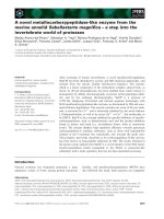
to be due to the oligomerization processes (Fig. 4A,B,
column 5).
Salt-induced effects on 17b-HSDcl at pH 2.0
The addition of a neutral electrolyte, such as NaCl,
could shield repulsion interactions in the highly posi-
tively charged 17b-HSDcl at pH 2.0. The effects on
the secondary structure of 17b-HSDcl of increasing
NaCl concentrations at pH 2.0 and 25 °C are shown
in Fig. 3A, and these indicate that an increase in NaCl
concentration induces an increase in b
S
structure. At
300 mm NaCl, the a
H
and b
S
structures increase from
5 to 14% and 20 to 52%, respectively (Table 1). The
inset in Fig. 3A shows the changes in molar ellipticity
followed at 215 nm vs. NaCl concentration. Clearly
the transition from the U
A
state (stable conformation,
acid unfolded) to the conformational state with a non-
native secondary structure, occurs in the NaCl concen-
tration range 150–300 mm. The thermal stability of
this newly formed soluble oligomers of 17b-HSDcl at
pH 2.0 in the presence of 300 mm NaCl (Fig. 4A,B,
column 4) was investigated by measuring the CD spec-
tra at different temperatures in the far-UV range
(Fig. 3B). The obtained results show that oligomers of
17b-HSDcl are resistant to temperatures up to 60 °C.
At temperatures above 60 °C, gradual changes in
molar ellipticity were seen (Fig. 3B), suggesting the
formation of larger aggregates or disruption of
17b-HSDcl oligomers containing significant amounts
of non-native secondary structure.
The results from native and SDS ⁄ PAGE confirm
that 17b-HSDcl at pH 2.0 does not oligomerize in the
absence of salt, whereas in the presence of 100 and
300 mm NaCl 17b-HSDcl is in oligomeric form
(Fig. 4A,B). The oligomers of 17b-HSDcl in the pres-
ence of NaCl are too large to penetrate into the gel
(Fig. 4A), whereas under the denaturing conditions
Conformational stability of 17b-HSDcl N. Poklar Ulrih and T. Lanis
ˇ
nik Riz
ˇ
ner
3930 FEBS Journal 273 (2006) 3927–3937 ª 2006 The Authors Journal compilation ª 2006 FEBS
they dissociate into monomeric form (Fig. 4B). At
NaCl concentrations > 350 mm, the precipitation of
17b-HSDcl has been observed in the cuvette.
Heat-induced denaturation of 17b-HSDcl at
pH 7.0–8.4
The pH titration of 17b-HSDcl at 25 °C indicated a
very narrow pH range at which the 17b-HSDcl tertiary
and secondary structures are intact (see above). In this
narrow pH range from 7 to 8.5, we studied thermal
stability of 17b-HSDcl from CD and UV melting
curves, from which the temperature of denaturation
(T
d
) and the van’t Hoff enthalpy of denaturation
(DH°
vH
)of17b-HSDcl were obtained, as described
previously [23]. DH°
vH
was calculated based on
assumption that the thermal denaturation of 17b-
HSDcl is a reversible two-state process. In fact, heat-
induced denaturation of 17b-HSDcl was reversible if
the experiment was stopped immediately after the
transition temperature. The degree of reversibility
decreased with the temperature to which the sample of
17b-HSDcl was heated (data not shown).
The thermodynamic profile of 17b-HSDcl is given in
Table 2. In the pH range 7.0–8.0, the T
d
and DH°
vH
of
17b-HSDcl do not change significantly. From the UV
melting profile, the T
d
for 17b-HSDcl at pH 7.5 is rel-
atively low, at 42.9 ± 0.5 °C; this is perhaps not so
surprising as it has been shown that the apparent opti-
mal temperature of enzymatic activity of 17b-HSDcl
is 28 °C at pH 7.0 [3]. Slightly higher T
d
and DH°
vH
values were determined from the CD melting profiles
(Table 2).
Urea and GuHCl effects on 17b-HSDcl at 25 °C
The effects of increasing concentrations of urea and
GuHCl on the structural properties of 17b-HSDcl were
investigated using far- and near-UV CD. The results
presented in Fig. 5A–D indicate that 17b-HSDcl
unfolds at very low concentrations of denaturants. The
denaturation concentrations at which half of the 17b-
HSDcl molecules are in the denaturated and a half in
the native state, C
d
, are 1.2 m for urea and 0.4 m for Gu-
HCl (determined from Fig. 5A,B). Because the denatu-
rant-induced unfolding of 17b-HSDcl was a reversible
two-state transition, it can be described in terms of its
equilibrium constant, K
d
°. From these K
d
° values, the
corresponding Gibbs free energies, DG
d
°,of17b-HSDcl
in solutions of different concentrations of urea and
GuHCl can be determined using the general relation:
DG
d
° ¼ –RT lnK
d
°. Numerous studies on urea and
GuHCl denaturation of proteins have shown that in the
denaturant concentration range in which the denatura-
tion of proteins can be followed, the DG
d
° of denatura-
tion can be expressed as a linear function of denaturant
concentration: DG
d
¼ DG
d
;H
2
O
mC [24,25]. For
17b-HSDcl, the calculated m-values at 25 °C and
pH 7.0 are )12.9 ± 0.6 kJÆLÆmol
)1
for urea and )14.4 ±
0.8 kJÆLÆmol
)1
for GuHCl, whereas corresponding val-
ues for are 15.3 ± 0.7 and 5.9 ± 0.3 kJÆmol
)1
, respect-
ively. These DG
d
;H
2
O
values obtained from analysing
the data for urea and GuHCl denaturation of 17b-
HSDcl give DG
d
;H
2
O
from GuHCl as threefold lower
than that from urea. Possible explanations for this pheno-
menon could arise from a lack of the native baseline
and the errors involved in the d ata c ollection and analysis.
AB
Fig. 3. Effects of NaCl on structural changes in 17b-HSDcl at pH 2.0. (A) NaCl concentration effects on far-UV CD spectra and molar ellipticity
followed at 215 nm (inset)of17b-HSDcl at pH 2.0. (B) Temperature effects on molar ellipticity followed at 235 nm (
) and 217 nm (s)of
17b-HSDcl in the presence of 300 m
M NaCl at pH 2.0 (in 10 mM glycine buffer).
N. Poklar Ulrih and T. Lanis
ˇ
nik Riz
ˇ
ner Conformational stability of 17b-HSDcl
FEBS Journal 273 (2006) 3927–3937 ª 2006 The Authors Journal compilation ª 2006 FEBS 3931
NADPH binding to 17b-HSDcl at 25 °C
The effects of the coenzyme NADPH on the confor-
mation of 17b-HSDcl were investigated at pH 7.3 in
NaCl ⁄ P
i
buffer. The influence of NADPH on the far-
UV CD spectrum of 17b-HSDcl at a molar ratio of
1 : 1 for NADPH ⁄ 17b-HSDcl (per monomeric unit)
can be seen in Fig. 6A. The changes in the secondary
structure of 17b-HSDcl after NADPH binding are not
significant (Fig. 6B), suggesting that the coenzyme
interrupts the structure of 17b-HSDcl only locally.
Binding of the coenzyme NADPH to 17b-HSDcl at
a molar ratio of 1 : 1 thermally stabilized 17b-HSDcl
for 0.6 ± 0.5 °C, whereas at higher NADH:17b-
HSDcl molar ratios, e.g. 5 : 1, the thermal stabiliza-
tion was 2.5 ± 0.5 °C. These data are in agreement
with the CD data, which indicate only minor struc-
tural rearrangements of 17b-HSDcl after NADPH
binding.
Discussion
For oligomeric proteins changes in secondary and ter-
tiary structures during native to denatured transitions
are usually accompanied by dissociation into subunits.
17b-HSDcl is a dimeric member of the SDR super-
family, in which neighbouring subunits are connected
via hydrophobic interactions and several salt bridges
involving amino acid residues His111 and Arg129. It
has been shown that Arg129 and His111 interact with
Asp121, Glu117 and Asp187 residues from the neigh-
bouring subunits [14]. Replacement of His111 with
Leu prevented dimer formation and caused loss of bio-
logical activity of 17b-HSDcl, whereas the His111Ala
mutation did not affect either dimerization or enzyme
activity. It has also been reported [3] and confirmed by
our measurements that 17b-HSDcl is active in the pH
range 7.0–9.0. The results reported here show that the
conformational changes coincide with the changes in
functional activity. A loss in enzymatic activity for
17b-HSDcl at pH values < 7.0, which is at first seen
as a slight change in the far-UV CD signal in the pH
range 7.0–6.0 and it is followed by significant change
in the CD and UV signal, can be ascribed to denatura-
tion of 17b-HSDcl. The results from electrophoretic
titration analysis show that 17b-HSDcl in the pH
range 7–3.5 moves through the gel in two forms.
Although the dimeric form of 17b-HSDcl is predomi-
nant, we proposed that partial dissociation is taking
place at pH values < 7.0 and it is likely to be induced
by protonation of the His111 residue that is involved
in dimerization. More precisely, it has been proposed
that dimerization takes place across the Q-axis and
involves the a-E and a-F helices of both subunits [14].
Our CD data show a slight increase in the amount of
a
H
structure in the pH range 7.0–6.5 and further sup-
port the results from electrophoretic titration analysis
that 17b-HSDcl partially dissociate into subunits at
pH values < 7.0.
Table 2. The thermodynamic profile of 17b-HSDcl obtained from UV
melting curves at different pH values in NaCl/P
i
buffer.
a
from CD
measurements. T
d
, temperature at denaturation midpoint; DT, the
width of the transition; DH°
vH
, van’t Hoff enthalpy of denaturation.
pH T
d
(°C) DT(°C) DH°
vH
(kJ ⁄ mol)
7.3
a
47.0 ± 0.5 7.2 ± 1 355 ± 30
7.0 41.8 ± 0.5 11.4 ± 1 289 ± 30
7.5 42.9 ± 0.5 11.7 ± 1 285 ± 30
8.0 42.8 ± 0.5 11.6 ± 1 288 ± 30
8.4 44.9 ± 0.5 12.0 ± 1 279 ± 30
BSA
A
12 3 4 5
st.B 12 3 4 5
Fig. 4. Native and SDS ⁄ PAGE electrophoretogram of 17b-HSDcl. A
total of 8 lg of recombinant 17b-HSDcl in the following solutions:
(1) NaCl ⁄ P
i
buffer, pH 7.3; (2) 10 mM glycine buffer, pH 2.0; (3)
10 m
M glycine buffer, 100 mM NaCl, pH 2.0; (4) 10 mM glycine buf-
fer, 300 m
M NaCl, pH 2.0 and (5) 1 M HCl were applied to (A)
native and (B) SDS ⁄ PAGE. BSA was used for comparison on native
PAGE. Prestained molecular markers were: 20, 26, 36, 47, 85 and
118 kDa, respectively.
Conformational stability of 17b-HSDcl N. Poklar Ulrih and T. Lanis
ˇ
nik Riz
ˇ
ner
3932 FEBS Journal 273 (2006) 3927–3937 ª 2006 The Authors Journal compilation ª 2006 FEBS
High aggregation ability is a conventional property
of non-native protein conformations. Examples include
aggregation following heat- or pH-induced denatura-
tion and aggregation of folding intermediates, and this
has been observed for numerous globular proteins.
Under conditions of extreme pH, the main forces that
unfold proteins are repulsion between charged groups
in the protein molecule. The 17b-HSDcl molecule is
highly charged (the absolute net charge at pH 2.0 esti-
mated from its amino acid composition is +31), and
therefore the acidic unfolded U
A
state does not aggre-
gate. The observed increase in molar ellipticity at pH
values < 2.0 indicates some electrostatically driven
structural changes in this protein molecule in response
to an increased concentration of Cl
–
ions [26], which is
further supported by the NaCl titration of 17b-HSDcl
at pH 2.0. After addition of salt, formation of b
S
structures is observed, as results of the oligomerization
processes. The presence of high concentrations of salt
has two effects on protein–protein interactions: First,
the presence of counterions around the charged groups
weakens the repulsion and allows intermolecular forces
become relatively strengthened and thus manifest
themselves [26]. In addition to this nonspecific effect of
salt as counterions, specific effects of salt on protein–
protein interactions indicate the presence of exposed
hydrophobic surfaces [26].
Additional information relating to the presence or
absence of a unique tertiary structure of a protein
molecule can be obtained from analysis of its urea-
and GuHCl-induced unfolding. Indeed, it has been
shown that the steepness of a denaturant-induced
unfolding curve depends strongly on whether a given
protein has a unique tertiary structure or whether it is
Fig. 5. Urea- and GuHCl-induced denaturation of 17b-HSDcl at 25 °C. (A) Urea and (B) GuHCl effects on far-UV and (C) near-UV CD spectra
of 17b-HSDcl. (D) Urea (d) and GuHCl (s) effects on molar ellipticity followed at 220 nm of 17b-HSDcl.
N. Poklar Ulrih and T. Lanis
ˇ
nik Riz
ˇ
ner Conformational stability of 17b-HSDcl
FEBS Journal 273 (2006) 3927–3937 ª 2006 The Authors Journal compilation ª 2006 FEBS 3933
already denatured and exists as a molten globule [27].
Urea and GuHCl induced unfolding of 17b-HSDcl at
C
d
values of 1.2 and 0.4 m, respectively. Similarly, a
C
d
value of 0.64 m was reported by Oppermann et al.
[28] for GuHCl denaturation of 3b ⁄ 17b-HSD from
Comamonas testosteroni, with a corresponding value of
15.1 kJÆmol
)1
which is comparable with our value of
15.3 kJÆmol
)1
[28]. Human placenta 17b-hydroxyster-
oid-dehydrogenase (17b-HSD type 1), which shares
21% amino acid identity with 17b-HSDcl and posses-
ses the same protein fold, has urea-induced conforma-
tional transitions with C
d
values of 1.5 and 5.8 m,
suggesting that the first transition is from the native
dimeric state to a molten-globule-like dimeric state,
and that the second transition leads to the fully dena-
tured state that is accompanied by the dissociation of
oligomeric molecules [29].
NADPH-dependent enzymes have one or two con-
served basic residues that interact electrostatically with
the ribose 2¢-phosphate group of the adenine nucleo-
tide of NADPH. One of these interacts with the sec-
ond glycine in the Gly-X-X-X-Gly-X-Gly motif, and
this is restricted to a Lys or Arg, which prevails in
lower organisms [30,31]. In 17b-HSDcl, Arg28 com-
pensates for the negative charge of the 2¢-phosphate
group of the adenine nucleotide of NADPH, whereas
Thr200 and Thr202 interact with the nicotinamide
moiety of NADPH [13]. Our results show that the co-
enzyme NADPH clearly binds to the free enzyme. The
dissociation constant, K
d
, of the enzyme-NADPH
complex of 1.6 lm has been previously determined by
fluorescence measurements at pH 8.0 in a 100 mm
phosphate buffer [13]. The changes in the secondary
structure of 17b-HSDcl after binding of NADPH are
not significant, although a slight increase in the aperio-
dic structure seen at the expense of b
S
structure is seen.
This local rearrangement of secondary structure does
not significantly affect the thermal stability of 17b-
HSDcl. Indeed, a previous report [28] that binding of
NAD
+
to 3b ⁄ 17b-HSD from C. testosteroni is influ-
enced by local structural changes, involving strand b
D
and turn b
A
to a
B
, supports our data.
In conclusion, our study indicates that 17b-HSDcl
is enzymatically active and thermodynamicly stable
over a narrow pH range, as would be the case for
other proteins in the SDR superfamily that function
as dimers or tetramers. The loss of enzymatic activ-
ity of 17b-HSDcl at pH values < 7.0 can be
ascribed to protonation ⁄ deprotonation equilibria of
numerous acidic amino acid residues causing the
denaturation of 17b-HSDcl. The combined results of
the unfolding of 17b-HSDcl suggest that it can take
on different conformational states at 25 °C, as sum-
marized by the scheme:
Agg(i) Agg(s) $ U
A
$ N
2
$ D
B
where Agg(i) is the insoluble aggregates of 17b-HSDcl
(pH £ 2.0 and concentration of NaCl > 300 m m);
Agg(s), soluble oligomers of 17b-HSDcl in the pres-
ence of salt (pH ¼ 2.0 and 150–300 mm NaCl); U
A
,
the acid-unfolded state (pH 2–3); N
2
, the native
dimeric state (pH 7–9); and D
B
, the base-denatured
state at pH > 10. Of note, the observed thermody-
namic stability of 17b-HSDcl at 25 °C, with a value of
15.3 kJÆmol
)1
(0.06 kJÆmol
)1
amino acid), is much
lower than for the majority of globular proteins
( 0.4 kJÆmol
)1
amino acid). The binding of the
Fig. 6. Coenzyme NADPH binding to 17b-HSDcl at 25 °C. (A) Coen-
zyme NADPH effects on far-UV CD spectra of 17b-HSDcl at 25 °C
at a molar ratio of 1 : 1. (B) The molar ellipticity of 17b-HSDcl at
220 nm, [Q]
220
, as a function of increasing NADPH concentration
expresed as molar ratio R ([NADPH] ⁄ [17b-HSDcl]) at 25 °C.
Conformational stability of 17b-HSDcl N. Poklar Ulrih and T. Lanis
ˇ
nik Riz
ˇ
ner
3934 FEBS Journal 273 (2006) 3927–3937 ª 2006 The Authors Journal compilation ª 2006 FEBS
coenzyme NADPH induces local structural reorganiza-
tion of 17b-HSDcl without significantly influencing this
thermal stability. These data thus show that the
absence of induction of thermal stability by NADPH
binding is the consequence of enthalpic compensation
of the disturbed intramolecular interactions by the
newly formed electrostatic and H-bonds with NADPH,
as was suggested by earlier structural modelling of
17b-HSDcl based on trihydroxynaphthalene reductase
from Magnaporthe grisea [11].
Experimental procedures
Materials
NADPH, glycine, hystidine and Mes were from Sigma-Ald-
rich (St. Louis, MO). GuHCl and urea were from Fluka
(Buchs, Switzerland). GuHCl was recrystallized from hot
ethanol before use. Na
2
HPO
4
and NaH
2
PO
4
were from
Kemika (Zagreb, Croatia), NaCl, NaOH and HCl for the
titration experiments and dimethyl formamide were from
Merck (Darmstadt, Germany).
Solutions
NADPH solutions in NaCl ⁄ P
i
buffer and double-distilled
water were prepared immediately before use. The concen-
trations of NADPH were determined spectrophotometrical-
ly at 340 nm and 25 °C, using an extinction coefficient of
65 000 m
)1
Æcm
)1
. NaCl ⁄ P
i
buffer (142.7 or 300 mm NaCl,
10 mm Na
2
HPO
4
, 1.8 mm NaH
2
PO
4
, pH 7.3), 10 mm gly-
cine buffer (pH 2.0), and double-distilled water were used
as solvents.
Recombinant 17b-HSDcl
Recombinant 17b-HSDcl was prepared as a GST-fusion
protein in the Escherichia coli strain JM107 and purified
using affinity chromatography on glutathione–Sepharose,
followed by cleavage with thrombin, as described previously
[4]. The 17b-HSDcl concentration was determined spectro-
photometrically at 280 nm and 25 °C using a calculated
extinction coefficient [32] of 20 065 m
)1
Æcm
)1
(per monomer
unit).
Enzymatic assay
Enzymatic activity of the recombinant 17b-HSDcl was
determined spectrophotometrically at 340 nm. We fol-
lowed the oxidation of 4-estrene-17b-ol-3-one to 4-est-
rene-3,17-dione (Sigma-Aldrich) in the presence of
NADP
+
in 100 mm phosphate buffer, pH 6–8.5 at 25 °C.
In all of the experiments, 1% dimethyl formamide was
present to enhance substrate solubility. The time course
of absorbance was measured for 450 s and initial veloci-
ties were determined.
Denaturation studies
Temperature-, pH-, urea- and GuHCl-induced denaturation
of 17b-HSDcl were monitored using a combination of UV
spectrophotometry and CD measurements.
UV spectrophotometry
The UV light absorbance values were measured using a
Hewlett Packard 8453 UV-VIS spectrophotometer (Hewlett-
Packard GmbH, Waldbronn, Germany) equipped with a
thermoelectrically controlled cell holder. The UV-absorption
spectra of 17b-HSDcl were measured after titration with
HCl or NaOH. The absorbance vs. temperature profiles of
17b-HSDcl were measured at 280 nm. Temperature was
increased in 1 °C increments, and protein samples were
allowed to equilibrate for 1 min at each temperature setting.
The temperature-induced denaturation profiles of 17b-HSDcl
were used to determine transition temperatures, T
d
and van’t
Hoff enthalpy of denaturation, DH
v.H
. The subsequent
absorbance vs. temperature profiles of 17b-HSDcl were used
to assess the reversibility of the protein denaturation.
CD
The CD spectra were measured using an AVIV Model
62 A DS spectropolarimeter (AVIV Associates, Lakewood,
NJ) equipped with a thermoelectrically controlled cell
holder. Cuvettes with path lengths of 1 mm were used for
far- (200–260 nm) and 10 mm for near-UV (240–310 nm)
measurements, with 17b-HSDcl concentrations of 0.25 and
0.75 mgÆmL
)1
, respectively. CD spectra were recorded
either as functions of temperature, between 10 and 95 °Cin
5 °C steps, or of pH (HCl or NaOH) or ion (NaCl), denat-
urant (urea, GuHCl) and coenzyme (NADPH) concentra-
tions. The latter were achieved by incremental additions of
the relevant reagents to a cuvette containing a known
amount of 17b-HSD at 25 °C. The mean residue ellipticity,
[h]
k
, was calculated by using the relation:
½H
k
¼
M
O
H
k
100 c 1
ð1Þ
in which M
o
is the mean residue molar mass (107.0 gÆmol
)1
for 17b-HSDcl), Q
k
is the measured ellipticity in degrees, c
is the concentration in gÆmL
)1
, and l is the path length in
decimetres. [ Q]
k
was expressed in deg cm
2
Ædmol
)1
. Secon-
dary structure content was calculated from the far-UV CD
spectra using contin software [21]. The degree of reversibil-
ity of the urea- and GuHCl-induced unfolding of
17b-HSDcl was determined by measuring the CD spectrum
of 17b-HSDcl after dialysing the sample of protein in urea
or GuHCl against buffer solution.
N. Poklar Ulrih and T. Lanis
ˇ
nik Riz
ˇ
ner Conformational stability of 17b-HSDcl
FEBS Journal 273 (2006) 3927–3937 ª 2006 The Authors Journal compilation ª 2006 FEBS 3935
pH measurements
The pH titration was performed at 25 °C using a 10-lL
Hamilton syringe (Hamilton Co., Reno, NV) equipped with
a Chaney adapter. The pH of protein solutions was meas-
ured separately using a pH-meter (model MA 5705; Iskra,
Slovenia) with an Ag ⁄ AgCl combination microelectrode
(Mettler, Toledo, Spain). The absolute error of pH meas-
urements was ± 0.01 pH units.
Native PAGE
Samples of 17 b-HSDcl in: (a) 10 mm glycine buffer, pH 2.0
in the absence or presence of NaCl (100, 300 mm); (b) 1 m
HCl; (c) in NaCl ⁄ P
i
buffer, pH 7.3. were analysed by native
PAGE. Discontinous native PAGE was performed on 9%
acrylamide gels according to Ornstein-Davis in 25 mm Tris,
190 mm glycine pH 8.3 buffer [33]. Continous native PAGE
was carried out on 9% acrylamide gels in 30 mm histidine,
30 mm Mes buffer pH 6.1 [34]. Following electrophoresis at
150 V, the proteins were stained using Coomassie Brilliant
Blue.
SDS/PAGE
Samples of 17b-HSD (see above) were analysed also using
SDS ⁄ PAGE. Eight micrograms were denatured in sample
buffer and then applied to 12% acrylamide gel [35]. After
ectrophoresis at 200 V, the proteins were stained using
Coomassie Brilliant Blue.
Electrophoretic titration analysis
Electrophoretic titration curve analysis is a 2D technique
that allows the determination of protein charge characteris-
tics. It was performed using a PhastGel IEF 3–9 media. In
the first dimension, the stable linear pH gradient was
generated. The gel was then rotated 90° clockwise and
17b-HSDcl was applied perpendicularly to the pH gradient
across the middle of the gel. After electrophoresis, the gel
was stained with Coomassie Brilliant Blue and documented.
The electrophoresis was performed in a PHAST System
(Amersham Pharmacia Biotech, Uppsala, Sweden), accord-
ing to the manufacturer’s instructions [36].
Acknowledgements
We thank Professor Tigran V. Chalikian of the Uni-
versity of Toronto, Canada, in whose laboratory the
CD measurements were performed, Mrs Melita Ana
Mac
ˇ
ek for performing the UV-titration experiments
and Mrs Irena Paves
ˇ
ic
ˇ
from Department of Biology at
Biotechnical Faculty of the University of Ljubljana for
performing the electrophoretic titration analysis.
References
1 Oppermann U, Filling C, Hult M, Shafgat N, Wu X,
Lindh M, Shafqat J, Nordling E, Kallberg Y, Persson B
et al. (2003) Short-chain dehydrogenases ⁄ reductases
(SDR): the 2002 update. Chem Biol Interact 143 ⁄ 144,
247–253.
2 Kallberg Y, Oppermann U, Jornvall H & Persson B
(2002) Short-chain dehydrogenase ⁄ reductase (SDR)
relationships: a large family with eight clusters common
to human, animal, and plant genomes. Protein Sci 11,
636–641.
3 Lanis
ˇ
nik Rizˇ ner T, Z
ˇ
akelj-Mavric
ˇ
M, Plemenitas
ˇ
A&
Zorko M (1996) Purification and characterization of
17b-hydroxysteroid dehydrogenase from the filamentous
fungus Cochliobus lunatus. J Steroid Biochem Mol Biol
59, 205–214.
4 Lanis
ˇ
nik Rizˇ ner T, Moeller G, Thole HH, Z
ˇ
akelj-Mav-
ric
ˇ
M & Adamski J (1999) A novel 17b-hydroxysteroid
dehydrogenase in the fungus Cochliobus lunatus: new
insights into the evolution of steroid-hormone signal-
ling. Biochem J 337, 425–431.
5 Lanis
ˇ
nik Rizˇ ner T, Stojan J & Adamski J (2001)
Searching for the physiological function of 17b-hy-
droxysteroid dehydrogenase from the fungus Cochliobo-
lus lunatus: studies of substrate specificity and
expression analysis. Mol Cell Endocrinol 171, 193–198.
6 Andersson A, Jordan D, Schneider G & Lundqvist Y
(1996) Crystal structure of the ternary complex of 1,3,8-
trihydroxynaphthalene reductase from Magnaporthe gri-
sea with NADPH and an active-site inhibitor. Structure
4, 1161–1170.
7 Thompson JE, Basarab GS, Andersson A, Lundqvist Y
& Jordan DB (1997) Trihydroxynaphthalene reductase
from Magnaporthe grisea: realization of an active center
inhibitor and elucidation of the kinetic mechanism.
Biochemistry 36, 1852–1860.
8 Thompson JE, Fahnestock S, Farral L, Liao D, Valent
B & Jordan DB (2000) The second naphthol reductase
of fungal melanin biosynthesis in Magnaporthe grisea –
tetrahydroxynaphthalene reductase. J Biol Chem 275,
34867–34872.
9 Liao D, Thompson JE, Fahnestock S, Valent B & Jor-
dan DB (2001) A structural account of substrate and
inhibitor specificity differences between two naphthol
reductases. Biochemistry 40, 8696–8704.
10 Jordan DB, Basarab GS, Liao D, Johnson WMP,
Winzenberg KN & Winkler DA (2001) Structure-based
design of inhibitors of the rice blast fungal enzyme tri-
hydroxynaphthalene reductase. Mol Graph Model 19,
434–447.
11 Lanis
ˇ
nik Rizˇ ner T, Adamski J & Stojan J (2000)
17b-Hydroxysteroid dehydrogenase from Cochliobolus
lunatus: model structure and substrate specificity. Arch
Biochem Biophys 384, 255–262.
Conformational stability of 17b-HSDcl N. Poklar Ulrih and T. Lanis
ˇ
nik Riz
ˇ
ner
3936 FEBS Journal 273 (2006) 3927–3937 ª 2006 The Authors Journal compilation ª 2006 FEBS
12 Lanis
ˇ
nik Rizˇ ner T, Stojan J & Adamski J (2001)
17b-Hydroxysteroid dehydrogenase from the fungus
Cochliobolus lunatus: structural and functional aspects.
Chem Biol Interact 130–132, 793–803.
13 Kristan K, Stojan J, Mo
¨
ller G, Adamski J & Lanis
ˇ
nik
Rizˇ ner T (2005) Coenzyme specificity in fungal 17b-hy-
droxysteroid dehydrogenase. Mol Cell Endocrinol 241,
80–87.
14 Kristan K, Deluca D, Adamski J, Stojan J & Lanis
ˇ
nik
Rizˇ ner T (2005) Dimerization and enzymatic activity of
fungal 17b-hydroxysteroid dehydrogenase from the
short-chain dehydrogenase ⁄ reductase superfamily. BMC
Biochem 6, 28.
15 Fomitcheva J, Baker ME, Anderson E, Lee GY & Aziz
N (1998) Characterization of Ke 6, a new 17 beta-
hydroxysteroid dehydrogenase, and its expression in
gonadal tissues. J Biol Chem 273, 22664–22671.
16 Li A, Tedde R, Krozowski ZS, Pala A, Li KX, Shackle-
ton CH, Mantero F, Palermo M & Stewart PM (1998)
Molecular basis for hypertension in the ‘type II variant’
of apparent mineralocorticoid excess. Am J Hum Genet
63, 370–379.
17 He XZ, Merz G, Mehta P, Schultz H & Yang SY (1999)
Human brain short chain l-3-hydroxyacyl coenzyme A
dehydrogenase is a single-domain multifunctional enzyme
– characterization of a novel 17 beta-hydroxysteroid
dehydrogenase. J Biol Chem 274, 15014–15019.
18 Lin SX, Han Q, Azzi A, Zhu D, Gangloff A & Camp-
bell RL (2000) 3D structure of human estrogenic 17
beta-HSD1: binding with various steroids. J Steroid
Biochem Mol Biol 69, 425–429.
19 Masuzaki H, Peterson J, Shinyama H, Morton NM,
Mullins JJ, Seckl JR & Flier JS (2001) A transgenic
model of visceral obesity and the metabolic syndrome.
Science 294, 2071–2072.
20 Sreerama N & Woody RW (2000) Circular dichroism of
peptides and proteins. In Circular Dichroism: Principles
and Applications (Berova N, Nakanishi K & Woody
RW, eds), pp. 601–620. Wiley, New York.
21 Provencher SW & Glo
¨
ckner J (1981) Estimation of
globular protein secondary structure from circular
dichroism. Biochemistry 20, 33–37.
22 Cassetta A, Budefeld T, Lanis
ˇ
nik Rizˇ ner T, Kristan K,
Stojan J & Lamba D (2005) Crystallization, X-ray dif-
fraction analysis and phasing of 17b-hydroxysteroid
dehydrogenase from the fungus Cochliobolus lunatus.
Acta Crystallogr F61, 1032–1034.
23 Poklar N, Lah J, Salobir M, Mac
ˇ
ek P & Vesnaver G
(1997) pH and temperature-induced molten globule-like
denatured states of equinatoxin II: a study by
UV-melting, DSC, far- and near-UV CD spectroscopy
and ANS fluorescence. Biochemistry 36, 14345–14352.
24 Schellman JA (1978) Solvent denaturation. Biopolymers
17, 1305–1322.
25 Green RF & Pace CN (1974) Urea and guanidine
hydrochloride denaturation of ribonuclease, lysozyme,
a-chymotrypsin, and b-lactoglobulin. J Biol Chem 249,
5388–5393.
26 Goto Y & Fink AL (1989) Conformational states of
beta-lactamase: molten-globule states at acidic and alka-
line pH with high salt. Biochemistry 28, 945–952.
27 Uversky VN, Kutyshenko VP, Protasova NY, Rogov
VV, Vassilenko KS & Gudkov AT (1996) Circularly
permuted dihydrofolate reductase possesses all the prop-
erties of the molten globule state, but can resume func-
tional tertiary structure by interaction with its ligand.
Protein Sci 5, 1844–1851.
28 Opperman UCT, Filling C, Berndt KD, Persson B,
Ladenstein R & Jo
¨
rnvall H (1997) Active site directed
mutagenesis of 3b ⁄ 17b-hydroxysteroid dehydrogenase
establishes differential effects on short-chain dehydroge-
nase ⁄ reductase reactions. Biochemistry 36, 34–40.
29 Mendoza-Herandez G & Rendon JL (1996) Human pla-
cental estradiol 17b-dehydrogenase: structural and cata-
lytic changes during urea-denaturation. Biochem
Biophys Acta 1297, 219–227.
30 Nakanishi M, Kakumoto M, Matsuura K, Deyashiki
Y, Tanaka N, Nonaka T, Mitsui Y & Hara A (1996)
Involvement of two basic residues (Lys-17 and Arg-39)
of mouse lung carbonyl reductase in NADP(H) binding
and fatty acid activation: site-directed mutagenesis and
kinetic analyses. J Biochem 120, 257–263.
31 Tanaka N, Nonaka T, Tanabe T, Yoshimoto T, Tsuru
D & Mitsui Y (1996) Crystal structures of the binary
and ternary complexes of 7 alpha-hydroxysteroid dehy-
drogenase from Escherichia coli. Biochemistry 35, 7715–
7730.
32 Pace CN, Vajdos LF, Grimsley G & Gray T (1995)
How to measure and predict the molar absorption coef-
ficient of a protein? Protein Sci 4, 2411–2423.
33 Laemmli UK (1970) Cleavage of structural proteins
during the assembly of the head of bacteriophage T4.
Nature 227, 680–685.
34 McLelain T (1982) Electrophoresis buffers for polyacryl-
amide gels at various pH. Anal Biochem 126, 94–99.
35 Ornstein L (1964) Disc electrophoresis I. Ann NY Acad
Sci 121, 321.
36 Pharmacia LKB Biotechnology (1987) Pharmacia Phast-
System Users’ Manual. Pharmacia LKB Biotechnology,
Uppsala, Sweden.
N. Poklar Ulrih and T. Lanis
ˇ
nik Riz
ˇ
ner Conformational stability of 17b-HSDcl
FEBS Journal 273 (2006) 3927–3937 ª 2006 The Authors Journal compilation ª 2006 FEBS 3937