Báo cáo khoa học: Ki-1⁄57 interacts with PRMT1 and is a substrate for arginine methylation pptx
Bạn đang xem bản rút gọn của tài liệu. Xem và tải ngay bản đầy đủ của tài liệu tại đây (971.87 KB, 16 trang )
Ki-1
⁄
57 interacts with PRMT1 and is a substrate for
arginine methylation
Dario O. Passos
1,2
, Gustavo C. Bressan
1,2
, Flavia C. Nery
1,3
and Jo
¨
rg Kobarg
1,2,3
1 Centro de Biologia Molecular Estrutural, Laborato
´
rio Nacional de Luz Sı
´
ncrotron, Campinas, Brazil
2 Departamento de Bioquı
´
mica, Universidade Estadual de Campinas, Brazil
3 Departamento Gene
´
tica e Evoluc¸a˜o, Universidade Estadual de Campinas, Brazil
Ki-1 ⁄ 57 was initially identified by the cross-reactivity
of the anti-CD30 mAb Ki-1 [1–5]. Initial studies on
the Ki-1 ⁄ 57 protein antigen itself revealed that it is
associated with Ser ⁄ Thr protein kinase activity [3] and
that it is located in the cytoplasm, at the nuclear pores
and in the nucleus, where it is frequently found in
association with the nucleolus and other nuclear bodies
[4]. Because Ki-1 ⁄ 57 was also found to bind to hya-
luronan and other negatively charged glycosaminogly-
cans, such as chondroitin sulfate, heparan sulfate and
RNA, although with lower affinity, it was also named
intracellular hyaluronan binding protein 4 (IHABP4)
Keywords
cellular localization; mapping;
post-translational modification; protein
arginine methylation; regulatory protein
Correspondence
J. Kobarg, Centro de Biologia Molecular
Estrutural, Laborato
´
rio Nacional de Luz
Sı
´
ncrotron, Rua Giuseppe Ma
´
ximo Scolfaro
10.000, C.P. 6192, 13084-971 Campinas – SP,
Brazil
Fax: +55 19 3512 1006
Tel: +55 19 3512 1125
E-mail:
(Received 3 May 2006, revised 6 June
2006, accepted 27 June 2006)
doi:10.1111/j.1742-4658.2006.05399.x
The human 57 kDa Ki-1 antigen (Ki-1⁄ 57) is a cytoplasmic and nuclear
protein, associated with Ser ⁄ Thr protein kinase activity, and phosphorylat-
ed at the serine and threonine residues upon cellular activation. We have
shown that Ki-1 ⁄ 57 interacts with chromo-helicase DNA-binding domain
protein 3 and with the adaptor ⁄ signaling protein receptor of activated
kinase 1 in the nucleus. Among the identified proteins that interacted with
Ki-1 ⁄ 57 in a yeast two-hybrid system was the protein arginine-methyl-
transferase-1 (PRMT1). Most interestingly, when PRMT1 was used as bait
in a yeast two-hybrid system we were able to identify Ki-1 ⁄ 57 as prey
among 14 other interacting proteins, the majority of which are involved in
RNA metabolism or in the regulation of transcription. We found that
Ki-1 ⁄ 57 and its putative paralog CGI-55 have two conserved Gly ⁄ Arg-rich
motif clusters (RGG ⁄ RXR box, where X is any amino acid) that may be
substrates for arginine-methylation by PRMT1. We observed that all
Ki-1 ⁄ 57 protein fragments containing RGG ⁄ RXR box clusters interact
with PRMT1 and are targets for methylation in vitro. Furthermore, we
found that Ki-1 ⁄ 57 is a target for methylation in vivo. Using immunofluo-
rescence experiments we observed that treatment of HeLa cells with an
inhibitor of methylation, adenosine-2 ¢ ,3¢-dialdehyde (Adox), led to a reduc-
tion in the cytoplasmic immunostaining of Ki-1 ⁄ 57, whereas its paralog
CGI-55 was partially redistributed from the nucleus to the cytoplasm upon
Adox treatment. In summary, our data show that the yeast two-hybrid
assay is an effective system for identifying novel PRMT arginine-methyla-
tion substrates and may be successfully applied to other members of the
growing family of PRMTs.
Abbreviations
Act D, actinomycin D; Adox, adenosine-2¢,3¢-dialdehyde; Daxx, Fas-binding protein; GST, glutathione S-transferase; IHABP4, intracellular
hyaluronan binding protein 4; Ki-1 ⁄ 57, 57 kDa Ki-1 antigen; PKC, protein kinase C; PRMT, protein arginine methyl transferase; RACK1,
receptor of activated kinase 1; RGG ⁄ RXR box, glycine ⁄ arginine-rich motif (where X is any amino acid); SAM, S-adenosyl-
L-methionine;
Topors, topoisomerase-binding protein.
3946 FEBS Journal 273 (2006) 3946–3961 ª 2006 The Authors Journal compilation ª 2006 FEBS
[6]. Another human protein, CGI-55, has an amino
acid sequence identity of 40.7% and a sequence simi-
larity of 67.4% with Ki-1 ⁄ 57 [7], suggesting that both
proteins could be paralogs. CGI-55 has also been
shown to bind to the 3¢-region of the mRNA encoding
the type-1 plasminogen activator inhibitor (PAI-1) and
was therefore also named PAI–RNA-binding protein 1
(PAI–RBP1) [8].
We have recently shown that both Ki-1 ⁄ 57 and
CGI-55 interact with the chromatin-remodeling factor
chromo-helicase DNA-binding domain protein 3 [7].
Furthermore, Ki-1 ⁄ 57, but not CGI-55, interacts with
the transcription factor MEF2C [9], p53 [10] and the
signaling adaptor protein receptor of activated pro-
tein C (RACK1) [11]. Recently, another group found
that RACK1 interacts with p73, a paralog of p53, and
that RACK1 reduces p73-mediated transcription by
direct physical binding with it [12].
Arginine methylation is a post-translational modifi-
cation of proteins in higher eukaryotes, the exact func-
tion of which is poorly understood. Several studies
have pointed out that arginine methylation of proteins
can regulate a wide range of protein functions, inclu-
ding nuclear export [13], nuclear import [14], and
interaction with nucleic acids [15] or other proteins
[16]. Functional outcomes of protein modification by
methylation are the remodeling of chromatin [17] or
the possible stabilization of specific mRNAs after cell
activation-mediated methylation of mRNA-stabilizing
proteins such as HuR [18]. The arginines can be mono-
or dimethylated in a symmetrical or asymmetrical fash-
ion. The target arginines of protein arginine methyl
transferases are often embedded in typical Gly ⁄ Arg-
rich motifs (RGG ⁄ RXR) [19]. These motifs can be
found principally in proteins involved in RNA process-
ing and transcriptional regulation. Protein arginine-
methyltransferase-1 (PRMT1) is the major arginine
methyltransferase in human cells, accounting for
> 85% of the methylation of cellular protein sub-
strates [20]. Although embryonic stem cells deficient
for the PRMT1 gene are viable in culture, mice lacking
the gene die during the embryonic phase [21], suggest-
ing that protein methylation is crucial for development
or differentiation.
Here, we report on the identification of an interac-
tion between Ki-1 ⁄ 57 and PRMT1 in reciprocal yeast
two-hybrid experiments and also confirm this interac-
tion using in vitro pull-down experiments with recom-
binant purified proteins. Furthermore, we performed
detailed mapping studies of the interaction and methy-
lation sites and show that Ki-1 ⁄ 57 is a substrate for
protein arginine methylation in vivo. Finally, we show
that treatment of cells with the methylation inhibitor
adenosine-2¢,3¢-dialdehyde (Adox) results in a reduc-
tion in the cytoplasmic labeling of Ki-1 ⁄ 57 in
immunofluorescence microscopy. By contrast, CGI-55,
the putative paralog of Ki-1 ⁄ 57, showed a partial
redistribution from the nucleus to the cytoplasm, upon
Adox treatment.
Results
Yeast two-hybrid screen with Ki-1
⁄
57 as bait
To identify Ki-1 ⁄ 57-interacting proteins, a yeast two-
hybrid system [22] was employed, utilizing a human
fetal brain cDNA library (Clontech, Palo Alto, CA).
In a first screen we used a fragment of the Ki-1 ⁄ 57
cDNA encoding amino acids 122–413 as bait. We
screened 2.0 · 10
6
cotransformants, which yielded 250
clones positive for both His3 and LacZ reporter con-
structs. We were able to obtain the sequences of 64
library plasmid DNA clones, two of which encoded
PRMT1. In a second round of screening, we used a
construction that encodes amino acids 1–150 of
Ki-1 ⁄ 57 fused to the C-terminus of LexA (pBTM116)
and tested it against the fetal brain cDNA library.
Screening ~ 2 · 10
6
cotransformants resulted in 66
DNA sequences, six of which encoded PRMT1.
PRMT1 represented 6% of all the sequenced clones
from both two-hybrid screens.
Yeast two-hybrid screen using PRMT1 as bait
We also performed a yeast two-hybrid screen with
PRMT1(1–344) as bait to test if the two-hybrid system
was suitable for screening a cDNA library for putative
new substrates for PRMT1 arginine methylation and
to test whether it would be possible to confirm the
observed interaction of Ki-1 ⁄ 57 with PRMT1 by invert-
ing bait–prey relations. We obtained 273 clones and iso-
lated 36 recombinant bait plasmids to sequence their
cDNA inserts. Table 1 lists all the proteins shown inter-
act with PRMT1 [23–36]. We not only were able to con-
firm the interaction with Ki-1 ⁄ 57, which was found to
be a PRMT1-interacting protein, but we did identify a
further 14 PRMT1-interacting proteins.
Some of these proteins have previously been identi-
fied as substrates for arginine methylation, including
CIRBP [29,37] and EWSR1 [31]. Others have been
associated either functionally or physically with
PRMT1, including tubulin [24] or ILF3 [36]. Most of
these proteins contain one (86%) or more (66%)
RGG ⁄ RXR boxes (Table 1). Two of the proteins are
ribosomal proteins that do not contain any typical
RGG ⁄ RXR box motifs in their sequences. It is known
D. O. Passos et al. Functional association of Ki-1 ⁄ 57 and PRMT1
FEBS Journal 273 (2006) 3946–3961 ª 2006 The Authors Journal compilation ª 2006 FEBS 3947
Table 1. PRMT1-interacting proteins as identified by yeast two-hybrid system screen. ND, not determined.
Protein interacting
with PRMT1
(synonym ⁄ s)
No. of
RGG ⁄ RXR
boxes
Insert
length
(bp)
a
Coded protein residues
(retrieved ⁄ complete
sequence)
Domain
composition
b
Function
c
Found
clones
d
Accession
number Ref.
Ki-1 ⁄ 57 (IHABP4) 14 1100 140–413 ⁄ 413 N-terminal Arg-rich region Unknown, possibly involved
in: signal transduction,
transcriptional regulation,
RNA metabolism, interacts
with several other proteins
(including RACK1, PKC, Daxx,
Topors, CHD3
2 NM_014282 5–11
PRMT1 (HRMT1L2 ⁄
ANM1 ⁄ HCP1 ⁄ IR1B4)
1 1500 1–343 ⁄ 343 catalytic core Methylates the guanidine nitrogens
of arginyl residues in glycine and
arginine-rich domains
5 NM_198318 23
Tubulin betapolypeptide 1 1500 291–445 ⁄ 445 – Major constituent of microtubules 15 NM_178012 24
Ubiquitin-conjugating
enzyme E21
1 1400 1–157 ⁄ 157 –
–
catalyzes attachment of ubiquitin-like
protein SUMO-1 to other proteins
1 NM_003345 25
hnRNP-A3 (FBRNP ⁄
D10S102 ⁄
2610510D13Rik)
11 1200 145–378 ⁄ 378 2 RNA recognition motifs (RRM)
C-terminal Gly-rich region
Plays a role in cytoplasmic
trafficking of RNA
1 NM_194247 26
Daxx (DAP6 ⁄ BING2 ⁄
Fas-binding protein)
5 900 555–740 ⁄ 740 Acid-rich domain
Ser ⁄ Pro ⁄ Thr-rich domain
Regulates JNK pathway, apoptosis
and transcription in PML ⁄ POD ⁄ ND10
nuclear bodies in concert with PML
1 NM_001350 27
Ribosomal protein L37a – 350 1–92 ⁄ 92 C4-type zinc finger-like domain Component of the 60S ribosomal
subunit
1 NM_000998 28
CIRBP (CIRP2 ⁄ CIRP) 7 1250 31–172 ⁄ 172 1 RRM, C-terminal
Gly-rich region
Cold-induced suppression of cell
proliferation
1 NM_001280 29
NSAP1 (hnRNPQ ⁄
SYNCRIP ⁄ pp68 ⁄
GRY-RBP ⁄ dJ3J17.2)
16 1800 390–623 ⁄ 623 3 RRM, C-terminal
Tyr ⁄
Gly-rich region
Component of ribonucleosomes and
heterogenous nuclear
ribonucleoproteins, processing of
precursor mRNA
1 NM_006372 30
EWSR1 (EWS) 25 ND0 1–713 ⁄ 713 1 RRM, zinc finger RanBP2-type,
N-terminal Gln ⁄ Thr ⁄ Ser ⁄ and
C-terminal Gly-rich regions
Tumorigenesis 1 NM_013986 31
Ribosomal protein S29 – 800 1–56 ⁄ 56 C2–C2 zinc finger-like domain Component of the 60S
ribosomal subunit
2 NM_001032 32
SFRS1
(ASF ⁄ SF2 ⁄ SRp30a)
15 ND 28–248 ⁄ 248 2 RRM, C-terminal
Gly ⁄ Ser ⁄ Arg -rich regions
premRNA splicing factor 1 NM_006924 33
Topors (TP53BPL ⁄ LUN) 32 ND 873–1045 ⁄ 1045 Zinc finger RING-type,
Ser ⁄ Arg ⁄ Lys-rich regions,
a leucine zipper
PML association, ubiquitination,
possible tumor suppressor
1 NM_005802 34
Functional association of Ki-1 ⁄ 57 and PRMT1 D. O. Passos et al.
3948 FEBS Journal 273 (2006) 3946–3961 ª 2006 The Authors Journal compilation ª 2006 FEBS
that other ribosomal proteins, such as yeast L12, are
substrates of arginine methylation, although they do
not contain RGG ⁄ RXR motifs [38]. Eight PRMT1-
interacting proteins, including Ki-1 ⁄ 57, are likely
candidate substrates for PRMT1 and have not been
described as substrates previously.
This seems to indicate that yeast two-hybrid screens
in general can be used to identify new PRMT sub-
strates in different tissues or cells. Furthermore, it is
worth noting that most of the proteins identified are
nuclear proteins either characterized as RNA-interact-
ing proteins (NSAP1, CIRBP, SFRS1) or implicated in
the regulation of transcription, e.g. Fas-binding protein
(Daxx) and topoisomerase-binding protein (Topors).
In addition, we found PRMT1 itself to be a prey, con-
firming that PRMT1 forms dimers [39]. Finally, it is
remarkable that many of the identified PRMT1-inter-
acting proteins, including Daxx, Topors, CIRBP and
SFRS1, also interacted with Ki-1 ⁄ 57 [10].
Prediction of putative methylation sites
in Ki-1
⁄
57 and CGI-55
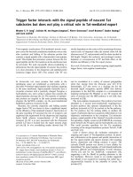
Analysis of the protein sequence of Ki-1 ⁄ 57 revealed
that it possessed several clusters of RGG ⁄ RXR box
motifs, which may be target sites for protein arginine
methylation by PRMT1 (Fig. 1). These clusters are
located at the N-terminus (amino acids 47, 55, 70), in
the central region (178–199) and on the extreme
C-terminus (369–383). Alignment with the putative
Ki-1 ⁄ 57 paralog CGI-55 showed that the central and
C-terminal clusters are conserved in both proteins
(Fig. 1A,B). The central cluster (178–199) in Ki-1 ⁄ 57
contains seven RGG ⁄ RXR motifs, three of which are
conserved in the corresponding cluster of CGI-55 (158–
179), which contains five of such motifs. The C-terminal
cluster in Ki-1 ⁄ 57 (369–383) contains four RGG ⁄ RXR
motifs, all of which are conserved in CGI-55 (352–365),
which contains an additional fifth motif (Fig. 1B).
Interaction and mapping of the interaction site
of Ki-1
⁄
57 with PRMT1
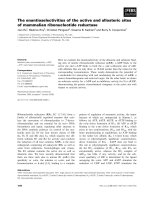
Next, we wanted to map the Ki-1 ⁄ 57 region involved in
the interaction with PRMT1 using the yeast two-hybrid
method (Fig. 2). Nine N- and C-terminal deletion con-
structs of the Ki-1 ⁄ 57 protein were fused to the LexA–
DNA-binding domain (Fig. 2A) and tested for their
ability to bind full-length PRMT1 (Fig. 2B–E). Interest-
ingly, the interactions of the N-terminus of Ki-1 ⁄ 57 (1–
150), its C-terminus (122–413) and a fragment spanning
its central region (151–260) with PRMT1 were each
stronger than that of full-length Ki-1 ⁄ 57 (Fig. 2B,C).
Table 1. (Continued).
Protein interacting
with PRMT1
(synonym ⁄ s)
No. of
RGG ⁄ RXR
boxes
Insert
length
(bp)
a
Coded protein residues
(retrieved ⁄ complete
sequence)
Domain
composition
b
Function
c
Found
clones
d
Accession
number Ref.
ZCCHC12 7 ND 191–412 ⁄ 412 CCHC zinc finger domain
(zinc-knuckle)
Nucleic acid binding, transcriptional regulation 1 NM_173798 35
ILF3 (MMP4 ⁄ MPP4 ⁄
NF90 ⁄ NFAR-1 ⁄
TCP80 ⁄ DRBP76 ⁄
MPHOSPH4 ⁄ NF-AT-90)
7 1100 546–894 ⁄ 894 2 double-stranded RNA-binding
motifs (DSRM), C-terminal
glycine-rich region
Transcription factor required for
expression of interleukin-2 in
T cells, binds RNA
2 NM_012218 36
a
Approximate length of the sequences retrieved from the library.
b
Other domains may be present.
c
Other functions may be known.
d
Number of times the clone ⁄ protein was found
among the 36 sequenced clones.
D. O. Passos et al. Functional association of Ki-1 ⁄ 57 and PRMT1
FEBS Journal 273 (2006) 3946–3961 ª 2006 The Authors Journal compilation ª 2006 FEBS 3949
The C-terminus (261–413) had approximately the same
affinity as full-length Ki-1 ⁄ 57 (Fig. 2B,C). When we tes-
ted further subdeletions of this C-terminal fragment
(Fig. 2D,E) we found that only the two subdeletions of
Ki-1 ⁄ 57 containing the predicted RGG ⁄ RXR box clus-
ter (369–383) interacted with PRMT1 (Fig. 2A,D,E).
Empty vector or constructions containing subdeletions
of Ki-1 ⁄ 57 lacking the C-terminal RGG ⁄ RXR box clus-
ter did not interact with PRMT1.
Next, we performed an in vitro pull-down assay with
the recombinant purified proteins 6xHis–K1 ⁄ 57 and
GST–PRMT1 to confirm the interaction (Fig. 2F).
The assay confirmed the specificity of the interaction,
since glutathione–Sepharose beads coupled with GST–
PRMT1 were able to coprecipitate 6xHis–Ki-1 ⁄ 57, but
not the control protein 6xHis–RACK1. The figure also
shows the equal loading and input controls of the
tested proteins.
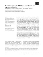
Fig. 1. Alignment of Ki-1 ⁄ 57 and CGI-55 and prediction of putative arginine methylation sites. (A) Protein sequence alignment of the putative
homologs Ki-1 ⁄ 57 and CGI-55. Boxes indicate putative arginine methylation sites that could be targets for PRMT1 and their boundaries are
marked with numbers. (B) Detailed representation of the two conserved multiple RGG ⁄ RXR boxes in the central region and at the C-termi-
nus of Ki-1 ⁄ 57 and CGI-55. In the central region, three of the seven RGG ⁄ RXR targets are strictly conserved in CGI-55. For the C-terminal
region, four of the five RGG ⁄ RXR motifs found in CGI-55 are conserved in Ki-1 ⁄ 57. The residue T375, located between two RGG motifs but
not found in CGI-55, is pointed out because it is a target residue for phosphorylation by PKC in vitro.
Functional association of Ki-1 ⁄ 57 and PRMT1 D. O. Passos et al.
3950 FEBS Journal 273 (2006) 3946–3961 ª 2006 The Authors Journal compilation ª 2006 FEBS
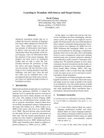
In vitro methylation of Ki-1
⁄
57 and CGI-55
by PRMT1
The interaction of Ki-1 ⁄ 57 with PRMT1 and the pres-
ence and conservation of the RGG ⁄ RXR box motifs
in the amino acid sequences of Ki-1 ⁄ 57 and CGI-55
suggest that these two proteins are likely targets of
arginine methylation by PRMT1. To test this hypo-
thesis we incubated Ki-1 ⁄ 57 and its putative paralog
CGI)55 as glutathione S-transferase (GST)-fusion
proteins with GST–PRMT1 in vitro and performed
a protein methylation assay. We found that Ki-1 ⁄ 57
and its putative paralog CGI-55 are good in vitro
substrates for protein arginine methylation by
PRMT1 (Fig. 3A), whereas control proteins like
PRMT1 itself (which contains a RXR motif at its
C-terminus), RACK1 and GST (as a fusion partner of
GST–PRMT1) were not methylated.
A
B
C
ED
F
Fig. 2. PRMT1 interacts with all RGG ⁄ RXR box-containing protein regions of Ki-1 ⁄ 57. (A) Schematic representation of PRMT1 (cloned in
pGAD424 in fusion with the Gal4-activation domain) and Ki-1 ⁄ 57 (1) and its deletion constructs 2–11 (cloned in pBTM116 in fusion with the
LexA–DNA-binding domain) used in the yeast two-hybrid assay. Fusion proteins are indicated by striped boxes and the putative RGG ⁄ RXR
boxes by black boxes, which indicate the involved amino acid regions. (B, D) The PRMT1 construct was transformed in L40 yeast cells. The
indicated deletion constructs of Ki-1 ⁄ 57 were cotransformed and tested for interaction by assessing their ability to grow on the -Trp, -Leu,
-His plates. The presence of plasmids was confirmed by growth of all cotransformants on -Trp, -Leu plates (data not shown). (C, E) Quantifi-
cation of the strength of interaction by measurement of the b-galactosidase activity in a liquid ONPG assay (see Experimental procedures for
details). The quantity of the produced yellow color is expressed in arbitrary units. (F) Pull-down assay for the confirmation of the interaction
between PRMT1 and Ki-1 ⁄ 57 in vitro. Recombinant purified GST–PRMT1 protein was coupled to glutathione–Sepharose beads. After wash-
ing the beads were incubated with either bacterially expressed and purified 6xHis-Ki-1 ⁄ 57 or the control protein 6xHis–RACK1. After wash-
ing, coprecipitated proteins were analyzed by western blot against the 6xHis tag or PRMT1 (for control of equal loading). Equal loading with
6xHis fusion proteins was controlled by SDS ⁄ PAGE stained using Coomassie Brilliant Blue. Selected molecular masses of the protein ladder
are indicated.
D. O. Passos et al. Functional association of Ki-1 ⁄ 57 and PRMT1
FEBS Journal 273 (2006) 3946–3961 ª 2006 The Authors Journal compilation ª 2006 FEBS 3951
Endogenous Ki-1
⁄
57 can be methylated in vitro
after Adox treatment of cells
When we isolated Ki-1 ⁄ 57 from the cytoplasmic and
nuclear fractions of L540 Hodgkin analogous cells by
immunoprecipitation and incubated it with recombin-
ant GST–PRMT1, we observed that it cannot be
methylated in vitro (Fig. 3B, lanes 3 and 4). We chose
L540 cells for the following experiments, because they
express a reasonable amount of Ki-1 ⁄ 57 protein,
A
B
C
Fig. 3. Both Ki-1 ⁄ 57 and its putative paralog CGI-55 are substrates of arginine methylation by PRMT1 in vitro and Ki-1 ⁄ 57 is methylated
in vivo. (A) In vitro methylation assay: PRMT1 was expressed and purified as a GST fusion protein in E. coli and incubated with the indicated
recombinant proteins, all expressed in and purified from E. coli.Anin vitro arginine-methylation assay was performed as described in Experi-
mental procedures. Methylated proteins were run out on SDS ⁄ PAGE (right side) and the gel was exposed to a X-ray film. PRMT1 itself and
RACK1 served as control proteins. (B) In vivo methylation assay: L540 Hodgkin-analogous cells were (lanes 1 and 2) or were not (lanes 3
and 4) incubated with the inhibitor of endogenous protein methylation Adox, lyzed and fractionated in nuclear (lanes 2 and 4) and cytoplas-
mic (lanes 1 and 3) fractions. Ki-1 ⁄ 57 was immunoprecipitated (lanes 1–4) and then submitted to methylation by PRMT1 in vitro. As a negat-
ive control we used mAb Ki-67 [44]. We immunoprecipitated its antigen (°), which was then submitted to in vitro methylation by PRMT1
(lanes 5). As expected it did not show any incorporation of radioactivity. The antigen recognized by Ki-67 is not known to be a substrate for
methylation by PRMT1. Proteins were run out on SDS ⁄ PAGE and their methylation assessed by autoradiography. A parallel gel was analyzed
by Coomassie Brilliant Blue staining. Lane 6: bacterial 6xHis-Ki-1 ⁄ 57 methylated in vitro was run out in order to facilitate localization of the
cellular Ki-1 ⁄ 57 protein band. The heavy and light chains of the antibodies (*) served as molecular mass markers (50 and 25 kDa) (C) In vivo
methylation of Ki-1 ⁄ 57. HeLa cells were or incubated or not with the inhibitor of endogenous protein methylation Adox, metabolically labeled
with
3
H-SAM, lyzed and fractionated in nuclear and cytoplasmic fractions. After Ki-1 ⁄ 57 immunoprecipitation from both fractions, samples
were assessed by autoradiography as described above. A parallel CGI-55 immunoprecipitation served as a control and did not result in the
detection of any radioactively labeled bands (data not shown).
Functional association of Ki-1 ⁄ 57 and PRMT1 D. O. Passos et al.
3952 FEBS Journal 273 (2006) 3946–3961 ª 2006 The Authors Journal compilation ª 2006 FEBS
which was also isolated and identified by protein
amino acid sequencing from these cells [5]. Methyla-
tion of Ki-1 ⁄ 57 isolated from L540 cells suggests that
is already methylated in vivo in these cells. The
in vitro methylation reaction is specific because the
control antigen, immunoprecipitated by anti-(Ki-67)
IgG, did not serve as a substrate for PRMT1 in vitro
(lane 5).
When we pretreated the L540 cells with Adox, an
inhibitor of the cellular synthesis of the methyl-group
donor molecule S-adenosyl-l-methionine (SAM), we
observed that Ki-1 ⁄ 57 was strongly methylated by
PRMT1 (Fig. 3B, lanes 1 and 2) in vitro. These results
show that Ki-1 ⁄ 57 already existed in a methylated
form in L540 cells. Most interestingly, we observed
that Ki-1 ⁄ 57 from the nucleus can be stronger methy-
A
B
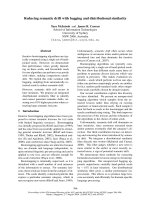
Fig. 4. Regions of Ki-1 ⁄ 57 containing RGG ⁄ RXR boxes are methylated by PRMT1 in vitro but methylation can be blocked by previous phos-
phorylation. (A) cDNAs encoding the Ki-1 ⁄ 57 protein fragments shown in schematic Fig. 2A were subcloned into the bacterial expression
vectors, expressed as GST- or 6xHis fusions in E. coli and purified. The indicated protein fragments and control proteins were submitted to
in vitro methylation using GST–PRMT1 and analyzed by autoradiography for incubated radioactive methyl groups. Loading of the reactions
was controlled by SDS ⁄ PAGE (Coomassie Brilliant Blue). Molecular masses of selected marker proteins are indicated on the right of both
left- and right-hand panels. Arrow-heads indicate the bands that correspond to the predicted molecular masses of the 6xHis- or GST-Ki-1 ⁄ 57
fragments. Asterisks indicate the position of 6xHis–RACK1 protein bands. The open circle indicates the GST protein band. (B) As (A) but with
or without previous phosphorylation of the indicated GST–Ki-1 ⁄ 57-fusion proteins, by PKC-Pan, in vitro.
D. O. Passos et al. Functional association of Ki-1 ⁄ 57 and PRMT1
FEBS Journal 273 (2006) 3946–3961 ª 2006 The Authors Journal compilation ª 2006 FEBS 3953
lated by PRMT1 in vitro, than Ki-1 ⁄ 57 from the cyto-
plasm (Fig. 3B, lanes 1–2).
Metabolic labeling of HeLa cells in vivo with radio-
active [
3
H]-SAM showed stronger methylation of Ki-
1 ⁄ 57 in the absence of the inhibitor Adox (Fig. 3C)
than in its presence. This can be explained by the
mode of action of the inhibitor Adox, which reduces
the amount of the endogenous methyl group donor
molecule SAM in the cells. As a consequence of this,
the small amount of externally added radioactively
labeled SAM may be suboptimal for an effective
methylation of Ki-1 ⁄ 57 in vivo. Interestingly, we did
not observe any radioactive labeling by methyl incor-
poration of the control immunoprecipitated protein
CGI-55 (data not shown). This suggests that either the
protein concentration of CGI-55 in HeLa cells is much
lower than that of Ki-1 ⁄ 57 or that the degree of
methylation of CGI-55 in vivo is much lower that of
Ki-1 ⁄ 57 and not detectable under the conditions tested
in Fig. 3C.
Mapping the protein regions of Ki-1
⁄
57 that are
methylated by PRMT1 in vitro
To address which of the described RGG ⁄ RXR box
clusters are possible targets for PRMT1 methylation,
we submitted a series of deletion proteins of bacteri-
ally derived Ki-1 ⁄ 57 to an in vitro methylation assay
with PRMT1 (Fig. 4A). We found that the N-ter-
minal (1–150), central (151–260) and C-terminal
(261–413) regions of Ki-1 ⁄ 57 are all strongly methy-
lated by PRMT1 (Fig. 4A, lanes 3, 5 and 6) in vitro.
This shows that all three major clusters of
RGG ⁄ RXR boxes (Fig. 1B) are possible targets for
arginine methylation by PRMT1. We also tested five
subdeletions of the C-terminal region of Ki-
1 ⁄ 57(261–413) (Fig. 2A). Only Ki-1 ⁄ 57(294–413) and
Ki-1 ⁄ 57(347–413), both of which contain the predic-
ted RGG ⁄ RXR box cluster, were methylated by
PRMT1 (Fig. 4A, lanes 13 and 15), suggesting that
the presence of this cluster is both necessary and
sufficient for methylation of the C-terminal region of
Ki-1 ⁄ 57.
To test whether the protein RACK1, which binds
to the C-terminus of Ki-1 ⁄ 57 [11], influences the
methylation reaction by PRMT1 it was added to the
assay (Fig. 4A, lanes 1, 7, 16, 17). We found that
the presence of RACK1, which is not itself methyla-
ted by PRMT1 (Fig. 4A, lane 8), had no influence
on the outcome of the methylation reaction. This
suggests that PRMT1 can still methylate the C-ter-
minal domain of Ki-1 ⁄ 57, although RACK1 is bound
to it.
Prior phosphorylation of Ki-1
⁄
57 can decrease its
methylation by PRMT1 in vitro
We previously reported that the Ki-1 ⁄ 57 C-terminus
is a target for phosphorylation by activated protein
kinase C (PKC) in vitro and in vivo [11]. Therefore, we
asked if there is an influence of the phosphorylation of
Ki-1 ⁄ 57 on its methylation by PRMT1. First we used
full-length protein 6xHis–Ki-1 ⁄ 57 previously phosphor-
ylated or not in vitro. We did not observe any differ-
ence in the amount of subsequent methylation of the
phosphorylated vs. nonphosphorylated form (data not
shown). We speculate that it may not be possible to
detect small local changes in the degree of methylation,
because the overall Ki-1 ⁄ 57 sequence has many puta-
tive methylation sites.
We therefore also phosphorylated two C-terminal
deletion constructs of the Ki-1 ⁄ 57 with 4b-phorbol 12-
myristate 13-acetate-activated PKC–Pan in vitro and
then methylated them with PRMT1 in vitro. We noted
that methylation of the larger fragment Ki-1 ⁄ 57(294–
413) is little influenced by prior phosphorylation, but
methylation of the smaller fragment Ki-1 ⁄ 57(347–413)
is significantly inhibited by previous phosphorylation
(Fig. 4B). Both constructs contain the conserved C-ter-
minal RGG ⁄ RXR box cluster 369–383, which con-
tains, in the middle two RGG motifs, the target
residue T375 for phosphorylation by PKC (Fig. 1C)
[11]. Introduction of a negative charge in this region
of the RGG box may lead to the observed inhibitory
influence on protein methylation by PRMT1. The lar-
ger inhibitory effect on the smaller fragment in com-
parison with the larger fragment may be explained by
a local effect of the phosphorylation and introduction
of a negative charge, which may be expected to be rel-
atively larger on a smaller protein fragment. Moreover,
interaction of PRMT1 with the smaller fragment is
weaker than with the larger one (compare Fig. 2A and
E). Therefore, the inhibitory influence of phosphoryla-
tion on this weaker interaction with the smaller frag-
ment may be more pronounced.
PRMT1 dimerization and its N-terminal domain
are necessary for the methylation of full-length
protein Ki-1
⁄
57
We also wanted to map the regions of PRMT1 that
are important for both its dimerization and its interac-
tion with Ki-1 ⁄ 57. Therefore, we generated a series of
truncations of PRMT1 and cloned them into the yeast
expression vector pGAD424 (Fig. 5A). We noted that
only one of the five PRMT1 deletions, which contains
both the catalytic core and the C-terminal domain,
Functional association of Ki-1 ⁄ 57 and PRMT1 D. O. Passos et al.
3954 FEBS Journal 273 (2006) 3946–3961 ª 2006 The Authors Journal compilation ª 2006 FEBS
PRMT1(35–344), was able to dimerize (Fig. 5B,C).
This can be explained by the presence of the dimeriza-
tion region of PRMT1 in the C-terminal domain. Pre-
vious studies have shown that this region is important
for the dimerization of PRMT1 and that PRMT1 is
catalytically active only in its dimerized form [39].
When the PRMT1 deletions were tested for interac-
tion with Ki-1 ⁄ 57, only the PRMT1 deletion (35–344)
showed significant interaction in a quantitative
b-galactosidase assay (Fig. 5E), although all deletions
showed residual growth in the plate assay (Fig. 5D).
Nonetheless, the interaction of deletion PRMT1(35–
344) decreased by 75% (Fig. 5E) in comparison with
full-length PRMT1. This suggests that the N-terminal
region of PRMT1 is important for recognition of full-
length protein substrates, and that PRMT1 dimeriza-
tion is necessary but not sufficient for effective binding
to a full-length protein substrate such as Ki-1 ⁄ 57.
A
B
CF
ED
Fig. 5. PRMT1 deletion lacking the N-terminal first 34 amino acids dimerizes but shows strongly reduced recognition of the full-length pro-
tein substrate Ki-1 ⁄ 57 and residual methylation activity in vitro. (A) Schematic representation of full-length PRMT1 (P) and the six PRMT1
deletion constructs pD1–pD6 used in the yeast two-hybrid studies (B–E) and in vitro methylation assays of Ki-1 ⁄ 57 (F). The diagonal striped
box indicates the Gal4 DNA-binding domain (AD), the vertical dotted box (35–175) in the middle of the PRMT1 protein represents the cata-
lytic domain and the dark box (176–211) the dimerization arm. The black box below indicates the LexA–DNA-binding domain (BD). (B) Six
PRMT1 deletion constructs (in vector pGAD424 fused to the Gal4 activation domain) were tested for their potential to dimerize with full-
length PRMT1 (cloned in fusion with the LexA–DNA-binding domain in vector pBTM116). The indicated PRMT1 constructs were cotrans-
formed into L40 yeast cells which were tested for interaction by assessing their ability to grow on the -Trp, -Leu, -His plates (right). Presence
of plasmids was tested by growth on -Trp, -Leu plates (left). (C, E) Quantification of the strength of indicated interactions by measurement
of the beta-galactosidase in a liquid ONPG assay (see Experimental procedures for details). The quantity of the produced yellow color is
expressed in arbitrary units. (D) The full-length Ki-1 ⁄ 57 construct (cloned in pBTM116 in fusion with the LexA–DNA-binding domain) was
transformed into L40 yeast cells. Full-length PRMT1 (P) or the indicated PRMT1 deletion construct (pD 1–pD6) all cloned in fusion with the
Gal4-AD in pGAD424, were cotransformed into L40 yeast cells which were tested for interaction as in (B) above. (F) In vitro methylation of
GST–Ki-1 ⁄ 57 by different GST–PRMT1 deletion constructs (also see panel A). Methylation was assessed by autoradiography and exposition
to X-ray film for 7 or 30 days. Protein loading was controlled by SDS ⁄ PAGE and anti-GST western blot as indicated. Molecular masses of
selected marker proteins are indicated on the right of the panels.
D. O. Passos et al. Functional association of Ki-1 ⁄ 57 and PRMT1
FEBS Journal 273 (2006) 3946–3961 ª 2006 The Authors Journal compilation ª 2006 FEBS 3955
Interestingly, the N-terminal portion of seven human
PRMTs varies most substantially, supporting the
hypothesis that these regions are somehow involved in
the specific recognition of different protein substrates
[40].
Next, we tested the in vitro methylation activity
of four GST–PRMT1 protein deletion constructs
expressed in Escherichia coli, using full-length GST–
Ki-1 ⁄ 57 fusion protein as a substrate (Fig. 5F). In
agreement with the interaction results described above,
we found that only full-length PRMT1 effectively
methylates Ki-1 ⁄ 57 in vitro, suggesting that both dimeri-
zation of PRMT1 and the presence of the N-terminal
domain are required for effective methylation (Fig. 5F;
autoradiography, 7 days exposure). Longer exposure of
the gel (30 days) revealed that all three PRMT1-deletion
constructs containing the catalytic core domain have a
substantial residual methylation activity on the full-
length Ki-1 ⁄ 57. By contrast, this was not observed
with the RGG ⁄ RXR box region of the protein
hnRNPQ ⁄ NSAP1 [41], which even after 30 days expo-
sure did not show any signs of radioactive labeling (data
not shown).
Immunofluorescence analysis of the localization
of Ki-1
⁄
57, CGI-55 and PRMT1
It has been described previously that the methylation
of proteins might be an important prerequisite for nuc-
lear import ⁄ export or to address proteins to distinct
cellular compartments [19]. Therefore, we performed
immunolocalization studies of Ki-1 ⁄ 57, CGI-55 and
PRMT1 in the absence and presence of the inhibitors
Adox and actinomycin D (Act D) (Fig. 6). In
untreated HeLa cells more Ki-1 ⁄ 57 is found in the
cytoplasm than in the nucleus, but its cytoplasmic
immunostaining is clearly reduced after treatment with
the methylation inhibitor Adox. This suggests that the
methylation status of Ki-1 ⁄ 57 can influence its distri-
bution between the nuclear and cytoplasmic compart-
ments. Treatment with Act D inhibits the synthesis
and consequently the export of mRNA from the nuc-
leus and we observed that it causes significant reduc-
tion in cytoplasmic imumunostaining for Ki-1 ⁄ 57. This
suggests that the localization of Ki-1 ⁄ 57 may be also
influenced by the localization and ⁄ or transport of
mRNA. By contrast, CGI-55, the putative paralog of
Ki-1 ⁄ 57, showed behavior contrary to that observed
for Ki-1 ⁄ 57. In untreated cells CGI-55 is found pre-
dominantly in the nucleus and after Adox treatment it
is partially redistributed to the cytoplasm. Act D has
a similar effect to Adox on CGI-55. Interestingly,
PRMT1 itself shows a predominantly nuclear staining
that did not change much during the treatment with
either Adox or Act D.
Discussion
When we found in yeast-two hybrid screens that a
significant part of the identified Ki-1 ⁄ 57-interacting
Fig. 6. Immunofluorescence analysis of the localization of the proteins Ki-1 ⁄ 57, CGI)55 and PRMT1. HeLa cells were grown on coverslips
and incubated for 16 h with or without Adox or for 3 h with actinomycin D at 37 °C, with the addition of cycloheximide and chloramphenicol
during the last 3 h in all conditions tested. Cells were fixed with 100% methanol and the indicated proteins were immunodetected with the
following primary antibodies: mouse Ki-1 mAb, mouse anti-(human CGI-550) mAb 10.5.6., and mouse anti-(human PRMT1) IgG ab7027. Fluo-
rescein-coupled antimouse (green) serum was used a secondary reagent. DAPI staining (blue) served to localize the position of the nucleus.
Cells were examined with a Nikon fluorescence microscope.
Functional association of Ki-1 ⁄ 57 and PRMT1 D. O. Passos et al.
3956 FEBS Journal 273 (2006) 3946–3961 ª 2006 The Authors Journal compilation ª 2006 FEBS
clones represents PRMT1, we speculated that arginine
methylation could be an important post-translational
modification for this protein. We were able to confirm
by other experiments that Ki-1 ⁄ 57 is a substrate for
arginine methylation by PRMT1 in vitro and in vivo.
Furthermore, we also performed a two-hybrid screen
with the protein PRMT1 as bait and found Ki-1 ⁄ 57
among 12 RGG ⁄ RXR box-containing interacting pro-
teins or putative substrates. This is not only a confirm-
ative result for the finding that Ki-1 ⁄ 57 is a substrate
for arginine methylation by PRMT1 but also indicates
that the yeast two-hybrid system might serve as an
effective method for identifying new substrates for
PRMTs in general. We are aware of only one other
study that used the yeast two-hybrid system to identify
possible PRMT substrates [36]. The yeast two-hybrid
system may prove very useful to identify specific and
common substrates for the at least nine different
human PRMTs [42].
Post-translational modifications of proteins are
important to modify and regulate their functions. We
previously found that Ki-1 ⁄ 57 is connected via the
adaptor protein RACK1 to activated PKC and that
PKC can phosphorylate it on distinct threonine resi-
dues located in its extreme C-terminus [11]. Because, in
addition to this cell-activation-dependent phosphoryla-
tion, Ki-1 ⁄ 57 is also post-translationally modified by
arginine methylation mediated by PRMT1, we wanted
to see if there is an influence of the previous phos-
phorylation on the methylation by PRMT1, in vitro.
In fact, T375, one of the putative target residues of
PKC, is located in the center of the second conserved
RGG ⁄ RXR box cluster at the C-terminus of Ki-1 ⁄ 57
(Fig. 1C) [11]. We found that phosphorylation of the
protein fragment Ki-1⁄ 57(347–413) can affect its subse-
quent arginine methylation by PRMT1.
The structure resolution of various PRMTs from
different species showed a structural conservation of
PRMTs catalytic core and that the PRMTs must
dimerize for catalytic activity [40]. Furthermore, the
amino acid sequences of at least eight human PRMTs
differ predominantly in their N-terminal regions [19].
We speculated that these regions might be important
for differential recognition of target substrates. The
structural requirements of PRMT1 for the recognition
and methylation activity have so far, only been ana-
lyzed for small peptide substrates [39]. Recognition
and methylation of an entire protein substrate may,
however, be different to that described for small pep-
tides. Hence, we performed interaction mapping and
in vitro methylation assays of Ki-1 ⁄ 57 with a series of
PRMT1 protein deletion constructs. We found that
PRMT1 lacking its 33 amino acid N-terminal domain
is capable of dimerizing to almost the same extend as
wild-type PRMT1. The interaction of this same
PRMT1 deletion with full-length Ki-1 ⁄ 57 was, how-
ever, inhibited by a significant 75%, suggesting that
the N-terminal region may be involved in the recogni-
tion of this substrate. In vitro methylation experiments
with PRMT1 deletion mutants expressed in E. coli
initially demonstrated that only full-length PRMT1 is
effectively methylating Ki-1 ⁄ 57. However, longer expo-
sure of the same X-ray film revealed that all three
mutant proteins that contained the catalytic core of
the protein still show significant activity to methylate
Ki-1 ⁄ 57, indicating that the catalytic domain alone, in
the absence of dimerization or N-terminal domains,
retains residual activity on a full-length protein sub-
strate. This seems to depend on the nature of the pro-
tein substrate involved, because we did not observe
any methylation of an isolated RGG ⁄ RXR box
domain of the protein hnRNPQ ⁄ NSAP1 [41], even
with longer exposure of the X-ray film, under the same
conditions (data not shown).
One question that arises from the finding that
Ki-1 ⁄ 57 is a substrate for PRMT1 arginine methyla-
tion is related to the functional consequence of methy-
lation for the protein. Because Ki-1 ⁄ 57 function is not
yet known, but methylation has been described as
essential for regulation of the subcellular, principally
nuclear, localization of a series of other proteins [19],
we set out to test the localization of Ki-1 ⁄ 57 with and
without methylation. We found that Ki-1 ⁄ 57, which is
normally found more in the cytoplasm than in the nuc-
leus, shows decreased cytoplasmic immunostaining
when cells are treated with the methylation inhibitor
Adox. This suggests that the methylation status of
Ki-1 ⁄ 57 can influence its distribution between nuclear
and cytoplamic compartments.
The fact that Act D, which inhibits the synthesis
of nuclear mRNA, also causes a decrease in cyto-
plasmic Ki-1 ⁄ 57 immunolabeling, suggests a possible
functional association with the export of RNA from
the nucleus. Clearly, more experiments are necessary,
however, these results together with other published
data encourage further experiments to tests such a
hypothesis: First, CGI-55 a putative homolog of
Ki-1 ⁄ 57 has been described as an mRNA-interacting
protein termed PAI–RBP1 [8]. Second, Ki-1 ⁄ 57 itself,
also termed IHABP4, has been reported to interact
with negatively charged extracellular macromolecules
such as hyaluronan and RNA in vitro [6]. Finally,
some early studies on Ki-1 ⁄ 57, using gold-labeled
Ki-1 antibody in electron microscopy analysis [4],
showed labeling of the nuclear envelope and of
‘spiral-shaped’ structures which were associated with
D. O. Passos et al. Functional association of Ki-1 ⁄ 57 and PRMT1
FEBS Journal 273 (2006) 3946–3961 ª 2006 The Authors Journal compilation ª 2006 FEBS 3957
the nuclear pores and appeared to ‘pass’ through
them.
The amino acid sequence of CGI-55 is very similar
to that of Ki-1 ⁄ 57 (41% identity, 67% similarity), sug-
gesting that the two proteins could be paralogs. In
immunofluorescence microscopy studies CGI)55,
which shows normally a predominan nuclear labeling,
showed increased cytoplasmic staining after Adox
treatment. This suggests that methylation has different
effects on the distribution of the two proteins between
the nucleus and cytoplasm. Future studies must
address the detailed regulation of the cellular localiza-
tion of these two proteins and if and how these pro-
teins are involved in RNA binding and possibly
metabolism.
Experimental procedures
Plasmid constructions
Several sets of oligonucleotides were designed to allow
subcloning of cDNAs encoding the indicated amino acid
sequences of the proteins studied. Cloning of the complete
cDNA encoding Ki-1 ⁄ 57 and RACK1, or their deletions, in
bacterial (pGEX, pET28a, or pProEx) and yeast expression
vectors (pBTM116, pGAD424, pACT2) has been described
previously [11]. Insertion of PRMT1 complete cDNA into
pGEX-5X-2 (GE Healthcare, Waukesha, WI) allowed to
express PRMT1(1–344) as a C-terminal fusion to GST
(GST–PRMT1). The cDNA of full-length PRMT1(1–344)
was inserted into pBTM116, and its indicated deletion con-
structs were inserted into vector pACT2. The deletion con-
structs of PRMT1 were also subcloned into bacterial
expression vector pGEX to allow their expression as GST-
tagged fusion proteins.
Yeast two–hybrid screenings and interaction
analysis
pBTM116-Ki-1 ⁄ 57(122–413) [11], pBTM116-Ki-1 ⁄ 57(1–150)
and pBTM116-PRMT1(1–344) vectors were used to express
fragments spanning the C- or N-terminus of Ki-1 ⁄ 57 or full-
length PRMT1, respectively, linked to the C-terminus of
LexA DNA-binding domain. Recombinant plasmids were
transfected in Saccharomyces cerevisiae strain L40. A human
fetal brain cDNA library (Clontech, Palo Alto, CA) expres-
sing GAL4 activation domain (AD) fusion proteins was
cotransfected with each one of these three recombinant
pBTM116 vector constructs in three separate screening
assays. Selection of transformants, b-galactosidase activity
test, plasmid DNA extraction and sequencing were per-
formed as described previously [11,41]. The quantitative
ONPG assay to assess the b-galactosidase activity was per-
formed as described previously [43].
Bacterial expression and protein purification
GST, GST–Ki-1 ⁄ 57, GST–PRMT1, 6xHis–RACK1 and
6xHis–Ki-1 ⁄ 57 full-length proteins or indicated deletions
were expressed in E. coli BL21-CodonPlus-RIL (Stratagene,
La Jolla, CA) and purified using glutathione–Sepharose 4B
(GE Healthcare, Waukesha, WI) or Ni-NTA Sepharose as
described before [41].
Western blot analysis, antibodies and cell culture
Proteins were separated by SDS ⁄ PAGE, transferred to a
poly(vinylidene difluoride) membrane and visualized by
immunochemiluminescence using a mouse anti-GST IgG (to
control equal loading of beads), mouse anti5xHis mAb
(Qiagen, Hilden, Germany) or mouse anti-(Ki-1 ⁄ 57) mAbs
A26 or Ki-1 and secondary anti-(mouse IgG)–HRP
conjugate. The anti-(Ki-1 ⁄ 57) mAbs A26 [5], Ki-1 [1] and
Ki-67 [44] have been described previously. Mouse anti-
(human PRMT1) IgG ab7027 was purchased from Ab-
cam, Inc. (Cambridge, MA). The anti-(CGI)55) mouse mAb
has been described previously [7]. HeLa cells and L540
Hodgkin analogous cells were cultivated as described previ-
ously [11].
Pull-down assay
Recombinant purified GST–PRMT1 (3 lg) protein was cou-
pled to glutathione–Sepharose beads. After washing beads
were incubated with either bacterially expressed and purified
6xHis–Ki-1 ⁄ 57 (1 lg) or the control protein 6xHis–RACK1
(1 lg). After six washes coprecipitated proteins were ana-
lyzed by western blot against the 5xHis tag or PRMT1 (for
control of equal loading) as described above. Equal loading
with 6xHis fusion proteins was controlled by SDS ⁄ PAGE
stained with Coomassie Brilliant Blue.
In vitro methylation and phosphorylation
Recombinant Ki-1 ⁄ 57, or the control proteins were incuba-
ted in NaCl ⁄ P
i
containing 1 mm EDTA, 1 mm phenyl-
methylsulfonyl fluoride and 2 lL of radiolabeled SAM
(2 lCi) (GE Healthcare) with or without GST–PRMT1
(bound to glutathione beads) for 1 h at 37 °C, as indicated
in the figures. Reactions were stopped by heating to 100 °C
for 5 min in sample buffer and then run on SDS ⁄ PAGE.
After fixing the gel for 20 min in 10% v ⁄ v both methanol
and acetic acid in water it was washed, and incubated in
amplifying solution (GE Healthcare) for 1 h 30 min, washed
again briefly, dried and exposed to Hyperfilm MP (GE
Healthcare) for 2 days or for the indicated times. In vitro
phosphorylation of Ki-1 ⁄ 57 was performed as described
previously [11] utilizing commercial PKC-Pan (Promega,
Madison, WI). PKC-Pan was purified from rat brain and
consists predominantly of the PKC isoforms a,b and c.
Functional association of Ki-1 ⁄ 57 and PRMT1 D. O. Passos et al.
3958 FEBS Journal 273 (2006) 3946–3961 ª 2006 The Authors Journal compilation ª 2006 FEBS
Preparation of cytoplasmic and nuclear extracts,
methylation assays with cellular Ki-1
⁄
57 and
metabolic labeling
L540 cells (5.0 · 10
7
) incubated or not with Adox (20 lm)
for 16 h, were lyzed for 1 h at 4 °C in 1 mL of modified
cytoplasmic buffer (20 mm Tris pH 8.0, 10 mm KCl, 0.1 mm
EDTA, 1.5 m m MgCl
2
, 0.5 mm dithiothrietol, 2 mm phenyl-
methylsulfonyl fluoride and protease inhibitors) [45]. After
centrifugation at 14 000 g, the nuclear fraction was lyzed in
1 mL nuclear buffer (20 mm Tris pH 8.0, 0.4 m NaCl,
0.1 mm EDTA, 1.5 mm MgCl
2
, 0.5 mm dithiothreitol, 25%
v ⁄ v glycerol) at 4 °C for 1 h. The cytoplasmic and nuclear
fractions were incubated for 2 h, at 4 °C, with 20 lL pro-
tein A–Sepharose beads (GE Healthcare), previously loaded
with the indicated antibodies overnight at 4 °C, washed three
times in cytoplasmic buffer and incubated with human
recombinant protein GST–PRMT1 and 4 lL of radiolabeled
SAM (4 lCi; GE Healthcare) in a final volume of 50 lL.
Finally, the reaction was stopped by adding 10 lLof6·
SDS ⁄ PAGE sample buffer and boiling at 100 °C for 5 min.
Western blots using the indicated antibodies were developed
by chemiluminescence as described previously [41] (Fig. 3B).
For the in vivo metabolic labeling experiment (Fig. 3C),
5.0 · 10
7
HeLa cells were preincubated with 20 lm Adox
as above for 16 h and subsequently labeled in vivo by incu-
bation with 20 lCiÆmL
)1
radiolabeled SAM, 10 mm cyclo-
hexamide and 10 mm chloramphenicol, under constant
agitation at 37 °C for 4 h, in the presence of freshly added
Adox (20 lm). Lysis, fractionation of nucleus and cyto-
plasm, immunoprecipitation and SDS ⁄ PAGE were per-
formed as above and autoradiography of the dried gel was
performed on a Hyperfilm MP at 80 °C for 6 months.
Immunofluorescence analysis
HeLa cells grown on glass cover slips were incubated or
not with Adox (100 lgÆmL
)1
) for 16 h or with Act D
(10 lgÆmL
)1
) for 3 h at 37 °C. To inhibit protein synthesis
we also added cycloheximide (100 lgÆ mL
)1
) and chloram-
phenicol (40 lgÆmL
)1
). Cells were fixed with 100% meth-
anol and immunostained with primary mouse mAbs Ki-1,
anti-(CGI-55) 10.5.6 or anti-PRMT1, and secondary anti-
body fluorescein anti-mouse IgG. Cells were examined with
a Nikon (Kanagawa, Japan) microscope. DAPI staining
was used to show the positions of the nuclei. Cells were
examined with a Nikon fluorescence microscope.
Acknowledgements
This work as supported financially by the Fundac¸ a
˜
o
de Amparo a
`
Pesquisa do Estado Sa
˜
o Paulo (FAP-
ESP), the Conselho Nacional de Pesquisa e Desen-
volvimento (CNPq) and the LNLS. We thank Maria
Eugenia R. Camargo for technical assistance, Dr
Carlos H. I. Ramos and Luciana R. Camillo for
DNA-sequencing.
References
1 Schwab U, Stein H, Gerdes J, Lemke H, Kirchner H,
Schaadt M & Diehl V (1982) Production of a monoclo-
nal antibody specific for Hodgin and Sternberg–Reed
cells of Hodgkin’s disease and a subset of normal lym-
phoid cells. Nature 299, 65–67.
2 Hansen H, Lemke H, Bredfeldt G, Ko
¨
nnecke I & Havs-
teen B (1989) The Hodgkin-associated Ki-1 antigen
exists in an intracellular and a membrane-bound form.
Biol Chem Hoppe-Seyler 370, 409–416.
3 Hansen H, Bredfeldt G, Havsteen B & Lemke H (1990)
Protein kinase activity of the intracellular but not the
membrane-associated form of the Ki-1 antigen (CD30).
Res Immunol 141, 13–31.
4 Rohde D, Hansen H, Hafner M, Lange H, Mielke V,
Hansmann ML & Lemke H (1992) Cellular localization
and processing of the two molecular forms of the Hodg-
kin-associated Ki-1 (CD30) antigen. Am J Pathol 140,
473–482.
5 Kobarg J, Schnittger S, Fonatsch C, Lemke H, Bowen
MA, Buck F & Hansen HP (1997) Characterization,
mapping and partial cDNA sequence of the 57 kDa
intracellular Ki-1 antigen. Exp Clin Immunogenet 14,
273–280.
6 Huang L, Grammatikakis N, Yoneda M, Banerjee SD
& Toole BP (2000) Molecular characterization of a
novel intracellular hyaluronan-binding protein. J Biol
Chem 275, 29829–29839.
7 Lemos TA, Passos DO, Nery FC & Kobarg J (2003)
Characterization of a new family of proteins that inter-
act with the C-terminal region of the chromatin-remo-
deling factor CHD-3. FEBS Lett 533, 14–20.
8 Heaton JH, Dlakic WM, Dlakic M & Gelehrter TD
(2001) Identification and cDNA cloning of a novel RNA-
binding protein that interacts with the cyclic nucleotide-
responsive sequence in the type-1 plasminogen activator
inhibitor mRNA. J Biol Chem 276, 3341–3347.
9 Kobarg CB, Kobarg J, Crosar-Alberto DP, Theizen TH
& Franchini KG (2005) MEF2C DNA-binding activity
is inhibited through its interaction with the regulatory
protein Ki-1 ⁄ 57. FEBS Lett 579, 2615–2622.
10 Nery FC, Rui E, Kuniyoshi TM & Kobarg J (2006)
Evidence for the interaction of the regulatory protein
Ki-1 ⁄ 57 with p53 and its interacting proteins. Biochem
Biophys Res Commun 341, 847–855.
11 Nery FC, Passos DO, Garcia VS & Kobarg J (2004) Ki-
1 ⁄ 57 interacts with RACK1 and is a substrate for the
phosphorylation by phorbol 12-myristate 13-acetate acti-
vated protein kinase C. J Biol Chem 279, 11444–11455.
12 Ozaki T, Watanabe K-I, Nakagawa T, Miyazaki K,
Takahashi M & Nakagawara A (2003) Function of p73,
D. O. Passos et al. Functional association of Ki-1 ⁄ 57 and PRMT1
FEBS Journal 273 (2006) 3946–3961 ª 2006 The Authors Journal compilation ª 2006 FEBS 3959
not of p53, is inhibited by the physical interaction with
RACK1 and its inhibitory effect is counteracted by
pRB. Oncogene 22, 3231–3242.
13 McBride AE, Cook JT, Stemmler EA, Rutledge KL,
McGrath KA & Rubens JA (2005) Arginine methyla-
tion of yeast mRNA-binding protein Npl3 directly
affects its function, nuclear export, and intranuclear
protein interactions. J Biol Chem 280, 30888–30898.
14 Xu W, Du Chen HK, Asahara H, Tini M, Emerson
BM, Montminy M & Evans RM (2001) A transcrip-
tional switch mediated by cofactor methylation. Science
294, 2507–25011.
15 Gary JD & Clarke S (1998) RNA and protein interac-
tions modulated by protein arginine methylation. Prog
Nucleic Acid Res Mol Biol 61, 65–131.
16 Bedford MT, Frankel A, Yaffe MB, Clarke S, Leder P
& Richard S (2000) Arginine methylation inhibits the
binding of proline-rich ligands to Src homology 3, but
not WW, domains. J Biol Chem 275, 16030–16036.
17 Stallcup MR (2001) Role of protein methylation in
chromatin remodeling and transcriptional regulation.
Oncogene 20, 3014–3020.
18 Li H, Park S, Kilburn B, Jelinek MA, Henschen-Edman
A, Aswand DW, Stallcup MR & Laird-Offringa TA
(2002) Lipopolysaccharide-induced methylation of HuR,
an mRNA-stabilizing protein, by CARM1. Coactivator-
associated arginine methyltransferase. J Biol Chem 277,
44623–44630.
19 McBride AE & Silver PA (2001) State of the arg: pro-
tein methylation at arginine comes of age. Cell 106, 5–8.
20 Tang J, Frankel A, Cook RJ, Sangduk K, Paik WK,
Williams KR, Clarke S & Herschmann HR (2000)
PRMT1 is the predominant type I protein arginine
methyltransferase in mammalian cells. J Biol Chem 275,
7723–7730.
21 Pawlak MR, Scherer CA, Chen J, Roshon MJ & Ruley
HE (2000) Arginine N-methyltransferase 1 is required
for early postimplantation mouse development, but cells
deficient in the enzyme are viable. Mol Cell Biol 20 ,
4859–4869.
22 Bartel PL & Fields S (1995) Analyzing protein–protein
interactions using two-hybrid system. Methods Enzymol
254, 241–263.
23 Scott HS, Antonarakis SE, Lalioti MD, Rossier C,
Silver PA & Henry MF (1998) Identification and
characterization of two putative human arginine methyl-
transferases (HRMT1L1 and HRMT1L2). Genomics 48,
330–340.
24 Yanagida M, Hayano T, Yamauchi Y, Shinkawa T,
Natsume T, Isobe T & Takahashi N (2004) Human
fibrillarin forms a sub-complex with splicing factor
2-associated p32, protein arginine methyltransferases,
and tubulins alpha 3 and beta 1 that is independent of
its association with preribosomal ribonucleoprotein
complexes. J Biol Chem 279, 1607–1614.
25 Watanabe TK, Fujiwara T, Kawai A, Shimizu F, Taka-
mi S, Hirano H, Okuno S, Ozaki K, Takeda S, Shimada
Y et al. (1996) Cloning, expression, and mapping of
UBE2I, a novel gene encoding a human homologue of
yeast ubiquitin-conjugating enzymes which are critical
for regulating the cell cycle. Cytogenet Cell Genet 1, 86–
89.
26 Alice SW, Kim MJ, Jianguo S, Trent PM, Mark JS,
Keith SH & Ross S (2002) Heterogeneous nuclear ribo-
nucleoprotein A3, a novel RNA trafficking response ele-
ment-binding protein. J Biol Chem 277, 18010–18020.
27 Ko YG, Kang YS, Park H, Seol W, Kim J, Kim T,
Park HS, Choi EJ & Kim S (2001) Apoptosis signal-reg-
ulating kinase 1 controls the proapoptotic function of
death-associated protein (Daxx) in the cytoplasm. J Biol
Chem 276, 39103–39106.
28 Hoof T, Fislage R & Tummler B (1992) Primary
sequence of the human ribosomal protein L37a. Nucleic
Acids Res 20, 5475.
29 Matsumoto K, Aoki K, Dohmae N, Takio K & Tsuji-
moto M (2002) CIRP2, a major cytoplasmic RNA-bind-
ing protein in Xenopus oocytes. Nucleic Acids Res 28,
4689–4697.
30 Grosset C, Chen CY, Xu N, Sonenberg N, Jacquemin-
Sablon H & Shyu AB (2000) A mechanism for transla-
tionally coupled mRNA turnover: interaction between
the poly(A) tail and a c-fos RNA coding determinant
via a protein complex. Cell 103, 29–40.
31 Belyanskaya LL, Gehrig PM & Gehring H (2001) Expo-
sure on cell surface and extensive arginine methylation
of Ewing sarcoma (EWS) protein. J Biol Chem 276,
18681–18687.
32 Chan YL, Suzuki K, Olvera J & Wool IG (1993) Zinc
finger-like motifs in rat ribosomal proteins S27 and S29.
Nucleic Acids Res 21, 649–655.
33 Zahler AM, Lane WS, Stolk JA & Roth MB (1992) SR
proteins: a conserved family of pre-mRNA splicing fac-
tors. Genes Dev 6, 837–847.
34 Rasheed ZA, Saleem A, Ravee Y, Pandolfi PP & Rubin
EH (2002) The topoisomerase 1 binding RING protein
Topors, is associated with promyelocytic nuclear bodies.
Exp Cell Res 277, 152–160.
35 Strausberg RL, Feingold EA, Grouse LH, Derge JG,
Klausner RD, Collins FS, Wagner L, Shenmen CM,
Schuler GD, Altschul SF et al. (2002) Generation and
initial analysis of more than 15,000 full-length human
and mouse cDNA sequences. Proc Natl Acad Sci USA
99, 16899–16903.
36 Jie T, Peter NK & Harvey RH (2000) Protein-arginine
methyltransferase I, the predominant protein-arginine
methyltransferase in cells, interacts with and is regulated
by interleukin enhancer-binding factor 3. J Biol Chem
275, 19866–19876.
37 Aoki K, Ishii Y, Matsumoto K & Tsujimoto M (2002)
Methylation of Xenopus CIRP2 regulates its arginine-
Functional association of Ki-1 ⁄ 57 and PRMT1 D. O. Passos et al.
3960 FEBS Journal 273 (2006) 3946–3961 ª 2006 The Authors Journal compilation ª 2006 FEBS
and glycine-rich region-mediated nucleocytoplasmic dis-
tribution. Nucleic Acids Res 30, 5182–5192.
38 Chern M-K, Chang K-N, Liu L-F, Tam T-CS, Liu Y-C,
Liang Y-L & Tam MF (2002) Yeast ribosomal protein
L12 is a substrate of protein-arginine methyltransferase
2. J Biol Chem 277, 15345–15353.
39 Zhang X & Cheng X (2003) Structure of the predominant
protein arginine methyltransferase PRMT1 and analysis
of its binding to substrate peptides. Structure 11, 509–520.
40 Zhang X, Zhou L & Cheng X (2000) Crystal structure
of the conserved core of protein arginine methyltransfer-
ase PRMT3. EMBO J 19, 3509–3519.
41 Moraes KCM, Quaresma AJC, Maehnss K & Kobarg J
(2002) Identification and characterization of proteins that
selectively interact with isoforms of the mRNA binding
protein AUF1 (hnRNP D). Biol Chem 384, 25–37.
42 Cook JR, Lee JH, Yang ZH, Krause CD, Herth N,
Hoffmann R & Pestka S (2006) FBXO11 ⁄ PRMT9, a
new protein arginine methyltransferase, symmetrically
dimethylates arginine residues. Biochem Biophys Res
Commun 342, 472–481.
43 Moura P, Rui E, Goncalves KA & Kobarg J (2005)
The cysteine residues of the hepatitis B virus onco-pro-
tein HBx are not required for its interaction with RNA
or with human p53. Virus Res 108, 121–131.
44 Gerdes J, Schwab U, Lemke H & Stein H (1983)
Production of a mouse monoclonal antibody reactive
with a human nuclear antigen associated with cell
proliferation. Int J Cancer 31, 13–20.
45 Baumann M, Gires O, Kolch W, Mischak H, Zeidler R,
Pich D & Hammerschmidt W (2000) The PKC targeting
protein RACK1 interacts with the Epstein-Barr virus
activator protein BZLF1. Eur J Biochem 267, 3891–
3901.
D. O. Passos et al. Functional association of Ki-1 ⁄ 57 and PRMT1
FEBS Journal 273 (2006) 3946–3961 ª 2006 The Authors Journal compilation ª 2006 FEBS 3961