Báo cáo khoa học: ˚ cDNA cloning and 1.75 A crystal structure determination of PPL2, an endochitinase and N-acetylglucosaminebinding hemagglutinin from Parkia platycephala seeds potx
Bạn đang xem bản rút gọn của tài liệu. Xem và tải ngay bản đầy đủ của tài liệu tại đây (499.84 KB, 13 trang )
cDNA cloning and 1.75 A
˚
crystal structure determination
of PPL2, an endochitinase and N-acetylglucosamine-
binding hemagglutinin from Parkia platycephala seeds
Benildo S. Cavada
1
, Frederico B. B. Moreno
2
, Bruno A. M. da Rocha
1,3
, Walter F. de Azevedo Jr
4
,
Rolando E. R. Castello
´
n
1
, Georg V. Goersch
1
, Celso S. Nagano
5
, Emmanuel P. de Souza
1
,
Kyria S. Nascimento
1
, Gandhi Radis-Baptista
1
, Plı
´nio
Delatorre
3
, Yves Leroy
6
, Marcos H. Toyama
7
,
Vicente P. T. Pinto
8
, Alexandre H. Sampaio
9
, Domingo Barettino
5
, Henri Debray
6
, Juan J. Calvete
5
and Libia Sanz
5
1 BioMol-Laboratory, Departamento de Bioquı
´
mica e Biologia Molecular, Universidade Federal do Ceara
´
, Fortaleza, Ceara
´
, Brazil
2 Departamento de Fı
´
sica, Universidade Estadual Paulista, UNESP, Sa˜o Jose
´
do Rio Preto, Sa˜ o Paulo, Brazil
3 Departamento de Cie
ˆ
ncias Fı
´
sicas e Biolo
´
gicas, Universidade Regional do Cariri, Fortaleza, Ceara
´
, Brazil
4 Faculdade de Biocie
ˆ
ncias, Centro de Pesquisas em Biologia Molecular e Funcional, PUCRS, Porto Alegre, Rio Grande do Sul, Brazil
5 Instituto de Biomedicina de Valencia, CSIC, Spain
6 Laboratoire de Chimie Biologique et Unite
´
Mixte de Recherche No. 8576 du CNRS, Universite
´
des Sciences et Technologies de Lille,
France
7 Departamento de Bioquı
´
mica, Instituto de Biologia, Universidade Estadual de Campinas (UNICAMP), Campinas, SP, Brazil
8 Faculdade de Medicina, Universidade Federal do Ceara
´
, Sobral, Brazil
9 Laboratorio de Bioquı
´
mica Marinha, Departamento de Engenharia de Pesca, Universidade Federal do Ceara
´
, Fortaleza, Ceara
´
, Brazil
Keywords
endochitinase; glycosyl hydrolase family 18;
Mimosoideae; Parkia platycephala; X-ray
crystal structure
Correspondence
B. S. Cavada, BioMol-Laboratory,
Departamento de Bioquı
´
mica e Biologia
Molecular, Universidade Federal do Ceara
´
,
Fortaleza, Ceara
´
, Brazil
Fax ⁄ Tel: +55 8540089818
E-mail:
H. Debray, Laboratoire de Chimie Biologique
et Unite
´
Mixte de Recherche du CNRS
N°8576, Universite
´
des Sciences et
Technologies de Lille, ba
ˆ
timent C-9,
59655 Villeneuve D’Ascq Cedex
Fax: +33 320436555
Tel: +33 320410108
E-mail:
(Received 22 May 2006, revised 26 June
2006, accepted 28 June 2006)
doi:10.1111/j.1742-4658.2006.05400.x
Parkia platycephala lectin 2 was purified from Parkia platycephala (Legumi-
nosae, Mimosoideae) seeds by affinity chromatography and RP-HPLC.
Equilibrium sedimentation and MS showed that Parkia platycephala
lectin 2 is a nonglycosylated monomeric protein of molecular mass
29 407 ± 15 Da, which contains six cysteine residues engaged in the for-
mation of three intramolecular disulfide bonds. Parkia platycephala lectin 2
agglutinated rabbit erythrocytes, and this activity was specifically inhibited
by N-acetylglucosamine. In addition, Parkia platycephala lectin 2 hydro-
lyzed b(1–4) glycosidic bonds linking 2-acetoamido-2-deoxy-b-d-glucopyra-
nose units in chitin. The full-length amino acid sequence of Parkia
platycephala lectin 2, determined by N-terminal sequencing and cDNA clo-
ning, and its three-dimensional structure, established by X-ray crystallo-
graphy at 1.75 A
˚
resolution, showed that Parkia platycephala lectin 2 is
homologous to endochitinases of the glycosyl hydrolase family 18, which
share the (ba)
8
barrel topology harboring the catalytic residues Asp125,
Glu127, and Tyr182.
Abbreviations
CTAB, cetyl triethylammonium bromide; GlcNac, N-acetyl-
D-glucosamine; GSP, gene-specific forward primer; HPAEC-PAD, high-pH anion
exchange chromatography with pulsed amperometric detection; PE, pyridylethylated; PPL1, Parkia platycephala lectin 1; PPL2, Parkia
platycephala lectin 2; PTC, phenylisothiocyanate; PTH, phenylthiohydantoin.
3962 FEBS Journal 273 (2006) 3962–3974 ª 2006 The Authors Journal compilation ª 2006 FEBS
Lectins comprise a heterogeneous class of (glyco)pro-
teins that possess one noncatalytic domain that binds
carbohydrates in a specific and reversible manner with-
out altering their covalent structure [1]. Lectins deci-
pher the glycocodes encoded in the structure of glycans
in processes such as cell communication, host defense,
fertilization, development, parasitic infection, tumor
metastasis, and plant defense against herbivores and
pathogens [2]. Mechanisms for sugar recognition have
evolved independently in a restricted number of protein
folds (e.g. jelly roll domain, C-type lectin fold, b-pro-
peller, b-trefoil motif, b-prism I and II domains, Ig
domains, b-sandwich, mixed ab structure, and hevein
domain) [1,3] (for a complete catalog of carbohydrate-
binding protein domains, please consult the 3D Lectin
Database at In
plants, most of the currently known lectins can be
placed in seven families of structurally and evolutionar-
ily related proteins [1]. The seed lectins of leguminous
plants constitute the largest and most thoroughly stud-
ied lectin family. These lectins have represented para-
digms for establishing the structural basis [4–9] and
thermodynamics [10–13] of selective sugar recognition.
Most studies on lectins from Leguminosae involve
members of the Papilionoideae subfamily, whereas
investigations on lectins of the other two subfamilies,
Caesalpinoideae and Mimosoideae, are scarce. Indeed,
to date, the only lectins from the Mimosoideae that
have been functionally and structurally characterized
are those from seeds of species of the genus Parkia,
including Parkia speciosa [14], Parkia javanica [15],
Parkia discolor [16] and the glucose ⁄ mannose-specific
lectin from Parkia platycephala seeds [17–21]. Parkia
(Leguminosae, Mimosoideae), regarded as the most
primitive group of leguminous plants [22], is a pantropi-
cal genus of trees comprising about 30 species found in
the neotropics from Honduras to south-eastern Brazil,
West Africa, the northern part of Malaysia and the
south of Thailand. Parkia platycephala is an important
forage tree growing in parts of north-eastern Brazil.
The seed lectin from Parkia platycephala is a 47.9-kDa
single-chain nonglycosylated mosaic protein composed
of three tandemly arranged jacalin-related b-prism
domains [19,20].
The sugar-binding specificity of Parkia platycephala
lectin towards mannose, an abundant building block
of surface-exposed glycoconjugates of viruses, bacteria,
and fungi, suggests a role for the Parkia platycephala
lectin in defense against plant pathogens [1]. Moreover,
the Parkia platycephala lectin also shows sequence
similarity with stress-upregulated and pathogen-upreg-
ulated defense genes of a number of different plants,
suggesting a common ancestry for jacalin-related
lectins and inducible defense proteins [19]. In addition
to using lectins, whose precise role in plant defense
remains to be determined [23,24, and references cited],
plants defend themselves against pathogens (i.e. fungi)
secreting pathogenesis-related enzymes, such as xylan-
ases and chitinases, which degrade the pathogen’s cell
wall [25–27]. In a previous article we have reported
the presence of an endochitinase in Parkia platycephala
seeds [28]. Now, we have determined its complete
amino acid sequence by a combination of Edman deg-
radation and cDNA cloning, and report its biochemi-
cal characterization and the determination of its crystal
structure. Our results show that this protein, termed
Parkia platycephala lectin 2 (PPL2), is homologous to
endochitinases of the glycosyl hydrolase family 18 that
exhibit rabbit erythrocyte-agglutinating, N-acetylgluco-
samine-binding and chitin-hydrolyzing activities.
Results and Discusion
PPL2, a nonglycosylated and monomeric
GlcNAc-binding hemagglutinin
PPL2 was purified from Parkia platycephala seeds
by affinity chromatography on either Red-Sepharose
(Fig. 1A) or chitin-Sepharose. The protein agglutinated
trypsin-treated rabbit erythrocytes (128 hemagglutinat-
ing units mg
)1
), and this activity was abolished
by 19 mm N-acetyl-d-glucosamine (GlcNac). Other
sugars, such as glucose, mannose, galactose, fucose
and N-acetyl-d-galactosamine, displayed only partial
hemagglutination inhibitory activity at much higher
concentrations (> 75 mm) than GlcNac. Moreover,
the glycoproteins bovine thyroglobulin, ovine submax-
illary mucin, bovine fetuin and bovine asialofetuin
were devoid of hemagglutination inhibitory activity.
Bovine thyroglobulin contains nine complex glycosyla-
tion sites and four high-mannose oligosaccharides [29].
Ovine submaxillary mucin is a glycoprotein bearing a
high density of O-linked oligosaccharides expressing si-
alyl Tn antigens and sialyl core 3 sequences [30].
Bovine fetuin contains three N-linked glycosylation
sites occupied with trisialylated, tetrasialylated or pen-
tasialylated trianntennary structures, and three mono-
sialylated or disialylated O-linked saccharides [31–33].
We thus concluded that PPL2 represented an N-ace-
tylglucosamine-binding hemagglutinin.
The apparent molecular masses of both native
and reduced PPL2 determined by SDS ⁄ PAGE were
30 kDa (Fig. 1A, insert). The molecular mass of
native PPL2, measured by MALDI-TOF MS, was
29 407 ± 15 Da (Fig. 1A). This value was not altered
upon incubation of the denatured, but nonreduced,
B. S. Cavada et al. PPL2, an endochitinase from Parkia platycephala
FEBS Journal 273 (2006) 3962–3974 ª 2006 The Authors Journal compilation ª 2006 FEBS 3963
protein with the alkylating reagent 4-vinylpyridine. On
the other hand, the same treatment after reduction of
the protein with dithiothreitol changed the molecular
mass of PPL2 to 30 052 ± 15 Da (Fig. 1B). The mass
increment of about 645 Da indicated that PPL2 had
incorporated six pyridylethyl groups. The combined
data clearly showed that PPL2 contained six cysteine
residues engaged in the formation of three intramolec-
ular disulfide bonds. Amino acid compositional analy-
sis of the purified protein (Table 1) was in agreement
with this conclusion.
The estimated apparent molecular mass for PPL2 on
a calibrated size-exclusion chromatographic column
was 12 kDa, indicating that the protein had an anom-
alous elution profile. Molecular mass determinations
by size-exclusion chromatography are dependent on
the hydrodynamic properties of the molecule, and, in
addition, interaction of the protein with the matrix
may also introduce large errors into the estimated
molecular mass. Thus, we carried out a more rigorous
analysis of the aggregation state of PPL2 employing
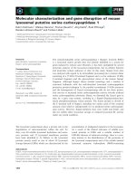
Fig. 1. Purification and molecular mass
determination of PPL2. (A) MALDI-TOF
mass determination of native PPL2 purified
by affinity chromatography as illustrated in
the insert. Insert: the fraction of a Parkia
platycephala seed homogenate precipated
with 60% saturation ammonium sulfate was
resuspended in 50 m
M Tris, pH 7.0, contain-
ing 100 m
M NaCl, and applied to a Red-
Sepharose column. Retained material was
eluted with 3
M NaCl. Fractions exhibiting
hemagglutinating activity (gray area) were
pooled. Right panel: SDS ⁄ PAGE of the
pooled hemagglutinin termed PPL2. Lane a,
molecular mass makers: glutamic dehydro-
genase (55.4 kDa), lactate dehydrogenase
(36.5 kDa), carbonic anhydrase (31.0 kDa),
trypsin inhibitor (21.5 kDa), lysozyme
(14.4 kDa), aprotinin (6.0 kDa). Lane b,
reduced PPL2. (B) MALDI-TOF mass deter-
mination of reduced and pyridylethylated
PPL2. Insert: apparent molecular masses of
native PPL2 determined by equilibrium sedi-
mentation analytic centrifugation in solutions
with different pH values.
Table 1. Amino acid composition [molÆ(mol protein)
)1
] of PPL2.
Asx, aspartic acid and asparagine; Glx, glutamic acid and glutamine.
Amino acid PPL2
Asx 34
Glx 16
Gly 22
Ser 27
His 2
Arg 5
Thr 13
Ala 20
Pro 11
Tyr 9
Val 12
Met 1
Cys 6
Ile 13
Leu 23
Phe 11
Lys 9
Trp 7
Total 241
PPL2, an endochitinase from Parkia platycephala B. S. Cavada et al.
3964 FEBS Journal 273 (2006) 3962–3974 ª 2006 The Authors Journal compilation ª 2006 FEBS
analytic ultracentrifugation equilibrium sedimentation,
a technique that is firmly based in thermodynamics
and does not therefore rely on calibration or on mak-
ing assumptions concerning the shape of the protein.
Using this approach, the apparent molecular mass of
the PPL2 lectin in solutions with pH in the range 2.5–
8.5 was 34 ± 3 kDa (Fig. 1B, insert). This figure, in
conjunction with the MS analyses, showed that the
protein behaved as a pH-independent monomeric pro-
tein.
Carbohydrate analysis performed by GLC (data not
shown) failed to show the presence of any amino or
neutral monosaccharide, strongly indicating that PPL2
was a nonglycosylated protein.
PPL2 displays chitinase activity
Edman degradation analysis of reduced and pyridyl-
ethylated protein yielded the first 42 amino acid resi-
dues of PPL2: GGIVVYWGQNGGEGTLTSTCESGL
YQIVNIAFLSQFGGGRRP. A blast analysis (http://
www.ncbi.nlm.nih.gov/blast/) revealed extensive (up to
approximately 75%) similarity with a large number of
plant chitinase sequences deposited in the publicly
accessible protein databases, such as the basic chitinase
III from Nicotiana tabacum (P29061), an acidic chi-
tinase from Glycine max (BAA77677), chitinase b from
Phytolacca americana (Q9S9F7), chitinase from Pso-
phocarpus tetragonolobus (BAA08708), chitinases from
Vitis vinifera (CAC14014), basic chitinase from Vigna
unguiculata (Q43684), and chitinase B from leaves of
pokeweed (Q9S9F7). All of these proteins are poly
[1,4-(N-acetyl-b-d-glucosaminide)] glycanhydrolases of
the glycosyl hydrolase family 18 (EC 3.2.1.14) [34]
( />whose prototype is hevamine, isolated from the rubber
tree [35,36].
The possible chitinase activity of PPL2 was investi-
gated by quantitative GC determination of the amount
of GlcNac released using chitin as substrate. PPL2
released 3 lg of GlcNacÆh
)1
Æ(mg protein)
)1
. In compar-
ison, commercial Streptomyces griseus chitinase exhib-
ited an activity of 80 lg of GlcNacÆh
)1
Æ(mg protein)
)1
,
and the GlcNac-specific agglutinins from wheat germ
(WGA) and Urtica dioica (UDA) did not show any
chitinase activity. Peracetylated GlcNac (retention time
33.60 min) was observed in the reaction mixtures con-
taining PPL2 or Streptomyces griseus chitinase but not
in those reaction mixtures to which WGA or UDA
were added. These results demonstrated that PPL2 was
indeed an active chitinase able to hydrolyze the b(1–4)
glycosidic bond linking the GlcNac units of chitin. In
order to determine whether PPL2 presented chitinase
activity only for the nonreducing end of chitin (exochi-
tinase activity) or also had the ability to hydrolyze
internal b(1–4) glycosidic linkages (an endochitinase
activity), 40 lL of the reaction mixture used for the
chitinase assay were analyzed by Dionex high-pH
anion exchange chromatography using a CarboPac
PA-100 column. The elution times of three major ana-
lytes present in the reaction mixture (3.93, 4.84 and
5.58 min) matched those of the standard carbohydrates
GlcNac, (GlcNac)
2
and (GlcNac)
3
(3.86, 4.84 and
5.58 min, respectively). This result demonstrated an
endochitinase activity for PPL2. The exact mechanism
of glycoside hydrolysis (e.g. with retention or not of
the b-anomeric configuration of the products) remains
to be established, however.
The finding that PPL2 exhibited GlcNac-dependent
hemagglutination and endochitinase activities was stri-
king but not without precedent. The acidic chitinase
BjCHI1 from Brassica juncea showed hemagglutination
ability [37]. However, BjCHI1 is a unique chitinase
with two chitin-binding domains, and both chitin-bind-
ing domains are essential for agglutination [38]. On the
other hand, PPL2 is a single-domain protein. Hence,
PPL2 may possess at least two carbohydrate-binding
sites. One of them probably corresponds to the cata-
lytic site, whereas the other one(s) remain to be char-
acterized.
Plant chitinases constitute a class of pathogenesis-
related proteins that play an important role in defense
against pathogens through degradation of chitin pre-
sent in the fungal cell wall and in insect cuticles
[37,39]. The first characterization of a chitinase in the
Mimosoideae subtribe, an antifungal chitinase from
Leucaena leucocephala has been reported only recently
[40]. This protein belongs to the class I chitinases of
the glycosyl hydrolase family 19, and is, thus, structur-
ally unrelated to PPL2.
It is noteworthy that the seeds of Parkia platycep-
hala contain two different lectins: the mannose ⁄ glu-
cose-specific PPL1 [19,21] and the GlcNac-binding
lectin with chitinase activity, PPL2, described here.
The fact that mannose is an abundant building block
of surface-exposed glycoconjugates of viruses, bacteria
and fungi supports the view that PPL, and other
mannose-recognizing lectins, play a role in plant def-
ense against pathogens [1]. Specifically, the planar
array of carbohydrate-binding sites on the rim of the
toroid-shaped structure of the Parkia platycephala
lectin dimer [21] immediately suggested a mechanism
to promote multivalent interactions leading to cross-
linking of carbohydrate ligands as part of the host
strategy against phytopredators and pathogens. The
presence of two unrelated lectins in plant seeds has
B. S. Cavada et al. PPL2, an endochitinase from Parkia platycephala
FEBS Journal 273 (2006) 3962–3974 ª 2006 The Authors Journal compilation ª 2006 FEBS 3965
been also reported in Canavalia ensiformis (Legumino-
sae): concanavalin A, a prototypic glucose ⁄ mannose-
specific legume lectin built by the jellyroll fold [1,7],
and concanavalin B, which, although it shares about
40% sequence identity with plant chitinases belonging
to glycosyl hydrolase family 18, has not been shown
to have any chitinase activity [41]. The lack of chi-
tinase activity of concanavalin B can be explained by
differences in the loops that form the substrate-bind-
ing cleft [42].
Sequencing of cDNA and genomic DNA for PPL2
Conserved amino acid sequences from glycosyl hydrol-
ase family 18 were used to design two degenerate prim-
ers that allowed us to PCR-amplify a specific product of
approximately 500 bp (pPPL2). Its sequence was then
used to design a gene-specific forward primer (GSP-
PPL2) to extend the sequence analysis of the PPL2
cDNA by 3¢RACE. Using the GSP-PPL2 and Qo prim-
ers, the sequence was extended in the 3¢ direction by
PCR walking. From these sequences (pPPL2 and
3¢RACE), two specific primers (PPL2f and PPL2r) were
designed that amplified a fragment of 800 bp corres-
ponding to the stretch between the conserved N-ter-
minal sequence
6
YWGQNGG
12
and the STOP codon
(Fig. 2). Using primers designed from the cDNA
sequence, the PPL2 gene was amplified from genomic
DNA of Parkia platycephala seedlings. The size of the
amplified genomic DNA was identical to that of the
cDNA, indicating that the PPL2 gene was devoid of in-
trons, as observed for other class III chitinase genes [43].
The complete amino acid sequence of PPL2 deter-
mined by the combination of N-terminal sequencing
and cDNA cloning contains 271 amino acid residues,
including the six conserved cysteine residues of class
III chitinases, and the putative catalytic residues of
class III plant chitinases, which in PPL2 correspond to
amino acid positions 125 (Asp) and 127 (Glu). The cal-
culated isotope-averaged molecular mass of the PPL2
sequence is 29 490.1 Da, which is about 86 ± 15 Da
greater than the molecular mass determined by
MALDI-TOF MS, suggesting that the native protein
may lack the C-terminal valine residue.
Overall three-dimensional structure of PPL2
Figure 3 displays the structure of PPL2. The 2F
o
) F
c
density map contoured at 1r showed that, with the
exception of a small loop between the a
4
and b
5
regions corresponding to residues from Asn144 to
Lys149, the majority of the protein residues were well
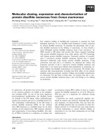
Fig. 2. cDNA and amino acid sequence of
PPL2. The nucleotide and the amino acid
sequences are numbered on the right side.
The underlined nucleotide sequences corres-
pond to primers used to clone and sequence
the full-length PPL2. The underlined amino
acid sequences 6–12 and 178–185 represent
the conserved polypeptide stretches from
which degenerate primers were initially
designed. The N-terminal amino acid
sequence determined by Edman degradation
is labeled. The six conserved cysteines of
class III chitinases are shadowed, and the
conserved residues of the active site of
family 18 of glycosyl hydrolases are boxed.
PPL2, an endochitinase from Parkia platycephala B. S. Cavada et al.
3966 FEBS Journal 273 (2006) 3962–3974 ª 2006 The Authors Journal compilation ª 2006 FEBS
fitted. The PPL2 model has good overall stereochemis-
try (Table 2), with no amino acid residues in the
disallowed region of the Ramachandran plot. The
PPL2 structure consists of a compact (b ⁄ a)
8
barrel
with dimensions of approximately 50 · 40 · 25 A
˚
,
including three disulfide bonds (Cys20–Cys67, Cys50–
Cys57 and Cys158–Cys187) and five cis peptide bonds.
Two of the cis peptide bonds of PPL2 (Gly147–Lys148
and Lys148–Lys149) are located in a region of poor
density, whereas the remaining three (Ala31–Phe32,
Phe160–Pro161 and Trp253–Asp254) are well defined
at the electron density. With the exception of four sul-
fate ions (Fig. 4), which presumably remained bound
to PPL2 throughout its purification protocol, as the
protein was precipitated by ammonium sulfate to sep-
arate it from pigments, no metal ions or ligands were
detected. Sulfate ions were assigned according to
Copley and Barton [44].
Structural comparison and analysis of conserved
motifs
The overall structural features of the PPL2 model are
conserved in other GH18 plant chitinases, i.e. hevam-
ine (Hevea brasiliensis) (PDB code 2HVM), the
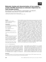
Fig. 3. Crystal structure of PPL2. (A) and (B) show two views
of the (ab)
8
barrel fold of PPL2. The a-helices (red) and b-strands
(yellow) are labeled from 1 to 8. Disulfide bonds are depicted in
blue. In (B), the active site cleft loops are located at the right face
of the model.
Table 2. Statistics of data collection, refinement and quality of the structure.
Overall resolution dataset Highest resolution dataset
Data collection
Total number of observations 95 262 12 669
Total number of unique observations 25 805 3521
R
merge
0.040 0.228
Highest resolution limit (A
˚
) 1.73 1.73
Lowest resolution limit (A
˚
) 32.31 1.83
Completeness (%) 95.5 90.4
Multiplicity 3.7 3.6
I ⁄ r (I) 13.1 2.4
Wavelength (A
˚
) 1.431
Space group P2
1
2
1
2
1
Cell parameters (A
˚
) a ¼ 55.19, b ¼ 59.95, c ¼ 76.70
Refinement
Resolution range (A
˚
) 1.73–32.31
R
factor
(%) 16.88
R
free
(%) 19.87
Number of nonhydrogen atoms in protein structure 2086
Number of sulfate ions 4
Number of water molecules 249
Root mean square deviations from ideal values
Bond lengths (A
˚
) 0.012
Bond angles (degrees) 1.48
Temperature factors
Average B-value for whole protein chain (A
˚
2
) 13.26
Average B-value for sulfate ions (A
˚
2
) 41.97
Average B-values for water molecules (A
˚
2
) 24.29
Ramachandran plot
Residues in most favored regions 195 (87.8%)
Residues in additional allowed regions 26 (11.7%)
Residues in generously allowed regions 1 (0.5%)
B. S. Cavada et al. PPL2, an endochitinase from Parkia platycephala
FEBS Journal 273 (2006) 3962–3974 ª 2006 The Authors Journal compilation ª 2006 FEBS 3967
xylanase inhibitor XIP-I from Triticum aestivum
(1TE1), and ConB (Canavalia ensiformis) (1CNV),
with which PPL2 shares 68%, 40% and 40%
sequence similarity, respectively (Fig. 4A). The three-
dimensional structure of PPL2 can be superimposed
onto those of hevamine, XIP-I and ConB, with root
mean square deviation (r.m.s.d.) for all Ca atoms of
0.90 A
˚
, 1.01 A
˚
and 1.14 A
˚
, respectively. In particular,
the two consensus motifs described for the glycosyl
hydrolase family 18, e.g. the presence of the abso-
lutely conserved strands b
3
and b
4
(Fig. 4A, boxed),
and the hydrogen bond network between residues
Asp120 and Gly121 and Val74 (Fig. 4A,B) [33], are
also conserved in PPL2. On the other hand, the lar-
gest structural divergence is associated with the active
site cleft loops, which comprise the residues linking
neighbor b-strands in the (ab)
8
barrel. Thus, whereas
with the exception of the b
6
a
6
loop, all the active site
cleft loops of PPL2 are highly conserved in hevamine,
and only few structural differences are evident when
comparing the b
2
a
2
and b
7
a
7
loops from PPL2 and
ConB, the active site cleft loops from XIP-I signifi-
cantly depart from those of PPL2.
The PPL2 chitin-binding site
X-ray studies have suggested that enzymes of the
GH18 family showing chitinase activity have conserved
Asp125, Glu127 and Tyr183 amino acids (hevamine
numbering) in their active sites. Their significance for
catalysis is not well understood, although it has been
suggested that Glu127 may act as a proton donor to
the cleavable glycosidic bond, and Asp125 and Tyr183
would contribute to the stabilization of the oxazolin-
ium intermediate [45]. In PPL2, these residues corres-
pond to Asp125, Glu127 and Tyr182 (Figs 2 and 4A).
Asp125 and Glu127 are located in the b
4
a
4
loop, and
Tyr182 at loop b
6
a
6
. The highly conserved, function-
ally relevant, structural features that are common to
PPL2 and hevamine suggest that these two chitinases
may share essentially the same catalytic mechanism. In
addition, our data showing that PPL2 strongly binds
GlcNac would support a hypothetical mechanism by
which the lectin hydrolyzes a chitin polymer by cycles
of anchoring, cleavage and being released from a
GlcNac-binding site, and anchoring to another
GlcNac-binding site. Clearly, detailed molecular and
structural studies are required to investigate this.
Experimental procedures
Isolation of PPL2
Mature seeds from Parkia platycephala were collected in the
state of Ceara
´
(north-eastern Brazil) and ground in a coffee
mill. The flour was defatted with n-hexane, air-dried at room
Fig. 4. Sulfate ions bound to crystallized
PPL2. (A)–(D) display details of the binding
of sulfate ions (S) 1–4 within the crystal
structure of PPL2. In each panel, the elec-
tron density assigned to the sulfate ions is
displayed in an insert. W, water molecule.
PPL2, an endochitinase from Parkia platycephala B. S. Cavada et al.
3968 FEBS Journal 273 (2006) 3962–3974 ª 2006 The Authors Journal compilation ª 2006 FEBS
temperature and kept dry for further use. Soluble proteins
were extracted overnight at room temperature by continuous
stirring with 1 : 15 (w ⁄ v) 500 mm HCl solution, containing
150 mm NaCl. Insoluble material was separated by centrifu-
gation (Ultracentrifuga Beckman modelo XL-1, Palo Alto,
CA) at 10 000 g for 20 min at 5 °C. The supernatant was
adjusted to pH 7.0 and left for 12 h at 4 °C. Precipitated
pigments were removed by centrifugation (Ultracentrifuga
Beckman modelo XL-1), and the supernatant was subjected
to precipitation with 60% saturated ammonium sulfate.
After centrifugation (Ultracentrifuga Beckman modelo XL-1),
the pellet was resuspended in a small volume of 50 mm Tris,
pH 7.0, containing 100 mm NaCl, dialyzed against this buf-
fer, and subjected to affinity chromatography on a Red-
Sepharose CL-4B column (26 · 1.5 cm) (Sigma-Aldrich, Sa
˜
o
Paulo, Brazil) equilibrated with the same buffer as described
previously for GlcNAc-specific enzymes [46]. Unbound
material was eluted by washing the column with equilibra-
tion buffer, and the retained fraction was desorbed with 3 m
NaCl in buffer, dialyzed against equilibrium buffer, and
assayed for hemagglutinating activity following a standard
procedure with trypsin-treated rabbit red blood cells [47]. To
this end, a two-fold dilution was prepared for each sugar
(1 m starting concentration) solution in 0.15 m NaCl con-
taining 5 mm CaCl
2
and 5 mm MnCl
2
. Each dilution had a
final volume of 0.2 mL. The purified lectin was diluted in
0.15 m NaCl to achieve 4 units of hemagglutinating activity
per mL. The lowest concentration of inhibitor exhibiting
agglutinating activity was termed the minimum inhibitory
concentration. Aliquots of 0.2 mL of the 4 unit solution of
the lectin were used for hemagglutination inhibition assay.
Monosaccharides (mannose, glucose, galactose, N-acetyl-
glucosamine, N-acetylgalactosamine, fucose) and glyco-
proteins (bovine thyroglobulin, ovine submaxillary mucin,
bovine fetuin, and asialofetuin) were tested for hemaggluti-
nation inhibitory activity.
Purification of PPL2
The protein fraction retained in the Red-Sepharose CL-4B
column was further fractionated by RP-HPLC and by chi-
tin affinity chromatography. For RP-HPLC, 3 mg of total
proteins was dissolved in 250 lL of 0.1% trifluoroacetic
acid (solution A) and centrifuged (Ultracentrifuga Beckman
modelo XL-1) at 4500 g for 2 min. The supernatant
was applied on a lBondapack C18 analytic column
(3.9 · 300 mm) (Waters, Milford, MA, USA) equilibrated
in solution A, and the column was developed using the fol-
lowing chromatographic conditions: 100% buffer A for
5 min, followed by gradients of 0–30% of solution B
(66.6% acetonitrile in A) for 5 min, 30–40% B for 30 min,
40–70% B for 5 min, 70–80% B for 10 min, 80–100% B
for 5 min, and 100% B for 10 min. The elution was monit-
ored at 280 nm. Fractions were collected manually, lyo-
philized and stored at ) 70 °C until used. For affinity
chromatography, the protein fraction retained in the Red-
Sepharose column was applied overnight to a chitin column
(2 · 5 cm) (Sigma-Aldrich) equilibrated in 50 mm
Tris ⁄ HCl, 150 mm NaCl, pH 7.2. Unbound material was
eluted by washing the column with equilibration buffer,
Fig. 5. Structural features of PPL2 and the GH18 family. (A) Multiple sequence alignment of PPL2, hevamine, XIP-I and ConB. Absolutely
conserved residues in the four proteins are shown in white over a red background. Conservative substitutions or residues conserved in at
least two proteins are depicted in pale red and boxed. Cysteine residues engaged in the formation of disulfide bonds (S–S) are conected by
discontinuous lines. The secondary structure elements of PPL2 are shown on top of the sequence alignment: arrows represent b-strands
and springs denote a-helices. (B) Detail of the network of hydrogen bonds between PPL2 residues Asp120, Gly121 and Val74, which repre-
sent a conserved structural motif of the GH18 family.
B. S. Cavada et al. PPL2, an endochitinase from Parkia platycephala
FEBS Journal 273 (2006) 3962–3974 ª 2006 The Authors Journal compilation ª 2006 FEBS 3969
and the retained fraction was desorbed with 50 mm
Tris ⁄ HCl, 3 m NaCl, pH 7.2.
Molecular mass determinations
Tricine-PAGE in a discontinuous gel and buffer system [48]
was used to estimate the apparent molecular mass of the
proteins. Samples were denatured for 10 min in sample
buffer containing 2.5% (w ⁄ v) SDS before electrophoresis.
After the run, the gels were stained with Coomassie
Brilliant Blue G (0.2%) in methanol ⁄ acetic acid ⁄ water
(4:1:6,v⁄ v) and destained in the same solution. Protein
molecular weight markers (GE Healthcare Biosciences AB,
Uppsala, Sweden) were included in each run.
The molecular masses of the native, reduced and carbam-
idomethylated proteins were determined by MALDI-TOF
MS using an Applied Biosystems (Foster City, CA, USA)
Voyager PRO-STR instrument operating at an accelerating
voltage of 25 kV in the linear mode and using 3,5-dimeth-
oxy-4-hydroxycinnamic acid (10 mgÆmL
)1
in 50% aceto-
nitrile) as the matrix.
The apparent molecular mass of the Parkia platycephala
lectin 2 in solutions of different pH was determined by size-
exclusion chromatography and by analytic ultracentrifuga-
tion equilibrium sedimentation using a Beckman XL-A
centrifuge with UV absorption scanner optics. For size-
exclusion chromatography, PPL2 (2 mgÆmL
)1
) was applied
to a Superose-12 HR10 ⁄ 30 column connected to an A
¨
KTA
HPLC system (GE-Healthcare Bioscience). The column was
equilibrated and eluted with 20 mm sodium phosphate buf-
fer, pH 7.2, containing 150 mm NaCl at a flow rate of
0.5 mLÆmin
)1
. Elution was monitored at 280 nm. Equilib-
rium sedimentation experiments were carried out at 20 °C
and 13 000 r.p.m. using an AN-50 Ti rotor. The protein
was dissolved at about 0.1 mgÆmL
)1
in the following buff-
ers, each containing 100 mm NaCl, 1 mm Cl
2
Mn, and
1mm Cl
2
Ca: 20 mm sodium citrate pH 2.5; 20 mm sodium
citrate, pH 3.5; 20 mm sodium citrate, pH 4.5; 20 mm Mes,
pH 5.5; 20 mm Mes, pH 6.5; 20 mm Tris ⁄ HCl, pH 7.5; and
20 mm Tris ⁄ HCl, pH 8.5.
Quantitation of free cysteine residues and
disulfide bonds
For quantitation of free cysteine residues and disulfide
bonds, the purified proteins dissolved in 10 lLof50mm
Hepes, pH 9.0, 5 m guanidine hydrochloride containing
1mm EDTA were heat-denatured at 85 °C for 15 min,
allowed to cool at room temperature, and incubated with
either 10 mm 4-vinylpyridine for 15 min at room tempera-
ture, or with 10 mm 1,4-dithioerythritol (Sigma-Aldrich) for
15 min at 80 °C; this was followed by addition of 4-vinyl-
pyridine at 25 mm final concentration and incubation for
1 h at room temperature. The pyridylethylated (PE) protein
was freed from reagents using a C18 Zip-Tip pipette tip
(Millipore Ibe
´
rica S.A., Madrid, Spain) after activation
with 70% acetonitrile (ACN) and equilibration in 0.1%
trifluoroacetic acid. Following protein adsorption and
washing with 0.1% trifluoroacetic acid, the PE-protein was
eluted onto the MALDI-TOF plate with 1 lL of 70%
ACN and 0.1% trifluoroacetic acid and subjected to MS
analysis as above.
The number of free cysteine residues (N
SH
) was deter-
mined from Eqn (1):
N
SH
¼ðM
PE
À M
NAT
Þ=105:1 ð1Þ
where M
PE
is the mass of the denatured but nonreduced
protein incubated in the presence of 4-vinylpyridine, M
NAT
is the mass of the native, HPLC-isolated protein, and 105.1
is the mass increment (in Da) due to the pyridylethylation
of one thiol group.
The number of total cysteine residues (N
Cys
) can be cal-
culated from Eqn (2):
N
Cys
¼ðM
Alk
À M
NAT
Þ=105:1 ð2Þ
where M
Alk
is the mass (in Da) of the fully reduced and
alkylated protein.
Finally, the number of disulfide bonds N
S–S
can be calcu-
lated from Eqn (3):
N
SÀS
¼ðN
cys
À N
SH
Þ=2 ð3Þ
Amino acid analysis and N-terminal amino acid
sequence determination
Amino acid analysis was performed on a Pico-Tag amino
acid analyzer (Waters) as described [49]. One nanomole of
purified protein was hydrolyzed in 6 m HCl ⁄ 1% phenol at
106 °C for 24 h. The hydrolyzate was reacted with 20 lL
of fresh derivatization solution (methanol ⁄ triethyl-
amine ⁄ water ⁄ phenylisothiocyanate, 7 : 1 : 1 : 1, v ⁄ v) for
1 h at room temperature, and the phenylisothiocyanate
(PTC)-amino acids were identified and quantitated on an
RP-HPLC column calibrated with a mixture of standard
PTC-amino acids (Pierce, Rockford, IL, USA). Cysteine
residues were determined as cysteic acid.
N-terminal sequencing of reduced and carboxymethylated
proteins was performed in an Applied Biosystems model
Procise 491 gas–liquid protein sequencer. The phenylthiohy-
dantoin (PTH) derivatives of the amino acids were identi-
fied with an Applied Biosystems model 450 microgradient
PTH analyzer.
Genomic DNA and RNA isolation, and cDNA
cloning
Genomic DNA from fresh leaves of 2-week-old seedlings of
Parkia platycephala grown from mature seeds was extracted
using the cetyl triethylammonium bromide (CTAB) proce-
dure [50].
PPL2, an endochitinase from Parkia platycephala B. S. Cavada et al.
3970 FEBS Journal 273 (2006) 3962–3974 ª 2006 The Authors Journal compilation ª 2006 FEBS
For RNA isolation, young Parkia platycephala buds were
immediately ground to a powder with a pestle in liquid
nitrogen. Total cellular RNA was isolated with Concert
Plant RNA reagent (Invitrogen S.A., Barcelona, Spain).
Single-stranded cDNAs were synthesized by reverse trans-
cription using oligo-dT
17
and MMLV reverse transcriptase
(Promega Biotech Ibe
´
rica, Madrid, Spain). Degenerated
primers were designed from conserved amino acid
sequences of plant chitinases YWGQNGG and WVQFY
NNP (sense primer 5¢-TAY TGG GAR AAY GGN GG-3¢,
and antisense primer 5¢-GG RTT RTT RTA RAA YTG
NAC CCA-3¢; the nomenclature follows the IUPAC code
for degeneracies). PCR amplification was performed with
1 U (International unit) of Taq DNA polymerase (HF,
Roche Diagnostics S.L., Barcelona, Spain) using the follow-
ing conditions: DNA was denatured at 94 °C for 4 min, and
this was followed by 30 cycles of denaturation (30 s at
94 °C), annealing (30 s at 50 °C) and extension (30 s at
72 °C), followed by a final extension for 10 min at 72 °C.
The amplified DNA fragment was cloned into the pGEM-T
vector (Invitrogen). The inserted DNA fragments were sub-
jected to sequencing on an Applied Biosystems model 377
DNA sequencing system using T7 and SP6 primers, and this
sequence was used for designing specific oligonucleotides for
completing the sequence by 3¢RACE. 3¢RACE was done as
described [51] using the Qt primer (5¢-CCA GTG AGC
AGA GTG ACG AGG ACT CGA GCT CAA GCT
16
-3¢)
for reverse transcription, and the sense primer GSP-PPL2
(5¢-CTG CTG CAC CAC AAT GTC CTT TTC-3¢) and the
antisense primer Qo (5¢-CCA GTG AGC AGA GTG
ACG-3¢) for PCR amplification. The 3¢RACE reaction
conditions were as those for cDNA amplification, except
that annealing was done at 60 °C. Using this informa-
tion, two specific primers were designed, PPL2-forward
(5¢-TAT TGG GGC CAG AAT GGA G-3¢) and PPL2-
reverse (5¢-TCAA ACA CTG GGC TTA ATT TTG G-3¢)
for amplifying and sequencing the full-length ORF of PPL2.
Assay for chitinase activity
Chitinase enzymatic assays were performed in Pyrex tubes
(7 mL) with Teflon-lined screw caps. The reaction mixtures
(total 1250 lL) contained 0.05 m sodium acetate buffer
(pH 5.5), 5 mg of washed chitin powder (blank), and either
25 lL of a PPL2 solution (1 mgÆmL
)1
)or10lL (0.5 lU)
of Streptomyces griseus family 19 chitinase (Sigma) (one
unit will liberate 1.0 mg of GlcNac from chitin per hour at
pH 6.0 at 25 °C in a 2 h assay) as positive control, both in
sodium acetate buffer. The negative control consisted of the
same reaction mixture, except that sodium acetate buffer
replaced the protein sample. Twenty-five microliters of
1mgÆmL
)1
solutions of two GlcNac-specific lectins, the
agglutinins from wheat germ (WGA) and Urtica dioica
(UDA), which are devoid of chitinase activity, were also
included in the assays as specificity controls. For calibration
and quantitation, a mixture of 1 lg of each, mannose and
GlcNac in sodium acetate buffer was used. The reaction
mixtures were incubated at 37 °C for 3 h and lyophilized.
GlcNac production was monitored and quantitated as per-
acetylated GlcNac by GC [Varian 3400 gas chromatograph
equipped with a flame ionization detector, a Ross injector
and a 30 m · 0.25 mm capillary column EC.Tm
)1
(100%
methylsilicone apolar phase of column, EC.Tm
)1
, 0.25 lm
film phase, Altech), 0.25 lm film phase (Altech, Fleming-
ton, NJ, USA)]. The injector and detector temperature was
250 °C, and the oven temperature program was 3 C°Æmin
)1
from 120 to 250 ° C. The carrier gas helium pressure was
1 bar. Briefly, released GlcNac was peracetylated by addi-
tion of 0.5 mL of acetic anhydride to the lyophilized sam-
ples, followed by incubation for 4 h at 100 °C. Samples
were then evaporated to dryness under a stream of nitrogen
and mild heating with a hair dryer. To eliminate salts and
proteins from the reaction mixture, 1.5 mL of chloroform
and 1 mL of distilled water were added to each tube. After
thorough vortexing, the aqueous upper phase was discarded
and the lower chloroform phase was extracted four times
with 1 mL of distilled water. The chloroform phases were
freed of water by filtration through small columns made of
a Pasteur pipette filled with anhydrous sodium sulfate. The
filtrates were collected in Pyrex tubes (7 mL) and evapor-
ated to dryness under a stream of nitrogen. Chloroform
(40 lL) was added to each tube, and 4 lL was injected in
the gas chromatograph for analysis.
GlcNac production (retention time 33.60 min) was
also monitored by GC ⁄ MS analysis performed on a Carlo
Erba GC 8000 gas chromatograph equipped with a
25 m · 0.32 mm CP-Sil 5CB low-bleed MS capillary col-
umn, 0.25 lm film phase (Chrompack France, Les Ullis,
France). The temperature of the Ross injector was 250 °C
and the samples were analyzed using the following tempera-
ture program: 120 °C for 3 min, then 3 C°Æmin
)1
until
250 °C. The column was coupled to a Finnigan Automass
II mass spectrometer. The analyses were performed either
in the electron impact mode (ionization energy 70 eV,
source temperature 150 °C) or in the chemical ionization
mode in the presence of ammonia (ionization energy
150 eV, source temperature 100 °C). Detection was per-
formed for positive ions.
High-pH anion exchange chromatography with
pulsed amperometric detection (HPAEC-PAD)
HPAEC-PAD was performed with a Dionex Series DX30
HPLC system (Dionex Corporation, Voisins Le Breton-
neux, France) equipped with a pulsed electrochemical detec-
tor, operating in the pulsed amperometric detection mode
with a gold working electrode and an Ag ⁄ AgCl reference
electrode. Electrode potential settings were E1 + 0.05 V,
E2 + 0.6 V and E3 ) 0.6 V, with 500, 3 and 7 ms applied
durations, respectively, and an integrated time period of
B. S. Cavada et al. PPL2, an endochitinase from Parkia platycephala
FEBS Journal 273 (2006) 3962–3974 ª 2006 The Authors Journal compilation ª 2006 FEBS 3971
0.10–0.48 s. Detection was set with a range of detection of
300 nC. Detector response was analyzed with a C-R8A
chromatopac integrator (Shimadzu, Kyoto, Japan). A
standard sample consisted of 0.1 lgÆ lL
)1
of each GlcNac,
N,N¢-diacetylchitobiose or N,N¢,N¢¢-triacetylchitotriose dis-
solved in water. Samples (12.5 lL) were injected in a Dio-
nex CarboPac PA-100 pellicular anion exchange column
running at a flow rate of 0.8 mLÆmin
)1
. Elution was per-
formed with buffer A (100 mm NaOH) for 1 min followed
by a linear gradient of 0–40% buffer B (100 mm NaOH
and 1 m sodium acetate) over 40 min.
Crystallization and structure determination
PPL2 was crystallized by the hanging drop vapor diffusion
method at 20 °C as described [28]. The crystals belong to
the P2
1
2
1
2
1
space group with one monomer in the asym-
metric unit. Crystals soaked in a cryoprotectant solution
containing 75% of mother liquor [0.2 m ammonium acet-
ate, 0.1 m trisodium citrate dehydrate, pH 5.6, and 30%
(w ⁄ v) PEG 4000] and 25% of glycerol were flash-frozen at
100 K in a liquid nitrogen stream. X-ray diffraction data
were collected at 1.73 A
˚
at the synchrotron radiation source
of Cpr station Laborato
´
rio Nacional de Luz Sı
´
ncrotron
(Campinas, Brazil). The data were processed and scaled
using mosflm and scala [52], respectively. Crystallographic
data are summarized in Table 2.
The PPL2 crystal structure was determined by molecu-
lar replacement using the amore software [52], using data
in the resolution range 15–3.0 A
˚
, and the hevamine
coordinates (PDB accession code 2HVM) as the search
model. Rotation and translation functions revealed one
molecule in the asymmetric unit. The position and orien-
tation of the molecule, as a single rigid body entity, were
refined for 20 cycles with refmac [52], using reflections
in the resolution range 32–1.73 A
˚
. Appropriate amino
acid changes were carried out to convert the molecular
model of hevamine into PPL2. Several steps of rebuild-
ing, interspersed with restrained refinement, using ref-
mac, yielded the current model at 1.73 A
˚
resolution.
Sulfate ion molecules were placed by inspection of the
F
o
–F
c
map. For each cycle of refinement, the stereo-
chemistry of the model was monitored with the
procheck incorporated into the CCP4 package [52].
Finally, water molecules were placed in the model over
several steps of refinement with arp ⁄ warp and inspected
manually. The atomic coordinates, fitted with xtalview
[52], are accesible from the Protein DataBank (http://
www.rcsb.org/pdb/) under code 2GSJ.
Acknowledgements
This work was supported by Conselho Nacional de
Desenvolvimento Cientı
´
fico e Tecnolo
´
gico (CNPq),
CAPES, FUNCAP, PADCT and Program CAPES ⁄
COFECUB no. 336 ⁄ 01, and grant BFU2004-
01432 ⁄ BMC from the Ministerio de Educacio
´
n y Cien-
cia, Madrid, Spain. B. S. Cavada, W. F. De Azevedo Jr
and A. H. Sampaio are senior investigators of CNPq.
References
1 Van Damme EJM, Peumans WJ, Barre A & Rouge
´
P
(1998) Plant lectins: a composite of several distinct
families of structurally and evolutionary related proteins
with diverse biological roles. Crit Rev Plant Sci 17, 575–
692.
2 Gabius H-J & Gabius S (1997) Glycoscience. Status and
Perspectives. Chapman & Hall, Weinheim.
3 Dodd RB & Drickamer K (2001) Lectin-like proteins in
model organisms: implications for evolution of carbohy-
drate-binding activity. Glycobiology 11, 71–79.
4 Rini JM (1995) Lectin structure. Annu Rev Biomol
Struct 24, 551–577.
5 Weis WI & Drickamer K (1996) Structural basis of lec-
tin-carbohydrate recognition. Annu Rev Biochem 65,
441–473.
6 Elgavish S & Shaanan B (1997) Lectin–carbohydrate
interactions: different folds, common recognition princi-
ples. Trends Biochem Sci 22, 462–467.
7 Loris R, Hamelryck T, Bouckaert J & Wyns L (1998)
Legume lectin structure. Biochim Biophys Acta 1383,
9–36.
8 Bouckaert J, Hamelryck T, Wyns L & Loris R (1999)
Novel structures of plant lectins and their complexes
with carbohydrates. Curr Opin Struct Biol 9, 572–577.
9 Vijayan M & Chandra N (1999) Lectins. Curr Opin
Struct Biol 9, 707–714.
10 Chervenak MC & Toone EJ (1995) Calorimetric analy-
sis of the binding of lectins with overlapping carbohy-
drate binding. Biochemistry 34, 5685–5695.
11 Dam TK, Cavada BS, Grangeiro TB, Santos CF, de
Sousa FAM, Oscarson S & Brewer CF (1998) Dioclei-
nae lectins are a group of proteins with conserved bind-
ing sites for the core trimannoside of asparagine-linked
oligosaccharides and differential specificities for complex
carbohydrates. J Biol Chem 273, 12082–12088.
12 Dam TK, Cavada BS, Grangeiro TB, Santos CF,
Ceccatto VM, de Sousa FAM, Oscarson S & Brewer
CF (2000) Thermodynamic binding studies of lectins
from the diocleinae subtribe to deoxy analogs of the
core trimannoside of asparagine-linked oligosaccharides.
J Biol Chem 275, 16119–16126.
13 Dam TK, Roy R, Das SK, Oscarson S & Brewer CF
(2000) Binding of multivalent carbohydrates to concana-
valin A and Dioclea grandiflora lectin. Thermodynamic
analysis of the ‘multivalency effect’. J Biol Chem 275,
14223–14230.
PPL2, an endochitinase from Parkia platycephala B. S. Cavada et al.
3972 FEBS Journal 273 (2006) 3962–3974 ª 2006 The Authors Journal compilation ª 2006 FEBS
14 Suvachittanont W & Peutpaiboon A (1992) Lectin from
Parkia speciosa seeds. Phytochemistry 31, 4065–4070.
15 Utarabhand P & Akkayanont P (1995) Purification of a
lectin from Parkia javanica beans. Phytochemistry 38,
281–285.
16 Cavada BS, Madeira SVF, Calvete JJ, Sousa LAG,
Bomfim LR, Dantas AR, Lopes MC, Grangeiro TB,
Freitas BT, Pinto VPT et al. (2000) Purification, chem-
ical, and immunochemical properties of a new lectin
from Mimosoideae (Parkia discolor). Prep Biochem
Biotech 30, 271–280.
17 Cavada BS, Santos CF, Grangeiro TB, Moreira da Silva
LIM, Campos MJO, de Sousa FAM & Calvete JJ
(1997) Isolation and partial characterization of a lectin
from Parkia platycephala Benth seeds. Physiol Mol Biol
Plant 3, 109–115.
18 Ramos MV, Cavada BS, Bomfim LR, Debray H,
Mazard A-M, Calvete JJ, Grangeiro TB & Rouge
´
P
(1999) Interaction of the seed lectin from Parkia platy-
cephala (Mimosoideae) with carbohydrates and complex
glycans. Prot Pept Lett 6, 215–222.
19 Mann K, Farias CM, Gallego del Sol FG, Santos CF,
Grangeiro TB, Nagano CS, Cavada BS & Calvete JJ
(2001) The amino-acid sequence of the glucose ⁄ man-
nose-specific lectin isolated from Parkia platycephala
seeds reveals three tandemly arranged jacalin-related
domains. Eur J Biochem 268, 4414–4422.
20 Gallego del Sol F, Go
´
mez J, Hoos C, Nagano CS,
Cavada BS, England P & Calvete JJ (2005) Energetics
of 5-bromo-4-chloro-3-indolyl-a-D-mannose binding to
the Parkia platycephala seed lectin and its use for MAD
phasing. Acta Cryst F 61, 326–331.
21 Gallego del Sol F, Nagano CS, Cavada BS & Calvete
JJ (2005) The first crystal structure of a Mimosoideae
lectin reveals a novel quaternary arrangement of a wide-
spread domain. J Mol Biol 353, 574–583.
22 Heywood VH (1971) Chemotaxonomy of the Legumino-
sae (Harborne JB & Boulter D, eds), pp. 1–29. Aca-
demic Press, London.
23 Chrispeels MJ & Raikhel NV (1991) Lectins, lectin
genes, and their role in plant defense. Plant Cell 3, 1–9.
24 Wang X & Ma Q (2005) Characterization of a jasmo-
nate-regulated wheat protein related to a b -glucosidase-
aggregating factor. Plant Physiol Biochem 43, 185–192.
25 Collinge DB, Kragh KM, Mikkelsen JD, Nielsen KK,
Rasmussen U & Vad K (1993) Plant chitinases. Plant J
3, 31–40.
26 Hamel F, Boivin R, Tremblay C & Bellemare G (1997)
Structural and evolutionary relationships among chiti-
nases of flowering plants. J Mol Evol 44, 614–624.
27 Kasprzewska A (2003) Plant chitinases ) regulation and
function. Cell Mol Biol Lett 8, 809–824.
28 Cavada BS, Castello
´
n RER, Vasconcelos GG, Rocha
BAM, Bezerra GA, Debray H, Delatorre P, Nagano
CS, Toyama M, Pinto VPT et al. (2005) Crystallization
and preliminary X-ray diffraction analysis of a new chi-
tin-binding protein from Parkia platycephala seeds. Acta
Crystallogr F 61, 841–843.
29 Rawitch AB, Pollock HG & Yang S-X (1993) Thyroglo-
bulin glycosylation: location and nature of the N-linked
oligosaccharide units in bovine thyroglobulin. Arch
Biochem Biophys 300, 271–279.
30 Hill HD Jr, Reynolds JA & Hill RL (1977) Purifica-
tion, composition, molecular weight, and subunit
structure of ovine submaxillary mucin. J Biol Chem
252, 3791–3798.
31 Spiro RG & Bhoyroo D (1974) Structure of the O-gly-
cosidically linked carbohydrate units of fetuin. J Biol
Chem 249, 5704–5717.
32 Green ED, Adelt G, Baenziger JU, Wilson S & Van
Halbeek H (1988) The asparagine-linked oligosacchar-
ides on bovine fetuin. Structural analysis of N-glyca-
nase-released oligosaccharide by 500-megahertz
1
H
NMR spectroscopy. J Biol Chem 263, 18253–18268.
33 Rohrer JS, Cooper GA & Townsend RR (1993) Identifi-
cation, quantitation, and characterization of glyco-
peptides in reversed-phase HPLC separations of
glycoprotein proteolytic digests. Anal Biochem 212,7–
16.
34 Henrissat B (1991) A classification of glycosyl hydrolas-
es based on amino acid sequence similarities. Biochem J
280, 309–316.
35 Jekel PA, Hartmann BH & Beintema JJ (1991) The pri-
mary structure of hevamine, an enzyme with
lysozyme ⁄ chitinase activity from Hevea brasiliensis latex.
Eur J Biochem 200 , 123–130.
36 Van Scheltinga ACT, Kalk KH, Beintema JJ & Dijkstra
BW (1994) Crystal structures of hevamine, a plant
defense protein with chitinase and lysozyme activity,
and its complex with an inhibitor. Structure 2, 1181–
1189.
37 Chye ML, Zhao KJ, He ZM, Ramalingam S & Fung
KL (2005) An agglutinating chitinase with two chitin-
binding domains confers fungal protection in transgenic
potato. Planta 220, 717–730.
38 Tang CM, Chye ML, Ramalingam S, Ouyang SW,
Zhao KJ, Ubhayasekera W & Mowbray SL (2004)
Functional analyses of the chitin-binding domains and
the catalytic domain of Brassica juncea chitinase
BjCHI1. Plant Mol Biol 56, 285–298.
39 Robertus JD & Monzingo AF (1999) The structure and
action of chitinases. EXS 87, 125–135.
40 Kaomek M, Mizuno K, Fujimura T, Sriyotha P &
Cairns JR (2003) Cloning, expression, and characteri-
zation of an antifungal chitinase from Leucaena leuco-
cephala de Wit. Biosci Biotechnol Biochem 67, 667–
676.
41 Hennig M, Jansonius JN, Van Scheltinga ACT, Dijkstra
BW & Schlesier BJ (1995) Crystal structure of concana-
valin B at 1.65 A resolution. An ‘inactivated’ chitinase
B. S. Cavada et al. PPL2, an endochitinase from Parkia platycephala
FEBS Journal 273 (2006) 3962–3974 ª 2006 The Authors Journal compilation ª 2006 FEBS 3973
from seeds of Canavalia ensiformis. J Mol Biol 254,
237–246.
42 Van Scheltinga ACT, Hennig M & Dijkstra BW (1996)
The 1.8 A
˚
resolution structure of hevamine, a plant
chitinase ⁄ lysozyme, and analysis of the conserved
sequence and structure motifs of glycosyl hydrolase
family 18. J Mol Biol 262, 243–257.
43 Lawton KA, Beck J, Potter S, Ward E & Ryals J (1994)
Regulation of cucumber class III chitinase gene expres-
sion. Mol Plant-Microbe Interact 7, 48–57.
44 Copley RR & Barton GJ (1994) A structural analysis
of phosphate and sulphate binding sites in proteins.
Estimation of propensities for binding and conserva-
tion of phosphate binding sites. J Mol Biol 242, 321–
329.
45 Bokma E, Rozeboom HJ, Sibbald M, Dijkstra BW &
Beintema JJ (2002) Expression and characterization of
active site mutants of hevamine, a chitinase from the
rubber tree Hevea brasiliensis. Eur J Biochem 269,
893–901.
46 Pastuszak I, Drake R & Elbein AD (1996) Kidney
N-acetylgalactosamine (GalNAc)-1-phosphate kinase, a
new pathway of GalNAc activation. J Biol Chem 271,
20776–20782.
47 Ainouz IL, Sampaio AH, Benevides NMB, Freitas
ALP, Costa FHF, Carvalho MR & Pinheirojoventino F
(1992) Agglutination of enzyme treated erythrocytes by
Brazilian marine algal extracts. Bot Mar 35, 475–479.
48 Scha
¨
gger H & von Jagow G (1987) Tricine–sodium
dodecyl sulfate–polyacrylamide gel electrophoresis for
the separation of proteins in the range from 1 to 100
kDa. Anal Biochem 166, 368–379.
49 Henrikson RL & Meredith SC (1984) Amino acid
analysis by reversed-phase high-performance liquid
chromatography: precolumn derivatization with phenyli-
sothiocyanate. Anal Biochem 136, 65–71.
50 Steenkamp J, Wiid I, Lourens A & van Helden P (1994)
Improved method for DNA extraction from Vitis vini-
fera. Am J Enol Vitic 45, 102–106.
51 Frohman MA & Martin GR (1989) Rapid amplification
of cDNA ends using nested primers. Techniques 1, 165–
170.
52 Collaborative Computational Project Number 4 (1994).
Acta Cryst D50, 760–763.
PPL2, an endochitinase from Parkia platycephala B. S. Cavada et al.
3974 FEBS Journal 273 (2006) 3962–3974 ª 2006 The Authors Journal compilation ª 2006 FEBS