Báo cáo khoa học: a-Conotoxin analogs with additional positive charge show increased selectivity towards Torpedo californicaand some neuronal subtypes of nicotinic acetylcholine receptors pdf
Bạn đang xem bản rút gọn của tài liệu. Xem và tải ngay bản đầy đủ của tài liệu tại đây (688.09 KB, 12 trang )
a-Conotoxin analogs with additional positive charge show
increased selectivity towards Torpedo californica and
some neuronal subtypes of nicotinic acetylcholine
receptors
Igor E. Kasheverov
1
, Maxim N. Zhmak
1
, Catherine A. Vulfius
2
, Elena V. Gorbacheva
2
,
Dmitry Y. Mordvintsev
1
, Yuri N. Utkin
1
, Rene
´
van Elk
3
, August B. Smit
3
and Victor I. Tsetlin
1
1 Shemyakin-Ovchinnikov Institute of Bioorganic Chemistry, Russian Academy of Sciences, Moscow, Russia
2 Institute of Cell Biophysics, Russian Academy of Sciences, Pushchino, Russia
3 Department of Molecular and Cellular Neurobiology, Center for Neurogenomics and Cognitive Research, Vrije Universiteit, Amsterdam,
the Netherlands
a-Conotoxins are a group of relatively short peptides
(12–19 amino acid residues, two disulfide bridges) from
the venom of poisonous marine snails of the Conus
genus [1]. In addition to peptides isolated from venom
new a-conotoxins have recently been identified by
cDNA cloning from venomous glands and have been
Keywords
acetylcholine-binding protein; acetylcholine-
elicited Cl
–
current; a-conotoxin analogs;
identified Lymnaea neurons; nicotinic
acetylcholine receptor
Correspondence
V. I. Tsetlin, Shemyakin-Ovchinnikov
Institute of Bioorganic Chemistry, Russian
Academy of Sciences, Miklukho-Maklaya
str. 16 ⁄ 10 Moscow, Russia
Tel ⁄ Fax: +7 495 335 57 33
E-mail:
(Received 28 March 2006, revised 16 June
2006, accepted 4 August 2006)
doi:10.1111/j.1742-4658.2006.05453.x
a-Conotoxins from Conus snails are indispensable tools for distinguishing
various subtypes of nicotinic acetylcholine receptors (nAChRs), and synthe-
sis of a-conotoxin analogs may yield novel antagonists of higher potency
and selectivity. We incorporated additional positive charges into a-conotox-
ins and analyzed their binding to nAChRs. Introduction of Arg or Lys res-
idues instead of Ser12 in a-conotoxins GI and SI, or D12K substitution in
a-conotoxin SIA increased the affinity for both the high- and low-affinity
sites in membrane-bound Torpedo californica nAChR. The effect was most
pronounced for [D12K]SIA with 30- and 200-fold enhancement for the
respective sites, resulting in the most potent a-conotoxin blocker of the
Torpedo nAChR among those tested. Similarly, D14K substitution in
a-conotoxin [A10L]PnIA, a blocker of neuronal a7 nAChR, was previously
shown to increase the affinity for this receptor and endowed
[A10L,D14K]PnIA with the capacity to distinguish between acetylcholine-
binding proteins from the mollusks Lymnaea stagnalis and Aplysia califor-
nica. We found that [A10L,D14K]PnIA also distinguishes two a7-like
anion-selective nAChR subtypes present on identified neurons of L. stag-
nalis: [D14K] mutation affected only slightly the potency of [A10L]PnIA to
block nAChRs on neurons with low sensitivity to a-conotoxin ImI, but
gave a 50-fold enhancement of blocking activity in cells with high sensitiv-
ity to ImI. Therefore, the introduction of an additional positive charge
in the C-terminus of a-conotoxins targeting some muscle or neuronal
nAChRs made them more discriminative towards the respective nAChR
subtypes. In the case of muscle-type a-conotoxin [D12K]SIA, the contribu-
tion of the Lys12 positive charge to enhanced affinity towards Torpedo
nAChR was rationalized with the aid of computer modeling.
Abbreviations
ACh, acetylcholine; AChBP, acetylcholine-binding protein; IC
50
, ligand concentration at which 50% inhibition is achieved; nAChR, nicotinic
acetylcholine receptor; n
H
, Hill coefficient.
4470 FEBS Journal 273 (2006) 4470–4481 ª 2006 The Authors Journal compilation ª 2006 FEBS
synthesized chemically [2–5]. a-Conotoxins have become
widely used tools in studies on nicotinic acetylcholine re-
ceptors (nAChRs) [6,7] because they can distinguish
between different nicotinic acetylcholine receptor
(nAChR) subtypes. For example, a-conotoxins GI, MI
and SIA selectively block muscle-type nAChRs, whereas
some others block distinct neuronal nAChRs, e.g.
a-conotoxins ImI and ImII target homo-oligomeric a7
nAChR [8], whereas a-conotoxins MII, PnIA, GIC
block heteromeric nAChR containing a3, a6 and b2
subunits [6]. A change in one or several residues of the
naturally occurring a-conotoxin might result in a change
in its nAChR subtype selectivity [9]. For example, the
A10L substitution in a-conotoxin PnIA switched its
selectivity from the a3b2 to the a7 nAChR [10,11].
Synthesis of diverse a-conotoxin analogs, mutations
in nAChRs and pair-wise mutation analysis have
enabled the identification of specific a-conotoxin
and ⁄ or nAChR residues taking part in ligand–receptor
interactions [12–15]. The crystal structure of the acet-
ylcholine-binding protein (AChBP) from the mollusk
Lymnaea stagnalis, which provides a high-resolution
structure for the extracellular domains of nAChRs
[16,17], has been used to build models for a-conotoxin
binding to distinct nAChRs [18]. Recently, crystal
structures have been solved for AChBP complexes
with two a-conotoxins: [A10L, D14K]PnIA, a double
mutant of a-conotoxin PnIA [19], and for a-conotoxin
ImI [20,21]. These structures provide a solid basis for
modeling the spatial structures of a-conotoxins with
the cognate nAChRs. Modeling may also be a start-
ing point for the rational design of new a-conotoxins
with higher affinity and better selectivity towards
nAChRs.
D14K substitution increased the affinity of the
starting [A10L]PnIA for chicken a7 nAChR and
L. stagnalis AChBP [19]. X-Ray data on the
AChBP)a-conotoxin complex were the basis for con-
structing a model for a7 nAChR complexes with
[A10L]PnIA and [A10L, D14K]PnIA [19]. We used the
X-ray data and cryoelectron microscopy structure of
Torpedo nAChR [22] to build a respective model for
a-conotoxin [D12K]SIA, wherein the Lys12 positive
charge gave the most dramatic increase in the affinity
for T. californica nAChR.
Anion-selective nAChRs in some identified neurons
of the fresh-water snail L. stagnalis and marine mol-
lusk Aplysia californica were found to resemble the a7
nAChRs of vertebrates in terms of their pharmacologi-
cal profile and the response kinetics to acetylcholine
(ACh) [23,24]. To further elucidate the significance of
a positive charge in the C-terminus of a-conotoxins
we compared the action of [A10L]PnIA and
[A10L,D14K]PnIA on a
7-like nAChRs in identified
Lymnaea neurons. This is of interest in light of the
recent cloning of a set of nAChR subunits from this
species and electrophysiological analysis of several of
them expressed in Xenopus oocytes [25,26].
Results and Discussion
Synthesis of a-conotoxins
New analogs of a-conotoxins GI, SI and SIA with
arginine, lysine and ⁄ or aspartate introduced at position
12 (Table 1) were synthesized using a solid-phase
method with the simultaneous formation of the two
disulfides. For a-conotoxin SIA, which has Asp12 in
this position, an additional D12S analog was also
synthesized. A series of a-conotoxin MI analogs was
similarly synthesized. In this case, we employed Lys-
scanning mutagenesis for the possibly complete set of
MI variants, excluding the substitutions of structurally
important amino acid residues (Cys, Pro). As a result,
three novel analogs of a-conotoxin MI with a lysine
residue introduced at position 5, 7 or 11 were obtained.
Simultaneous formation of the disulfides decreases
the number of stages and usually gives higher peptide
yields, although this is sometimes accompanied by the
production of incorrectly bridged isomers [27]. When
several isomers were formed, the peptide with correctly
formed disulfide bridges was assumed to have a higher
potency to bind to the membrane-bound T. californica
nAChR in the radioligand-binding assay (see below).
It is known that incorrect disulfide formation in
a-conotoxins that target the muscle-type nAChRs
entails a decrease in the affinity [28]. However, the
enhanced affinity of the incorrectly formed isomer of
a-conotoxin AuIB, targeting one neuronal-type
nAChR, was revealed previously [29] and makes the
method less predictive. Therefore, all new synthesized
analogs were also characterized using CD spectroscopy
to detect secondary structure changes in the ‘incorrect’
isomers (see below). In 13 syntheses of muscle-type
conotoxins we found the generation of isomers only in
two cases – one additional minor peak for [S12D]GI
and two for SIA (all peaks had correct molecular
masses).
a-Conotoxin [A10L]PnIA, known to act on a7
nAChR [10,11] was obtained by solid-phase peptide
synthesis using the simultaneous formation of two
disulfides as described previously [30]. In the case of
[A10L,D14K]PnIA, orthogonal protecting groups were
used for correct pair-wise closing of disulfides to
exclude the formation of other isomers (see Experi-
mental procedures). The structures of all synthesized
I. E. Kasheverov et al. Novel a-conotoxin analogs
FEBS Journal 273 (2006) 4470–4481 ª 2006 The Authors Journal compilation ª 2006 FEBS 4471
peptides were verified by MALDI analysis (Table 1)
and purity by RP-HPLC (data not shown).
CD spectroscopy
CD spectra were obtained for aqueous solutions of
native a-conotoxins GI, MI, SI, SIA and their analogs
[S12R ⁄ K ⁄ D]GI, [H5K]MI, [S12R]SI, [D12S⁄ K]SIA, as
well as for one isomer of [S12D]GI and two isomers
of SIA, which were produced in noticeable quantities
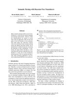
during peptide syntheses. As an example the spectra of
a-conotoxin GI analogs are presented in Fig. 1. Amino
acid substitutions at position 12 did not result in any
noticeable alterations in peptide secondary structure.
However, the second (minor) isomer of [S12D]GI dis-
played a remarkable change in spectral characteristics
(inset in Fig. 1). Similarly, the spectra of the SI and
SIA analogs with substitution at position 12 (as well
as [H5K]MI) were identical to that of the respective
naturally occurring a-conotoxins. However, both
minor isomers of SIA had spectra resembling that of
minor [S12D]GI isomer (data not shown).
The available literature data indicate that single
amino acid substitutions do not markedly change the
CD curves of a-conotoxins. However, breaking the
Cys–Cys disulfide bonds in a-conotoxin ImI [31] or
x-conotoxin MVIIA [32], or changing the size of the
disulfide-confined peptide loops by introduction of
an additional amino acid residue in a-conotoxin ImI
[33], resulted in a remarkable change in CD spectra,
with shifting of the ellipticity minimum into the
195–200 nm region. This shift resembles that seen for
minor isomers of both [S12D]GI and SIA (see curve
4a in the inset of Fig. 1). Taken together, these results
indicate that analysis of biological activities (Fig. 2)
has been carried out on a series of a-conotoxins with
correctly closed disulfides.
Binding of synthesized a-conotoxin analogs to
membrane-bound Torpedo nAChR
The activity of analogs was evaluated in competition
with radioiodinated a-conotoxins GI or MI for binding
Fig. 1. CD spectra of a-conotoxins GI (1, solid line), [S12K]GI (2,
dotted line), [S12R]GI (3, dash-dot line) and main isomer of
[S12D]GI (4, dash line) in water. Inset: CD spectra of two [S12D]GI
isomers – the main (4, solid line) and minor (4a, dash line) ones.
Table 1. The structures of synthesized naturally occurring a-conotoxins and their analogs. All a-conotoxins have amidated C-termini as well
as disulfide bridges Cys1–Cys3 and Cys2–Cys4. The substituted residues in the analogs are indicated in bold type.
a-Conotoxin Sequence
Mol. mass, MH
+
Calculated MALDI-measured
GI ECCNPACGRHYSC 1438.6 1438.4
[S12R]GI ECCNPACGRHYRC 1507.7 1506.6
[S12K]GI ECCNPACGRHYKC 1479.7 1480.9
[S12D]GI ECCNPACGRHYDC 1466.6 1465.5
SI ICCNPACGPKYSC 1354.6 1353.5
[S12R]SI ICCNPACGPKYRC 1423.7 1422.4
SIA YCCHPACGKNFDC 1456.7 1455.6
[D12S]SIA YCCHPACGKNFSC 1428.7 1427.5
[D12K]SIA YCCHPACGKNFKC 1469.9 1469.3
MI GRCCHPACGKNYSC 1494.7 1494.4
[H5K]MI GRCCKPACGKNYSC 1485.7 1484.6
[A7K]MI GRCCHPKCGKNYSC 1553.2 1554.1
[N11K]MI GRCCHPACGKKYSC 1508.8 1507.7
[A10L]PnIA
a
GCCSLPPCALNNPDYC 1664.7 1664.7
[A10L,D14K]PnIA
a
GCCSLPPCALNNPKYC 1677.8 1677.6
a
Described in Celie et al. [19].
Novel a-conotoxin analogs I. E. Kasheverov et al.
4472 FEBS Journal 273 (2006) 4470–4481 ª 2006 The Authors Journal compilation ª 2006 FEBS
to membrane-bound T. californica nAChR (Fig. 2).
Both tracers bound specifically to the Torpedo receptor
with equal high affinity: K
d
values for
125
I-labeled GI
and MI were 24 ± 3 and 28 ± 6 nm, respectively. By
contrast to a-conotoxin GI and M1, a-conotoxin SI
has an equal potency to both sites in the Torpedo
nAChR [34], whereas a-conotoxin SIA binds to only
one site [35] as revealed by competition with
125
I-labe-
led a-bungarotoxin. That is why we did not prepare the
radioactive forms of these peptides, and
125
I-labeled GI
was used as a tracer to test the SI and SIA analogs. In
these experiments the synthetic a-conotoxins GI, SI,
SIA and MI were used as controls. The respective lig-
and concentrations at which 50% inhibition is achieved
(IC
50
values) are presented in Table 2.
The introduction of a positively charged amino acid
residue instead of a neutral one in position 12 of
a-conotoxins GI and SI resulted in a three- to seven-
fold increase in the affinity to both binding sites of
the Torpedo nAChR (Fig. 2A,B; Table 2). The most
remarkable was the D12K mutation in a-conotoxin
SIA: the binding efficiencies to the high- and low-affin-
ity sites increased for the [D12K]SIA analog by 35 and
260 times, respectively (Fig. 2C; Table 2). This increase
was due mainly to removal of the negatively charged
amino acid residue in this position, because substitu-
tion with neutral Ser also resulted in affinity enhance-
ment to both sites (25 and 65 times, respectively).
Conversely, the introduction of a negative charge in
position 12 of a-conotoxin GI caused a considerable
decrease in the affinity for the receptor (Fig. 2A;
Table 2). However, the introduction of an additional
positive charge (Lys) at position 11 of a-conotoxin MI
(which corresponds spatially to residue 10 of a-cono-
toxins GI, SI and SIA) affected the peptide activity
only slightly, whereas H5K or A7K mutations wor-
sened the binding characteristics of these analogs
(Fig. 2D; Table 2).
It should be noted that all three minor isomers
(of [S12D]GI and SIA) showed more than tenfold
Fig. 2. Inhibition of
125
I-labeled a-conotoxins GI (A–C) and MI (D) binding to membrane-bound Torpedo nAChR with indicated a-conotoxins
and their analogs. Final concentrations of the radioligand and toxin-binding sites of receptor were 280 and 230 n
M, respectively. The data
shown are the averages of two independent experiments. The inhibition curves were fitted using
ORIGIN 6.1 (MicroCal Software Inc.) in the
frames of a two-site competition model for all peptides (with one exception for [A7K]MI). The respective IC
50
values are presented in
Table 2.
I. E. Kasheverov et al. Novel a-conotoxin analogs
FEBS Journal 273 (2006) 4470–4481 ª 2006 The Authors Journal compilation ª 2006 FEBS 4473
decreased efficacies, compared with the major com-
pounds, in competition with radiolabeled a-conotoxin
GI for the T. californica nAChR binding (data not
shown).
Both PnIA variants at concentrations of up to
100 lm were inactive in competition with
125
I-labeled
GI for binding to the membrane-bound Torpedo
nAChR (data not shown).
We synthesized mainly the modified a-conotoxins
targeting the muscle-type nAChR. Literature data on
the role of charged residues in this group of a-cono-
toxins are in part contradictory. Several researchers
have shown that charged groups at the N-termini of
a-conotoxins GI, MI and SI exert only a weak influence
on the activity [33,35–39]. The important role of Arg9
in the interaction with a high-affinity a-conotoxin-
binding site on the Torpedo nAChR has been convin-
cingly demonstrated: R9P and R9A substitutions in
a-conotoxin GI resulted in a two to three order of
magnitude loss in the affinity for the a ⁄ c site, whereas
the reverse substitutions P9R and P9K in a-conotoxin
SI enhanced the affinity for this site [34,35,40]. How-
ever, when Ala or Pro residues in a-conotoxin MI were
substituted for the Lys10, whose spatial disposition is
close to that of Arg9 in a-conotoxin GI, the interac-
tion with the high-affinity a ⁄ c-binding site was affected
to a much less degree [38–40]. In addition, acylation of
Lys10 with azidobenzoyl or benzoylbenzoyl groups
practically did not change the capacity of the respect-
ive derivatives to interact with the membrane-bound
Torpedo nAChR [41].
Of all known muscle-type a-conotoxins, only
a-conotoxin SIA interacts exclusively with one a ⁄ c site
on the Torpedo nAChR [35]. Interestingly, this peptide
contains a negatively charged residue (Asp12) in the
C-terminal part of the molecule (Table 1) whose role
has not been examined previously. D12S substitution
resulted in a 25- and 65-fold increase in the affinity for
the high- and low-affinity binding sites, respectively
(Fig. 2C; Table 2). Introduction of a positive charge
(Lys) at this position resulted in an additional fourfold
increase in affinity for the low-affinity site (Fig. 2C;
Table 2). Substitution of Lys or Arg for Ser12 in
a-conotoxins GI and SI gave a reliable enhancement
(three- to sevenfold) of the affinity for both binding
sites (Fig. 2A,B; Table 2). By contrast, introduction of
a negative charge at this position (S12D) in a-conotox-
in GI brought about a marked decrease in the affinity
(Fig. 2A; Table 2). It is noteworthy that use of
125
I-
labeled a-conotoxin GI in these experiments, instead
of the usual
125
I-labeled a-bungarotoxin [34,35,39,40],
revealed the differences in potency to two sites for
a-conotoxin SI and made possible the detection of a
low-affinity binding site for a-conotoxin SIA in the
Torpedo nAChR. From literature data it is known that
the affinities of muscle-type a-conotoxins to the Tor-
pedo nAChRs binding sites (tested in competition with
125
I-labeled a-bungarotoxin) vary from one to three
orders of magnitude [12,34,35,39]. The given explan-
ation for this scatter is the influence of the receptor
state, test conditions, etc. It is therefore not surprising
that using a different tracer in the radioligand assay
may result in different binding parameters for a-cono-
toxins. This was shown previously for one a-conotoxin
GI analog on the Torpedo receptor [41]. There is
convincing evidence (the crystal structures of the
a-cobratoxin and a-conotoxin complexes with acetyl-
choline-binding proteins) [19–21,42] that the binding
sites for these two groups of competitive antagonists
overlap, but are not identical.
Grafting positive charges on to other positions
of a-conotoxin amino acid sequence resulted in
Table 2. Activity of a-conotoxins and their analogs tested in compe-
tition binding assays. Using the membrane-bound Torpedo nAChR,
the inhibitory activities of a-conotoxins GI, SI, SIA or MI and their
analogs were evaluated in competition with
125
I-labeled a-conotox-
ins GI (GI, SI, SIA and their analogs) or MI (MI and its analogs): see
respective inhibition curves presented in Fig. 2. IC
50
values were
calculated using
ORIGIN 6.1 in the frames of both one- and two-site
models using the joint data from two or three independent experi-
ments for each a-conotoxin. The choice was made in favor of the
model giving the minimal ‘reduced chi-squared’ parameter comple-
mented with reasonable SE values and taking into consideration
the Hill coefficients (n
H
). For all muscle-type conotoxins (with one
exception), a two-site model was found the best. In the case of
[A7K]MI analog the program failed to fit the data to a two-site
model, so the respective IC
50
value was generated in the frames of
one-site model and ascribed to both sites. Both PnIA variants were
inactive in competition with
125
I-labeled a-conotoxins GI at 100 lM
(4 ± 2% of inhibition).
a-Conotoxin n
H
IC
50
,lM
high affinity site low affinity site
GI 0.64 ± 0.04 1.6 ± 0.7 9.3 ± 3.7
[S12R]GI 0.68 ± 0.04 0.49 ± 0.25 1.7 ± 1.0
[S12K]GI 0.56 ± 0.06 0.29 ± 0.13 3.2 ± 1.1
[S12D]GI 0.52 ± 0.07 12.0 ± 2.9 230 ± 50
SI 0.53 ± 0.06 4.0 ± 1.2 58 ± 25
[S12R]SI 0.59 ± 0.04 1.0 ± 0.3 8.2 ± 3.6
SIA 0.32 ± 0.06 3.5 ± 1.3 440 ± 150
[D12S]SIA 0.46 ± 0.03 0.13 ± 0.02 6.6 ± 0.6
[D12K]SIA 0.52 ± 0.05 0.10 ± 0.05 1.7 ± 0.7
MI 0.55 ± 0.04 0.26 ± 0.07 6.6 ± 0.7
[H5K]MI 0.53 ± 0.04 9.1 ± 1.8 130 ± 60
[A7K]MI 0.83 ± 0.06 54 ± 4 54 ± 4
[N11K]M 0.68 ± 0.06 0.24 ± 0.16 3.7 ± 0.7
[A10L,D14K]PnIA – – 100
[A10L]PnIA – – 100
Novel a-conotoxin analogs I. E. Kasheverov et al.
4474 FEBS Journal 273 (2006) 4470–4481 ª 2006 The Authors Journal compilation ª 2006 FEBS
weakening of the binding capacity as seen for a series
of Lys-analogs of a-conotoxin MI (Fig. 2D; Table 2).
Our experiments revealed a new site in the muscle-
type a-conotoxins, wherein the presence of a charge
considerably affects the efficiency of interaction with
the Torpedo nAChR: a positive charge in the C-termi-
nus increases the affinity for both binding sites,
whereas a negative charge drastically decreases it.
In contrast to muscle-type a-conotoxins, negatively
charged amino acid residues can be found in the C-ter-
minus of many a-conotoxins acting on neuronal
nAChRs, namely in AuIA, AuIB, AnIA ⁄ C, EpI,
Vc1.1, PnIA ⁄ B [43]. Substitutions in the neuronal
a-conotoxins were used to modify their selectivity [9–
11]: A10L mutation in PnIA enhanced its affinity for
rat and chicken a7 nAChR and weakened it for a3b2,
converting the parent peptide from a3b2-preferring to
a7-preferring [10,11,44]. However, no studies on the
role of the above-mentioned negative charges in the
C-terminus were performed earlier. Based on
[A10L]PnIA, we recently synthesized a new analog that
bears an additional D14K substitution [19]. This sub-
stitution increased the affinity of the ‘double mutant’
for the chicken a7 nAChR and for L. stagnalis
AChBP, but not for A. californica AChBP [19]. In this
study we show that [A10L,D14K]PnIA exhibits high
affinity for one subtype of a7-like nAChRs in L. stag-
nalis neurons, discriminating two nAChRs.
a-Conotoxin blockade of Cl
–
currents elicited by
ACh in identified neurons of L. stagnalis
PnIA analogs were tested on the identified neurons
(LP1–3, RP2,3) from left and right parietal ganglia of
L. stagnalis. The responses to ACh of these neurons
result from an increase in only Cl
–
conductance, as
revealed by I–V relationship determination at various
Cl
–
concentrations in the internal solution (C.A. Vulf-
ius et al. unpublished results). The AChRs in parietal
Lymnaea neurons resemble a7 nAChRs of vertebrates
in terms of the efficacy of choline, cytisine, and nico-
tine (all of them are full agonists) and their high sensi-
tivity to a-conotoxin ImI [23]. Two groups of cells
distinct in terms of desensitization kinetics and sensi-
tivity to ImI (IC
50
288 ± 27 and 10.3 ± 1.3 nm,
respectively) have been recognized [23].
Both PnIA mutants inhibited the ACh-elicited cur-
rent but had a weaker potency than a-conotoxin ImI
(Fig. 3), in contrast to their much higher affinity for
Lymnaea AChBP [19]. The residual unblocked current
was of approximately the same amplitude in the pres-
ence of saturating concentrations of ImI or either of
the two PnIA mutants. The relative potency of the
PnIA variants differed significantly in two types of
neurons. There was no large distinction between
[A10L]PnIA and [A10L,D14K]PnIA on cells with low
sensitivity to a-conotoxin ImI (Fig. 3A; IC
50
30 and
15 lm, respectively). However, in neurons with high
sensitivity to ImI, the [D14K] mutation increased the
affinity 50-fold (Fig. 3B; IC
50
400 nm compared with
20 lm for [A10L]-variant). Average IC
50
ratios for
ImI ⁄ [A10L,D14K]PnIA ⁄ [A10L]PnIA were 1 : 60 : 120
and 1 : 13 : 670 in two groups of cells. Thus, only
those nAChRs which can be blocked by a-conotoxin
ImI at very low concentrations discriminate between
[A10L]PnIA and [A10L,D14K]PnIA. These results
support our previous suggestion about the existence of
two distinct populations of ImI-sensitive nAChRs in
the Lymnaea neurons [23].
Enhancement of the affinity of the [D14K] mutant
for nAChRs in a group of cells with high sensitivity to
a-conotoxin ImI is comparable with the increase in the
affinity for Lymnaea AChBP and chicken a7 nAChR
[19], but the effect in the case of Lymnaea nAChRs is
much more pronounced. In contrast, introduction of a
positive charge at position 14 does not seem important
for the interaction with nAChRs in neurons with low
sensitivity to ImI just as for the interaction with Aply-
sia AChBP [19]. Thus, a positive charge seems to be
important for the interaction with some but not all
neuronal nAChRs.
Twelve nAChR subunits (A–L) have recently been
identified in the CNS of L. stagnalis and three (A, B,
and I) have been expressed in Xenopus laevis oocytes
yielding functional homopentameric nAChRs [25,26].
It is interesting to compare the heterologously
expressed nAChRs with nAChRs in the Lymnaea
neurons differing in the affinity for a-conotoxins
ImI and [A10L,D14K]PnIA. Pharmacological profiles
of heterologously expressed nAChR-A and native
nAChRs are very similar, but the A-homomer mediates
cation conductance [25]. Anion-selective nAChR-B can
be activated by choline, nicotine and cytisine (all three
drugs being full agonists) [25], and blocked slightly by
100 nm a-conotoxin ImI (the maximal concentration
used). Therefore, nAChR-B might be a candidate for
native nAChR which has low sensitivity to a-conotox-
in ImI and does not discriminate two PnIA variants.
However, more probably, nAChRs with low or high
sensitivity to ImI in parietal neurons may be formed
with the participation of some other subunits. Alter-
natively, some unidentified factors can influence
a-conotoxin pharmacology on o ocyte-express ed nAChRs
as has been earlier suggested from the comparison of
a-conotoxin EpI and AuIB effects on the recombinant
and native a3- and a7-containing nAChRs [45].
I. E. Kasheverov et al. Novel a-conotoxin analogs
FEBS Journal 273 (2006) 4470–4481 ª 2006 The Authors Journal compilation ª 2006 FEBS 4475
Modeling a-conotoxin complexes with
T. californica nAChR
X-Ray structures of Aplysia AChBP in complexes with
[A10L,D14K]PnIA [19] and ImI [20,21] provide the
basis for modeling the a-conotoxin complexes with
those nAChRs that are blocked specifically by the
respective a-conotoxins. Using these X-ray crystal
structures and the cryoelectron microscopy structure of
4A
˚
resolution for the Torpedo marmorata nAChR
[22], we performed computer modeling of the
[D12K]SIA complex with the T. californica nAChR
(Fig. 4). The aim was to envisage the structure of mus-
cle a-conotoxins with their target receptors and to
explain a dramatic increase in the affinity contributed
by the D12K substitution. The NMR structure of a
homologous a-conotoxin SI [46] was used for docking
experiments. We modeled only complexes with an a–c
interface of the receptor because the structure of some
fragments of the d subunit still remains unsolved and
the reliability of the complexes of ligands bound to the
a–d interface is lower.
According to our calculations, the fold of the
a-conotoxin analog remains practically unchanged when
serine or lysine are substituted for D12. The averaged
rmsd is 0.19–0.22 A
˚
. Increased flexibility in the N-ter-
minus in [D12S]SIA and especially in [D12K]SIA,
compared with native SIA, was detected. The main dif-
ference is seen in the C- and N-termini. In the case of
native toxin, the position of the C-terminus is stabil-
ized by the ionic pair (salt bridge) between the N-ter-
minus and the side chain of the aspartate D12. In
the case of [D12S]SIA, the ionic link is changed to the
H-bond, which provides slightly more flexibility to the
N-terminus. The introduced lysine side chain is orien-
ted mainly to the C-terminus, forming H-bond with it,
being also directed to the aromatic ring of Y1. How-
ever, in general, the conformation of the mutant toxins
is very similar to that of the wild-type molecule.
Docking and fast molecular dynamics simulations
demonstrated a similar position for SIA and its analogs
in the binding pocket. We found that all a-conotoxins
are kept in the binding pocket mostly by Van der
Waals’ interactions and by stacking of their disulfide
bridges with aromatic residues of the receptor
(Table 3), similarly to what has been demonstrated for
Fig. 3. Comparison of blocking activity of three a-conotoxins on
Lymnaea neurons with low (A) and high (B) sensitivity to ImI. The
insets show the ACh-elicited currents recorded from 4 neurons in
control (solid lines) and after 5 min pretreatment with a-conotoxins
ImI, [A10L]PnIA or [A10L,D14K]PnIA (dotted lines). Concentrations
of a-conotoxins (in lM) are marked left to the corresponding traces.
Calibrations are the same for all oscillograms. The plots are inhibi-
tion curves for three a-conotoxins. Uninhibited currents were nor-
malized by the control response just before treatment with the
a-conotoxin. The points are either the mean ± SE from 3 to 9
experiments or the mean from duplicates. The curves were fitted
to the Hill equation. The IC
50
and Hill coefficient (n
H
) values are
0.25 l
M and 1.16 (n ¼ 9 cells) for ImI, 15 lM and 0.62 (n ¼ 6) for
[A10L,D14K]PnIA, 30 l
M and 0.64 (n ¼ 7) for [A10L]PnIA in cells
with low sensitivity to ImI (A); 0.03 l
M and 0.81 (n ¼ 7) for ImI,
0.4 l
M and 0.49 (n ¼ 2) for [A10L,D14K]PnIA, 20 lM and 0.71
(n ¼ 5) for [A10L]PnIA in cells with high sensitivity to ImI (B).
Novel a-conotoxin analogs I. E. Kasheverov et al.
4476 FEBS Journal 273 (2006) 4470–4481 ª 2006 The Authors Journal compilation ª 2006 FEBS
a-conotoxins [A10L,D14K]PnIA and ImI bound to
AChBP [19–21]. The main difference between SIA and
its analogs was found for the mutated residue of the
toxin. The D12 side chain plays no role in binding, at
least its side chain forms no bonds with the receptor
residues. In the case of [D12S]SIA, the abovementioned
increased flexibility of the N-terminus permits
[D12S]SIA to enter deeper into the pocket and to form
closer and stronger contacts (mainly Van der Waals)
with the receptor. The [D12K]SIA occupies the position
in the nAChR pocket similar to that of [D12S]SIA, but
in addition a new ionic interaction is observed: K12 is
directly interacting with E57 of the nAChR c-subunit,
whereas several amino acid residues (Q59, Y117 and
some other) facilitate the formation of this bond
(Fig. 4). The reason why both SIA mutants have identi-
cal affinities for the a ⁄ c site (Table 2) may be that,
according to docking experiments, the [D12S] analog is
entering the binding site somewhat deeper and is form-
ing stronger Van der Waals’ contacts, which may give a
potential energy gain comparable with that of the ionic
bond formed by the [D12K] variant.
In summary, our results show that introduction of a
positive charge to the C-terminus of a-conotoxins gives
new analogs of distinct selectivity whose mode of
action, with the purpose of future design of novel
antagonists, can be rationalized in the light of the
available X-ray data.
Experimental procedures
Materials
nAChR-enriched membranes from the electric organ of
T. californica used in the radioligand assays [47] were
kindly provided by Prof F. Hucho (Free University of
Berlin, Germany). All iodinations of conotoxins were per-
formed using chloramine T (Serva, Heidelberg, Germany)
and Na [
125
I] (Izotop, Moscow, Russia). Monoiodinated
(3-[
125
I]iodotyrosyl
54
)-a-bungarotoxin (~ 2000 CiÆmmol
)1
)
was from Amersham Biosciences (Little Chalfont, UK).
a-Cobratoxin was purified from crude venom of Naja kaou-
thia as described previously [48].
Synthesis of a-conotoxin analogs
All peptides except for PnIA variants were synthesized on
Rink-resin using Fmoc-strategy and trityl protection of the
cysteine thiol groups. Coupling of amino acids was carried
out with the hydroxybenzotriazole–carbodiimide procedure.
Deprotection with simultaneous cleavage of the peptides
from resin was achieved using a mixture of trifluoro-
acetic acid, ethanedithiol, m-cresole, and dimethylsulfide
Table 3. Amino acid interactions between nAChR (a–c interface)
and bound a-conotoxins SIA, [D12S]SIA and [D12K]SIA. The addi-
tional interactions for the analogs are placed in square brackets.
Designations by type: normal, direct Van der Waals interactions;
underlined, H-bond (toxin residue atom ID-receptor residue atom
ID); bold, ionic pair; in parentheses – doubtful or weak.
Toxin residue
Receptor residue
a-subunit c-subunit
Tyr1
Trp170(OH-NE1)
Cys2–Cys7 Tyr190, Tyr198
Cys3–Cys13 Trp55
His4
Tyr93(ND1-OH)
(Tyr190)
Pro5 Trp149, Thr150
Arg79(O-NH1 ⁄ 2)
Ala6 Thr150,
Asp152(O-N) Arg79(O-NH1 ⁄ 2)
Lys9 Asp76, (Leu109),
Tyr111(NZ-OH)
Asn10 Cys192–Cys193
Phe11
Tyr117(O-OH), Leu119
Asp12 Tyr117
[Ser12] [Trp55, Tyr117]
[Lys12] [Tyr117, Glu57(NZ-OE1 ⁄ 2)]
Cys13
Thr36(SG-OG1)
Fig. 4. A model for complexes of a-conotoxin SIA and its [D12K]-
analog with the Torpedo nAChR extracellular domain. The extracel-
lular domains of a- (left) and c- (right) subunits are in pink and tan.
a-Conotoxin SIA and [D12K]SIA molecules are shown by green and
blue sticks (-C-S-S-C-bridges in yellow), respectively. Aromatic
amino acid residues of the Torpedo nAChR forming its ligand-bind-
ing site at the a–c interface are colored with orange. Some resi-
dues of the c-subunit close to the second loop of the toxin
molecule (Table 3) are numbered. Ionic pair between analog Lys12
and c-subunit Glu57 side chains is in red. The side chain of the
Lys9 residue and the N- and C-termini of toxins are marked in
green.
I. E. Kasheverov et al. Novel a-conotoxin analogs
FEBS Journal 273 (2006) 4470–4481 ª 2006 The Authors Journal compilation ª 2006 FEBS 4477
(9 : 0.3 : 0.3 : 0.3 v ⁄ v ⁄ v ⁄ v) for 40 min at 25 °C. The crude
linear peptides were dissolved in 50% isopropanol, titrated
to pH 9.0 with N-ethyldiisopropylamine, and left at 25 °C
[27]. The oxidation process, as monitored by reaction with
Ellman’s reagent, was complete in 18 h, and then the pH
was decreased to 5.0 with acetic acid. RP-HPLC on a semi-
preparative C
18
column was used to purify one predomin-
ant peak or in some cases of several isomers; each of them
was characterized by CD spectra and tested for ability to
bind to the membrane-bound T. californica nAChR (see
below).
[A10L]PnIA and [A10L,D14K]PnIA were synthesized on
a Rink polymer using the O-benzothiazol-1-yl-N,N,N¢,N¢-
tetramethyluronium tetrafluoroborate ⁄ N,N-diisopropyleth-
ylamine method for activation of Fmoc-amino acids. In the
[A10L]PnIA synthesis, all thiols were protected by a Trt
group. In the [A10L,D14K]PnIA synthesis, Trt was used for
Cys3 and Cys16, and tBu for Cys2 and Cys8. Deblocking of
peptides was carried out with trifluoroacetic acid as des-
cribed above. Linear peptides were purified by RP-HPLC on
a Reprosil-Pur C
18
column (250 · 10 mm) using an acetonit-
rile gradient from 10 to 40% in 30 min. Two disulfide brid-
ges in [A10L]PnIA were closed simultaneously in 0.1 m
NH
4
CO
3
solution [30]. The required product was isolated by
HPLC and characterized with the aid of MALDI MS. When
synthesizing [A10L,D14K]PnIA, disulfide bridges were
formed selectively. First, after removal of the peptide from
the polymer, oxidation on air at pH 8.5 in the isopropa-
nol ⁄ water mixture was used to form a disulfide between
Cys3 and Cys16. Then, using a silyl chloride ⁄ sulfoxide
method [49], tBu protection was removed from Cys2 and
Cys8 with simultaneous formation of the respective disulfide.
MALDI-TOF analysis was carried out on a Reflex III
mass spectrometer (Bruker, Bremen, Germany) using 2,5-
dihydroxybenzoic acid as a matrix.
CD spectroscopy
CD spectra were recorded on a JASCO J-810 spectropola-
rimeter (JASCO International Co., Tokyo, Japan). The
results were expressed as molar ellipticity, [Q] (degÆcm
2
Æ
dmol
)1
), determined as [Q] ¼ Q·100 · MRW ⁄ (c · L),
where Q is the measured ellipticity in degrees at a wave-
length k, MRW is the mean amino acid residue weight cal-
culated for each a-conotoxin as the division of peptide
molecular mass by the number of amino acid residues, c is
the peptide concentration in mgÆmL
)1
, and L is the light
path length in cm. The instrument was calibrated with
(+))10-camphorsulfonic acid, assuming [Q]
291
¼ 7820
degÆcm
2
Ædmol
)1
[50].
Radioligand assays
125
I-Iodination of a-conotoxins GI and MI was carried out
by the chloramine T method as described previously [41].
For competition binding assays, suspensions of nAChR-
rich membranes (230 nm a-bungarotoxin binding sites pre-
pared in 50 mm Tris ⁄ HCl buffer, pH 8.0, containing
1mgÆmL
)1
of BSA) were incubated for 1 h with various
amounts of the a-conotoxin analogs, followed by an addi-
tional 35 min incubation with 280 nm
125
I-labeled a-cono-
toxin GI or
125
I-labeled a-conotoxin MI. Nonspecific
binding was determined by preincubation of the membranes
with a 200-fold excess of a-cobratoxin. The membrane sus-
pensions were applied to glass GF ⁄ F filters (Whatman,
Maidstone, UK) presoaked in 0.25% polyethylenimine, and
the unbound radioactivity was removed from the filter by
washes (3 · 3 mL) with 50 mm Tris ⁄ HCl buffer, pH 8.0.
The inhibition curves obtained are presented in Fig. 2, the
IC
50
values given in Table 2.
Data analyses were performed using origin 6.1 (Micro-
Cal Software Inc, Northampton, MA). The competition
curves of
125
I-labeled GI ⁄ MI binding inhibition with
a-conotoxin analogs were fit both to one-site or two-site
models.
Electrophysiology
Experiments were carried out on identified giant neurons
(LP1–3, RP2,3) isolated from L. stagnalis right or left pari-
etal ganglia after mild enzymatic digestion (protease from
Streptomyces griseus, Sigma, St Louis, MO, 2 mgÆmL
)1
,
50 min at room temperature). Neurons were internally per-
fused and voltage-clamped at )60 mV. The composition of
the internal and external solutions, techniques of ACh
application and cell incubation with the toxins were as des-
cribed previously [23]. ACh-induced currents were digitized
and sampled online on a Pentium PC via a home-made
operational amplifier supplying a virtual ground and a
Digidata1200 B interface (Axon Instruments Inc., Foster
City, CA). Acquisition and analysis of the data were made
using pclamp6 (Axon Instruments Inc.). IC
50
values were
determined as the toxin concentration required to reduce
by half the current fraction sensitive to this toxin.
Model building
The model of the extracellular domains of the T. californica
nAChR subunits was constructed using modeller 7v7
( with the sequence align-
ment from LGIC database ( />srv/LGICdb/LGICdb.php) on the basis of the crystal
structures of L. stagnalis AChBP complexes with nicotine
(1UW6) and carbamylcholine (1UV6), the X-ray crystal
structure of the complex of A. californica AChBP with
a-conotoxin [A10L,D14K]PnIA [19] and the T. marmorata
nAChR cryo-electron microscopy structure (2BG9), as will
be published in more detail elsewhere.
The models of the SIA, [D12S]SIA and [D12K]SIA were
built using the X-ray crystal structure of a-conotoxin SI
Novel a-conotoxin analogs I. E. Kasheverov et al.
4478 FEBS Journal 273 (2006) 4470–4481 ª 2006 The Authors Journal compilation ª 2006 FEBS
(1HJE). All crystal structures were from the Protein Data
Bank ( Point mutations were
introduced in the molecule with spdbviewer 3.7 sp5 (http://
swissmodel.expasy.org/spdbv/) mutation instrument. The
structure verification was carried out with what_check
( Then the struc-
tures were relaxed (300 steps of steepest descent with cutoff
10 A
˚
) with tinker ( using
AMBER¢99 force field [51] during minimization and
molecular dynamics simulations. Rather short (100 pico-
second) trajectories were calculated at the temperature
300 K and dielectric permittivity e ¼ 1. Time step of integ-
ration procedures were taken as small as 1 femtosecond.
Radius of truncation for Coulomb interactions was 20 A
˚
.
No periodic boundaries were applied. Lennard–Jones inter-
actions were calculated only up to 16 A
˚
(at that, from 15
to 16 A
˚
a polynomial switch function was applied). Berend-
sen thermostat was applied [52].
Docking simulations and selection of solutions
Docking simulations were performed under hex 4.2b
( Thus flexible ligand was
docked to the rigid receptor. Visual analysis in the spdb
viewer followed to reject false-positive solutions. The posi-
tion of the toxin in the binding pocket proposed by the
program was considered valid if there was a contact of
toxin Lys9 residue with cTyr111 found by the pair-wise
mutagenesis studies [40]. Molecular dynamics procedures
were run over the solutions after this selection using the
same parameters as was described in the previous section.
Acknowledgements
This research was supported by the Russian Founda-
tion for Basic Research (06-04-49198; 05-04-48932),
partially by the Civilian Research and Development
Foundation grant RB1-2028, and by a grant of RFBR-
NWO (047.015.016) to ABS and VIT, grant MCB
RAN to VIT. We also express our thanks to Prof N.
Unwin for providing the coordinates of the Torpedo
nAChR, Prof F. Hucho for fruitful discussions, and
Dr Irina A. Kudelina for help with CD measurements.
References
1 Terlau H & Olivera BM (2004) Conus venoms: a rich
source of novel ion channel-targeted peptides. Physiol
Rev 84, 41–68.
2 McIntosh JM, Dowell C, Watkins M, Garrett JE,
Yoshikami D & Olivera BM (2002) a-Conotoxin GIC
from Conus geographus, a novel peptide antagonist of
nicotinic acetylcholine receptors. J Biol Chem 277,
33610–33615.
3 Ellison M, McIntosh JM & Olivera BM (2003) a-Cono-
toxins ImI and ImII. Similar a7 nicotinic receptor
antagonists act at different sites. J Biol Chem 278, 757–
764.
4 Dowell C, Olivera BM, Garrett JE, Staheli ST, Watkins
M, Kuryatov A, Yoshikami D, Lindstrom JM & McIn-
tosh JM (2003) a -Conotoxin PIA is selective for a6
subunit-containing nicotinic acetylcholine receptors.
J Neurosci 23, 8445–8452.
5 Azam L, Dowell C, Watkins M, Stitzel JA, Olivera BM
& McIntosh JM (2005) a-Conotoxin BuIA, a novel pep-
tide from Conus bullatus, distinguishes among neuronal
nicotinic acetylcholine receptors. J Biol Chem 280, 80–87.
6 Nicke A, Wonnacott S & Lewis RJ (2004) a-Conotoxins
as tools for the elucidation of structure and function of
neuronal nicotinic acetylcholine receptor subtypes. Eur
J Biochem 271, 2305–2319.
7 Tsetlin VI & Hucho F (2004) Snake and snail toxins
acting on nicotinic acetylcholine receptors: fundamental
aspects and medical applications. FEBS Lett 557, 9–13.
8 Ellison M, Gao F, Wang HL, Sine SM, McIntosh JM
& Olivera BM (2004) a-Conotoxins ImI and ImII target
distinct regions of the human a7 nicotinic acetylcholine
receptor and distinguish human nicotinic receptor sub-
types. Biochemistry 43, 16019–16026.
9 McIntosh JM, Azam L, Staheli S, Dowell C, Lindstrom
JM, Kuryatov A, Garrett JE, Marks MJ & Whiteaker P
(2004) Analogs of a-conotoxin MII are selective for
a6-containing nicotinic acetylcholine receptors. Mol
Pharmacol 65, 944–952.
10 Luo S, Nguyen TA, Cartier GE, Olivera BM, Yoshika-
mi D & McIntosh JM (1999) Single-residue alteration in
a-conotoxin PnIA switches its nAChR subtype selectiv-
ity. Biochemistry 38, 14542–14548.
11 Hogg RC, Miranda LP, Craik DJ, Lewis RJ, Alewood
PF & Adams DJ (1999) Single amino acid substitutions
in a-conotoxin PnIA shift selectivity for subtypes of the
mammalian neuronal nicotinic acetylcholine receptor.
J Biol Chem 274, 36559–36564.
12 Chiara DC, Xie Y & Cohen JB (1999) Structure of the
agonist-binding sites of the Torpedo nicotinic acetylcho-
line receptor: affinity-labeling and mutational analyses
identify cTyr-111 ⁄ dArg-113 as antagonist affinity deter-
minants. Biochemistry 38, 6689–6698.
13 Quiram PA, Jones JJ & Sine SM (1999) Pairwise inter-
actions between neuronal a7 acetylcholine receptors and
a-conotoxin ImI. J Biol Chem 274, 19517–19524.
14 Bren N & Sine SM (2000) Hydrophobic pairwise inter-
actions stabilize a-conotoxin MI in the muscle acetyl-
choline receptor binding site. J Biol Chem 275 , 12692–
12700.
15 Quiram PA, McIntosh JM & Sine SM (2000) Pairwise
interactions between neuronal a7 acetylcholine receptors
and a-conotoxin PnIB. J Biol Chem 275, 4889–4896.
I. E. Kasheverov et al. Novel a-conotoxin analogs
FEBS Journal 273 (2006) 4470–4481 ª 2006 The Authors Journal compilation ª 2006 FEBS 4479
16 Brejc K, van Dijk WJ, Klaassen RV, Schuurmans M,
van Der Oost J, Smit AB & Sixma TK (2001) Crystal
structure of an ACh-binding protein reveals the ligand-
binding domain of nicotinic receptors. Nature 411, 269–
276.
17 Celie PH, van Rossum-Fikkert SE, van Dijk WJ, Brejc
K, Smit AB & Sixma TK (2004) Nicotine and carba-
mylcholine binding to nicotinic acetylcholine receptors
as studied in AChBP crystal structures. Neuron 41, 907–
914.
18 Dutertre S & Lewis RJ (2004) Computational
approaches to understand a-conotoxin interactions at
neuronal nicotinic receptors. Eur J Biochem 271, 2327–
2334.
19 Celie PHN, Kasheverov IE, Mordvintsev DY, Hogg
RC, van Nierop P, van Elk R, van Rossum-Fikkert SE,
Zhmak MN, Bertrand D, Tsetlin V et al. (2005) Crystal
structure of nicotinic acetylcholine receptor homolog
AChBP in complex with an a-conotoxin PnIA variant.
Nat Struct Mol Biol 12, 582–588.
20 Hansen SB, Sulzenbacher G, Huxford T, Marchot P,
Taylor P & Bourne Y (2005) Structures of Aplysia
AChBP complexes with nicotinic agonists and antago-
nists reveal distinctive binding interfaces and conforma-
tions. EMBO J 24 , 3635–3646.
21 Ulens C, Hogg RC, Celie PH, Bertrand D, Tsetlin V,
Smit AB & Sixma TK (2006) Structural determinants of
selective a-conotoxin binding to a nicotinic acetylcholine
receptor homolog AChBP. Proc Natl Acad Sci USA
103, 3615–3620.
22 Unwin N (2005) Refined structure of the nicotinic acet-
ylcholine receptor at 4 A
˚
resolution. J Mol Biol 346,
967–989.
23 Vulfius CA, Tumina OB, Kasheverov IE, Utkin YN &
Tsetlin VI (2005) Diversity of nicotinic receptors med-
iating Cl
–
current in Lymnaea neurons distinguished
with specific agonists and antagonist. Neurosci Lett 373,
232–236.
24 Kehoe JS & McIntosh JM (1998) Two distinct nicotinic
receptors, one pharmacologically similar to the verte-
brate a7-containing receptor, mediate Cl
–
currents in
Aplysia neurons. J Neurosci 18, 8198–8213.
25 van Nierop P, Keramidas A, Bertrand S, van Minnen J,
Gouwenberg Y, Bertrand D & Smit AB (2005) Identifi-
cation of molluscan nicotinic acetylcholine receptor
(nAChR) subunits involved in formation of cation- and
anion-selective nAChRs. J Neurosci 25, 10617–10626.
26 van Nierop P, Bertrand S, Munno DW, Gouwenberg
Y, van Minnen J, Spafford JD, Syed NI, Bertrand D &
Smit AB (2006) Identification and functional expression
of a family of nicotinic acetylcholine receptor subunits
in the central nervous system of the mollusc Lymnaea
stagnalis. J Biol Chem 281, 1680–1691.
27 Zhmak MN, Kasheverov IE, Utkin YN, Tsetlin VI,
Vol’pina OM & Ivanov VT (2001) Efficient synthesis of
natural a-conotoxins and their analogs. Bioorgan Khim
27, 83–88.
28 Nishiuchi Y & Sakakibara S (1982) Primary and sec-
ondary structure of conotoxin GI, a neurotoxic trideca-
peptide from a marine snail. FEBS Lett 148, 260–262.
29 Dutton JL, Bansal PS, Hogg RC, Adams DJ, Alewood
PF & Craik DJ (2002) A new level of conotoxin diver-
sity, a non-native disulfide bond connectivity in a -cono-
toxin AuIB reduces structural definition but increases
biological activity. J Biol Chem 277, 48849–48857.
30 Hu SH, Gehrmann J, Guddat LW, Alewood PF, Craik
DJ & Martin JL (1996) The 1.1 A
˚
crystal structure of
the neuronal acetylcholine receptor antagonist, a-cono-
toxin PnIA from Conus pennaceus. Structure 4, 417–423.
31 Lamthanh H, Jegou-Matheron C, Servent D, Menez A
& Lancelin JM (1999) Minimal conformation of the
a-conotoxin ImI for the a7 neuronal nicotinic acetylcho-
line receptor recognition: correlated CD, NMR and
binding studies. FEBS Lett 454, 293–298.
32 Price-Carter M, Hull MS & Goldenberg DP (1998)
Roles of individual disulfide bonds in the stability and
folding of an x-conotoxin. Biochemistry 37, 9851–9861.
33 Kasheverov IE, Zhmak MN, Maslennikov IV, Utkin
YN & Tsetlin VI (2003) Comparative study on selectiv-
ity of a-conotoxins GI and ImI using their synthetic
analogues and derivatives. Neurochem Res 28, 599–606.
34 Groebe DR, Gray WR & Abramson SN (1997) Deter-
minants involved in the affinity of a-conotoxins GI and
SI for the muscle subtype of nicotinic acetylcholine
receptors. Biochemistry 36, 6469–6474.
35 Hann RM, Pagan OR, Gregory LM, Jacome T &
Eterovic VA (1997) The 9-arginine residue of a -cono-
toxin GI is responsible for its selective high affinity for
the a ⁄ c agonist site on the electric organ acetylcholine
receptor. Biochemistry 36
, 9051–9056.
36 Hashimoto K, Uchida S, Yoshida H, Nishiuchi Y,
Sakakibara S & Yukari K (1985) Structure–activity rela-
tions of conotoxins at the neuromuscular junction. Eur
J Pharmacol 118, 351–354.
37 Almquist RG, Kadambi SR, Yasuda D, Weitl F, Polgar
W & Toll LR (1989) Paralytic activity of (des-Glu1)
conotoxin GI analogs in the mouse diaphragm. Int J
Peptide Protein Res 34, 455–462.
38 Jacobsen RB, DelaCruz RG, Grose JH, McIntosh JM,
Yoshikami D & Olivera BM (1999) Critical residues
influence the affinity and selectivity of a-conotoxin MI
for nicotinic acetylcholine receptors. Biochemistry 38,
13310–13315.
39 Papineni RVL, Sanchez JU, Baksi K, Willcockson IU &
Pedersen SE (2001) Site-specific interactions of a-cono-
toxin MI with the nicotinic acetylcholine receptor.
J Biol Chem 276, 23589–23598.
40 Velez-Carrasco W, Valdes S, Agresar L, Lettich A,
Guerra AY & Hann RM (2004) a-Conotoxin residues
that interact at close range with c-tyrosine-111 and
Novel a-conotoxin analogs I. E. Kasheverov et al.
4480 FEBS Journal 273 (2006) 4470–4481 ª 2006 The Authors Journal compilation ª 2006 FEBS
mutant d-tyrosine-113 on the Torpedo nicotinic acetyl-
choline receptor. Biochemistry 43, 12700–12708.
41 Kasheverov I, Rozhkova A, Zhmak M, Utkin Y, Iva-
nov V & Tsetlin V (2001) Photoactivatable a-conotoxins
reveal contacts with all subunits as well as antagonist-
induced rearrangements in the Torpedo californica acet-
ylcholine receptor. Eur J Biochem 268, 3664–3673.
42 Bourne Y, Talley TT, Hansen SB, Taylor P & Marchot
P (2005) Crystal structure of a Cbtx–AChBP complex
reveals essential interactions between snake a-neurotox-
ins and nicotinic receptors. EMBO J 24, 1512–1522.
43 Loughnan ML & Alewood PF (2004) Physico-chemical
characterization and synthesis of neuronally active
a-conotoxins. Eur J Biochem 271, 2294–2304.
44 Hogg RC, Hopping G, Alewood PF, Adams DJ & Ber-
trand D (2003) a -Conotoxins PnIA and [A10L]PnIA
stabilize different states of the a7-L247T nicotinic acet-
ylcholine receptor. J Biol Chem 278, 26908–26914.
45 Nicke A, Samochocki M, Loughnan ML, Bansal PS,
Maelicke A & Lewis RJ (2003) a-Conotoxins EpI and
AuIB switch subtype selectivity and activity in native
versus recombinant nicotinic acetylcholine receptors.
FEBS Lett 554, 219–223.
46 Benie AJ, Whitford D, Hargittai B, Barany G & Janes
RW (2000) Solution structure of a-conotoxin SI. FEBS
Lett 476, 287–295.
47 Schiebler W, Lauffer L & Hucho F (1977) Acetylcholine
receptor enriched membranes: acetylcholine binding
and excitability after reduction in vitro. FEBS Lett 81,
39–42.
48 Kukhtina VV, Weise C, Osipov AV, Starkov VG, Titov
MI, Esipov SE, Ovchinnikova TV, Tsetlin VI & Utkin
YN (2000) MALDI mass spectrometry for identification
of new proteins in the snake poisons. Bioorg Khim 26,
803–807.
49 Fujiwara Y, Yokotani J, Akaji K & Kiso Y (1996)
Synthesis of human C-type natriuretic peptide 22 using
chlorotrityl resin and tetrafluoroboric acid deprotection.
Chem Pharm Bull (Tokyo) 44, 1326–1331.
50 Schippers P & Dekkers H (1981) Direct determination
of absolute circular dichroism data and calibration of
commercial instruments. Anal Chem 53, 778–788.
51 Wang JM, Cieplak P & Kollman PA (2000) How well
does a restrained electrostatic potential (RESP) model
perform in calculating conformational energies of
organic and biological molecules? J Comp Chem 21,
1049–1074.
52 Berendsen HJC, Postma JPM, Vangunsteren WF,
Dinola A & Haak JR (1984) Molecular dynamics with
coupling to an external bath. J Chem Phys 81, 3684–
3690.
I. E. Kasheverov et al. Novel a-conotoxin analogs
FEBS Journal 273 (2006) 4470–4481 ª 2006 The Authors Journal compilation ª 2006 FEBS 4481