Báo cáo khoa học: The antibody to GD3 ganglioside, R24, is rapidly endocytosed and recycled to the plasma membrane via the endocytic recycling compartment ppt
Bạn đang xem bản rút gọn của tài liệu. Xem và tải ngay bản đầy đủ của tài liệu tại đây (638.3 KB, 15 trang )
The antibody to GD3 ganglioside, R24, is rapidly
endocytosed and recycled to the plasma membrane via
the endocytic recycling compartment
Inhibitory effect of brefeldin A and monensin
Ramiro Iglesias-Bartolome
´
, Pilar M. Crespo, Guillermo A. Gomez and Jose L. Daniotti
Centro de Investigaciones en Quı
´
mica Biolo
´
gica de Co
´
rdoba, CIQUIBIC (UNC-CONICET), Departamento de Quı
´
mica Biolo
´
gica,
Universidad Nacional de Co
´
rdoba, Argentina
Gangliosides are complex glycosphingolipids contain-
ing one or more sialic acid residues, which are mainly
located at the outer leaflet of the plasma membrane of
eukaryotic cells. They participate in cell-surface events
such as modulation of growth factor receptors and
cell-to-cell and cell-to-matrix interactions [1–5]. They
are synthesized in the lumen of the Golgi complex by
a complex system of membrane-bound glycolipid
acceptors, glycosyltransferases, and sugar nucleotide
transporters [6]. After their synthesis, gangliosides
leave the Golgi complex via the lumenal surface of
transport vesicles. We recently demonstrated that the
ganglioside NeuAca2,8NeuAca2,3Galb1,4Glc ceramide
(GD3) is transported from the trans-Golgi network
(TGN) to the plasma membrane via a Rab11-
independent, brefeldin A (BFA)-insensitive exocytic
Keywords
endocytic recycling; gangliosides; glycolipid
antibodies; intracellular transport; R24
antibody
Correspondence
J.L. Daniotti, CIQUIBIC (UNC-CONICET),
Departamento de Quı
´
mica Biolo
´
gica,
Facultad de Ciencias Quı
´
micas, Universidad
Nacional de Co
´
rdoba, Ciudad Universitaria,
5000 Co
´
rdoba, Argentina
Fax: +54 3514334074
Tel: +54 3514334171
E-mail:
(Received 15 January 2006, revised 14
February 2006, accepted 20 February 2006)
doi:10.1111/j.1742-4658.2006.05194.x
Gangliosides are sialic acid-containing glycosphingolipids present on mam-
malian plasma membranes, where they participate in cell-surface events
such as modulation of growth factor receptors and cell-to-cell and cell-to-
matrix interactions. Antibodies to gangliosides have been associated with
a wide range of clinically identifiable acute and chronic neuropathy syn-
dromes. In addition, antibodies to tumor-associated gangliosides are being
used as therapeutic agents. Their binding to and release from cell mem-
branes and intracellular destinations have not so far been extensively exam-
ined. In this study, we characterized in both GD3 ganglioside-expressing
Chinese hamster ovary (CHO)-K1 and SK-Mel 28 melanoma cells the
intracellular trafficking and subcellular localization of the mouse monoclo-
nal antibody to GD3, R24. By biochemical techniques and detailed confo-
cal microscopic analysis, we demonstrate that the GD3–R24 antibody
complex is rapidly and specifically internalized by a dynamin 2-independent
pathway and then accumulates in the endocytic recycling compartment. In
addition, we show that the R24 antibody exits the recycling compartment
en route to the plasma membrane by a dynamin 2-dependent pathway sen-
sitive to brefeldin A and monensin. Taken together, our results indicate
that the GD3–R24 complex is endocytosed in GD3-expressing cells, accu-
mulates in the recycling endosome, and is transported back to the plasma
membrane via a route that involves clathrin-coated vesicles.
Abbreviations
BFA, Brefeldin A; CHO, Chinese hamster ovary; DMEM, Dulbecco’s modified Eagle’s medium; GFP, green fluorescent protein; GalNAc-T,
UDP-GalNAc–LacCer ⁄ G3 ⁄ GD3 N-acetylgalactosaminyltransferase; GD3, NeuAca2,8NeuAca2,3Galb1,4Glc ceramide; GM3,
NeuAca2,3Galb1,4Glc ceramide; Sial-T2, CMP-NeuAc–GM3 sialyltransferase; TGN, trans-Golgi network; YFP, yellow fluorescent protein.
1744 FEBS Journal 273 (2006) 1744–1758 ª 2006 The Authors Journal compilation ª 2006 FEBS
pathway [7]. After their arrival at the plasma mem-
brane, gangliosides undergo endocytosis. Once inter-
nalized, glycosphingolipids can be: (a) recycled to the
plasma membrane directly from early endosomes; (b)
sorted from endosomes to the Golgi complex, where
they can be reglycosylated; or (c) degraded at the lyso-
somal level [8].
Antibodies to more than 20 different glycolipids,
including gangliosides, have been associated with a
wide range of clinically identifiable acute and chronic
neuropathy syndromes, including Guillain–Barre
´
syn-
drome (anti-GM1, anti-GM2, anti-GQ1b), chronic
idiopathic ataxic neuropathy (anti-GM3, anti-GD1b,
anti-GD3, anti-GQ1), Miller–Fisher syndrome (anti-
GQ1b) and multifocal motor neuropathy (anti-GM1)
[9,10]. In addition, antibodies to tumor-associated
gangliosides are being used as therapeutic agents, for
example, anti-GD2 for neuroblastoma [11] and anti-
GD3 for melanoma [12,13]. Several targeted therapies
need the antibody to remain at the cell surface to
mediate antibody-dependent and complement-depend-
ent cytotoxicity. However, rapid internalization of the
antibodies to intracellular compartments is desired for
cytotoxic effects of toxins or cytotoxic drugs conju-
gated with the antibody [14]. Neither the binding and
release of ganglioside antibodies from cell membranes
nor their intracellular destinations have so far not been
extensively tested.
In this study, we characterized in both GD3-
expressing Chinese hamster ovary (CHO)-K1 and SK-
Mel 28 melanoma cell lines the binding, intracellular
trafficking, and subcellular localization of the mouse
monoclonal antibody to GD3, R24(IgG3). By bio-
chemical techniques, immunofluorescence and confocal
microscopic analysis, we demonstrate that the
GD3–R24 complex was rapidly and specifically inter-
nalized and accumulated mainly in a perinuclear
compartment. This compartment was positive for
expression of the GTPase Rab11 and internalized
transferrin, which are two recycling endosome mark-
ers, but not for UDP-GalNAc–LacCer ⁄ G3 ⁄ GD3
N-acetylgalactosaminyltransferase (GalNAc-T), a TGN
marker. In addition, we show that R24 exited the
recycling compartment by a dynamin 2-dependent
pathway sensitive to BFA and monensin. Interestingly,
after 60 min of endocytosis of the R24 antibody, most
of the antibody was recovered from the culture
medium. Taken together, our results indicate that
the GD3–R24 complex is rapidly endocytosed in
CHO-K1 and SK-Mel 28 melanoma cells, accumulates
in the recycling endosome, and is transported back
to the plasma membrane via a route involving
clathrin-coated vesicles.
Results
GD3–R24 antibody complex is rapidly internalized
in GD3-expressing CHO-K1 cells
We established an antibody-binding technique to track
the fate of surface GD3–R24 after its internalization in
a GD3-expressing CHO-K1 cell clone (clone 2),
already established in our laboratory by stable expres-
sion of CMP-NeuAc–GM3 sialyltransferase (Sial-T2)
cDNA [7,15,16]. Briefly, cells from clone 2 were incu-
bated for 10 min on ice to inhibit intracellular trans-
port and then with R24 [17] for 45 min on ice. Then,
cells where extensively washed with cold buffer to
remove unbound antibody, and the temperature chan-
ged to 37 °C to restore transport and thereby allow
endocytosis of GD3–R24 for different times. Confocal
microscopic analysis revealed that R24 bound to live
cells at 4 °C had a plasma membrane punctate distri-
bution (Fig. 1A), as previously demonstrated in this
cell line [18]. After induction of endocytosis by chan-
ging the temperature to 37 °C, GD3–R24 was found in
vesicles all over the cytoplasm, and at 15 min it began
to show a perinuclear distribution. After 30 min at
37 °C, the intracellular pool of R24 became more con-
centrated in the perinuclear region and the plasma
membrane mark almost disappeared (Fig. 1A). At
60 min after the beginning of endocytosis, almost all
cells were negative for R24 staining unless the anti-
body was present constantly in the cell culture med-
ium. It should be mentioned that, at the steady-state,
most of the GD3 ganglioside was found to be present
in the plasma membrane in cells from clone 2,
although a fraction was also observed in endosomal
structures [18]. Endocytosis of R24 seems to be specif-
ically mediated by GD3, as wild-type CHO-K1 cells,
which only express the GM3 ganglioside, did not bind
(Fig. 1B, 45 min at 4 °C) and internalize R24, even
with antibody in the culture medium for 2 h at 37 °C
(Fig. 1B).
Next, we performed a western blot to quantify the
proportion of endocytosed R24 at different times
(Fig. 1C). Thus, cells were incubated with R24 at 4 °C
to label the surface pool of GD3 ganglioside, and then
shifted to 37 °C to restore transport. Cells at 15, 30,
60 and 90 min were acid stripped to remove mem-
brane-bound antibody, harvested, and the presence of
internalized R24 analyzed by western blot. To evaluate
the efficiency of surface stripping by acid washing, one
sample was acid-treated directly after incubation at
4 °C; under these conditions the antibody was com-
pletely removed from the cell surface (result not
shown). About 50% of the antibody bound to the cell
R. Iglesias-Bartolome
´
et al. Endocytic trafficking of an antibody to GD3
FEBS Journal 273 (2006) 1744–1758 ª 2006 The Authors Journal compilation ª 2006 FEBS 1745
surface at 4 °C was efficiently endocytosed after
15 min at 37 °C. After 30 min, the pool of intracellular
antibody had decreased to 25%, and, at 60 and 90 min
after internalization, the antibody concentration was
below the limit of detection by western blot. These
results confirm the immunofluorescence and confocal
microscopic findings. Taken together, the results show
that R24 is rapidly internalized in GD3-expressing
CHO-K1 cells, intracellularly accumulated, and later
degraded and ⁄ or depleted from subcellular compart-
ments.
R24 antibody is not targeted and degraded in
lysosomes
To assess whether the R24 antibody was targeted to
lysosomes during endocytic transport, we performed
a colocalization analysis of internalized antibody
with the acidotropic probe LysoTracker Red. Results
shown in Fig. 2A clearly indicate that endocytosed
R24 did not significantly colocalize with the lyso-
some marker at 30 min. Comparable results were
also obtained at 15 and 45 min after endocytosis
(results not shown). R24 was found colocalized to a
minor extent with the acidotropic probe in small
acidic organelles, which probably represents endo-
somes. These results suggest that little R24 anti-
body enters lysosomes or that the epitope for the
R24 antibody is rapidly lost on transport into lyso-
somes.
To evaluate further whether the antibody is degra-
ded in lysosomes, internalization and intracellular
transport of R24 was performed in the presence of
NH
4
Cl and chloroquine, which are inhibitors of lyso-
some degradation. Cells from clone 2 were allowed to
internalize R24 in the absence (control) or presence
of 30 mm NH
4
Cl or 60 lm chloroquine over 90 min
at 37 °C. Cells were then harvested and the presence
of R24 analyzed by western blot. As clearly shown in
Fig. 2B, the inhibitors could not prevent cellular
depletion of R24 antibody. Taken together, these
results indicate that most of the R24 antibody is
not targeted to and degraded in lysosomes after its
internalization.
A
BC
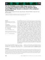
Fig. 1. R24 is rapidly and specifically endocytosed in GD3-expressing CHO-K1 cells. (A) CHO-K1 cells (GD3+) were incubated with R24 for
45 min at 4 °C. After washing of the cells, the temperature was shifted to 37 °C to allow endocytosis of the complex GD3–R24, and cells
were fixed at 15, 30 and 60 min. R24 antibody was detected by using anti-mouse IgG conjugated with Alexa
488
. Single confocal sections of
0.7 lm were taken parallel to the coverslip. Cell boundaries (white lines) are indicated at 30 and 60 min. The perinuclear region is also indic-
ated (arrows) at 15 and 30 min. The fluorescence micrographs shown are representative of five independent experiments. (B) Wild-type
CHO-K1 cells (wt, GD3–) were incubated with R24 for 45 min at 4 °C. Then the temperature was shifted to 37 °C, and cells were fixed at
2 h. R24 antibody detection was carried out as indicated in (A). (C) Cells from clone 2 (GD3+) were incubated with R24 at 4 °C to label the
surface pool of GD3 ganglioside, and then shifted to 37 °C to restore transport. Cells at 15, 30, 60 and 90 min were acid stripped to remove
membrane-bound antibody, harvested, and the presence of internalized R24 analyzed by western blot. The expression of Sial-T2–hemagglut-
inin (GD3 synthetase) in the same membrane was analyzed as a control of protein loading. IgG (L), Light chain of IgG. Scale bars: 20 lm.
Endocytic trafficking of an antibody to GD3 R. Iglesias-Bartolome
´
et al.
1746 FEBS Journal 273 (2006) 1744–1758 ª 2006 The Authors Journal compilation ª 2006 FEBS
The internalized R24 antibody is targeted and
transiently accumulated at the recycling
endosome
After internalization, a significant fraction of R24 was
located in a perinuclear region that resembles the
Golgi complex or the recycling endosomes as these or-
ganelles have a pericentriolar distribution in CHO-K1
cells [19]. In an effort to identify the juxtanuclear com-
partment where the endocytosed R24 antibody is con-
centrated, we performed extensive colocalization with
markers of both recycling endosome and Golgi com-
plex (Fig. 3). After 30 min of internalization, no colo-
calization was observed between R24 and GalNAc-T
fused to yellow fluorescent protein (YFP), a TGN
marker. However, we observed extensive colocalization
between R24 and the GTPase Rab11, an established
recycling endosome marker [19]. In addition, we also
found substantial overlapping of R24 with coendocyto-
sed Alexa
647
-transferrin in a perinuclear compartment,
demonstrating that a significant fraction of endocyto-
sed R24 in CHO-K1 cells was present in the recycling
endosome. The fraction of perinuclear labeling of R24
that does not overlap with transferrin and Rab11
would be associated with different recycling endosome
membranes, as it has been suggested, on the basis of
cellubrevin and endocytosed transferrin juxtanuclear
localization, that these compartments may be subdivi-
ded into distinct populations [20].
The R24 antibody is recycled to the plasma
membrane and released into the culture medium
As shown previously, we found that most of the endo-
cytosed R24 was not targeted to lysosomes but transi-
ently accumulated at the recycling endosome. After 60
or 90 min of R24 endocytosis, we could not detect the
internalized antibody by biochemical and immunologi-
cal techniques. An explanation for these results is that
R24 may be recycling from the pericentriolar endocytic
compartment to the plasma membrane where it is
released into the culture medium.
To address this issue, cells from clone 2 were incuba-
ted for 10 min on ice to inhibit intracellular transport
and then with R24 for 45 min on ice. Afterwards, cells
were allowed to internalize the antibody for 20 min at
A
B
Fig. 2. R24 antibody is not degraded in lysosomes. (A) CHO-K1 cells (GD3+) were incubated with R24 for 45 min at 4 °C. After washing of
the cells, the temperature was shifted to 37 °C to allow endocytosis of the complex GD3–R24, and cells were fixed at 30 min. R24 antibody
was detected by using anti-mouse IgG conjugated with Alexa
488
. For lysosome staining, cells were incubated with 0.2 lM acidotropic probe
LysoTracker Red DND-99 for 15 min at 37 °C before fixation. Single confocal sections of 0.7 lm were taken parallel to the coverslip. Cell
boundaries (white lines) are indicated. Scale bar: 20 lm (B) GD3-expressing CHO-K1 cells were incubated with R24 for 45 min at 4 °C
(0 min). After washing of the cells, the temperature was shifted to 37 °C to allow internalization of R24 antibody in the absence or presence
of 30 m
M NH
4
Cl or 60 lM chloroquine over 90 min at 37 °C. Then cells were harvested and the presence of R24 antibody analyzed by west-
ern blot. The expression of Sial-T2–hemagglutinin (GD3 synthetase) in the same membrane was analyzed as a control of protein loading.
R. Iglesias-Bartolome
´
et al. Endocytic trafficking of an antibody to GD3
FEBS Journal 273 (2006) 1744–1758 ª 2006 The Authors Journal compilation ª 2006 FEBS 1747
37 °C, and then the temperature was changed again to
4 °C. The cell surface was then stripped of any remain-
ing antibody with acid wash. At this point, cells only
contained R24 in intracellular compartments. Subse-
quently, prewarmed culture medium was added to the
cells, which were maintained at 37 °C to restore intra-
cellular transport. Cells and culture medium were
recovered at different times, and the presence of R24 in
both samples was analyzed by western blot. As shown
in Fig. 4, at the beginning of the time-course experi-
ment (stripped cells, 0 min) the antibody was present
only in the cell fraction. At 15 min, it was detected in
both fractions (cells and culture medium), and at
60 min most of it was recovered from the culture med-
ium. The antibody recovered from the culture medium
was found to have the expected molecular mass (whole
molecule) in gels run under nonreducing conditions
(results not shown). Together, these results indicate
that R24, once internalized, is recycled to the plasma
membrane and released into the culture medium.
R24 antibody recycling is sensitive to BFA and
dependent on dynamin activity
It has previously been shown that transferrin receptor
recycling as well as the formation of clathrin-coated
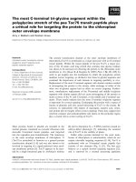
Fig. 3. The internally accumulated R24 antibody colocalizes with recycling endosome markers but not with the Golgi marker GalNAc-T.
CHO-K1 cells (GD3+) transiently expressing both GalNAc-T-YFP (upper row, pseudo colored green) and wild-type GFP-Rab11 (GFP-Rab11
wt, middle row) were incubated with anti-GD3 IgG for 45 min at 4 °C. After washing of the cells, the temperature was shifted to 37 °Cto
allow endocytosis of the complex GD3–R24 for 30 min. R24 antibody was detected by using rhodamine-conjugated goat anti-mouse IgG.
In another set of experiments, uptake of Alexa
647
-transferrin (Alexa
647
-Tf, pseudo colored green) was monitored simultaneously with R24
endocytosis (lower row). Expression of Rab11 and GalNAc-T was detected by the intrinsic fluorescence of GFP and YFP, respectively. Cell
boundary (white line) is indicated in the upper row. Insets in merge panels show details at higher magnification. The fluorescence micro-
graphs shown are representative of three independent experiments. Scale bars: 10 lm.
Endocytic trafficking of an antibody to GD3 R. Iglesias-Bartolome
´
et al.
1748 FEBS Journal 273 (2006) 1744–1758 ª 2006 The Authors Journal compilation ª 2006 FEBS
pits on the recycling endosome is inhibited in the pres-
ence of BFA [21–23]. If clathrin-coated vesicles play a
role in R24 recycling, BFA should interfere with this
pathway. To test this hypothesis, GD3-expressing
CHO-K1 cells were incubated on ice with R24 for
45 min. The cells were then incubated on ice in the
absence (control) and presence of 2 lgÆmL
)1
BFA for
15 min. At the end of this period, cells where washed
to remove unbound antibody, and then prewarmed
culture medium supplemented with BFA, when neces-
sary, was added, and the cells were transferred to
37 °C to allow endocytosis for different times.
Results shown in Fig. 5A indicate that BFA did not
affect R24 internalization, because the fraction of
internalized and accumulated antibody at 30 min was
similar in both control and BFA-treated cells. As
shown previously, 90 min after internalization we
could not detect the presence of intracellular antibody;
however, in BFA-treated cells a significant fraction of
the antibody remained accumulated in a perinuclear
compartment, consistent with the requirement of clath-
rin-coated vesicles for efficient R24 recycling. The
intracellular accumulation of R24 at 90 min was also
detectable by western blot experiments (Fig. 5B).
Under these conditions, we also found in BFA-treated
cells substantial overlapping of R24 with co-endocyto-
sed Alexa
647
-transferrin and with GalNAc-T-YFP, a
TGN marker (Fig. 5C, first row). These results clearly
demonstrate a fusion between the recycling endosomal
system and the TGN in the presence of BFA, as previ-
ously described [24].
Dynamins function in the pinching off of clathrin-
coated vesicles. It has previously been demonstrated
that the transferrin receptor egresses recycling endo-
somes, at least in part, by endosome-derived clathrin-
coated vesicles in a dynamin-dependent manner [22].
To study the requirement for dynamin in R24 depar-
ture from recycling endosome, GD3-expressing CHO-
K1 cells were transiently transfected to express both
wild-type (wtDyn2) and the dominant-negative form of
dynamin-2 (Dyn2K44 A), a mutant defective in GTP
loading and hydrolysis. After 24 h, cells were incuba-
ted with R24 at 4 °C for 45 min and then induced to
internalize the antibody by shifting the temperature to
37 °C. Cells were fixed at 30, 60 and 90 min, and the
presence of R24 was visualized by immunofluorescence
and confocal microscopic analysis. Results shown in
Fig. 6 demonstrate that wild-type dynamin-2 did not
affect R24 internalization (30 min) and later depletion
(60 and 90 min). On the other hand, the dominant-
negative version of dynamin-2 did not have much
effect on R24 internalization (30 min), but it signifi-
cantly affected the departure of R24 from the recycling
endosome (60 and 90 min). Taken together our data
indicate that recycling endosome-derived, clathrin-coa-
ted vesicles play a role in the endosomal recycling
pathway of the R24 antibody.
R24 antibody recycling is also sensitive to
monensin
It was previously proposed that endosomal acidifica-
tion is a prerequisite for the actual formation of the
carrier structures that move the TGN proteins
(TGN38 or furin) from the endosome back to the
TGN [25]. In an attempt to learn more about the
mechanism of endosomal recycling of R24, we
examined the effect of monensin, an ionophore that
dissipates pH gradients across organelle membranes
[26], on R24 recycling.
A
B
Fig. 4. R24 antibody is recycled to the plasma membrane and
released into the culture medium. (A) GD3-expressing CHO-K1 cells
were incubated with R24 antibody for 45 min on ice. Afterwards,
cells were allowed to internalize the antibody for 20 min at 37 °C,
and then the temperature was shifted again to 4 °C. The cell sur-
face was then stripped of any remaining antibody with acid wash
(0 min). Cells were then incubated at 37 °C to restore intracellular
transport, and cells and culture medium were recovered at 15 and
60 min. The presence of R24 antibody in both samples was ana-
lyzed by western blot as indicated in Experimental procedures. (B)
The relative contribution of bands in each condition was calculated
using the computer software
SCION IMAGE on the scanned film
shown in (A). Ponceau S staining was used to normalize levels of
proteins seeded in each lane. The band intensity for R24 antibody
at 0 min (cellular fraction) was arbitrarily taken as 1. Results are
representative of four independent experiments.
R. Iglesias-Bartolome
´
et al. Endocytic trafficking of an antibody to GD3
FEBS Journal 273 (2006) 1744–1758 ª 2006 The Authors Journal compilation ª 2006 FEBS 1749
GD3-expressing CHO-K1 cells were incubated with
the antibody for 45 min on ice. Then cells were incu-
bated on ice for another 15 min in the absence (con-
trol) or presence of 10 lm monensin. At the end of
this period, the unbound antibody was removed by
washing, and internalization was allowed to continue
A
B
C
Endocytic trafficking of an antibody to GD3 R. Iglesias-Bartolome
´
et al.
1750 FEBS Journal 273 (2006) 1744–1758 ª 2006 The Authors Journal compilation ª 2006 FEBS
at 37 °C for different times. Results shown in Fig. 5A
indicate that monensin did not affect R24 internalizat-
ion, as it was similar in both control and treated cells
at 30 min. As described above, at 90 min after inter-
nalization we could not detect intracellular antibody in
untreated cells; however, in monensin-treated cells a
significant fraction of the antibody remained accumu-
lated in a perinuclear compartment. The intracellular
Fig. 6. R24 antibody recycling is dependent
on dynamin activity. GD3-expressing CHO-
K1 cells were transiently transfected to
express both wild-type (wtDyn2) and the
dominant-negative form of dynamin-2
(Dyn2K44 A). After 24 h, cells were incub-
ated with the R24 antibody at 4 °Cfor
45 min and then allowed to internalize the
antibody by shifting the temperature at
37 °C. Cells were fixed at 30, 60 and
90 min, and the presence of R24 analyzed
by using rhodamine-conjugated goat anti-
mouse IgG. Single confocal sections of
0.7 lm were taken parallel to the coverslip.
Arrows indicate dynamin-transfected cells.
Cell boundaries (white lines) are indicated at
30, 60 and 90 min. Scale bars: 10 lm.
Fig. 5. R24 antibody recycling is sensitive to BFA and monensin. (A) GD3-expressing CHO-K1 cells were incubated on ice with the R24 anti-
body for 45 min. Then cells were incubated on ice in the absence (control) and presence of 2 lgÆmL
)1
BFA or 10 lM monensin for 15 min.
Cells where washed to remove unbound antibody and then prewarmed culture medium supplemented with BFA or monensin, when neces-
sary, was added and cells transferred to 37 °C to allow endocytosis for 30 and 90 min. R24 antibody was detected by using anti-mouse
IgG conjugated with Alexa
488
. Single confocal sections of 0.7 l m were taken parallel to the coverslip. Cell boundaries (white lines) are indic-
ated at 30 and 90 min. (B) GD3-expressing CHO-K1 cells were incubated with R24 for 45 min at 4 °C (0 min). After washing of the cells, the
temperature was shifted to 37 °C to allow internalization of R24 antibody in the absence (C) or presence of 2 lgÆmL
)1
BFA or 10 lM monen-
sin (Mon) over 30 and 90 min at 37 °C. Then cells were harvested and the presence of R24 antibody was analyzed by western blot in sam-
ples containing equal amounts of total proteins. (C) Same experiment as in (A) except that GalNAc-T-YFP was transiently expressed 24 h
before R24 and Alexa
647
-transferrin (Alexa
647
-Tf) internalization. Insets in merge panels show details at higher magnification. Triple color imag-
ing of single fixed CHO-K1 cells were taken with filters for rhodamine, Cy5 and YFP (first, second, and third panels from left, respectively).
The fourth panel is a merging of these images. Triple colocalization is visualized in white, and colocalization between R24 antibody and
Alexa
647
-transferrin is visualized in pink. Scale bars: 10 lm.
R. Iglesias-Bartolome
´
et al. Endocytic trafficking of an antibody to GD3
FEBS Journal 273 (2006) 1744–1758 ª 2006 The Authors Journal compilation ª 2006 FEBS 1751
accumulation of R24 in monensin-treated cells was
also observed in western blot experiments at 30 and
90 min (Fig. 5B). We also found in monensin-treated
cells overlapping of R24 antibody with co-endocytosed
Alexa
647
-transferrin. However, in contrast with the
effect of BFA, in monensin-treated cells we only
observed a slight colocalization between R24 antibody
and GalNAc-T-YFP, a TGN marker (Fig. 5C, second
row). Our initial interpretation of these results is that
they indicate that, like TGN38 and the TGN protease
furin, endosomal acidification is probably required for
R24 antibody to exit the recycling endosome.
R24 antibody is also internalized, recycled to
plasma membrane, and released into culture
medium in SK-Mel 28 melanoma cells
To investigate if the internalization and recycling path-
way of R24 antibody is a common feature that occurs
in other cell types, we analyzed the endocytic transport
of this antibody in SK-Mel 28 melanoma cells. The
radioactive labeling pattern of gangliosides from
SK-Mel 28 cells is shown in Fig. 7A. As previously
reported [17], GD3 and GM3 were the major ganglio-
sides expressed in this cell line, which run as doublets
because of differences in ceramide structure. Melan-
oma cells were incubated with R24, the unbound anti-
body was removed by washing, and internalization
was induced by transferring the cells to 37 °C for dif-
ferent periods of time. As shown in Fig. 7B, R24 was
efficiently internalized in SK-Mel 28 cells at 15, 30 and
60 min. Colocalization between endocytosed R24 and
transferrin was observed after 30 min (results not
shown). In this cell line, tubules from endocytic recyc-
ling are distributed more widely throughout the cyto-
plasm. Compared with in CHO-K1 cells, the antibody
showed a lower rate of intracellular disappearance. In
western blot experiments we observed that, at 60 and
90 min, a fraction of the antibody still remained asso-
ciated with intracellular structures whereas a significant
fraction was found in the culture medium (Fig. 7C).
Discussion
We have followed the entire endocytic itinerary of
the mouse monoclonal antibody to GD3, R24 in
GD3-expressing CHO-K1 and SK-Mel 28 cells. We
found that endocytosed R24 first appears in a diffuse,
A
B
C
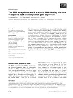
Fig. 7. R24 antibody is internalized, recycled to the plasma membrane, and released into culture medium in SK-Mel 28 melanoma cells. (A)
SK-Mel 28 melanoma cells were metabolically labeled from [
14
C]galactose for 24 h. Lipid extracts were prepared, purified, chromatographed
and visualized as indicated in Experimental procedures. The positions of cochromatographed glycolipid standards are indicated. The asterisk
indicates the position of an unidentified lipid. (B) SK-Mel 28 melanoma cells were incubated with R24 for 45 min at 4 °C. After washing of
the cells, the temperature was shifted to 37 °C to allow endocytosis of the complex GD3–R24, and cells were fixed at 15, 30 and 60 min.
R24 antibody was detected by using anti-mouse IgG conjugated with Alexa
488
. Single confocal sections of 0.7 lm were taken parallel to the
coverslip. (C) SK-Mel 28 melanoma cells were incubated with R24 antibody for 45 min on ice. Cells were allowed to internalize the antibody
for 20 min at 37 °C, and then the temperature was shifted again to 4 °C. The cell surface was then stripped of any remaining antibody with
acid wash (0 min). Then cells were incubated at 37 °C to restore intracellular transport, and cells and culture medium were recovered at 30,
60 and 90 min. The presence of R24 antibody in both samples was analyzed by western blot as indicated in Experimental procedures. Scale
bars: 20 lm.
Endocytic trafficking of an antibody to GD3 R. Iglesias-Bartolome
´
et al.
1752 FEBS Journal 273 (2006) 1744–1758 ª 2006 The Authors Journal compilation ª 2006 FEBS
punctate distribution in the cytoplasm that is consis-
tent with the sorting compartment of the early endo-
somes. Subsequently, R24 appeared concentrated in a
pericentriolar distribution which we have characterized
as the recycling endosome. After that, the antibody is
recycled to the plasma membrane and released into the
culture medium by a BFA ⁄ monensin-sensitive, dynam-
in-dependent recycling pathway. In addition, no evi-
dence was found for targeting and degradation of R24
antibody in lysosomes. These observations suggest that
R24 follows an endocytic pathway typical of other
recycling proteins, such as the model recycling protein
transferrin receptor. Nevertheless, in this study we des-
cribe for the first time the entire endocytic itinerary of
a glycolipid antibody.
Antibody-binding techniques are extensively used
to follow endocytic transport of proteins such as
glycosylhoshatidylinositol-anchored proteins [27–29],
major histocompatibility complex class I protein and
interleukin 2 a-subunit receptor [30], integrin b1 [31]
and cation-independent mannose 6-phosphate receptor
[32]. Antibodies tend not to have a significant effect
on the endocytic behavior of the proteins studied
[31,32]. Lysosome and endosome compartments have
an acidic pH, which both promotes the dissociation of
ligands such as low-density lipoprotein from their
receptors and the proper function of hydrolytic
enzymes. We demonstrate that the association of R24
with the GD3 ganglioside was unaffected even after
1 h at pH 5 or 6 (Fig. S1), suggesting that the GD3–
R24 complex could not be disrupted in acidic organ-
elles. Taking all these antecedents together, it is plaus-
ible that the itinerary of the R24 antibody reflects the
intracellular transit of the disialo ganglioside GD3. In
this vein, it has been demonstrated that exogenous
glycosphingolipids can be internalized and directed to
the Golgi apparatus, where they can be reglycosylated
and then delivered back to the cell surface [8,33]. Also,
internalized sphingolipid analogs (BODIPY-and NBD-
labeled lipids) can be recycled to the plasma mem-
brane via endosomes or through the Golgi complex
[34,35]. However, it should be taken into consideration
that the quantitative and qualitative behavior of the
analog lipids is quite different from long-chain cellular
lipids, as the ability of short fluorescent lipids to
diffuse spontaneously between different membranes is
not generally shared by their endogenous lipid
counterparts [36]. Even the binding of antibodies and
toxins to endogenously synthesized lipids may perturb
the natural behavior of these molecules. In a similar
way, it was reported that cholera toxin alters the
internalization mechanism of a fluorescent GM1
ganglioside [37].
It has been reported that transferrin receptor and its
ligand transferrin recycle to the plasma membrane with
the same kinetics as certain lipids [34] and independ-
ently of the transferrin receptor cytoplasmic domain
[38]. These data suggest that recycling of molecules from
endosome to plasma membrane can occur without
active recruitment of cytosolic coat proteins (bulk flow
process). However, more recently dynamin-dependent
transferrin receptor recycling by endosome-derived
clathrin-coated vesicles was reported [22]. Supporting
this observation, results obtained in this work using
BFA and the dominant-negative form of dynamin
indicate that recycling endosome-derived, clathrin-
coated vesicles may play a role in the endosomal recyc-
ling pathway of the R24 antibody. Previous studies
using a cross-linkable form of clathrin light chain indi-
cated that, once internalized, the return of the transfer-
rin receptor to the cell surface was largely insensitive
to clathrin cross-linking [39], consistent with the notion
that clathrin does not play a role in trafficking mole-
cules from the endosome back to the plasma mem-
brane. However, these results do not entirely exclude
the possibility that clathrin-coated pits may be oper-
ating in the transport of transferrin receptor back to
the plasma membrane in the absence of the cross-
linker. Alternatively, clathrin independent recycling
pathways may also be involved in the recycling of
molecules to the plasma membrane, as discussed
below. If it is assumed that R24 is associated with the
luminal membrane-bounded GD3 during the recycling
pathway, the complex must be segregated into
specialized domains to be sequestered by nascent clath-
rin-coated vesicles. Three properties are key to lipid
sorting: headgroup interactions, lipid shape and mem-
brane-order parameters [40]. GD3–R24 interactions
with a protein may cause the lipid to be sorted on the
basis of the characteristics of the protein, and such a
mechanism is important for the trafficking of gly-
cosylphosphatidylinositol-anchored proteins [41]. We
recently demonstrated that GD3 ganglioside, like gly-
cosylphosphatidylinositol-anchored proteins, is mainly
expressed in glycosphingolipid-enriched microdomain
(also called detergent-resistant membranes or rafts),
dynamic assemblies of cholesterol, saturated phospho-
lipids, and sphingolipids [16,18]. The partition of GD3
into glycosphingolipid-enriched microdomains may
represent a positive sorting signal for the correct sub-
cellular trafficking, as previously described for other
lipid-anchored proteins [42,43]. Finally, it is known
that dynamin participates in both clathrin-mediated
endocytosis and caveolae internalization [37]. Thus, the
lack of effect of the dominant-negative form of
dynamin on R24 internalization (Fig. 6) suggests that
R. Iglesias-Bartolome
´
et al. Endocytic trafficking of an antibody to GD3
FEBS Journal 273 (2006) 1744–1758 ª 2006 The Authors Journal compilation ª 2006 FEBS 1753
endocytosis of the GD3–R24 complex may occur
through clathrin-independent and caveolar-independ-
ent processes.
Results from this work indicate that BFA-treated
cells failed to recycle a fraction of endocytosed R24
antibody. A simple interpretation of these results is
that R24 antibody may recycle via a single route and
that BFA is inefficient or reduces the rate of R24
recycling. However, another explanation is that two
recycling pathways might be operating for R24 anti-
body, as previously demonstrated for endocytosed
transferrin receptor [23]. The first involves passage
through a recycling endosome. Transport from this
compartment to the plasma membrane involves clath-
rin-coated vesicles which bud off from the recycling
endosome. Results indicate that BFA and the domin-
ant-negative form of dynamin interfere with this path-
way. The second recycling pathway bypasses the
recycling endosome and involves direct transfer from
the sorting endosome to the plasma membrane. On
this subject, we demonstrate that R24 antibody is
endocytosed and accumulated in sorting endosomes at
16 °C colocalizing to some extent with the GTPase
Rab5 (Fig. S2). A similar analysis is valid for the inter-
pretation of results obtained using the carboxylic iono-
phore monensin because, in monensin-treated cells, a
proportion of endocytosed R24 antibody failed to
recycle to the plasma membrane. This intracellular
pool of R24 was found overlapping with co-endocyto-
sed Alexa
647
-transferrin, strongly suggesting that
monensin perturbs the departure of R24 from the
recycling endosome and hence that endosomal acidifi-
cation is probably required for the formation of the
carrier structures that move R24.
Results from this work also indicate that R24, once
internalized, is recycled to the plasma membrane and
released into the culture medium. How can it be
explained that R24 is not associated with the plasma
membrane after recycling and release into the extracel-
lular medium? If R24 antibody is associated with GD3
during the entire endocytic trafficking, the complex
could be dissociated in the plasma membrane probably
by a shift in the equilibrium in the absence of a signifi-
cant concentration of free antibody in the medium.
However, this appears improbable, as we showed that
the association of R24 with GD3 was unaffected over
1 h at pH 7.4 (Fig. S1). On the other hand, evidence is
accumulating that supports a physiological role for
exosomes in the removal of the transferrin receptor
during reticulocyte development, although exosomes
have been isolated from the cell culture of many non-
haematopoietic cells [44]. However, we also exclude
the possibility that the GD3–R24 complex is released
into the extracellular medium through vesicle-mediated
secretion (exosomes) as R24 present in the culture
medium was not pelleted by centrifugation at
100 000 g (Fig. S3). Further work is required to define
the precise mechanism involved in the release of R24
antibody into the extracellular medium.
The recycle pathway described in this work is likely
to be of considerable biological and immunological
significance. Antibodies to GD3 are being used as thera-
peutic agents for melanoma [13]. However, the rapid
internalization of R24 antibody observed in GD3-
expressing cells may be potentially detrimental to its
therapeutic use as it could not be linked to pathways
of complement-dependent and cellular-dependent anti-
cancer activity. It would be possible to exploit the
internalization feature for selective delivery of cyto-
toxic agents to GD3-expressing cancer cells. Finally,
the results of this study may provide a better under-
standing of how antibodies to glycosphingolipids
associated with neuropathies harm the different com-
partments of the peripheral nerve [9]. However, more
information is needed about the rate and routes of
intracellular transport of antibodies to glycosphingo-
lipids associated with neurological disorders.
Experimental procedures
Cell lines, cell culture and DNA transfection
The following cells were used: wild-type CHO-K1 cells
(ATCC, Manassas, VA, USA); CHO-K1 clone 2, a stable
Sial-T2 (tagged at the C-terminus with the nanopeptide epi-
tope of the viral hemagglutinin) transfectant expressing the
ganglioside GD3 [15,16]; and the SK-Mel 28 melanoma cell
line (ATCC). Cells were grown and maintained at 37 °Cin
5% CO
2
in Dulbecco’s modified Eagle’s medium (DMEM)
supplemented with 10% fetal bovine serum and antibiotics.
Transient transfections were carried out with 1 lg per
35-mm dish using cationic liposomes (Lipofectamine;
Invitrogen, Carlbad, CA, USA) essentially according to the
manufacturer’s instructions and incubated for 24 h at 37 °C
with the transfection reagent and DNA mixture.
Expression plasmids
The GTPase Rab11a–GFP wild-type construct was kindly
provided by M. Colombo (Universidad Nacional de Cuyo,
Mendoza, Argentina); plasmids coding for wild-type dyn-
amin-2 (wtDyn2), dynamin-2 K44A (Dyn2K44 A) and
GFP-Rab5 were supplied by J. Bonifacino (NICHD,
National Institutes of Health, Bethesda, MD, USA). The
construct containing the cDNA coding for the N-terminal
domain (cytosolic tail, transmembrane domain, and a few
Endocytic trafficking of an antibody to GD3 R. Iglesias-Bartolome
´
et al.
1754 FEBS Journal 273 (2006) 1744–1758 ª 2006 The Authors Journal compilation ª 2006 FEBS
amino acids from the stem region) of GalNAc-T fused to
the N-terminus of the YFP (GalNAc-T-YFP) was obtained
by subcloning the corresponding cDNA fragments into
the plasmid pEYFP-N1 (Clontech, Mountain View, CA,
USA) [6].
Cell labeling and internalization assays
Cells transiently transfected or not with plasmids indicated
above were incubated on ice for 10 min to inhibit intra-
cellular transport. Then, cells were incubated on ice for
45 min with hybridoma (ATCC No. HB-8445) supernatant
containing R24 antibody in order to label GD3 ganglioside
expressed on the cell surface. Afterwards, cells were washed
three times with cold NaCl ⁄ P
i
, transferred to 37 °C with
fresh prewarmed complete DMEM to allow antibody inter-
nalization for different times, and finally harvested by scra-
ping or fixed in 3% paraformaldehyde in NaCl ⁄ P
i
for
30 min at 4 °C. Where indicated, growth medium was sup-
plemented with 30 mm NH
4
Cl, 60 lm chloroquine,
2 lgÆmL
)1
BFA or 10 lm monensin. BFA and monensin
were present for at least 15 min before R24 endocytosis
was allowed and for different times after the temperature
change. Control cells were supplemented with the same
amount of vehicle. For transferrin internalization, cells were
first incubated for 90 min in DMEM without fetal bovine
serum, then incubated at 4 °C in cold DMEM containing
10 lgÆmL
)1
Alexa
647
-transferrin (Molecular Probes, Eugene,
OR, USA) and R24 antibody for 45 min, and then trans-
ferred to 37 °C with prewarmed DMEM, without fetal
bovine serum, supplemented with 10 lgÆmL
)1
Alexa
647
-
transferrin and processed at different times. For lysosome
staining, cells were incubated in DMEM without fetal
bovine serum supplemented with 0.2 lm acidotropic probe
LysoTracker Red DND-99 (Molecular Probes) for 15 min
at 37 °C before fixation. Where indicated, noninternalized
antibody remaining at the cell surface was removed by acid
stripping with 0.5% acetic acid buffer, pH 3.0, containing
0.5 m NaCl for 1 min on ice.
R24 recycling assay
Cells from clone 2 (GD3-expresing CHO-K1 cells) or
SK-Mel 28 cells were incubated for 10 min on ice to inhibit
intracellular transport and then with the R24 antibody for
45 min on ice. Cells were then transferred to 37 °C for
20 min to allow R24 endocytosis. Cell surface-bound anti-
body was then removed by acid stripping at 4 °C, and cells
were extensively washed with cold NaCl ⁄ P
i
. Then they were
incubated at 37 °C with 1 mL prewarmed fresh DMEM in
order to restore transport of internalized antibody. Cells
and culture medium were recovered at different times. Pro-
teins from the culture medium were precipitated with chlo-
roform ⁄ methanol (1 : 4, v ⁄ v) and resuspended in NaCl ⁄ P
i
.
The presence of R24 in cells and culture medium was
assessed by western blot.
R24 recycling in BFA-treated and monensin-treated
cells
Clone 2 cells were incubated on ice for 10 min to inhibit
intracellular transport and then with R24 antibody for
60 min with 2 lgÆmL
)1
BFA or 10 lm monensin present in
the culture medium for the last 15 min. Subsequently, cells
were washed three times with cold NaCl ⁄ P
i
and transferred
to 37 °C with fresh prewarmed complete DMEM supple-
mented or not with BFA or monensin as indicated. At dif-
ferent times, cells were harvested or fixed as described
below. The presence of the R24 antibody was assessed by
western blot or in cells by immunofluorescence and confo-
cal microscopic analysis.
Confocal immunofluorescence microscopy
Cells grown on coverslips were washed twice with NaCl ⁄ P
i
,
fixed in 3% paraformaldehyde in NaCl ⁄ P
i
for 30 min at
4 °C and permeabilized with 0.1% Triton X-100 ⁄ 200 mm
glycine in NaCl ⁄ P
i
for 10 min at 4 °C. Then, cells were
washed with NaCl ⁄ P
i
and exposed to secondary antibodies
for 90 min at 37 °C. Secondary antibodies were Alexa
488
-
conjugated goat anti-mouse IgG (Santa Cruz Biotechno-
logy, Santa Cruz, CA, USA) or rhodamine-conjugated goat
anti-mouse IgG (Sigma-Aldrich, St Louis, MO, USA), both
diluted 1 : 1000. After final washes with 1% BSA in
NaCl ⁄ P
i
, cells were mounted in FluorSave reagent (Calbio-
chem, EMD Biosciences, La Jolla, CA, USA). Cells stained
with LysoTracker or transferrin were fixed with 4% para-
formaldehyde in NaCl ⁄ P
i
for 30 min at room temperature,
incubated with 50 mm NH
4
Cl for 10 min, permeabilized
with 0.1% saponin ⁄ 2% BSA in NaCl ⁄ P
i
and then proc-
essed as indicated above.
Confocal images were collected using a Carl Zeiss LSM5
Pascal laser-scanning confocal microscope (Carl Zeiss, Jena,
Germany) equipped with an argon ⁄ helium ⁄ neon laser and a
100 · 1.4 numerical aperture, oil immersion objective (Zeiss
Plan-Apochromat). Single confocal sections of 0.7 lm were
taken parallel to the coverslip (xy sections). Images were
acquired and processed with the Zeiss lsm image software.
Final images were compiled with adobe photoshop 7.0. The
fluorescence micrographs shown in this manuscript are
representative of at least three independent experiments.
Electrophoresis and western blotting
Proteins were resolved by electrophoresis in 12% polyacryl-
amide gels under reducing or nonreducing conditions and
then transferred electrophoretically to nitrocellulose mem-
branes for 1 h at 300 mA. Protein bands in nitrocellulose
R. Iglesias-Bartolome
´
et al. Endocytic trafficking of an antibody to GD3
FEBS Journal 273 (2006) 1744–1758 ª 2006 The Authors Journal compilation ª 2006 FEBS 1755
membranes were visualized by Ponceau S staining. For
immunoblotting, nonspecific binding sites on the nitrocellu-
lose membrane were blocked with 5% defatted dry milk in
400 mm NaCl ⁄ 0.05% Tween 20 ⁄ 100 mm Tris ⁄ HCl, pH 7.5
buffer. Anti-hemagglutinin was used at a dilution of
1 : 1000. R24 and anti-hemagglutinin were detected directly
with an anti-mouse IgG coupled to horseradish peroxidase
and combined with the chemiluminescence detection kit
(ECL Plus; Amersham Biosciences, Little Chalfont, UK)
and Hyperfilm MP films (Amersham Biosciences). Mole-
cular mass was calculated based on calibrated standards
(BenchMark prestained protein ladder; Invitrogen) run in
every gel. The relative contribution of individual bands was
calculated using the computer software scion image (Scion
Corporation, Frederick, MD, USA) on scanned films of
low exposure images. Final images were compiled with
adobe photoshop 7.0.
Metabolic labeling, lipid extraction and
chromatography
For glycolipid analysis, cells in culture (3 · 10
5
cells per
35 mm dish) were labeled with 2 lCiÆmL
)1
d-[U-
14
C]galac-
tose ([
14
C]Gal; 329.5 mCiÆmmol
)1
; DuPont NEN, Boston,
MA, USA) for 24 h. After being washed with cold
NaCl ⁄ P
i
, cells were harvested by scraping from the plate
and pelleted by centrifugation. Lipids were extracted from
the cell pellet with chloroform ⁄ methanol (2 : 1, v ⁄ v) and
freed from water-soluble contaminants by passing through
a Sephadex G-25 column (Amersham Biosciences). The
lipid extract was supplemented with appropriate amounts
of standard gangliosides and developed on high-perform-
ance thin layer chromatography (Merck, Whitehouse Sta-
tion, NJ, USA) using chloroform ⁄ methanol ⁄ 0.2% CaCl
2
(60 : 36 : 8, by vol.) as solvent. Standard gangliosides were
visualized by exposing the plate to iodine vapors. Rou-
tinely, 2000–4000 cpm was spotted on each lane. Radioact-
ive gangliosides were visualized using a Fuji Photo Film
Bio Imagen analyzer (Tokyo, Japan) or visualized by
fluorography after dipping the plate in 0.4% melted 2,5-di-
phenyloxazole in 2-methylnaphthalene and exposure to a
radiographic film at )70 °C, usually for 5–7 days [2].
Acknowledgements
This work was supported in part by Grants from Sec-
retarı
´
a de Ciencia y Tecnologı
´
a (SECyT)-Universidad
Nacional de Co
´
rdoba, Consejo Nacional de Investigaci-
ones Cientı
´
ficas y Te
´
cnicas (CONICET, grant No. PIP
5151), Fundacio
´
n Antorchas (Grant No. 14116-112)
and Agencia Nacional de Promocio
´
n Cientı
´
fica y Tec-
nolo
´
gica (FONCYT, Grant No. 01-13522). We thank
G. Schachner and S. Deza for technical assistance with
cell culture, and C. Mas for excellent assistance with
confocal microscopy and image analysis. R.I.B.,
P.M.C. and G.A.G. are recipients of CONICET
(Argentina) Fellowships. J.L.D. is Career Investigator
of CONICET (Argentina).
References
1 Hakomori S, Handa K, Iwabuchi K, Yamamura S &
Prinetti A (1998) New insights in glycosphingolipid
function: ‘glycosignaling domain’, a cell surface assem-
bly of glycosphingolipids with signal transducer mole-
cules, involved in cell adhesion coupled with signaling.
Glycobiology 8, xi–xix.
2 Zurita AR, Maccioni HJ & Daniotti JL (2001) Modula-
tion of epidermal growth factor receptor phosphoryla-
tion by endogenously expressed gangliosides. Biochem J
355, 465–472.
3 Miljan EA & Bremer EG (2002) Regulation of growth
factor receptors by gangliosides. Sci STKE 2002, RE15,
doi: 10.1126/stke.2002.160.re15.
4 Proia RL (2003) Glycosphingolipid functions: insights
from engineered mouse models. Philos Trans R Soc
Lond B Biol Sci 358, 879–883.
5 Zurita AR, Crespo PM, Koritschoner NP & Daniotti
JL (2004) Membrane distribution of epidermal growth
factor receptors in cells expressing different gangliosides.
Eur J Biochem 271, 2428–2437.
6 Giraudo CG, Daniotti JL & Maccioni HJ (2001) Physi-
cal and functional association of glycolipid N-acetyl-
galactosaminyl and galactosyl transferases in the Golgi
apparatus. Proc Natl Acad Sci USA 98, 1625–1630.
7 Crespo PM, Iglesias-Bartolome R & Daniotti JL (2004)
Ganglioside GD3 traffics from the trans-Golgi network
to plasma membrane by a Rab11-independent and bre-
feldin A-insensitive exocytic pathway. J Biol Chem 279,
47610–47618.
8 Tettamanti G (2004) Ganglioside ⁄ glycosphingolipid
turnover: new concepts. Glycoconj J 20, 301–317.
9 Willison HJ & Yuki N (2002) Peripheral neuropathies
and anti-glycolipid antibodies. Brain 125, 2591–2625.
10 Zhang X & Kiechle FL (2004) Review: glycosphingo-
lipids in health and disease. Ann Clin Lab Sci 34, 3–13.
11 Thomas PB, Delatte SJ, Sutphin A, Frankel AE &
Tagge EP (2002) Effective targeted cytotoxicity of neu-
roblastoma cells. J Pediatr Surg 37, 539–544.
12 Lee FT, Rigopoulos A, Hall C, Clarke K, Cody SH,
Smyth FE, Liu Z, Brechbiel MW, Hanai N, Nice EC,
et al. (2001) Specific localization, gamma camera ima-
ging, and intracellular trafficking of radiolabelled chi-
meric anti-G (D3) ganglioside monoclonal antibody
KM871 in SK-MEL-28 melanoma xenografts. Cancer
Res 61, 4474–4482.
13 Choi BS, Sondel PM, Hank JA, Schalch H, Gan J,
King DM, Kendra K, Mahvi D, Lee L-Y, Kim K et al.
Endocytic trafficking of an antibody to GD3 R. Iglesias-Bartolome
´
et al.
1756 FEBS Journal 273 (2006) 1744–1758 ª 2006 The Authors Journal compilation ª 2006 FEBS
(2005) Phase I trial of combined treatment with ch14.18
and R24 monoclonal antibodies and interleukin-2 for
patients with melanoma or sarcoma. Cancer Immunol
Immunother, doi: 10.1007/s00262-005-0069-7.
14 Guillemard V & Saragovi HU (2004) Novel approaches
for targeted cancer therapy. Curr Cancer Drug Targets
4, 313–326.
15 Daniotti JL, Martina JA, Giraudo CG, Zurita AR &
Maccioni HJ (2000) GM3 alpha2,8-sialyltransferase
(GD3 synthase): protein characterization and sub-golgi
location in CHO-K1 cells. J Neurochem 74, 1711–
1720.
16 Crespo PM, Zurita AR & Daniotti JL (2002) Effect of
gangliosides on the distribution of a glycosylphosphati-
dylinositol-anchored protein in plasma membrane from
Chinese hamster ovary-K1 cells. J Biol Chem 277,
44731–44739.
17 Pukel CS, Lloyd KO, Travassos LR, Dippold WG,
Oettgen HF & Old LJ (1982) GD3, a prominent gang-
lioside of human melanoma. Detection and characterisa-
tion by mouse monoclonal antibody. J Exp Med 155,
1133–1147.
18 Crespo PM, Zurita AR, Giraudo CG, Maccioni HJ &
Daniotti JL (2004) Ganglioside glycosyltransferases and
newly synthesized gangliosides are excluded from deter-
gent-insoluble complexes of Golgi membranes. Biochem
J 377, 561–568.
19 Gomez GA & Daniotti JL (2005) H-Ras dynamically
interacts with recycling endosomes in CHO-K1
cells: involvement of Rab5 and Rab11 in the trafficking
of H-Ras to this pericentriolar endocytic compartment.
J Biol Chem 280, 34997–35010.
20 Teter K, Chandy G, Quinones B, Pereyra K, Machen T
& Moore HP (1998) Cellubrevin-targeted fluorescence
uncovers heterogeneity in the recycling endosomes.
J Biol Chem 273, 19625–19633.
21 Stoorvogel W, Oorschot V & Geuze HJ (1996) A novel
class of clathrin-coated vesicles budding from endo-
somes. J Cell Biol 132, 21–33.
22 van Dam EM & Stoorvogel W (2002) Dynamin-depen-
dent transferrin receptor recycling by endosome-derived
clathrin-coated vesicles. Mol Biol Cell 13, 169–182.
23 van Dam EM, Ten Broeke T, Jansen K, Spijkers P &
Stoorvogel W (2002) Endocytosed transferrin receptors
recycle via distinct dynamin and phosphatidylinositol
3-kinase-dependent pathways. J Biol Chem 277, 48876–
48883.
24 Lippincott-Schwartz J, Yuan L, Tipper C, Amherdt M,
Orci L & Klausner RD (1991) Brefeldin A’s effects on
endosomes, lysosomes, and the TGN suggest a general
mechanism for regulating organelle structure and mem-
brane traffic. Cell 67, 601–616.
25 Chapman RE & Munro S (1994) Retrieval of TGN pro-
teins from the cell surface requires endosomal acidifica-
tion. EMBO J 13, 2305–2312.
26 Mollenhauer HH, Morre DJ & Rowe LD (1990) Altera-
tion of intracellular traffic by monensin; mechanism,
specificity and relationship to toxicity. Biochim Biophys
Acta 1031, 225–246.
27 Nichols BJ, Kenworthy AK, Polishchuk RS, Lodge R,
Roberts TH, Hirschberg K, Phair RD & Lippincott-
Schwartz J (2001) Rapid cycling of lipid raft markers
between the cell surface and Golgi complex. J Cell Biol
153, 529–541.
28 Sabharanjak S, Sharma P, Parton RG & Mayor S
(2002) GPI-anchored proteins are delivered to recy-
cling endosomes via a distinct cdc42-regulated, cla-
thrin-independent pinocytic pathway. Dev Cell 2, 411–
423.
29 Polishchuk R, Di Pentima A & Lippincott-Schwartz J
(2004) Delivery of raft-associated, GPI-anchored pro-
teins to the apical surface of polarized MDCK cells by
a transcytotic pathway. Nat Cell Biol 6, 297–307.
30 Naslavsky N, Weigert R & Donaldson JG (2003) Con-
vergence of non-clathrin- and clathrin-derived endo-
somes involves Arf6 inactivation and changes in
phosphoinositides. Mol Biol Cell 14, 417–431.
31 Powelka AM, Sun J, Li J, Gao M, Shaw LM, Sonnen-
berg A & Hsu VW (2004) Stimulation-dependent recy-
cling of integrin beta1 regulated by ARF6 and Rab11.
Traffic 5, 20–36.
32 Lin SX, Mallet WG, Huang AY & Maxfield FR
(2004) Endocytosed cation-independent mannose 6-
phosphate receptor traffics via the endocytic recycling
compartment en route to the trans-Golgi network and
a subpopulation of late endosomes. Mol Biol Cell 15,
721–733.
33 Furukawa K, Thampoe IJ, Yamaguchi H & Lloyd KO
(1989) The addition of exogenous gangliosides to cul-
tured human cells results in the cell type-specific expres-
sion of novel surface antigens by a biosynthetic process.
J Immunol 142, 848–854.
34 Mayor S, Presley JF & Maxfield FR (1993) Sorting of
membrane components from endosomes and subsequent
recycling to the cell surface occurs by a bulk flow pro-
cess. J Cell Biol 121, 1257–1269.
35 Pagano RE (2003) Endocytic trafficking of glycosphingo-
lipids in sphingolipid storage diseases. Philos Trans R Soc
Lond B Biol Sci 358, 885–891.
36 Wang TY & Silvius JR (2000) Different sphingolipids
show differential partitioning into sphingolipid ⁄ choles-
terol-rich domains in lipid bilayers. Biophys J 79, 1478–
1489.
37 Singh RD, Puri V, Valiyaveettil JT, Marks DL, Bittman
R & Pagano RE (2003) Selective caveolin-1-dependent
endocytosis of glycosphingolipids. Mol Biol Cell 14,
3254–3265.
38 Johnson LS, Dunn KW, Pytowski B & Mcgraw TE
(1993) Endosome acidification and receptor trafficking:
bafilomycin A1 slows receptor externalization by a
R. Iglesias-Bartolome
´
et al. Endocytic trafficking of an antibody to GD3
FEBS Journal 273 (2006) 1744–1758 ª 2006 The Authors Journal compilation ª 2006 FEBS 1757
mechanism involving the receptor’s internalization
motif. Mol Biol Cell 4, 1251–1266.
39 Moskowitz HS, Heuser J, Mcgraw TE & Ryan TA
(2003) Targeted chemical disruption of clathrin function
in living cells. Mol Biol Cell 14, 4437–4447.
40 Maxfield FR & Mcgraw TE (2004) Endocytic recycling.
Nat Rev Mol Cell Biol 5, 121–132.
41 Kjoller L, Simonsen AC, Ellgaard L & Andreasen PA
(1995) Differential regulation of urokinase-type-1 inhibi-
tor complex endocytosis by phorbol esters in different
cell lines is associated with differential regulation of
alpha 2-macroglobulin receptor and urokinase receptor
expression. Mol Cell Endocrinol 109 , 209–217.
42 Brown DA & Rose JK (1992) Sorting of GPI-anchored
proteins to glycolipid-enriched membrane subdomains
during transport to the apical cell surface. Cell 68, 533–
544.
43 Keller P, Toomre D, Diaz E, White J & Simons K
(2001) Multicolour imaging of post-Golgi sorting and
trafficking in live cells. Nat Cell Biol 3, 140–149.
44 de Gassart A, Geminard C, Hoekstra D & Vidal M
(2004) Exosome secretion: the art of reutilizing non-
recycled proteins? Traffic 5, 896–903.
Supplementary material
The following supplementary material is available
online:
Fig. S1. Effect of pH on GD3 ganglioside–R24 anti-
body association. CHO-K1 cells (GD3+) were incub-
ated with R24 for 45 min at 4 °C (To). After being
washed, cells were incubated in NaCl ⁄ Pi solution at
pH 7.4, 6.0 or 5.0 for 1 h at 4 °C. Then cells and cul-
ture medium (NaCl ⁄ Pi solution) were recovered, and
the presence of R24 antibody in both samples was ana-
lyzed by western blot as indicated in Experimental pro-
cedures.
Fig. S2. R24 antibody is endocytosed and accumulated
in sorting endosomes at 16 °C. (A) GD3-expressing
CHO-K1 cells were incubated at 16 °C for 15 min and
then incubated with the R24 antibody at 16 °C for
1 h. Then cells were acid stripped to remove mem-
brane-bound antibody, fixed (left panel) or transferred
to 37 °C and fixed at 30 min (right panel). The pres-
ence of R24 analyzed by using Alexa
488
-conjugated
goat anti-mouse IgG. Cell boundaries (white lines) are
indicated. (B) GD3-expressing CHO-K1 cells were
transiently transfected with GFP-Rab5 wild-type
(GFP-Rab5wt). After 24 h, cells were incubated at 16
°C for 15 min and then with R24 at 16 °C for 1 h.
Cells were then acid stripped to remove membrane-
bound antibody and fixed. The presence of R24 was
analyzed by using rhodamine-conjugated goat anti-
mouse IgG (red). Expression of Rab5 was detected by
the intrinsic fluorescence of GFP (green). Single confo-
cal sections of 0.7 lm were taken parallel to the cover-
slip. Insets in the merge panel show details at higher
magnification. The upper inset shows a clear segrega-
tion of R24 antibody and Rab5 in sorting endosomes.
On the other hand, the lower inset shows endosomes
where colocalization between R24 antibody and Rab5
is clearly visualized (yellow areas). Scale bars: 10 lm.
Fig. S3. R24 antibody released into the culture med-
ium after recycling is not associated with exosomes.
CHO-K1 cells were incubated with R24 antibody for
45 min on ice. Afterwards, cells were allowed to intern-
alize the antibody for 20 min at 37 °C, and then the
temperature was shifted again to 4 °C. The cell surface
was then stripped of any remaining antibody with acid
wash (0 min). Then cells were incubated at 37 °Cto
restore intracellular transport, and cells and culture
medium were recovered at 30 and 60 min. The culture
medium was then centrifuged at 100 000 g for 2 h, and
the pellet (P) and supernatant (SN) fractions were
recovered. The presence of R24 antibody was analyzed
in all samples (cells, P and SN) by western blot as indic-
ated in Experimental procedures.
This material is available as part of the online article
from
Endocytic trafficking of an antibody to GD3 R. Iglesias-Bartolome
´
et al.
1758 FEBS Journal 273 (2006) 1744–1758 ª 2006 The Authors Journal compilation ª 2006 FEBS