Báo cáo khoa học: Characterization of a novel long-chain acyl-CoA thioesterase from Alcaligenes faecalis docx
Bạn đang xem bản rút gọn của tài liệu. Xem và tải ngay bản đầy đủ của tài liệu tại đây (437.58 KB, 14 trang )
Characterization of a novel long-chain acyl-CoA
thioesterase from Alcaligenes faecalis
Puja Shahi*
†
, Ish Kumar*
‡
, Ritu Sharma, Shefali Sanger and Ravinder S. Jolly
Institute of Microbial Technology, Chandigarh, India
Long-chain acyl-CoA thioesterases (EC 3.1.2.2) hydro-
lyze acyl-CoA esters to nonesterified fatty acids and
coenzyme A (CoASH) [1]. These are ubiquitously
expressed in bacteria, yeast, plants and mammals, and
in most cell compartments, such as endoplasmic reticu-
lum, cytosol, mitochondria and peroxisomes. Several
unrelated thioesterases have been purified to homogen-
eity from plants, animals, and bacteria, and the
cDNAs encoding several of them have been cloned
and sequenced [2–7]. Although the physiological func-
tions of these enzymes remain largely unknown, it is
speculated that they regulate lipid metabolism by
maintaining appropriate concentrations of acyl-CoA,
CoASH, and nonesterified fatty acids. The only estab-
lished function for acyl-CoA thioesterases is in the
termination of fatty acid synthesis in eukaryotes [8].
Two thioesterases, I and II, that cleave acyl-CoA
molecules in vitro have been characterized from
Keywords
Alcaligenes faecalis; immunogold electron
microscopy; long-chain acyl-CoA;
p-nitrophenyl esters; thioesterase
Correspondence
R. S. Jolly, Institute of Microbial
Technology, Sector 39, Chandigarh 160 036,
India
Fax: +91 172 269 0585
Tel: +91 172 269 0908
E-mail:
*These authors contributed equally to this
paper
†Present address
Department of Physiology and Biophysics,
University of Iowa, Iowa city, IA 52242,
USA
‡Present address
Department of Chemistry, Wesleyan Univer-
sity, Middletown, CT 06459, USA
(Received 19 January 2006, revised 12
March 2006, accepted 22 March 2006)
doi:10.1111/j.1742-4658.2006.05244.x
A novel long-chain acyl-CoA thioesterase from Alcaligenes faecalis has
been isolated and characterized. The protein was extracted from the cells
with 1 m NaCl, which required 1.5-fold, single-step purification to yield
near-homogeneous preparations. In solution, the protein exists as homo-
meric aggregates, of mean diameter 21.6 nm, consisting of 22-kDa sub-
units. MS ⁄ MS data for peptides obtained by trypsin digestion of the
thiosterase did not match any peptide from Escherichia coli thioesterases or
any other thioesterases in the database. The thioesterase was associated
exclusively with the surface of cells as revealed by ultrastructural studies
using electron microscopy and immunogold labeling. It hydrolyzed satur-
ated and unsaturated fatty acyl-CoAs of C
12
to C
18
chain length with V
max
and K
m
of 3.58–9.73 lmolÆmin
)1
Æ(mg protein)
)1
and 2.66–4.11 lm, respect-
ively. A catalytically important histidine residue is implicated in the active
site of the enzyme. The thioesterase was active and stable over a wide
range of temperature and pH. Maximum activity was observed at 65 °C
and pH 10.5, and varied between 60% and 80% at temperatures of
25–70 °C and pH 6.5–10. The thioesterase also hydrolyzed p-nitrophenyl
esters of C
2
to C
12
chain length, but substrate competition experiments
demonstrated that the long-chain acyl-CoAs are better substrates for thio-
esterase than p-nitrophenyl esters. When assayed at 37 and 20 °C, the affin-
ity and catalytic efficiency of the thioesterase for palmitoleoyl-CoA and
cis-vaccenoyl-CoA were reduced approximately twofold at the lower tem-
perature, but remained largely unaltered for palmitoyl-CoA.
Abbreviations
DTNB, 5,5¢-dithiobis(2-nitrobenzoic acid); TEM, transmission electron microscopy.
2374 FEBS Journal 273 (2006) 2374–2387 ª 2006 IMTECH
Escherichia coli. Thioesterase I, encoded by the tesA
gene, is a periplasmic protein of 20.5 kDa and has an
active site similar to serine proteases, consistent with
its inhibition with di-isopropyl fluorophosphate [2,9].
Thioesterase II, encoded by the tesB gene, is a tetra-
meric protein with identical subunits of 32 kDa and is
insensitive to inhibition with di-isopropyl fluorophos-
phate.
A histidine residue present at position 58 in thioest-
erase II has been implicated in the cleavage of the
thioester bond [10,11]. Two thioesterases with striking
similarities in their physical properties to thioesterase I
and II have been reported from photosynthetic bac-
teria, Rhodopseudomonas sphaeroides [12,13].
Cho and Cronan [2] prepared null mutants of both
tesA and tesB in an attempt to determine the role of
these thioesterases. The deletion of tesA, tesB or both
genes has no effect on the growth or lipid composition
of E. coli. However, the possibility of another enzyme
taking over the function of both enzymes or the pres-
ence of a third thioesterase in E. coli has not been
ruled out. The overexpression of either tesA or tesB to
levels that greatly exceed normal also has no effect on
the growth of E. coli [2,11]. It has also been shown
that E. coli thioesterases are unable to cleave acyl-
ACPs in vivo [14].
Recently, Schulz and coworkers [15] provided some
evidence for the function of cytoplasmic thioesterase of
E. coli in b-oxidation. They showed that oleate is
mostly degraded via the classical, isomerase-dependent
pathway in E. coli, but that a small amount of 2-trans-
5-cis-tetradecadienoyl-CoA is diverted from the path-
way by conversion into 3,5-cis-tetradecadienoyl-CoA
by D
3
,D
2
-enoyl-CoA isomerase. The 3,5- intermediate,
which would strongly inhibit b-oxidation if allowed to
accumulate, is hydrolyzed and the resultant 3,5-tetra-
decadienoate is excreted into the growth medium. In
another study, Zheng et al. [16] coexpressed thioest-
erase II with (R)-3-hydroxydecanoyl–acyl carrier pro-
tein-CoA transacylase (PhaG, encoded by the phaG
gene) to clarify the physiological role of thioesterase II.
3-Hydroxydecanoic acid was produced in E. coli by
mobilizing PhaG. By using an isogenic tesB (encoding
thioesterase II)-negative knockout E. coli strain, CH01,
it was found that the expression of tesB and phaG can
up-regulate each other. In addition, 3-hydroxydecanoic
acid was synthesized from glucose or fructose by
recombinant E. coli harboring phaG and tesB. This
study supports the hypothesis that the physiological
role of thioesterase II in E. coli is to prevent the
abnormal accumulation of intracellular acyl-CoA.
We have isolated a thioesterase from Alcaligenes
faecalis ISH108 and demonstrated its application in
chemoselective and racemization free deacylation of
thiol esters [17]. A. faecalis was isolated from soil sam-
ples during routine screening of micro-organisms for
various biotransformation applications. In this paper,
we describe the intracellular localization and character-
ization of the thioesterase. The wild-type expression of
protein was sufficiently large to obtain milligram quan-
tities of the protein from about 20 g of cells, which
required only 1.5-fold, single-step purification to
obtain a near homogeneous preparation.
Results
Isolation and purification of thioesterase
The thioesterase extracted from the cells with 1 m
NaCl, as described in Experimental procedures, exhib-
ited a specific activity of 4.72 lmolÆmin
)1
Æ(mg pro-
tein)
)1
. The enzyme could also be extracted by
suspending freshly grown cells in 50 mm Tris ⁄ HCl
buffer saturated with butanol at pH 7.4 [specific activ-
ity 2.5 lmolÆmin
)1
Æ(mg protein)
)1
]. Other extraction
systems, used to extract thioesterase from the cells,
included 1 m NaCl with 0.1% Triton X-100 [specific
activity 2.82 l molÆ min
)1
Æ(mg protein)
)1
] and butan-
1-ol-saturated Tris ⁄ HCl buffer containing 0.1% Triton
X-100 [specific activity 2.43 lmolÆmin
)1
Æ(mg pro-
tein)
)1
].
As the specific activity in 1 m NaCl extract was the
highest, it was selected as the method of choice for the
isolation of the enzyme. SDS ⁄ PAGE of the NaCl
extract, run under reducing conditions, showed a prom-
inent band (> 90%) at 22 kDa, which suggested that
further purification of the protein could be achieved by
size exclusion chromatography. Preliminary investiga-
tion using Sephadex G-75 (fractionation range
3–80 kDa) revealed that the native size of the protein
was much larger as it moved into the void volume of
the column. This allowed ultrafiltration with a 50-kDa
Centricon membrane (Amicon, Bedford, MA, USA)
for concentration of the samples or buffer change. In
the first attempt, Sephacryl S-300 (fractionation range
10–1500 kDa) was selected for the purification of
protein. The NaCl extract obtained was desalted and
concentrated using 50-kDa Centricon membranes. The
concentrated sample was loaded on the column pre-
equilibrated with 50 mm Tris ⁄ HCl buffer (pH 7.6)
containing 150 mm NaCl. The column was eluted with
the same buffer at a flow rate of 24 mLÆh
)1
. Thioest-
erase activity moved near the void volume of the col-
umn (Fig. 1A). Finally, the protein was purified on a
Sepharose CL-4B (fractionation range 60–20 000 kDa)
column (Fig. 1B). SDS ⁄ PAGE of the purified protein,
P. Shahi et al. Thioesterase of Alcaligenes faecalis
FEBS Journal 273 (2006) 2374–2387 ª 2006 IMTECH 2375
run under reducing conditions, showed a single band at
22 kDa (Fig. 2A). Sepharose CL-4B chromatography
resulted in 1.5-fold purification with 50% yield.
Molecular mass of thioesterase
The elution profile of the protein on gel filtration col-
umns indicated the protein to be a large homomeric
aggregate of 22-kDa subunits. On the Sepharose CL-4B
column, the thioesterase was eluted just after the higher-
molecular-mass (2000 kDa) fraction of dextran blue
and much before the standard protein, thyroglobulin
(669 kDa) (Fig. 1C). The inability of the protein to move
in native PAGE (anodic disc-PAGE using Tris ⁄ glycine,
as electrophoresis buffer, pH 8.3, or cathodic disc PAGE
using alanine ⁄ acetic acid buffer, pH 4.5), run under non-
reducing conditions, is also consistent with this observa-
tion. Finally, the aggregated structure of native protein
was established by transmission electron microscopy
(TEM). The purified sample was concentrated by
repeated ultrafiltration using a Centricon 50-kDa mem-
brane and suspended in water at a concentration of
600 lgÆmL
)1
. TEM was performed on a carbon grid
using 2% aqueous uranyl acetate and 2% phosphotung-
stic acid at pH 8.0. An electron micrograph showed
granular structures with a mean diameter of 21.6 nm
(Fig. 3A). The size distribution is shown in Fig. 3B.
Intracellular localization of thioesterase
We carried out electron microscopic immunogold labe-
ling studies with ultrathin sections of Alcaligenes cells
to localize thioesterase at the ultrastructural level.
Polyclonal antibodies, AbTE-N and AbTE-D, raised
against purified native enzyme and the piece of gel cor-
responding to the 22-kDa monomeric protein on
SDS ⁄ PAGE, respectively, were assayed for their specif-
icity by western blotting. AbTE-D antibodies were
used to rule out any nonspecific binding that might
have occurred with AbTE-N because of the aggregated
nature of the native protein. The purified enzyme was
run on SDS ⁄ PAGE, and, after electroblotting on to
nitrocellulose membrane, it was probed with AbTE-N
and AbTE-D (Fig. 2C). Both antibodies identified the
22-kDa band corresponding to the monomer of thio-
esterase enzyme on denaturing gel.
Alcaligenes was grown to mid-exponential phase,
and, after several dehydration steps, embedded in LR
White resin, which was then dehydrated in several
steps. Optimal ultrastructural preservation required
inclusion of 0.2% glutaraldehyde in the fixative; the
reactivity of the antibody was not affected by glutaral-
dehyde fixation. Thin sections cut using an ultramicro-
tome were incubated with primary antibodies followed
by nanogold labeled secondary antibody as described
in Experimental procedures and visualized under the
transmission electron microscope.
Fraction No
A
B
C
0 5 10 15 20 25 30
A
mn 082
A
mn 082
A
mn 082
0.0
0.1
0.2
0.3
0.4
0.5
0.6
L
m/lomn
0
100
200
300
400
500
600
700
800
Fraction No
51015202530
0.0
0.1
0.2
0.3
0.4
Lm/nim/lo
m
n
0
100
200
300
400
500
600
Fraction No
20 30 40 50 60 70
0.0
0.1
0.2
0.3
0.4
0.5
0.6
a
b
c
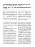
Fig. 1. Elution profile of thioesterase on gel filtration chromatogra-
phy. (A) 13 · 425 mm Sephacryl S-300 column; (B) 13 · 290 mm
Sepharose S-4B column. The column was pre-equilibrated with
100 m
M Tris ⁄ HCl buffer, pH 7.6, containing 150 mM NaCl at a flow
rate of mLÆh
)1
. Then 250 lg protein was loaded and eluted in the
same buffer. Fractions of 2.0 mL each were collected and assayed
for thioesterase activity as described in Experimental procedures. (d)
activity units. (C) 13 · 290 mm Sepharose S-4B column, flow rate
10 mLÆh
)1
. Peak A, Blue dextran; B, thioesterase; C, thyroglobulin.
Thioesterase of Alcaligenes faecalis P. Shahi et al.
2376 FEBS Journal 273 (2006) 2374–2387 ª 2006 IMTECH
Different fields were observed, and the gold particles
were found to be exclusively present on the surface of
the cells. The immunogold labeling was much more
abundant for cells where AbTE-N (Fig. 4A,B) was used
as primary antibody than for those where AbTE-D
(Fig. 4C) was used. No labeling occurred in the control
cells where primary antibodies were derived from pre-
immunized serum (Fig. 4D).
Does A. faecalis have multiple thioesterases?
Total thioesterase activity could not be extracted from
the cells with NaCl even after several treatments. To
find the presence of any other thioesterase, 10 g cells,
suspended in phosphate buffer, pH 7.0 (total volume
25 mL) were sonicated in three batches of equal volume
and fractionated into soluble and particulate fractions
by ultracentrifugation (100 000 g for 4 h). Most of the
thioesterase activity (> 80%) was present in the soluble
fraction, but the particulate fraction was also found to
be active. SDS ⁄ PAGE of both these fractions,
run under reducing conditions showed the presence of a
22-kDa protein (Fig. 2B). The particulate fraction on
incubation with 1 mL phosphate buffer, pH 7.0, con-
taining 1 m NaCl, released most of the activity. Total
proteins were combined and, after removal of particu-
lates, were concentrated to 2.5 mL by ultrafiltration
(3-kDa membrane; filtrate was devoid of thioesterase
activity). Then 1 mL of the concentrated protein was
applied to a Sephacryl S-300 gel-filtration column
(13 · 530 mm), pre-equilibrated with Tris ⁄ HCl buffer,
pH 7.5, containing 150 mm NaCl at a flow rate of
24 mLÆh
)1
and eluted in the same buffer. Fractions of
1.0 mL volume were collected and assayed, but thioest-
erase activity was detected only in the void volume.
SDS ⁄ PAGE of the void volume under reducing condi-
tions showed the presence of only 22-kDa protein.
Thus, we could identify only one thioesterase activity in
A. faecalis, in contrast with E. coli and Rhodopseudo-
monas sphaeroides, each of which contained two thio-
esterases; in addition, a third one has been implicated
in E. coli [2].
Mass spectrometry
Tandem MS was performed by Midwest Bio Services
(Overland Park, KS, USA) on an LCQ Deca XP Plus
ion trap mass spectrometer (ThermoFinnigan, Arcade,
NY, USA). SDS ⁄ PAGE of purified protein was run
under reducing conditions. The thioesterase band at
22 kDa from the Coomassie-stained gel was excised
and subjected to in-gel trypsinization. The resulting
peptide mixture was concentrated on a peptide trap col-
umn and washed to remove salts and other impurities.
The peptides were separated on a microcapillary C18
reverse-phase chromatography column, and the eluted
peptides sprayed directly into the mass spectrometer.
MS ⁄ MS spectral data were obtained and analyzed by
comparing them with the NCBI nonredundant protein
sequence database using turbosequest and peeks
online software (informaticssolutions.
com:8080/peaksonline/). The observed MS ⁄ MS spectra
did not match any peptide from E. coli thioesterases or
any other thioesterase in the database. The following
14.2
20.1
24.0
29.0
36.0
45.0
66.0
66.0
45.0
36.0
29.0
24.0
20.1
14.2
kDa
kDa
2 1
1
3
2
1
2
3
A
B
C
Fig. 2. Purification of thioesterase, fraction-
ation of thioesterase activity and western
blot with antibodies to thioesterase. (A) Pro-
tein samples run on 12.5% SDS ⁄ PAGE after
purification. Lane 1, molecular mass marker;
lane 2, purified enzyme. (B) Fractionation of
thioesterase. The particulate and soluble
fractions, obtained by ultracentrifugation of
sonicated cells, were run on gel. Lane 1,
purified thioesterase; lane 2, membrane
fraction; lane 3, soluble fraction. (C) West-
ern blotting. Lane 1, marker; lane 2, antibod-
ies raised against purified protein (AbTE-N);
lane 3, antibodies raised against gel purified
and denatured protein (AbTE-D) were used
as primary antibody followed by horseradish
peroxidase-conjugated anti-rabbit IgG.
P. Shahi et al. Thioesterase of Alcaligenes faecalis
FEBS Journal 273 (2006) 2374–2387 ª 2006 IMTECH 2377
peptide fragments were obtained: YYDDNIWIAL
DYCDYYQLTHKPASLEK, LTKDAKYLEKAKET
YAWTK, AKETYA + WTKKHLCDPTDHLYWD
NINLKGKVSK, VSKEKYAYNSGQMIQAGVLLY
EETGD + EQYLR, which showed identity with a
putative hydrolase from Bacteroides thetaiotaomicron
VPI-5482 (accession gi|29339943|gb|AAO77738.1|) [18].
Attempts at preparation of thioesterase null
mutants by chemical mutagenesis
An attempt was made to obtain thioesterase null mutant
by N-methyl-N¢-nitrosoguanidine mutagenesis using the
standard protocol [19], but without any success.
Approximately 10 000 colonies were screened in one set
for any mutation in the gene encoding thioesterase
enzyme with the help of plate assay [20]. The mutants
giving negative or ambiguous thioesterase assay results
were further analyzed for thioesterase activity by the
5,5¢-dithio-bis-(2-nitrobenzoic acid) (DTNB) method.
No mutant could be found that lacked thioesterase
activity.
Substrate specificity of thioesterase
Thioesterase-catalyzed hydrolysis of acyl-CoA
derivatives
The effect of chain length on the activity of thio-
esterase was studied. The initial rate of hydrolysis of a
series of saturated acyl-CoA derivatives at different
concentrations in the range 1.5–60 lm by thioesterase
(0.2 lg) was determined. At saturating concentrations,
stearoyl-CoA (C
18:0
) was the most active substrate,
with the rate of hydrolysis decreasing with decreasing
chain length. The acyl-CoAs of chain length longer
than C
18
, were not studied. The rates of hydrolysis of
palmitoyl-CoA (C
16:0
) and myristoyl-CoA (C
14:0
)at
saturating concentrations were similar.
The enzyme showed very little activity towards octa-
noyl-CoA, which required a higher concentration of
enzyme for detectable activity. No activity was
observed with acyl-CoAs having chain length smaller
than C
8
. The thioesterase also possessed activity
towards unsaturated long-chain acyl-CoA derivatives.
V
max
and K
m
values were determined by least-squares
analysis of double-reciprocal plots of the data obtained
from the corresponding Michaelis–Menten plots.
V
max
and K
m
values were in the range 3.58–9.73
lmolÆmin
)1
Æ(mg protein)
)1
and 2.66–4.11 lm, respect-
ively (Table 1).
Thioesterase-catalyzed hydrolysis of p-nitrophenyl
esters
The assay mixture (1 mL) consisted of 400 lm p-nitro-
phenyl derivative in 0.1 m phosphate buffer, pH 7.2,
containing 0.1 m NaCl. The reaction was started by
the addition of 0.2 lg thioesterase. Initial rates were
determined by measuring the increase in A
346
(e ¼
4800 m
)1
Æcm
)1
), the isosbestic point of the p-nitrophe-
nol ⁄ p-nitrophenoxide couple, as described [21]. The
effect of the chain length of the p-nitrophenyl esters
on the activity of thioesterase was studied (Table 2).
p-Nitrophenyl propanoate (C
3:0
) was found to be the
most active substrate, with the activity decreasing
sharply with increasing or decreasing chain length.
Diameter
(
nm
)
10 15 20 25 30 35 40
selcitraP fo rebmuN
0
10
20
30
40
50
A
B
Fig. 3. TEM. The purified thioesterase was desalted and concentra-
ted by repeated ultrafiltration using a Centricon 50-kDa membrane
and suspended in water at a concentration of 600 lgÆmL
)1
.TEM
was performed on the carbon grid using 2% aqueous uranyl acet-
ate and 2% phosphotungstic acid at pH 8.0. (A) Electron micro-
graph showing granular particles with mean diameter 21.6 nm. (B)
Size distribution of the thioesterase particles in TEM. Particle size
distribution was evaluated by measuring the diameter of 100 parti-
cles. The diameter was the mean of two right angled axes.
Thioesterase of Alcaligenes faecalis P. Shahi et al.
2378 FEBS Journal 273 (2006) 2374–2387 ª 2006 IMTECH
p-Nitrophenyl acetate (C
2:0
) and p-nitrophenyl hexano-
ate (C
6:0
) were 30% as active as their (C
3:0
) counter-
part. p-Nitrophenyl dodecanoate (C
12:0
) showed less
than 10% of the maximum activity, whereas p-nitro-
phenyl esters with chain length of more than C
12
were
not hydrolyzed by the thioesterase. p-Nitrophenyl pro-
panoate was a threefold more active substrate than the
best acyl-CoA substrate, stearoyl-CoA. It is interesting
to note that chain length specificity of Alcaligenes thio-
esterase for p-nitrophenyl esters was opposite to that
for acyl-CoA derivatives.
The thioesterase activity and p-nitrophenyl esterase
activities appear to be co-resident for the following
reasons. (a) Diethyl pyrocarbonate completely inhib-
ited both activities. (b) p-Nitrophenyl propanoate
provided protection against inhibition of thioesterase
activity by diethyl pyrocarbonate to the extent of 74%.
The experiment was performed as follows. To a pre-
cooled solution of enzyme (100 lg) in 1 mL phosphate
buffer, pH 6.0, was added 50 lm p-nitrophenyl prop-
ionate, immediately followed by 1 mm diethyl pyrocar-
bonate (from 1 m ethanol stock solution). A parallel
experiment was run as a control in which the addition
of substrate was omitted. The sample was incubated
for 5 min, and a 5-lL aliquot of each sample was
withdrawn and assayed for activity by the DTNB
method using stearoyl-CoA as substrate. (c) In an
analogous manner, when stearoyl-CoA was used as
AB
CD
Fig. 4. Transmission electron micrographs
of immunogold-labeled Alcaligenes treated
with various primary antibodies. Alcaligenes
cells were embedded in LR white resin as
described in Experimental procedures. Thin
sections were incubated with primary anti-
body raised against thioesterase, followed
by anti-rabbit IgG with conjugated nanogold
particles, and samples were analyzed under
the electron microscope. Different fields
were viewed. Arrowheads denote gold parti-
cles. (A) Primary antibody AbTE-N raised
against purified native thioesterase. (B)
Enlarged view of a single cell (bar, 200 nm).
(C) Primary antibody AbTE-D, raised against
a gel piece corresponding to the thioest-
erase band in SDS ⁄ PAGE. (D) Control in
which preimmune serum was used as pri-
mary antibody.
P. Shahi et al. Thioesterase of Alcaligenes faecalis
FEBS Journal 273 (2006) 2374–2387 ª 2006 IMTECH 2379
protecting agent and activity was assayed with p-nitro-
phenyl propanoate as substrate, 93% protection
against inhibition was observed.
Long-chain acyl-CoAs are better substrates than
aryl esters
Stearoyl-CoA provided better protection against inhi-
bition of thioesterase activity by diethyl pyrocarbonate
than p-nitrophenyl propanoate, as shown above, which
indicated that the long-chain acyl-CoAs are better sub-
strates for the enzyme than aryl esters. This was fur-
ther confirmed by substrate competition experiments.
Aryl esterase activity against p-nitrophenyl propionate
(400 lm) was inhibited by 52% when it was measured
in the presence of 100 lm stearoyl-CoA, whereas the
presence of 200 lm p-nitrophenyl propionate in the
assay mixture of stearoyl-CoA (50 lm) had no effect
on the thioesterase activity, measured by the DTNB
method.
Implication of a histidine residue in the active
site of thioesterase
The effect of various protein-modifying agents on the
activity of the thioesterase was studied (Table 3). Phe-
nylmethanesulfonyl fluoride, a serine-active agent, had
no significant effect on the activity of the enzyme.
N-Bromosuccinimide caused complete loss of activity at
1mm concentration, indicating the presence of catalyti-
cally important residues such as tyrosine, tryptophan
and histidine. However, dimethyl (2-hydroxy-5-nitro-
benzyl)sulfonium bromide, a tryptophan-modifying
agent, and N-acetylimidazole, a tyrosine inhibitor, did
not have any significant effect on the thioesterase activ-
ity. Diethyl pyrocarbonate, a histidine-modifying agent,
caused total loss of activity at 1 mm concentration. The
thiol-reactive agent DTNB did not have a significant
effect on the thioesterase activity, allowing the use of
DTNB in the thioesterase assay. Picrylsulfonic acid, a
lysine-modifying agent, also had no effect on the activ-
ity of the enzyme. These results indicate the presence of
a catalytically important histidine residue in the
enzyme. The presence of a histidine residue was further
supported by the following experiments.
(a) Reversal of inhibition by hydroxylamine. The
activity of the enzyme could be partially recovered by
the treatment of diethyl pyrocarbonate-inhibited
enzyme with hydroxylamine. An aliquot of enzyme,
inactivated with 1 mm diethyl pyrocarbonate at 4 °C,
was incubated at 25 °C, for 8 h with 250 mm hydrox-
ylamine and assayed for thioesterase activity by the
DTNB method after extensive dialysis, as described in
Experimental procedures. Its activity was expressed as
a percentage of that obtained from an experiment,
run in parallel, in which same amount of active
enzyme (no inhibitor added) was incubated with
250 mm hydroxylamine for 8 h at 25 °C and assayed
for activity in the same manner. The treatment of
the diethyl pyrocarbonate-inhibited enzyme with
Table 1. Thioesterase catalyzed hydrolysis of acyl-CoA derivatives.
A solution of acyl-CoA derivative was prepared in 100 m
M Tris ⁄ HCl
buffer, pH 7.6, at various concentrations in the range 1.5–60 l
M.
The reaction was started by the addition of an aliquot of enzyme
(0.2 lg), and initial rates of hydrolysis were measured by the DTNB
method as described in Experimental procedures. V
max
and K
m
val-
ues were determined by least-squares analysis of double-reciprocal
plots of the data obtained from the corresponding Michaelis–
Menten plots. Sr. No., serial number.
Sr. No. Substrate K
m
(lM)
V
max
[lmolÆmin
)1
Æ
(mg protein)
)1
]
1 Lauroyl-CoA 3.48 ± 0.3 3.58 ± 0.3
2 Myristoyl-CoA 2.66 ± 0.2 6.61 ± 0.5
3 Palmitoyl-CoA 4.11 ± 0.2 7.17 ± 0.3
4 Stearoyl-CoA 3.59 ± 0.1 9.73 ± 0.2
5 Myristoleoyl-CoA 3.39 ± 0.3 4.77 ± 0.4
6 Palmitoleoyl-CoA 3.90 ± 0.1 5.57 ± 0.3
7 cis-Vaccenoyl-CoA 2.84 ± 0.3 6.05 ± 0.5
Table 2. Thioesterase-catalyzed hydrolysis of aromatic esters. The assay mixture (1 mL) consisted of 400 lM p-nitrophenyl derivative in
0.1
M phosphate buffer, pH 7.2, containing 0.1 M NaCl. The reaction was started by the addition of 0.2 lg thioesterase. Initial rates were
determined by measuring the increase in A
346
(e ¼ 4800 M
)1
Æcm
)1
), the isobestic point of the p-nitrophenol ⁄ p-nitrophenoxide couple.
Sr. No. p-Nitrophenyl ester
Specific activity
[lmolÆmin
)1
Æ(mg protein)
)1
]
Relative
activity (%)
1 p-Nitrophenyl acetate 9.16 30.14
2 p-Nitrophenyl propanoate 30.52 100
3 p-Nitrophenyl butanoate 15.87 51.02
4 p-Nitrophenyl hexanoate 9.77 33.16
5 p-Nitrophenyl dodecanoate 2.44 8.67
6 p-Nitrophenyl palmitate Not detected –
7 p-Nitrophenyl stearate Not detected –
Thioesterase of Alcaligenes faecalis P. Shahi et al.
2380 FEBS Journal 273 (2006) 2374–2387 ª 2006 IMTECH
hydroxylamine in this way resulted in 58.7% recovery
of activity.
(b) A 35.7% increase in absorption at k
245
corres-
ponding to N-ethoxycarbonylation of histidine on
inactivation of thioesterase with diethyl pyrocarbonate
for 1 h was observed in differential spectra obtained as
described in Experimental procedures.
(c) Stearoyl-CoA, a substrate of the enzyme provided
93% protection against inhibition with diethyl pyro-
carbonate, as shown above.
Effect of temperature and pH on the activity of
the thioesterase
Optimum pH and pH stability
For determination of the optimum pH of the enzyme,
its activity was measured at various pHs ranging from
5.5 to 10.5 at 30 °C (Fig. 5A). The maximum activity
of thioesterase was obtained at pH 10.5. For pH 5.5–
8.0, phosphate buffers were used, in which 80% of
maximum activity was retained. For pH 7.2–9.0,
Tris ⁄ HCl buffers were used. Although the trend of
activity in the pH range 7.2–8.0 was the same as that
in phosphate buffer, the activity was 20% less than
that in phosphate buffer. For pH 9.0–10.5, sodium
carbonate buffer was used. At pH 9.0, the activity was
40% less in carbonate buffer than in Tris buffer.
The effects of buffer composition on enzyme activity
have been reported previously [22]. These effects may
be due to the effects of the buffer on the oligomeriza-
tion status of the enzyme [23]. An alternative explan-
ation is that the binding affinity of the enzyme for the
substrate is modified, presumably because of differ-
ences in the interaction of the buffer ions with the
binding site [24]. Activity at pH 10.5 was maximum
and set as 100%. Activity above pH 10.5 was not stud-
ied. Controls were used in each case to compensate for
chemical hydrolysis, which was substantial at higher
pH. Although maximum activity was obtained at
pH 10.5, all studies were carried out at pH 7.0–7.5 as
the substrates are prone to degradation under basic
conditions.
To evaluate pH stability, the enzyme was incubated
in different buffers, pH 5.5–10.5, at 30 °C for 20 h.
The remaining activity is expressed as a percentage of
the activity relative to the activity in the corresponding
buffer at time zero. Thioesterase retained almost
Table 3. Effect of protein-modifying reagents on thioesterase activ-
ity. Purified and dialyzed thioesterase at a concentration of
20 lgÆmL
)1
was incubated with each reagent at 25 °C for 15 min
and dialyzed against 50 m
M Tris ⁄ HCl buffer, pH 7.5, at 4 °C with
four buffer changes for 12 h. Residual activity, percentage of the
original activity, was calculated by the DTNB method as described
in Experimental procedures.
Sr. No. Reagent (1 m
M) Residual activity (%)
1 N-Bromosuccinimide 0.0
2 Phenylmethanesulfonyl fluoride 90.7
3 N-Acetylimidazole 90.7
4 Iodoacetamide 98.9
5 Diethyl pyrocarbonate 0.0
6 Dimethyl (2-hydroxy-5-nitrobenzyl)
sulfonium bromide
89.0
7 Picrylsulfonic acid 89.0
8 p-Chloromercuribenzoate 88.3
95,5¢-Dithiobis-(2-nitrobenzoic acid) 98.4
10 Dithiothreitol 80.0
11 None 100
pH
567891011
)%( ytivitca laudiseR
0
20
40
60
80
100
120
pH
567891011
)%( ytivitcA evitaleR
20
40
60
80
100
120
A
B
Fig. 5. (A) Effect of pH on the activity of thioesterase. Assays were
performed at 30 °C in various buffers at different pH. The activity
in carbonate buffer at pH 10.5 was set as 100%, all other values
are relative to it. (B) pH stability of thioesterase. A predetermined
amount of thioesterase was incubated in different buffers for 20 h
at 30 °C and assayed for thioesterase activity. The remaining activ-
ity is expressed as percentage of activity relative to the activity in
the corresponding buffer at time zero. (d) phosphate buffer; (.)
Tris ⁄ HCl buffer; (n) carbonate buffer.
P. Shahi et al. Thioesterase of Alcaligenes faecalis
FEBS Journal 273 (2006) 2374–2387 ª 2006 IMTECH 2381
complete activity at pH 5.5–6.0 (Fig. 5B). It retained
80% activity at pH 7.0–7.5 and was relatively unsta-
ble under alkaline conditions. After 20 h incubation at
pH 10.5 in carbonate buffer, only 20% activity was
retained.
Thermal properties of the thioesterase
Thioesterase activity was determined at different tem-
peratures in phosphate buffer at pH 7.0. Maximum
thioesterase activity occurred at 65 °C (Fig. 6A).
About 60–80% of maximum activity was retained at
temperatures of 25–70 °C. There was sharp decline in
activity at 75–80 °C; the enzyme retained 20% of
maximum activity at 80 °C.
To evaluate temperature stability, enzyme in phos-
phate buffer, pH 7.0 was incubated for 3 h at different
temperatures. Thioesterase activity was assayed at
30 °C by the DTNB method as described in Experi-
mental procedures. The activity at zero time at 30 °C
is assumed to be 100%, and all other values are
expressed relative to it. Most of the enzyme activity
was retained at 25–50 °C (Fig. 6B). After 3 h, 70% of
the activity remained at 70 °C. At 80 °C, most of the
activity was lost after 3 h.
Effect of metal ions on thioesterase activity
The effect of various bivalent metal ions on thioest-
erase activity was studied by the DTNB method as
described in Experimental procedures. Zn
2+
showed
concentration-dependent partial inhibition of the
enzyme. It had no effect on the activity at 1 mm con-
centration but caused 40% inhibition at 10 mm.Hg
2+
and Cu
2+
caused complete inhibition of enzyme activ-
ity at 1 mm concentration. The activity was enhanced
by 10–20% when enzyme assays were performed in the
presence of Mg
2+
,Ni
2+
or Co
2+
.
Effect of temperature on the kinetics of
thioesterase-catalyzed hydrolysis of
palmitoyl-CoA, palmitoleoyl-CoA and
cis-vaccenoyl-CoA
The cells were grown at 25, 30 and 37 °C and assayed
for thioesterase activity by the DTNB method. No
difference in activity was observed, ruling out a tem-
perature-dependent change in expression levels of thio-
esterase. The kinetics of thioesterase activity was
determined at 20 and 37 °C with palmitoyl-CoA,
palmitoleoyl-CoA or cis-vaccenoyl-CoA as substrate
(Table 4). With palmitoyl-CoA as substrate, thioest-
erase showed only marginal changes in V
max
and K
m
values at both the temperatures. However, approxi-
mately twofold reduced affinity and catalytic efficiency
was observed when palmitoleoyl-CoA or cis-vaccenoyl-
CoA was the substrate at the lower temperature.
Discussion
Two thioesterases, I and II, that cleave acyl-CoA mol-
ecules in vitro have been characterized from E. coli.
Thioesterase I is a periplasmic protein of 20.5 kDa and
has an active site similar to serine proteases [2,9]. Thio-
esterase II is a tetrameric protein with identical
subunits of 32 kDa and is insensitive to inhibition with
di-isopropyl fluorophosphate. A histidine residue in
thioesterase II has been implicated in the cleavage of
the thioester bond [10,11]. In comparison, thioesterase
from A. faecalis exists as large homomeric granular
aggregates (21.6 nm average diameter) of 22-kDa sub-
units (Figs 2A and 3). Phenylmethanesulfonyl fluoride,
a serine-active reagent, failed to inhibit the catalytic
activity of thioesterase. A. faecalis thioesterase was
digested with trypsin, and the resulting peptides separ-
ated on a microcapillary C18 reverse-phase chromato-
graphy column. MS ⁄ MS data were obtained and
analyzed by comparing them with the NCBI non-
redundant protein sequence database. The observed
Temperature (
o
C)
20 30 40 50 60 70 80 90
)%( ytivitca laudiseR/evitaleR
0
20
40
60
80
100
120
A
B
Fig. 6. Thermal properties of thioesterase. (A) Optimum tempera-
ture of thioesterase activity. Assays were performed in 50 m
M
phosphate buffer, pH 7.0. The relative activity is expressed as per-
centage of maximum activity attained under the experimental
conditions. (B) Thermostability of thioesterase. A predetermined
amount of thioesterase was incubated for 3 h at different tempera-
tures and then assayed for thioesterase activity in 50 m
M phos-
phate buffer at 30 °C. The activity at zero time at 30 °C is assumed
to be 100%; all other values are expressed relative to it.
Thioesterase of Alcaligenes faecalis P. Shahi et al.
2382 FEBS Journal 273 (2006) 2374–2387 ª 2006 IMTECH
MS ⁄ MS spectra did not match any peptide from
E. coli thioesterases or any other thioesterase in the
database. Thioesterase was found to be associated
exclusively with the surface of cells as revealed by
ultrastructural studies using electron microscopic im-
munogold labeling studies (Fig. 4).
A histidine residue has been implicated in the
active site of the enzyme based on the following
observations. (a) Incubation of thioesterase with
1mm diethyl pyrocarbonate resulted in complete loss
of enzyme activity. (b) An increase in absorption at
k
245
corresponding to N-ethoxycarbonylation of histi-
dine on inactivation with diethyl pyrocarbonate was
observed when differential spectra were recorded.
(c) Approximately 60% of the enzyme activity could
be recovered by the treatment of diethyl pyrocarbo-
nate-inhibited enzyme with hydroxylamine. (d) Stea-
royl-CoA, a substrate of the thioesterase, provided
93% protection against inactivation by diethyl
pyrocarbonate.
Alcaligenes thioesterase was active in and stable to a
wide range of temperatures and pH values. Maximum
activity was observed at 65 °C and pH 10.5 and varied
between 60% and 80% at 25–70 °C and pH 6.5–10
(Figs 5 and 6). Enzyme activity remained unaltered
after incubation in phosphate buffer for 3 h at 50 °C.
Thioesterase hydrolyzed saturated and unsaturated
acyl-CoAs of C
10
to C
18
chain length with V
max
and
K
m
values in the range 3.58–9.73 lmolÆmin
)1
Æ(mg pro-
tein)
)1
and 2.66–4.11 lm, respectively (Table 1). At
saturating concentrations, stearoyl-CoA (C
18:0
) was the
most active substrate, with the rate of hydrolysis
decreasing with decreasing chain length. Thioesterase
also has chymotrypsin-like activity and was able to
hydrolyze p-nitrophenyl esters of C
2
to C
12
chain
length. The most active substrate was C
3
, with the
activity falling sharply with increase or decrease in
chain length. Long-chain p-nitrophenyl esters were not
hydrolyzed by the enzyme. The odd-chain C
3
esters are
unlikely to be natural substrates of the enzyme. In any
case, the substrate competition experiments clearly
demonstrated that the long-chain acyl-CoAs are better
substrates than p-nitrophenyl esters.
The ratio of saturated ⁄ unsaturated fatty acid in mem-
brane phospholipids is tightly controlled in a tempera-
ture-dependent manner in micro-organisms, which
allows proper thermal regulation of membrane fluidity
[25–27]. Thermal regulation of membrane fluidity is
common to all organisms. Lower growth temperatures
result in an increase in the number of unsaturated
phospholipids in the membrane. E. coli can synthesize
phospholipids almost entirely from exogenous fatty
acids supplied by the growth medium. The satur-
ated ⁄ unsaturated fatty acids in membrane phospho-
lipids, synthesized from exogenous fatty acids is similar
to de novo ratio in a temperature-controlled fashion
[28]. A site for thermal regulation must therefore exist at
the level of utilization of exogenous fatty acids, in addi-
tion to a well-defined site for thermal regulation in
de novo fatty acid synthesis [26]. Previous literature sug-
gests that such a regulation is likely to be at the enzyme
and not gene level [29]. Starting from exogenous fatty
acids, the incorporation is known to involve first the
formation of acyl-CoA derivatives. Therefore, the possi-
bility exists that a thioesterase may be involved in this
thermal regulation, if it is able to control the ratios of
saturated and unsaturated fatty acyl-CoAs in a tem-
perature-dependent manner. V
max
⁄ K
m
values for palmi-
toyl-CoA were 1.74 and 1.57 at 37 and 20 °C,
respectively (Table 4). The corresponding values for
palmitoleoyl-CoA were 1.43 and 0.64, and 2.13 and 0.92
for cis-vaccenoyl-CoA. The K
m
values for palmitoyl-
CoA at 37 and 20 °C were 4.11 and 3.85, respectively,
whereas the corresponding values for palmitoleoyl-CoA
were 3.90 and 7.20, and 2.84 and 5.51 for cis-vaccenoyl-
CoA. Whereas the affinity and catalytic efficiency of Al-
caligenes thioesterase were reduced by about twofold
for palmitoleoyl-CoA and cis-vaccenoyl-CoA at lower
temperature, these remained largely unaltered for palmi-
toyl-CoA, which should result in a higher ratio of unsat-
urated ⁄ saturated fatty acyl-CoA at lower temperature
compared with higher temperature. In principle, the
Table 4. Effect of temperature on the kinetics of thioesterase-catalyzed hydrolysis of palmitoyl-CoA, palmitoleoyl-CoA and cis-vaccenoyl-
CoA. A solution of acyl-CoA derivative was prepared in 100 m
M Tris ⁄ HCl buffer, pH 7.6, at various concentrations in the range 1.5–60 lM.
The reaction was started by the addition of an aliquot of enzyme (0.2 lg), and initial rates of hydrolysis were measured by the DTNB method
as described in Experimental procedures. V
max
and K
m
values were determined by least-squares analysis of double-reciprocal plots of the
data obtained from the corresponding Michaelis–Menten plots.
Temp (°C)
Palmitoyl-CoA Palmitoleoyl-CoA cis-Vaccenoyl-CoA
K
m
V
max
V
max
⁄ K
m
K
m
V
max
V
max
⁄ K
m
K
m
V
max
V
max
⁄ K
m
20 3.85 6.05 1.57 7.20 4.60 0.64 5.51 5.07 0.92
37 4.11 7.17 1.74 3.90 5.57 1.43 2.84 6.05 2.13
P. Shahi et al. Thioesterase of Alcaligenes faecalis
FEBS Journal 273 (2006) 2374–2387 ª 2006 IMTECH 2383
reduced affinity and decreased catalytic efficiency of
thioesterase for unsaturated fatty acyl-CoA at lower
temperature compared with higher temperature may
account for the temperature-dependent regulation of
membrane phospholipids. A similar observation has
been made to define the probable mechanisms by which
temperature fixes the limit of growth for the psychro-
philic yeast, Rhodotorula aurantiaca A19, isolated
from Antarctic ice, which is unable to grow at moderate
temperatures (> 20 °C) [30]. A. faecalis provides an
interesting system to explore such a possibility.
Experimental procedures
Materials
Medium components, peptones, beef extract, agar powder,
etc. were purchased from Himedia Laboratories Pvt. Ltd.
(Bombay, India). Metal salts, NaCl and buffer reagents
were from E. Merck (Mumbai, India). Co-A derivatives,
p-nitrophenol derivatives, DTNB, chemicals for modifica-
tion of amino-acid residues such as diethyl pyrocarbonate,
iodoacetamide, dithiothreitol, N-bromosuccinimide, etc.,
chromatographic matrices, electrophoresis reagents such as
acrylamide, bisacrylamide, bromophenol blue, SDS, Coo-
massie blue, 2-mercaptoethanol, etc. were purchased from
Sigma-Aldrich, St Louis, MO, USA.
Bacterial strain
The strain isolated from soil samples was identified as a
bacterium, A. faecalis according to Bergey’s Manual [31],
and was designated A. faecalis ISH108. The strain has been
deposited with Microbial Type Culture Collection, MTCC
(Institute of Microbial Technology, Chandigarh, India)
( accession num-
ber MTCC7733).
Cell growth and protein extraction from
A. faecalis
Cells were routinely grown for 18 h (A
600
¼ 4.00) at 30 °C
at 200 r.p.m. in shaking flasks in medium containing 1%
peptone and 0.5% beef extract at pH 7.0. The wet cells
were suspended in 50 mm phosphate buffer containing 1 m
NaCl at pH 7.0 at a concentration of 2 gÆmL
)1
and incuba-
ted at 30 °C at 200 r.p.m. for 2 h. The debris was removed
by centrifugation, and the supernatant, enriched in thioest-
erase activity, was used in these studies.
Assay of thioesterase activity
Thioesterase activity was measured by following the
increase in A
412
(e ¼ 7684 m
)1
Æcm
)1
), when free CoASH
generated during deacylation of acyl-CoA reacted with
DTNB as previously described [32]. In brief, each assay
contained 100 mm Tris ⁄ HCl, pH 7.6, 0.4 mm DTNB and
100 lm acyl-CoA. The reaction was started by the addition
of an aliquot of enzyme (0.2 lg) to a final volume of
0.5 mL. The contents were incubated at 37 °C, and change
in absorbance was measured at 1 min interval for 5 min.
TEM of thioesterase
Protein eluted from a Sepharose CL-4B column was dia-
lyzed against water and concentrated by ultrafiltration
using 50-kDa Centricon filters. Protein samples were
analyzed by negative-staining electron microscopy. Carbon-
coated grids (Electron Microscopy Sciences, Fort Washing-
ton, PA, USA) were floated with a 20 lL drop of sample
(600 lgÆmL
)1
) and negatively stained with phosphotungstic
acid and uranyl acetate. Grids were viewed in a JEOL 1200
EXII transmission electron microscope (JEOL DATUM
Ltd., Tokyo, Japan). Random fields were photographed at
80 K. Particle size distribution was determined by measur-
ing the diameter of 100 particles. The diameter was the
mean of two right-angled axes.
Immunogold electron microscopy of A. faecalis
containing thioesterase
Polyclonal antibodies were raised in rabbit using either the
pure native enzyme (AbTE-N) or the gel piece excised from
an SDS ⁄ polyacrylamide gel (AbTE-D). The specificity of
both antibodies was determined by western blotting using
the standard protocol. ELISA was used to determine the
antibody titer after every booster dose. The titer of anti-
bodies was determined after each booster dose. 98-well
ELISA plates were incubated overnight with 1 lg purified
protein. Primary antibody followed by horseradish peroxi-
dase-conjugated goat anti-rabbit IgG was used for the
colorimetric assay. After the addition of substrate (tetra-
methylbenzidine ⁄ H
2
O
2
), the absorbance was recorded at
450 nm (1 ⁄ X dilution ¼ X titer of antibody). The titer of
AbTE-N and AbTE-D was found to be 2 000 000 and
180 000, respectively, after the third booster dose.
Cells were grown in rich medium containing 1% peptone
and 0.5% beef extract in a 100-mL flask for 6 h. The culture
was re-inoculated in a 1-L flask with 2–3% preinoculum
and grown until the late exponential phase for better expres-
sion of the enzyme. These were harvested by centrifugation
at 5000 g at 4 °C and washed 4 times with Dulbecco’s phos-
phate-buffered saline (NaCl ⁄ P
i
), resuspended and kept at
4 °C in 0.5% paraformaldehyde and 0.5% glutaraldehyde
for 30 min. These were washed with NaCl ⁄ P
i
and a suspen-
sion was made in 2% agarose solution. The agarose blocks
were cut into small pieces and dehydrated with a graded ser-
ies of ethanol and embedded in LR White resin (polymeriza-
tion at 60 °C for 48–74 h). Ultrathin sections cut with a
Thioesterase of Alcaligenes faecalis P. Shahi et al.
2384 FEBS Journal 273 (2006) 2374–2387 ª 2006 IMTECH
Reichert Ultracut Ultramicrotome (Leica Reichart Jung,
Vienna, Austria) were picked up on 200-mesh nickel grids.
Nonspecific sites were blocked with 0.1 m NaCl ⁄ P
i
with 3%
casein and 0.25% Tween 20 for 2 h at room temperature
(blocking buffer). The grids carrying the ultrathin sections
were then washed in 0.05% Tween 20 in NaCl ⁄ P
i
(washing
buffer) and incubated overnight with rabbit anti-thioesterase
IgG (diluted 1 : 1000 in 1 : 10 diluted blocking buffer) at
4 °C. The grids were washed in washing buffer and incuba-
ted for 2 h at room temperature with goat anti-rabbit IgG
conjugated to 10-nm gold spheres (diluted 1 : 20 in 1 : 10
diluted blocking buffer).
The grids were then washed in washing buffer and subse-
quently in 0.1 m phosphate buffer. The sections were fixed
with 1% glutaraldehyde in phosphate buffer for 15 min and
washed with milliQ water. The samples were then stained in
2% aqueous uranyl acetate for 30 min at room temperature
in the dark. This was followed by a final wash with milliQ
water. The grids thus prepared were examined in a JEOL
1200 EXII transmission electron microscope (operating
voltage 60–80 kV), and random fields were photographed.
The prints of the micrographs were made at the desired
magnification for further analysis. Controls included the
labeling of each set of samples with preimmune serum (i.e.
normal rabbit serum) instead of anti-thioesterase serum.
Enzyme-catalyzed hydrolysis of acyl-CoA
derivatives
Thioesterase-catalyzed hydrolysis of various long-chain
acyl-CoAs was studied as previously described [32]. A stock
solution of each acyl-CoA derivative was prepared in
100 mm Tris ⁄ HCl buffer, pH 7.6, at a concentration of
1mm. The calculated amount of this solution was added
to 0.5 mL Tris ⁄ HCl buffer (100 mm), pH 7.6, to give a con-
centration in the range 1.5–60 lm. The exact concentration
of substrate was determined spectrophotometically from
A
232
using a molar absorption coefficient (e)as
9400 m
)1
Æcm
)1
. The reaction was started by the addition of
an aliquot of enzyme (0.2 lg). The initial rate of hydrolysis
was measured by the DTNB method as described above.
Enzyme-catalyzed hydrolysis of p-nitrophenyl
esters
Thioesterase-catalyzed hydrolysis of various p-nitrophenyl
esters was studied as previously described [21]. The assay
mixture (1 mL) consisted of 400 lm p-nitrophenyl ester in
100 mm phosphate buffer, pH 7.2, containing 0.1 m NaCl.
The reaction was started by the addition of 0.2 lg enzyme.
The initial rate was determined by measuring the increase
in A
346
, the isobestic point of the p-nitrophenol ⁄ p-nitro-
phenoxide couple, and using a molar absorption coefficient
of 4800 m
)1
Æcm
)1
.
Effect of amino-acid-modifying agents on
thioesterase activity
Diethyl pyrocarbonate was diluted to 1 m with cold ethanol.
Further dilution was performed with 0.1 m phosphate
buffer, pH 6.0, containing 1 mm EDTA and 5% ethanol.
N-Bromosuccinimide was purified by recrystallization from
hot water followed by drying in benzene. N-bromosuccini-
mide and phenylmethanesulfonyl fluoride were dissolved in
alcohol as stock solution. The stock solution of iodoaceta-
mide, N-acetylimidazole, dimethyl (2-hydroxy-5-nitro-
benzyl)sulfonium bromide (Me
2
NbSBr) were prepared in
distilled water. The stock solution of p-chloromercuribenzo-
ate was prepared in 10 mm NaOH as a concentrated solution
and diluted with distilled water before use. The individual
experiments were performed with 20 lgÆmL
)1
thioesterase at
25 °C (except diethyl pyrocarbonate, which was performed
at 4 °C) in 100 mm Tris ⁄ HCl, pH 7.6 (except for Me
2
NbSBr
and diethyl pyrocarbonate which were performed in 50 mm
phosphate buffer, pH 6.0). After 15 min, the samples were
extensively dialyzed (four buffer changes) against 100 mm
Tris ⁄ HCl buffer, pH 7.6, for 12 h at 4 °C and assayed for
activity by the DTNB method, as described above.
Recovery of activity with hydroxylamine
An aliquot of enzyme (5 lg) was inactivated with 1 mm
diethyl pyrocarbonate at 4 °C as described above. It was
incubated at 25 °C, for 8 h with 250 mm hydroxylamine. A
parallel experiment was run as a control in which the same
amount of active enzyme (no inhibitor added) was incuba-
ted with 250 mm hydroxylamine for 8 h at 25 ° C. The sam-
ples were dialyzed at 4 °C for 15 h against Tris ⁄ HCl buffer,
pH 7.6 (four buffer changes) and assayed for activity by
the DTNB method, as described above.
Differential spectra of enzyme inhibition with
diethyl pyrocarbonate
Enzyme (1.5 mg) was added to 2.2 mL 50 mm phosphate
buffer, pH 6.0, and the contents cooled to 4 °C. Then 1 mL
of the solution was transferred to both reference and sample
cuvette. A stock solution of diethyl pyrocarbonate prepared
as above in alcohol was added to the sample cuvette at a
final concentration of 0.3 mm. An equal amount of alcohol
was added to the reference cuvette. The UV spectra (200–
400 nm) were recorded at 15 min intervals for 1 h. The
increase in A
245
was recorded, and the activity of both sam-
ples assayed by the DTNB method, as described above.
Acknowledgements
We thank the Council of Scientific and Industrial
Research, New Delhi for supporting this work and the
P. Shahi et al. Thioesterase of Alcaligenes faecalis
FEBS Journal 273 (2006) 2374–2387 ª 2006 IMTECH 2385
award of Research Fellowships to P.S., I.K., R.S. and
S.S. We acknowledge the help provided by Dr Manoj
Raje of IMTECH, Chandigarh in the electron micro-
scopic studies.
References
1 Walsh KA (1970) Methods Enzymol 19, 41–63.
2 Cho H & Cronan JE Jr (1993) Escherichia coli thioester-
ase I, molecular cloning and sequencing of the structural
gene and identification as a periplasmic enzyme. J Biol
Chem 268, 9238–9245.
3 Dormann P, Kridl JC & Ohlrogge JB (1994) Cloning
and expression in Escherichia coli of a cDNA coding for
the oleoyl-acyl carrier protein thioesterase from corian-
der (Coriandrum sativum L.). Biochim Biophys Acta
1212, 134–136.
4 Hwang CS & Kolattukudy PE (1993) Molecular cloning
and sequencing of thioesterase B cDNA and stimulation
of expression of the thioesterase B gene associated with
hormonal induction of peroxisome proliferation. J Biol
Chem 268, 14278–14284.
5 Loader NM, Woolner EM, Hellyer A, Slabas AR &
Safford R (1993) Isolation and characterization of two
Brassica napus embryo acyl-ACP thioesterase cDNA
clones. Plant Mol Biol 23, 769–778.
6 Naggert J, Witkowski A, Wessa B & Smith S (1991)
Expression in Escherichia coli, purification and
characterization of two mammalian thioesterases
involved in fatty acid synthesis. Biochem J 273,
787–790.
7 Naggert J, Williams B, Cashman DP & Smith S (1987)
Cloning and sequencing of the medium-chain S-acyl
fatty acid synthetase thioester hydrolase cDNA from rat
mammary gland. Biochem J 243, 597–601.
8 Mayes PA (1990) Biosynthesis of fatty acids. In Harpers
Biochemistry, 22nd edn (Murray RK, Granner DK,
Mayes PA & Rodwell VW, eds), pp. 199–205. Appleton
& Lange, San Mateo, CA.
9 Barnes EM Jr & Wakil SJ (1968) Studies on the
mechanism of fatty acid synthesis. XIX. Preparation
and general properties of palmitoyl thioesterase. J Biol
Chem 243, 2955–2962.
10 Bonner WM & Bloch K (1972) Purification and proper-
ties of fatty acyl thioesterase I from Escherichia coli.
J Biol Chem 247, 3123–3133.
11 Naggert J, Narasimhan ML, DeVeaux L, Cho H,
Randhawa ZI, Cronan JE Jr, Green BN & Smith S
(1991) Cloning sequencing and characterization of
Escherichia coli thioesterase II. J Biol Chem 266, 11044–
11050.
12 Seay T & Lueking DR (1986) Purification and proper-
ties of acyl coenzyme A thioesterase II from Rhodopseu-
domonas sphaeroides. Biochemistry 25, 2480–2485.
13 Boyce SG & Lueking DR (1984) Purification and char-
acterization of a long-chain acyl-CoA thioesrerase from
Rhodopseudomonas sphaeroides. Biochemistry 26, 2740–
2746.
14 Spencer AK, Greenspan AD & Cronan JE (1978)
Thioesterases I and II of Escherichia coli. Hydrolysis of
native acyl-acyl carrier protein thioesters. J Biol Chem
253, 5922–5926.
15 Ren Y, Aguirre J, Ntamack AG, Chu C & Schulz H
(2004) An alternative pathway of oleate beta-oxidation
in Escherichia coli involving the hydrolysis of a dead
end intermediate by a thioesterase. J Biol Chem 279,
11042–11050.
16 Zheng Z, Gong Q, Liu T, Deng Y, Chen J-C & Chen
G-Q (2004) Thioesterase II of Escherichia coli plays an
important role in 3-hydroxydecanoic acid production.
Appl Environ Microbiol 7, 3807–3813.
17 Kumar I & Jolly RS (2001) A thioesterase for chemo-
selective hydrolysis of Sacyl sulfanylalkanoates. Org
Lett 2, 283–285.
18 Xu J, Bjursell MK, Himrod J, Deng S, Carmichael LK,
Chiang HC, Hooper LV & Gordon JI (2003) A genomic
view of the human–Bacteroides thetaiotaomicron symbi-
osis. Science 299, 2074–2076.
19 Miller JH (1972) Experiments in Molecular Genetics, pp.
125–128. Cold Spring Harbor Laboratory Press, Cold
Spring Harbor, NY.
20 Kumar I (2000) Application of biocatalysis in the syn-
thesis of enantiomerically pure pharmaceuticals. PhD
Thesis, Panjab University, Chandigarh.
21 Rua ML, Diaz-Maurino VM, Otero C & Ballesteros A
(1993) Purification and characterization of two distinct
lipases from Candida cylindracea. Biochim Biophys Acta
1156, 181–189.
22 Srikantaiah MV, Noble N, Orenstein L & Morin RJ
(1981) Effect of buffer constituents on rat liver 3-
hydroxy-3-methyl glutaryl coenzyme A reductase. Lipids
16, 934–936.
23 Kleczkowski LA, Martz F & Wilczynska M (2005) Fac-
tors affecting oligomerization status of UDP-glucose
pyrophosphorylase. Phytochemistry 66, 2815–2821.
24 Salifi VA, Micosante G & Strom R (1977) The inactiva-
tion of beta-lactamase I from B. Cereus by dicloxacillin:
effect of buffer composition on the kinetic characteris-
tics of the modified enzyme. Ital J Biochem 26, 37–43.
25 Baldassare JJ, Rhinehart KB & DF.Silbert (1976)
Modification of membrane lipid: physical properties in
relation to fatty acid structure. Biochemistry 15, 2986–
2994.
26 Cronan JE Jr & Gelmann EP (1975) Physical properties
of membrane lipids: biological relevance and regulation.
Bacteriol Rev 39, 232–256.
27 Ghaneker AS & Nair PM (1973) Evidence for the
existence of an aerobic pathway for synthesis of
Thioesterase of Alcaligenes faecalis P. Shahi et al.
2386 FEBS Journal 273 (2006) 2374–2387 ª 2006 IMTECH
monounsaturated fatty acids by Alcaligenes faecalis.
J Bacteriol 114, 618–624.
28 Cronan JE Jr (1975) Thermal regulation of the mem-
brane lipid composition of Escherichia coli. Evidence for
the direct control of fatty acid synthesis. J Biol Chem
250, 7074–7077.
29 Garwin JL & Cronan JE Jr (1980) Thermal modulation
of fatty acid synthesis in Escherichia coli does not involve
de novo enzyme synthesis. J Bacteriol 141, 1457–1459.
30 Sabri A, Bare G, Jacques P, Jabrane A, Ongena M,
Van Heugen JC, Devreese B & Thonart P (2001) Influ-
ence of moderate temperatures on myristoyl-CoA meta-
bolism and acyl-CoA thioesterase activity in the
psychrophilic antarctic yeast Rhodotorula aurantiaca.
J Biol Chem 276, 12691–12696.
31 Holt JG, Krieg NR, Sneath PHA, Staley JT & Williams
ST, eds. (1994) Bergey’s Manual of Systematic Bacterio-
logy, Vol. 1. Williams & Wilkins, Baltimore.
32 Barnes EM Jr, Wakil SJ & Swindell AC (1970) Purifica-
tion and properties of a palmityl thioesterase II from
Escherichia coli. J Biol Chem 245, 3122–3128.
P. Shahi et al. Thioesterase of Alcaligenes faecalis
FEBS Journal 273 (2006) 2374–2387 ª 2006 IMTECH 2387