Báo cáo khoa học: Yeast oxidative stress response Influences of cytosolic thioredoxin peroxidase I and of the mitochondrial functional state pot
Bạn đang xem bản rút gọn của tài liệu. Xem và tải ngay bản đầy đủ của tài liệu tại đây (572.74 KB, 12 trang )
Yeast oxidative stress response
Influences of cytosolic thioredoxin peroxidase I and of the
mitochondrial functional state
Ana P. D. Demasi
1
, Gonc¸alo A. G. Pereira
1
and Luis E. S. Netto
2
1 Departamento de Gene
´
tica e Evoluc¸a˜ o – IB – UNICAMP, Campinas, Brazil
2 Departamento de Gene
´
tica e Biologia Evolutiva – IB – USP, Sa
´
o Paulo, Brazil
Aerobic organisms are constantly exposed to reactive
oxygen species (ROS), generated as normal metabolism
byproducts, especially by respiration [1,2]. To modu-
late ROS concentrations and to counteract their dele-
terious effects, there are several antioxidant systems
with an apparent functional redundancy, which may
provide an evolutionary advantage. Saccharomyces
cerevisiae cells possess multiple H
2
O
2
detoxifying
enzymes, such as catalases, cytochrome c peroxidase,
glutathione peroxidases, glutaredoxins and peroxire-
doxins, described as many isoforms and in distinct cel-
lular compartments [3–6]. So far, their specific roles
Keywords
hydrogen peroxide; gene expression;
mitochondrial dysfunction; oxidative stress;
thioredoxin peroxidase
Correspondence
G. Amarante Guimara˜es Pereira, Laborato
´
rio
de Geno
ˆ
mica e Expressa˜o – IB UNICAMP,
CP 6109, CEP 13083–970, Campinas-SP,
Brazil
Fax: + 55 19 37886235
Tel: + 55 19 37886237 ⁄ 6238
E-mail:
(Received 30 September 2005, revised
12 December 2005, accepted 20 December
2005)
doi:10.1111/j.1742-4658.2006.05116.x
We investigated the changes in the oxidative stress response of yeast cells
suffering mitochondrial dysfunction that could impair their viability.
First, we demonstrated that cells with this dysfunction rely exclusively on
cytosolic thioredoxin peroxidase I (cTPxI) and its reductant sulfiredoxin,
among other antioxidant enzymes tested, to protect them against H
2
O
2
-
induced death. This cTPxI-dependent protection could be related to its
dual functions, as peroxidase and as molecular chaperone, suggested by
mixtures of low and high molecular weight oligomeric structures of cTPxI
observed in cells challenged with H
2
O
2
. We found that cTPxI deficiency
leads to increased basal sulfhydryl levels and transcriptional activation of
most of the H
2
O
2
-responsive genes, interpreted as an attempt by the cells
to improve their antioxidant defense. On the other hand, mitochondrial
dysfunction, specifically the electron transport blockage, provoked a huge
depletion of sulfhydryl groups after H
2
O
2
treatment and reduced the H
2
O
2
-
mediated activation of some genes otherwise observed, impairing cell def-
ense and viability. The transcription factors Yap1 and Skn7 are crucial for
the antioxidant response of cells under inhibited electron flow condition
and probably act in the same pathway of cTPxI to protect cells affected by
this disorder. Yap1 cellular distribution was not affected by cTpxI defici-
ency and by mitochondrial dysfunction, in spite of the observed expression
alterations of several Yap1-target genes, indicating alternative mechanisms
of Yap1 activation ⁄ deactivation. Therefore, we propose that cTPxI is
specifically important in the protection of yeast with mitochondrial dys-
function due to its functional versatility as an antioxidant, chaperone and
modulator of gene expression.
Abbreviations
AhpC, alkyl hydroperoxide reductase; cTPxI, cytosolic thioredoxin peroxidase I, which is synonymous with Tsa1 and YML028W; 2-Cys Prx,
peroxiredoxins with two conserved cysteines involved in the catalytic mechanism; DTNB, 5,5¢-dithio-bis(2-nitrobenzoic acid); FCCP,
p-trifluoromethoxycarbonylcyanide phenylhydrazone; NP-SH, nonprotein sulfhydryl groups; PB-SH, protein bound sulfydryl group; PKA,
protein kinase A; Prxs, peroxiredoxins; ROS, reactive oxygen species; t-BOOH, t-butylhydroperoxide; TSA, thiol-specific antioxidant; Ybp1,
Yap1-binding protein.
FEBS Journal 273 (2006) 805–816 ª 2006 The Authors Journal compilation ª 2006 FEBS 805
have not been well established and are suggested to be
related to their differential regulation.
The transcription factors Yap1, Skn7, Msn2 and
Msn4 are the main regulators of S. cerevisiae oxidative
stress response [7–10]. Yap1 confers to cells the ability
to tolerate oxidants like H
2
O
2
, t-butyl hydroperoxide,
diamide, diethylmaleate and cadmium [11] by the activa-
tion of the expression of genes encoding most anti-
oxidant enzymes and components of the cellular
thiol-reducing pathways, comprising approximately 32
proteins of the H
2
O
2
stimulon [12]. Skn7 co-operates in
the activation of at least 15 of the Yap1 target proteins
in response to H
2
O
2
and t-butyl hydroperoxide, but not
to cadmium [7]. Contrary to Yap1, this transcriptional
regulator does not participate in the regulation of meta-
bolic pathways that regenerate the main cellular redu-
cing power, glutathione and NADPH [7]. Msn2 and
Msn4 activate genes whose promoters contain the stress
responsive element (STRE: CCCCT) after several envi-
ronmental challenges, including oxidative stress. Despite
an overlap with the Yap1 regulon (eight proteins), the
Msn2 ⁄ 4 regulon comprises a small number of antioxid-
ant enzymes, but several heat-shock proteins, meta-
bolism enzymes and proteases, and is involved with
activities of the ubiquitin and proteasome degradation
pathways [13]. Msn2 ⁄ 4 are negatively regulated by the
Ras-cAMP-protein kinase A (PKA) pathway [14], which
has been suggested to negatively influence also Yap1-
regulated transcription [8,15].
The involvement of mitochondria in the response of
yeast to oxidative stress is not well understood, despite
the fact that these organelles are the primary cellular
source of ROS. In S. cerevisiae cells, external mito-
chondrial NADH dehydrogenases [16], coenzyme Q
[17] and succinate dehydrogenase [18] were identified
as sites of ROS production in mitochondria. Muta-
tions or drugs that block terminal steps of the respirat-
ory chain further increase ROS generation due to the
accumulation of reduced electron carriers [19–21].
Even so, it was demonstrated that mitochondrial func-
tion is required for resistance to oxidative stress
[22,23].
Peroxiredoxins (Prxs) are abundant, ubiquitously
distributed peroxidases that reduce H
2
O
2
and peroxy-
nitrite at the expense of thiol compounds [24–26]. They
can be divided into 1-Cys and 2-Cys Prxs, based on
the number of cysteine residues involved in catalysis. It
has been shown that 2-Cys Prxs participate in the
regulation of H
2
O
2
-mediated signal transduction [27–
32]. In addition, two recent reports have demonstrated
that 2-Cys Prxs can act alternatively as peroxidases
and as molecular chaperones [33,34]. The peroxidase–
chaperone functional switch is established by a shift of
the cTPxI oligomeric state from low to high molecular
weight complexes, which is triggered by oxidative and
heat stress [33,34].
Cytosolic thioredoxin peroxidase I (cTPxI), encoded
by TSA1, is a 2-Cys Prx and is one of the five Prxs
described in S. cerevisiae. It was shown that cTPxI is
essential for the H
2
O
2
defense of yeast with dysfunc-
tional mitochondria [35]. Here, we describe results
indicating that cells with this dysfunction rely exclu-
sively on cTPxI and its reductant sulfiredoxin, among
other antioxidant enzymes tested, to protect them
against H
2
O
2
-induced death. Our results indicated two
possibilities (not mutually exclusive) for this cTPxI-
dependent protection: (a) the dual functions of cTPxI
as peroxidase and as molecular chaperone, suggested
by mixtures of low and high molecular weight oligo-
meric structures observed in cells challenged with
H
2
O
2
and (b) the capacity of cTPxI to interfere with
the expression of various Yap1-target genes.
Results
Effects of gene deletion and mitochondrial
perturbation on the oxidative stress response
We have previously shown that cTPxI is an important
component of the defense of cells with mitochondrial
dysfunction against H
2
O
2
[35]. Using the same experi-
mental approach, we have compared the sensitivities of
tsa1D and other mutants deficient in different antioxid-
ant enzymes to H
2
O
2
when mitochondrial function
was affected by antimycin A treatment (inhibitor of
complex III). We observed that loss of any of these
enzymes, cytosolic thioredoxin peroxidase II, alkyl
hydroperoxide reductase, mitochondrial thioredoxin
peroxidase I, cytochrome c peroxidase, glutathione
reductase, catalase T and catalase A, did not alter the
growth, either after the single treatments or after the
associations of both treatments (Fig. 1). On the other
hand, the deficiency of sulfiredoxin, a low molecular
weight protein that reduces the overoxidized forms of
cTPxI [36], did alter the growth when cells were trea-
ted with antimycin A plus H
2
O
2
(Fig. 1). Therefore,
the response of yeast with dysfunctional mitochondria
was very dependent on cTPxI and its reductant sulfi-
redoxin, among other antioxidants.
Next, we evaluated the behavior of tsa1D cells and
other mutants in response to H
2
O
2
when respiration
was altered with p-trifluoromethoxycarbonylcyanide
phenylhydrazone (FCCP), an oxidative phosphoryla-
tion uncoupler that can carry protons across the mito-
chondrial inner membrane, thus promoting proton
gradient collapse. Contrary to antimycin A, FCCP
cTPxI and respiratory function in antioxidant defense A. P. D. Demasi et al.
806 FEBS Journal 273 (2006) 805–816 ª 2006 The Authors Journal compilation ª 2006 FEBS
treatment accelerates the electron flow through the
respiratory chain and diminishes endogenous ROS
generation by mitochondria [37–39]. No increased sen-
sitivity to H
2
O
2
could be detected in any of the strains
treated with FCCP (Fig. 1).
The phenotype of mutant strains in response to
other oxidants besides H
2
O
2
was also investigated. All
strains presented similar sensitivity to t-butylhydro-
peroxide (t-BOOH) treatment, but tsa1D was slightly
more sensitive than the others to this oxidant when
antimycin A was also present in the media (Fig. 1).
Interestingly, tsa1D cells were not sensitive to diamide,
even in the presence of antimycin A (Fig. 1). Only
glr1D was more sensitive than wild-type cells to di-
amide and this effect was increased when cells were
also exposed to antimycin A (Fig. 1). This result was
expected, as diamide only induces generation of disul-
fide bridges [40] and glutathione reductase reduces the
disulfide form of glutathione. This point will be further
explored in the discussion section.
In summary, the results presented here indicated
that cTPxI exhibits a very specific defense of yeast with
dysfunctional mitochondrial in situations in which this
organelle presents electron transport impediment and ⁄
or produces high levels of superoxide.
Oxidation of sulfhydryl groups
Sulfhydryl groups, including nonprotein (NP-SH),
mostly represented by glutathione, and protein bound
(PB-SH), are abundant in cells and can be oxidized by
ROS. Therefore, they have been widely used as indica-
tors of oxidative stress [41–43]. To determine whether
TSA1 deletion and the mitochondrial dysfunction can
generate stressful conditions, the levels of sulfhydryl
groups in the reduced state were evaluated.
The first remarkable observation was that tsa1
mutant presented a pronounced increase in basal sul-
fhydryl groups compared with the wild-type strain,
especially in the NP-SH content (Fig. 2). In spite of
the high basal sulfhydryl content present in tsa1D cells,
exposure to H
2
O
2
promoted a significant loss of these
WT1
srx1
tsa1
tsa2
ahp1
prx1
ccp1
glr1
WT1
WT2
ctt1
cta1
lortnocH
2
O
2
A it
n
a
H
2
O
2
+
A
it
n
a
H
2
O
2
+
PC
CF
P
CC
F
t HOO
B
-
t
H
O
O
B
-
A
i
t
na +
e
d
i
m
a
i
d
e
d
i
mai
d
A
i
t
n
a +
∆
∆
∆
∆
∆
∆
∆
∆
∆
Fig. 1. Comparison of yeast mutants’ sensitivities to several stressful conditions. Growth of the strains BY4741, wild-type 1 (WT1), YPH250,
wild-type 2 (WT2) and mutants indicated on YPD plates containing no chemicals as a control, 1.2 m
M H
2
O
2
, 1.2 mM t-BOOH, 1.2 mM
diamide, 0.1 lgÆmL
)1
antimycin A (anti A), 2.5 lgÆmL
)1
FCCP, 5.0 lgÆmL
)1
singly or in association. For each strain, 12 lL of overnight culture
diluted to 0.2 OD
600nm
units and four subsequent 1 : 5 dilutions were spotted on plates. Growth was monitored after 2 days for all plates
except for diamide, after 5 days. Only the three last dilutions are represented in the figure.
B
P
P
N
BP
PNB
P
P
N
B
PPNBPP
N
B
PPN
0
20
40
60
80
100
120
140
160
180
TW
tsa1
Relative sulphydryl groups (%)
Hlo
r
t
noc
2
O
2
A
it
nA
A itnA
H
+
2
O
2
PCCF
P
C
CF
H
+
2
O
2
∆
Fig. 2. Comparison of sulfhydryl group levels in wild-type (WT) and
tsa1D cells exposed to several stressful conditions. Cell protein
extracts of strains BY4741 (WT) and tsa1D, obtained after exposi-
tion to 1.2 m
M H
2
O
2
, 0.1 lgÆmL
)1
antimycin A (anti A) or
2.5 lgÆmL
)1
FCCP, singly or in association, were assayed for thiol
groups by spectrophotometric test using DTNB. Absorbance was
read at 412 nm. Results are relative to the concentration of these
groups in control cells (100%) that were not exposed to any agent,
and represent average of 3 independent experiments. PB, protein
bound sulfydryl; NP, nonprotein sulfydryl.
A. P. D. Demasi et al. cTPxI and respiratory function in antioxidant defense
FEBS Journal 273 (2006) 805–816 ª 2006 The Authors Journal compilation ª 2006 FEBS 807
groups, reaching levels similar to those observed in
wild-type cells, much less affected. Antimycin A treat-
ment alone caused little increase of sulfhydryl groups
in both strains, when compared with the control situ-
ation. However, the association of antimycin A with
H
2
O
2
led to a huge depletion in NP-SH, as well as
PB-SH levels, only in cells lacking cTPxI (Fig. 2).
Only a limited loss of sulfhydryl groups was observed
for both strains treated with FCCP alone, and no
additional decreases in these levels were found with
the addition of H
2
O
2
, even in cells lacking cTPxI
(Fig. 2). These results indicated that tsa1 mutant cells
with dysfunctional mitochondria suffered intensive
oxidative stress only when this dysfunction is accom-
panied with electron flow obstruction and ⁄ or
increased endogenous ROS production (antimycin A
treatment).
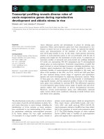
Switching of cTPxI oligomeric states in vivo
cTPxI and cTPxII can act as peroxidases and as
molecular chaperones, depending on changes of their
quaternary structures triggered by oxidative stress and
heat shock exposure [33,34]. When cTPxI appears
mainly as oligomeric protein structures of low molecu-
lar weight, this protein possesses mainly peroxidase
activity, whereas high molecular weight complexes
behave mainly as chaperones [33,34]. The specificity of
cTPxI in the protection of cells with dysfunctional
mitochondria (Fig. 1) might be related to the ability of
this protein to possess these two activities. Therefore,
we compared cTPxI oligomeric structures in vivo under
situations of normal and inhibited mitochondrial func-
tion, as it is hard to measure chaperone activity
in vivo.
Under control conditions, cTPxI appeared as a mix-
ture of complexes with molecular weight below
272 kDa and after treatment of yeast cells with H
2
O
2,
a considerable part of these species were converted to
HMW complexes of about 500 kDa or even higher
(Fig. 3), as previously described [33,34]. These switches
of cTPxI quaternary structures induced by H
2
O
2
were
not affected by any of the inhibitors of mitochondrial
function (Fig. 3). Similar results were obtained with
0.5 mm H
2
O
2
alone or in association with the mito-
chondrial function inhibitors (not shown).
Since it was well demonstrated that the conversion of
cTPxI to different oligomerization states is implicated
with its chaperone ⁄ peroxidase switching [33,34], we
suggest that the chaperone activity of this protein,
in addition to its peroxidatic function, is probably
involved with its specific role in the antioxidant defense
of yeast with mitochondrial dysfunction.
cTPxI influences the expression of genes involved
in yeast oxidative stress response: mitochondrial
function contribution
cTPxI participates of H
2
O
2
-mediated signaling proces-
ses, including regulation of gene expression [27–30].
Therefore, we have evaluated possible influences of
cTPxI and of the functional state of the mitochondria in
the expression of selected yeast antioxidant genes. In
this manner, we expected to obtain some clues to better
understand cTPxI importance in the response of cells
with mitochondrial dysfunction to oxidative stress.
It could be readily observed that the expression levels
of several genes were increased in cells lacking cTPxI
(Fig. 4). Four gene subsets could be delineated: (a)
genes with increased basal expression levels in tsa1D
cells: GSH1, GSH2, GLR1, PRX1, SOD1, GPX2 ,
AHP1, TRR1, SSA1; (b) genes with increased H
2
O
2
-
induced expression levels in tsa1D cells: CCP1, CTT1,
TRX2, TRX3, SOD2, GRX5, POS5; (c) genes without
expression alteration in tsa1D cells: ZWF1, IDP1,
GPX3, HSP104 (not shown); and (d) genes without
545
272
132
66
45
(kDa)
control
H
2
O
2
Anti A
Anti A + H
2
O
2
FCCP
FCCP + H
2
O
2
Fig. 3. Protein structures of cTPxI in vivo. Native-PAGE analysis of
crude protein extracts obtained from BY4741 (WT) cultures after
exposing cells during 40 min to no agent as a control, 1.2 m
M
H
2
O
2
,0.1lgÆmL
)1
antimycin A (anti A), 1.2 mM H
2
O
2
plus
0.1 lgÆmL
)1
antimycin A, 2.5 lgÆmL
)1
FCCP and 2.5 lgÆmL
)1
FCCP
plus 1.2 m
M H
2
O
2
, were separated on 9% native-PAGE and subjec-
ted to western blotting with a polyclonal anti-cTPxI IgG.
cTPxI and respiratory function in antioxidant defense A. P. D. Demasi et al.
808 FEBS Journal 273 (2006) 805–816 ª 2006 The Authors Journal compilation ª 2006 FEBS
Increased
basal
expression
levels in
tsa1
∆
cells
Increased
H
2
O
2
-
induced
expression
levels in
tsa1
∆
cells
Protein function Gene Expression levels Regulation
a
WT tsa1
∆
Actin
ACT1
Mitochondrial
function
influence
b
γ-glutamyl
cysteine synthase
GSH1 Yap1
+
Mitochondrial
thioredoxin peroxidase I
PRX1 Yap1, Msn2/4
+
Yap1
Glutathione peroxidase II
GPX2
+/-
Cytochrome c peroxidase
(mitochondrial)
CCP1 Yap1, Skn7
+
Catalase T
CTT1
Yap1, Skn7,
Msn2/4
+
Thioredoxin II
TRX2 Yap1, Skn7
+
Thioredoxin III
(mitochondrial)
TRX3
+
Manganese superoxide
dismutase (mitochondrial)
SOD2 Yap1, Skn7
+/-
Glutaredoxin 5
(mitochondrial)
GRX5
+
Cytosolic thioredoxin
peroxidase I
TSA1 Yap1, Skn7
-
Glutathione
reductase
GLR1 Yap1
-
Coper/zinc superoxide
dismutase
SOD1 Yap1, Skn7
-
Alkyl hydroperoxide
reductase
AHP1 Yap1, Skn7
-
1 2 3 4 5 6 7 8
a
Data from [7] and [13].
b
In H
2
O
2
- induced expression in tsa1 mutant cells (compare lanes 6 and 8)
H
2
O
2
-
+
-
+
-
+
-
+
anti A
++
++
Thioredoxin
reductase I
TRR1 Yap1, Skn7
+/-
POS5
Mitochondrial
NADH kinase
+/-
Glutathione
synthetase
GSH2
-
SSA1
Heat shock protein
of HSP70 family
Yap1, Skn7
+
Fig. 4. Expression of genes in wild-type and tsa1D cells exposed to several stressful conditions. Northern blot analysis of RNA isolated by
the hot acid phenol method from yeast strains BY4741 (WT) and tsa1D grown on YPD to mid-log phase, treated during 40 min with no agent
as a control or with 1.2 m
M H
2
O
2
and 0.1 lgÆmL
)1
antimycin A (anti A), singly or in association, as indicated in the figure. The symbols
–, + or ± in the last column denote absence, strong or mild influence of mitochondrial function on gene expression (comparison of band
intensities between lanes 6 and 8).
A. P. D. Demasi et al. cTPxI and respiratory function in antioxidant defense
FEBS Journal 273 (2006) 805–816 ª 2006 The Authors Journal compilation ª 2006 FEBS 809
detectable expression in both wild-type and tsa1D cells:
TSA2, CTA1, DOT5, TRX1, TTR1 (not shown). Genes
that belong to subsets (c) and (d) encode glucose-6-
phosphate dehydrogenase, mitochondrial isocitrate de-
hydrogenase, glutathione peroxidase III, heat shock
protein 104, thioredoxin peroxidase II, catalase A, nuc-
lear thioredoxin peroxidase, thioredoxin I and glutare-
doxin II, respectively. Among genes described in subset
(a), GSH1, GPX2, AHP1 and TRR1 expression levels
were further induced by H
2
O
2
(Fig. 4). In addition,
most if not all genes that presented altered expression in
tsa1 mutant are regulated by Yap1 (Fig. 4), indicating
that cTPxI could affect Yap1 activity. Furthermore,
Skn7 co-operates in the control of many of these genes
(Fig. 4) and constitute another transcription factor
whose activation might be influenced by cTPxI.
It is worth noting the influence of the functional
state of mitochondria in the H
2
O
2
-induced expression
levels of various genes, at least in tsa1 mutant (Fig. 4,
compare lanes 6 and 8), suggesting that respiratory-
compromised cells fail to activate some H
2
O
2
respon-
sive genes transcription at the same degree of respirat-
ory-competent ones. The H
2
O
2
-induced expression
levels of GSH1, PRX1, CCP1, CTT1, TRR1 were
affected the most by the defective mitochondria, while
those of GPX2, TRX2, TRX3, SOD2 GRX5, POS5
were influenced at a lower level.
The treatment with antimycin A alone, in all cases,
led to expression levels resembling those observed in
control cells (Fig. 4). These results are in agreement
with genome-wide studies that did not find significant
differences in the expression of antioxidant genes in
cells with mitochondrial dysfunction [44,45].
Participation of transcription factors in the
antioxidant defense of cells with normal or
impaired mitochondrial function
In order to identify transcription factors involved in
the response of cells with dysfunctional mitochondria
to oxidative stress, we evaluated H
2
O
2
sensitivity of
deletion mutants for the regulators most frequently
associated with oxidative stress response: Yap1, Skn7,
Msn2 and Msn4.
Single or double mutants for Yap1 and Skn7 were
very sensitive to H
2
O
2
, although deletion of YAP1
gene appeared to be more deleterious than the SKN7
gene deletion (Fig. 5). This high sensitivity was already
expected given the diversity of antioxidant enzymes
regulated by these factors [7]. The association of H
2
O
2
with antimycin A totally inhibited growth of these
mutants. In contrast, no further growth inhibition of
yap1D and skn7D was achieved by the association of
FCCP with H
2
O
2
, relative to H
2
O
2
alone (Fig. 5). No
significant growth retardation of yap1D and skn7D rel-
ative to wild-type counterparts was observed when
these cells were treated with antimycin A alone or with
FCCP alone. Therefore, the phenotypes of yap1D and
skn7D, were similar to those of tsa1D described in
Fig. 1, suggesting that Yap1, Skn7 and cTPxI act in
the same pathway in the response of yeast with dys-
functional mitochondria to oxidative stress. Because
cell growth was more affected by the deletion of YAP1
and SKN7 genes than by deletion of cTPxI (Fig. 5),
we suggest that other enzymes whose genes are regula-
ted by these factors could also be involved in the anti-
oxidant defense of respiratory-incompetent cells.
On the other hand, Msn2 and Msn4 appear to not
play significant role in the response of cells to H
2
O
2
or
to either of the compounds that interfere with mitoch-
ondrial function (antimycin A or FCCP), as no consid-
erable growth alterations for their deletion mutants
were detected (Fig. 5). Since Ras-cAMP-PKA pathway
inhibits Msn2 ⁄ 4 under catabolic repressing conditions
[14], the sensitivity of msn2Dmsn4D was also evaluated
in the absence of glucose. In this case, cells were grown
in raffinose medium. Again, these mutants grew simi-
larly to the wild-type cells (data not shown), dismissing
the involvement of Msn2 ⁄ 4 in the response of respirat-
ory incompetent cells to oxidative stress.
control anti A FCCP
yap1
∆
skn7
∆
skn7
∆
WT
yap1
∆
msn2
∆
msn4
∆
H
2
O
2
H
2
O
2
+
anti A
H
2
O
2
+
FCCP
Fig. 5. Sensitivities of mutants lacking transcription factors to several stressful conditions. Growth of the strains BY4741 (WT), and mutants
indicated on YPD plates containing no chemicals as a control, 0.8 m
M H
2
O
2
,0.1lgÆmL
)1
antimycin A (anti A), and 2.5 lgÆmL
)1
FCCP singly
or in association. Proceedings were performed as described in Fig. 1.
cTPxI and respiratory function in antioxidant defense A. P. D. Demasi et al.
810 FEBS Journal 273 (2006) 805–816 ª 2006 The Authors Journal compilation ª 2006 FEBS
Yap1 cellular distribution
It is well known that Yap1 is accumulated in the nuc-
leus of cells exposed to oxidative stress and, as a con-
sequence, the expression of its target genes is activated
[11,46,47]. We observed that mitochondrial dysfunction
negatively affects the H
2
O
2
-induced expression levels
of various Yap1-target genes in tsa1 mutant cells
(Fig. 4, compare lanes 6 and 8), which could account
for the decreased capacity of yeast to cope oxidative
stress (Fig. 1). To check this possibility, we examined
the distribution of Yap1 in the cells expressing GFP-
Yap1 fusion protein.
No significant difference in the cellular GFP-Yap1
distribution was observed between the wild-type and
tsa1 mutant cells in all of the conditions tested
(Fig. 6). In spite of the increased basal expression lev-
els of a variety of genes in tsa1 cells, we did not
observe GFP-Yap1 accumulation in the nucleus of
these cells, corroborating results previously obtained
[27]. Therefore, GFP-Yap1 is located in nucleus and
cytoplasm of both wild type and tsa1D cells in control
conditions (Fig. 6). Antimycin A treatment alone did
not lead to a nuclear accumulation of GFP-Yap1
(Fig. 6), which is in agreement with the similar expres-
sion levels of genes from control and antimycin A-trea-
ted (Fig. 4, compare lanes 1 with 3 and 5 with 7).
Antimycin A treatment did not alter the nuclear
Yap1 accumulation induced by H
2
O
2
, in neither the
wild-type nor in tsa1D cells (Fig. 6). Hence, the dimin-
ished H
2
O
2
-induced expression levels of some Yap1-
target genes observed in cells with impaired
mitochondrial function can not be attributed to alter-
ation in Yap1 cellular distribution. Probably other fac-
tors, such as ability of Yap1 to bind DNA [51], are
also involved in the activation of genes involved in the
response of yeast to oxidative stress. These possibilities
are further discussed below.
Discussion
It was previously demonstrated that cTPxI is essential
for the antioxidant defense of cells with mitochondrial
dysfunction [35]. Remarkably, we have shown here
that cTPxI is very specific among several other antioxi-
dants in the protection of cells with respiratory incom-
petence against peroxides (Fig. 1). The protective
action of cTPxI was prominent in situations of elec-
tron flow impediment. This was demonstrated by the
severe growth retardation (Fig. 1) and by the large
depletion of sulfydryl content (Fig. 2) of tsa1 mutant
treated with H
2
O
2
in association with antimycin A,
effects that were not observed when these cells were
exposed to H
2
O
2
+ FCCP. As it has long been shown,
while antimycin A augments ROS generation by
defective mitochondria [19–21], FCCP diminishes it
[37–39]. Therefore, it is possible that cTPxI could be
specifically important when internal ROS production is
elevated. In agreement, it was demonstrated that a
bacterial peroxiredoxin, alkyl hydroperoxide reductase
WT
tsa1
∆
control
H
2
O
2
Anti A
Anti A
+H
2
O
2
Fig. 6. Cellular distribution of GFP-tagged
Yap1. Cells of the strains JD7–7C (WT) and
tsa1D carrying expression plasmids for the
GFP-YAP1 fusion gene were exposed to 1.2
m
M H
2
O
2
and 0.1 lgÆmL
)1
antimycin A (anti
A), separately or in association, and confocal
laser scanning microscopy was carried out
as described under ‘Experimental proce-
dures’. The left panels show the fluorescent
images and the right panels show the trans-
mission images. All of the data shown are
representative of at least three independent
experiments, all of which gave similar
results.
A. P. D. Demasi et al. cTPxI and respiratory function in antioxidant defense
FEBS Journal 273 (2006) 805–816 ª 2006 The Authors Journal compilation ª 2006 FEBS 811
(AhpC), is the primary scavenger of endogenous H
2
O
2
[48]. Altered ATP levels do not appear to influence res-
piratory deficient yeast antioxidant defenses, since cells
treated with FCCP, which leads to a more pronounced
energy limitation due to the higher cytoplasmic ATP
hydrolysis rate [37] did not present alteration in H
2
O
2
sensitivity, even for the tsa1 mutant (Fig. 1).
The peroxidatic function of cTPxI probably overlaps
to some extent with other H
2
O
2
detoxifying enzymes,
but the recent finding that this protein possesses chap-
erone activity under stressful conditions provides a
very tempting explanation for the distinctive role of
cTPxI in protecting cells with mitochondrial dysfunc-
tion against oxidative stress. Indeed, our data showed
that cTPxI appears not only as low molecular weight,
but mostly as high molecular weight complexes in cells
exposed to H
2
O
2
alone or in association with antimy-
cin A (Fig. 3), suggesting that it may be acting as per-
oxidase and as chaperone under these conditions, as
this functional and structural correlation was already
well demonstrated [33,34]. Its high molecular weight
form with chaperone activity could protect essential
proteins from denaturation or could mediate activa-
tion of downstream defense signaling cascades that
prevent H
2
O
2
-induced cell death. Actually, it was dem-
onstrated that oxidant-mediated proper folding of
Yap1 is required for transcriptional activation and for
the nuclear accumulation of this regulator during
stress [49].
Another phenomenon that could be related to the
unique phenotype of tsa1 mutant cells is that most
thiol proteins are inactivated when oxidized to sulfinic
acids, because this oxidation state of cysteine residues
is not reducible by classical reducing agents such as
glutathione and thioredoxin. In contrast, the sulfinic
acid form of cTPxI can be specifically reduced by sul-
firedoxin [36]. In fact, srx1D presented similar pheno-
type of tsa1D cells (Fig. 1). The fact that antimycin A,
but not diamide, increased the sensitivity of tsa1
mutant cells to peroxides (Fig. 1) gave support to the
notion that higher oxidation states of cysteines might
be taking place in the cTPxI dependent response to
oxidative stress. This is because diamide is an oxidant
that gives rise only to disulfides [40], whereas peroxides
generate disulfides as well as sulfenates (Cys-SOH),
sulfinates (Cys-SO
2
H) and sulfonates (Cys-SO
3
H). The
hypotheses raised here to explain the unique tsa1D
phenotype are not mutually exclusive. Actually, sulfinic
acid formation in cTPxI by H
2
O
2
was suggested as a
trigger event for the switch of this peroxiredoxin from
a peroxidase to a chaperone enzyme [33].
2-Cys Prxs have been implicated in the regulation of
stress-induced gene expression [27–32]. A previous
work has shown that expression levels of GSH1, GLR1
and GPX2 were increased in a tsa1 mutant and this
effect was dependent on Yap1, since transcriptional
activation of these genes were not observed in tsa1D ⁄
yap1D double mutants [27]. Our data confirmed these
results, but indicate that there may be more mecha-
nisms involved in this TSA1-dependent regulation. For
some genes, the transcription increase seemed to be
stress-independent, while for others, it was dependent
on H
2
O
2
(Fig. 4). It is most relevant that, in several
cases, the H
2
O
2
induction was reduced by the presence
of antimycin A, suggesting that the functional state of
the mitochondria is somehow sensed by regulatory
mechanisms. Perhaps the decreased levels of these
enzymes, and other yet not detected, could be respon-
sible for the reduced viability of tsa1D cells grown in
the presence of H
2
O
2
plus antimycin A (Fig. 1).
The existence of a connection between cTPxI and
Yap1 is becoming evident, although its molecular basis
is not yet clear. It was demonstrated that Yap1 is
retained in the nucleus in the presence of H
2
O
2
and
thereby it interacts with the target genes [11,46,47].
This retention is dependent of the oxidation of Yap1
cysteine residues by H
2
O
2
, that modifies its conforma-
tion and hinders its interaction with Crm1, which oth-
erwise would export this factor to cytoplasm. H
2
O
2
oxidizes Yap1 in a process mediated by Gpx3 ⁄ Orp1, a
glutathione peroxidase homologue with thioredoxin
peroxidase activity [11], with a still unclear participa-
tion of Ybp1 [50]. Interestingly, it was demonstrated in
cells with a truncated form of Ybp1, that cTPx1 can
replace Gpx3 ⁄ Orp1 in the oxidation of Yap1 thus pro-
moting its nuclear retention [30]. Moreover, in Schizo-
saccharomyces pombe, a 2-Cys Prx, but not a GPx,
directly oxidizes Pap1 (a Yap1 homologue) provoking
the nuclear retention of this transcription factor [51].
Despite the observed alterations in the expression
of Yap1-target genes, the cellular localization of this
transcription factor was not altered by either the
TSA1 deletion or by the inhibition of mitochondrial
function (Fig. 6). Inoue et al. [27] have also demon-
strated that the expression of a reporter gene fused
to a Yap1-dependent promoter was significantly
increased in the absence of cTPxI by a mechanism
independent of Yap1 nuclear retention. There is a
precedent showing that Yap1 binding activity may be
affected. It was shown that the accessibility of Yap1
to the GSH1 promoter could be repressed by Cbf1, a
DNA-binding protein that binds to elements in the
vicinity of Yap1 binding site [52]. Alternatively, the
increased reducing power of tsa1 mutant, achieved by
the expression elevation of GSH1, GSH2, GLR1 and
TRR1 and detected by our analysis (Fig. 2), could
cTPxI and respiratory function in antioxidant defense A. P. D. Demasi et al.
812 FEBS Journal 273 (2006) 805–816 ª 2006 The Authors Journal compilation ª 2006 FEBS
contribute to the reduction of Yap1, hence diminish-
ing the oxidized Yap1 ‘lifetime’, thus avoiding its
accumulation in the nucleus. In fact, Wiatrowski and
Carlson [53] did not observe Yap1 accumulation in
the nucleus of cells shifted from glucose to glycerol,
otherwise observed, in the presence of glutathione
externally added.
Another striking point in the adaptation of cells
to H
2
O
2
is the redirection of carbohydrate flux from
hexose phosphate pool (glycolysis) to the pentose
phosphate pathway to the regeneration of NADPH
[12] which, in turn, is responsible for the maintenance
of both thioredoxin and glutathione in their reduced
states. As cells treated with antimycin A rely only on
glycolysis to produce ATP, the carbohydrate meta-
bolism redirection after H
2
O
2
treatment would be
affected and the generation of NADPH would be
diminished. The depletion of sulfhydryl groups
occurred in tsa1 cells treated with antimycin A plus
H
2
O
2
(Fig. 2) could corroborate with this hypothesis.
These multiple activities of cTPxI (peroxidase, chap-
erone and redox signaling) might be related to the cen-
tral roles of this protein in prevention of yeast against
genotoxic processes [54,55]. Here, we have shown some
alterations that could impair the oxidative stress
response of yeast cells with mitochondrial dysfunction
and that cTPxI is specifically important in their anti-
oxidant defense. Although the mitochondrial inhibition
procedures used here were extreme, these approaches
have been largely employed in bioenergetics studies and
have provided valuable information. Moreover, the
nonphysiological doses of peroxides used here were due
to the high redundancy of the yeast antioxidant systems.
Nevertheless, our results suggest that peroxiredoxins,
especially those with high similarity to the yeast cTPxI
could exert a decisive role in the establishment of
mitochondrial dysfunction-related diseases, although
further studies are necessary to ascertain this relation-
ship. In support of this hypothesis, peroxiredoxins have
been implicated in the development of different kinds of
cancer [56–60] and neurodegenerative diseases [61,62].
Experimental procedures
Yeast strains and growth conditions
The following S. cerevisiae strains were used in this study:
JD7–7C (MATa ura3–52 leu2 trpA K +) and tsa1D (MATa
ura3–52 leu2 trpA K + tsa1D::LEU2) were obtained from
Chae [63] (National Institute of Health, Bethseda, MD,
USA); BY4741 (MATa; his3D1; leu2D0; met15D0; ura3D0),
tsa1D (MATa; his3D1; leu2D0; met15D0; ura3D0; tsa1D ::
Kan Mx4), prx1D (MATa; his3D1; leu2D0; met15D0;
ura3D0; prx1D::Kan Mx4) tsa2D (MATa; his3D1; leu2D0;
met15D0; ura3D0; tsa2D::Kan Mx4), ahp1D (MATa;
his3D1; leu2D0; met15D0; ura3D0; ahp1D::Kan Mx4), ccp1D
(MATa; his3D1; leu2D0; met15D 0; ura3D0; ccp1D::Kan
Mx4), glr1D (MATa; his3D1; leu2D0; met15D0; ura3D0;
glr1D::Kan Mx4), yap1D (MATa; his3D1; leu2D0; met15D0;
ura3D0; yap1D::Kan Mx4), skn7D (MATa; his3D1; leu2D0;
met15D0; ura3D0; skn7D::Kan Mx4) were obtained from
EUROSCARF (University of Frankfurt, Germany);
YPH250 (MATa trp-D1 his3-D200 lys2–801 leu2-D1 ade2–
101 ura3–52), ctt1 D (MATa trp-D1 his3-D200 lys2–801 leu2-
D1 ade2–101 ctt1::URA3), cta1D (MATa his3-D200 lys2–
801 leu2-D1 ade2–101 ura3–52 cta1::TRP1) were obtained
from Izawa [64] (Kyoto University, Japan), and W303–1a
(MATa, ade2, can1, his3, leu2, trp1, ura3) and msn2Dmsn4D
(MATa, ade2, can1, his3, leu2, trp1, ura3, msn2::HIS3,
msn4::TRP1) were obtained from Boy-Marcotte [65] (Uni-
versite Paris-Sud, France).
Cells were grown at 30 °C on YPD medium (1% yeast
extract, 2% bacto-peptone, 2% glucose). For most analysis,
cells were harvested by centrifugation at mid-log phase,
usually at an OD
600nm
between 0.8 and 1.4.
Determination of tolerance to different oxidants
Spot test: cells were first grown in YPD media until a con-
centration of approximately 10
7
cellsÆmL
)1
, and then dilu-
ted to OD
600nm
¼ 0.2. Four subsequent 1 : 5 dilutions of
these cell suspensions were realized and a 12 lL droplet of
each was plated on YPD-agar medium containing 1.2 mm
H
2
O
2
, or 1.2 mm t-BOOH, 1.2 mm diamide, 0.1 lgÆmL
)1
antimycin A, or 2.0 lgÆmL
)1
FCCP, separately or in associ-
ation. Plates were then incubated 2 days. Only the three
highest dilutions were represented in the figures.
Determination of sulfhydryl groups
PB-SH levels were measured according to the method of
Sedlak and Lindsay [66], by subtracting the NP-SH content
from the total sulfhydryl (T-SH) content. Cells of the strains
JD7–7C and tsa1D were grown on YPD and, after treat-
ments with 1.2 mm H
2
O
2
, 0.1 lgÆmL
)1
antimycin A and
2.0 lgÆmL
)1
FCCP, separately or in association (as des-
cribed in the figure), approximately 2 · 10
6
cells from each
culture were collected. Protein extracts were obtained in
0.02 m EDTA pH 4.7 with glass beads addition followed by
centrifugation at 17 900 g for 15 min. The T-SH concentra-
tions were determined by absorption levels at 412 nm after
incubating 200 lL aliquots of protein extracts supernatants
with 780 lL 0.2 m Tris pH 8.2 and 20 lL5mm DTNB for
30 min. The NP-SH contents were determined in the super-
natant, after proteins precipitation with 5% trichloroacetic
acid (final concentration) by incubating 450 lL supernatant,
900 lL 0.4 m Tris pH 8.9 and 26 lL5mm DTNB for
5 min. Absorption levels were measured at 412 nm.
A. P. D. Demasi et al. cTPxI and respiratory function in antioxidant defense
FEBS Journal 273 (2006) 805–816 ª 2006 The Authors Journal compilation ª 2006 FEBS 813
Determination of the switching of cTPxI
structures in vivo
Cells grown on YPD were treated during 40 min with
1.2 mm H
2
O
2
, 0.1 lgÆmL
)1
antimycin A and 2.0 lgÆmL
)1
FCCP, separately or in association. The corresponding
whole cell extracts, obtained as described by Ausubel et al.
[67], were separated by 9% native-PAGE (15 · 15 cm gels,
overnight running) and subjected to immunoblotting with
an anti-cTPxI antibody. The nondenatured protein molecu-
lar weight marker kit was purchased from Sigma. As posit-
ive control, recombinant cTPxI was also present in the gels.
DNA manipulation
To generate the probes for northern blot analysis, the
DNA sequences of the selected antioxidant genes were PCR
amplified from the collection ExClones
TM
, Yeast ORF
Expression Clones, Research Genetics (Invitrogen, Madi-
son, WI, USA). The clones containing the expression plas-
mids corresponding to the ORFs of interest (YPL091W,
YJL101C, YML028W, YLR109W, YDR353W, YDR453C,
YDR513W, YNL241C, YCL035C, YFL039C, YHR008C,
YIL010W, YLR043C, YGR088W, YJR104C, YBL064C,
YDR256C, YDL066W, YPL188W, YOL049W, YAL005C,
YLL026W and YIR037W) were grown separately on YPD
medium, and DNA of each clone was extracted as des-
cribed by Ausubel et al. [67]. PCR was carried out using
the following primers: 5¢-GAATTCCAGCTGACCACC-3¢
and 5¢GATCCCCGGGAATTGCCAT-3¢. The resulting
PCR products were purified and the sequences were con-
firmed previously to the probes preparation.
RNA isolation and analysis
Total yeast RNA was extracted by the method of hot acid
phenol method and northern blotting was performed as
previously described [67]. The
32
P-labeled probes were pre-
pared by random primed synthesis [67]. Actin was used as
loading control and no significant difference was found rel-
ative to ribosomal RNA (not shown).
Localization of GFP-tagged Yap1
The expression plasmids for the green fluorescent protein
(GFP) fused to Yap1 were kindly provided by Kuge [68].
They were transferred to cells of the strains JD7–7C and
tsa1D. Cells were grown on YPD to mid-log phase, concen-
trated into 25 lL of medium, treated with 1.2 mm H
2
O
2
and 0.1 lgÆmL
)1
antimycin A separately or in association,
and 5 lL of each culture were spotted on to glass slides.
Confocal laser scanning microscopy analysis was performed
using a Zeiss LSM510 Axiovert 200 m microscope (Carl
Zeiss MicroImaging, Inc., Thornwood, NY, USA) by
exciting cells with 488 nm laser (Argon ⁄ 2).
Acknowledgements
We thank Dr Shusuke Kuge for providing strains and
plasmids. We also thank Hugo Metz for technical
assistance with the confocal laser scanning microscopy
analysis and Lyndel Meinhardt for his comments on
the manuscript. Special thanks to Vasco dos Santos
Dias (in memoriam). This work was supported by
grants from the Brazilian Agencies FAPESP and
CNPq.
References
1 Hallywell B & Gutteridge JMC (1989) In Free Radicals
in Biology and Medicine (Hallywell, B & Gutteridge,
JMC, eds), 2nd edn. Claredon Press, Oxford.
2 Finkel T & Holbrook NJ (2000) Oxidants, oxidative
stress and the biology of ageing. Nature 408, 239–247.
3 Jamieson DJ (1998) Oxidative stress responses of the
yeast Saccharomyces cerevisiae. Yeast 14, 1511–1527.
4 Grant CM (2001) Role of the glutathione ⁄ glutaredoxin
and thioredoxin systems in yeast growth and response
to stress conditions. Mol Microbiol 39, 533–541.
5 Collinson EJ & Grant CM (2003) Role of yeast gluta-
redoxins as glutathione S-transferases. J Biol Chem 278,
22492–22497.
6 Park SG, Cha MK, Jeong W & Kim IH (2000) Distinct
physiological functions of thiol peroxidase isoenzymes
in Saccharomyces cerevisiae. J Biol Chem 275, 5723–
5732.
7 Lee J, Godon C, Lagniel G, Spector D, Garin J, Laba-
rre J & Toledano MB (1999) Yap1 and Skn7 control
two specialized oxidative stress regulons in yeast. J Biol
Chem 274, 16040–16046.
8 Charizanis C, Juhnke H, Krems B & Entian K-D (1999)
The mitochondrial cytochrome c peroxidase Ccp1 of
Sacharomyces cerevisiae is involved in conveying an
oxidative stress signal to the transcription factor Pos9
(Skn7). Mol Gen Genet 262, 437–447.
9 Costa V & Moradas-Ferreira P (2001) Oxidative stress
and signal transduction in Saccharomyces cerevisiae:
insights into ageing, apoptosis and diseases. Mol Aspects
Med 22, 217–246.
10 Moye-Rowley WS (2002) Transcription factors regula-
ting the response to oxidative stress in yeast. Antioxid
Redox Signal 4, 123–140.
11 Delaunay A, Pflieger D, Barrault M, Vinh J & Toledano
MB (2002) A thiol peroxidase is an H
2
O
2
receptor and
redox transducer in gene activation. Cell 111, 471–481.
12 Godon C, Lagniel G, Lee J, Buhler JM, Kieffer S, Per-
rot M, Boucherie H, Toledano MB & Labarre J (1998)
The H
2
O
2
stimulon in Saccharomyces cerevisiae. J Biol
Chem 273, 22480–22489.
13 Hasan R, Leroy C, Isnard A-D, Labarre J, Boy-Mar-
cotte E & Toledano MB (2002) The control of the yeast
cTPxI and respiratory function in antioxidant defense A. P. D. Demasi et al.
814 FEBS Journal 273 (2006) 805–816 ª 2006 The Authors Journal compilation ª 2006 FEBS
H
2
O
2
response by the Msn2 ⁄ 4 transcription factors.
Mol Microbiol 45, 233–241.
14 Gorner W, Durchschlag E, Martinez-Pastor MT, Est-
ruch F, Ammerer G, Hamilton B, Ruis H & Schuller C
(1998) Nuclear localization of the C2H2 zinc finger pro-
tein Msn2p is regulated by stress and protein kinase A
activity. Genes Dev 12, 586–597.
15 Fernandes L, Rodrigues-Pousada C & Struhl K (1997)
Yap, a novel family of eight bZIP proteins in Saccharo-
myces cerevisiae with distinct biological functions. Mol
Cel Biol 17, 6982–6993.
16 Davidson JF & Schiestl RH (2001) Mitochondrial
respiratory electron carriers are involved in oxidative
stress during heat stress in Saccharomyces cerevisiae.
Mol Cel Biol 21, 8483–8489.
17 Guo J & Lemire BD (2003) The ubiquinone-binding site
of the Saccharomyces cerevisiae succinate-ubiquinone
oxidoreductase is a source of superoxide. J Biol Chem
278, 47629–47635.
18 Outten CE & Culotta VC (2003) A novel NADH kinase
is the mitochondrial source of NADPH in Saccharo-
myces cerevisiae. EMBO J 22, 2015–2024.
19 Boveris A & Chance B (1973) The mitochondrial gen-
eration of hydrogen peroxide. Biochem J 134, 707–716.
20 Turrens JF (1997) Superoxide production by the mito-
chondrial respiratory chain. Bioscience Reports 17, 3–8.
21 Cadenas E & Davies KJ (2000) Mitochondrial free radi-
cal generation, oxidative stress and aging. Free Rad Biol
Med 29, 222–230.
22 He CH, Masson J & Ramotar D (1996) Functional
mitochondria are essential for Saccharomyces cerevisiae
cellular resistance to bleomycin. Curr Genet 30, 279–
283.
23 Grant CM, MacIver FH & Dawes IW (1997) Mitochon-
drial function is required for resistance to oxidative
stress in the yeast Saccharomyces cerevisiae. FEBS Lett
410, 219–222.
24 Netto LES, Chae HZ, Kang SW, Rhee SG & Stadtman
ER (1996) Removal of hydrogen peroxide is involved
with the antioxidant properties of thiol specific antioxi-
dant (TSA): TSA possesses thiol peroxidase activity.
J Biol Chem 271, 15315–15321.
25 Hofmann B, Hecht H & Flohe
´
L (2002) Peroxiredoxins.
Biol Chem 383, 347–364.
26 Wood ZA, Schroder E, Harris JR & Poole LB (2003)
Structure, mechanism and regulation of peroxiredoxins.
Trends Biochem Sci 28, 32–40.
27 Inoue Y, Matsuda T, Sugiyama K, Izawa S & Kimura
A (1999) Genetic analysis of glutathione peroxidase in
oxidative stress response of Saccharomyces cerevisiae.
J Biol Chem 274, 27002–27009.
28 Ross SJ, Findlay VJ, Malakasi P & Morgan BA (2000)
Thioredoxin peroxidase is required for the
transcriptional response to oxidative stress in budding
yeast. Mol Biol Cell 11, 2631–2642.
29 Veal EA, Findlay VJ, Day AM, Bozonet SM, Evans
JM, Quinn J & Morgan BA (2004) A 2-cys peroxire-
doxin regulates peroxide-induced oxidation and activa-
tion of a stress-activated MAP kinase. Mol Cell 15,
129–139.
30 Okazaki S, Naganuma A & Kuge S (2005) Peroxire-
doxin-mediated redox regulation of the nuclear localiza-
tion of Yap1, a transcription factor in budding yeast.
Antioxid Redox Signal 7, 327–334.
31 Jin DY, Chae HZ, Rhee SG & Jeang KT (1997) Reg-
ulatory role for a novel human thioredoxin peroxidase
in NF-kappaB activation. J Biol Chem 272, 30952–
30961.
32 Haridas V, Ni J, Meager A et al. (1998) TRANK, a
novel cytokine that activates NF-kappaB and c-Jun
N-terminal kinase. J Immunol 161 , 1–6.
33 Jang HH, Lee KO, Chi YH et al. (2004) Two enzymes
in one: two yeast peroxiredoxins display oxidative
stress-dependent switching from a peroxidase to a
molecular chaperone function. Cell 117, 625–635.
34 Moon JC, Hah YS, Kim WY et al. (2005) Oxidative
stress-dependent structural and functional switching of a
human 2-Cys peroxiredoxin isotype II that enhances
HeLa cell resistance to H2O2-induced cell death. J Biol
Chem 280, 28775–28784.
35 Demasi APD, Pereira GAG & Netto LES (2001) Cyto-
solic thioredoxin peroxidase I is essencial for the anti-
oxidant defense of yeast with dysfunctional
mitochondria. FEBS Lett 509, 430–434.
36 Biteau B, Labarre J & Toledano MB (2003) ATP-
dependent reduction of cysteine-sulphinic acid by S. cer-
evisiae sulphiredoxin. Nature 425, 980–984.
37 Nicholls DG & Budd SL (2000) Mitochondria and neu-
ronal survival. Physiol Rev 80, 315–360.
38 Korshunov SS, Skulachev VP & Starkov AA (1997) High
protonic potential actuates a mechanism of reactive oxy-
gen species in mitochondria. FEBS Lett 416, 15–18.
39 Barros MH, Bandy B, Tahara EB & Kowaltowski AJ
(2004) Higher respiratory activity decreases mitochon-
drial reactive oxygen release and increases life span in
Saccharomyces cerevisiae. J Biol Chem 279, 49883–
49888.
40 Kosower NS, Kosower EM, Wertheim B & Correa WS
(1969) Diamide, a new reagent for the intracellular oxi-
dation of glutathione to the disulfide. Biochem Biophys
Res Commun 37, 593–596.
41 Garrido EO & Grant CM (2002) Role of thioredoxins
in the response of Saccharomyces cerevisiae to oxidative
stress induced by hydroperoxides. Mol Microbiol 43,
993–1003.
42 Wong C, Siu K & Jin D (2004) Peroxiredoxin-null yeast
cells are hypersensitive to oxidative stress and are geno-
mically unstable. J Biol Chem 279, 23207–23213.
43 Monteiro G, Kowaltowski AJ, Barros MH & Netto LE
(2004) Glutathione and thioredoxin peroxidases mediate
A. P. D. Demasi et al. cTPxI and respiratory function in antioxidant defense
FEBS Journal 273 (2006) 805–816 ª 2006 The Authors Journal compilation ª 2006 FEBS 815
susceptibility of yeast mitochondria to Ca(2+)-induced
damage. Arch Biochem Biophys 425, 14–24.
44 Traven A, Wong JM, Xu D, Sopta M & Ingles CJ
(2001) Interorganellar communication: altered nuclear
gene expression profiles in a yeast mitochondrial DNA
mutant. J Biol Chem 276, 4020–4027.
45 Epstein CB, Waddle JA, Hale Dave V 4th, Thornton J,
Macattee TL, Garner HR & Butow RA (2001) Gen-
ome-wide responses to mitochondrial dysfunction. Mol
Biol Cell 12, 297–308.
46 Kuge S, Toda T, Iizuka N & Nomoto A (1998) Crm1
(XpoI) dependent nuclear export of the budding yeast
transcription factor yAP-1 is sensitive to oxidative
stress. Genes Cells 3, 521–532.
47 Delaunay A, Isnard AD & Toledano MB (2000) H
2
O
2
sensing through oxidation of the Yap1 transcription
factor. EMBO J 19, 5157–5166.
48 Seaver LC & Imlay JA (2001) Alkyl hydroperoxide reduc-
tase is the primary scavenger of endogenous hydrogen
peroxide in Escherichia coli. J Bacteriol 183, 7173–7181.
49 Gulshan K, Rovinsky SA, Coleman ST & Moye-Row-
ley WS (2005) Oxidant-specific folding of Yap1 regu-
lates both transcriptional activation and nuclear
localization. J Biol Chem 280, 40524–40533.
50 Veal EA, Ross SJ, Malakasi P, Peacock E & Morgan
BA (2003) Ybp1 is required for the hydrogen peroxide-
induced oxidation of the Yap1 transcription factor.
J Biol Chem 278, 30896–30904.
51 Bozonet SM, Findlay VJ, Day AM, Cameron J, Veal
EA & Morgan BA (2005) Oxidation of a eukaryotic
2-Cys peroxiredoxin is a molecular switch controlling
the transcriptional response to increasing levels of
hydrogen peroxide. J Biol Chem 280, 23319–23327.
52 Wheeler GL, Trotter EW, Dawes IW & Grant CM
(2003) Coupling of the transcriptional regulation of glu-
tathione biosynthesis to the availability of glutathione
and methionine via the Met4 and Yap1 transcription
factors. J Biol Chem 278, 49920–49928.
53 Wiatrowski HA & Carlson M (2003) Yap1 accumulates
in the nucleus in response to carbon stress in Saccharo-
myces cerevisiae. Eukaryotic Cell 2, 19–26.
54 Huang M, Rio A, Nicolas A & Kolodner RD (2003) A
genomewide screen in Saccharomyces cerevisiae for
genes that suppress the accumulation of mutations. Proc
Natl Acad Sci USA 100, 11529–11534.
55 Smith S, Hwang J, Banerjee S, Majeed A, Grupta A &
Myung K (2004) Mutator genes for suppression of
gross chromosomal rearrangements identified by a
genome-wide screening in Saccharomyces cerevisiae.
Proc Natl Acad Sci USA 101, 9039–9044.
56 Noh DY, Ahn SJ, Lee RA, Kim SW, Park IA & Chae
HZ (2001) Overexpression of peroxiredoxin in human
breast cancer. Anticancer Res 21, 2085–2090.
57 Shen C & Nathan C (2002) Nonredundant antioxidant
defense by multiple two cysteine peroxiredoxins in
human prostate cancer cells. Mol Med 8, 95–102.
58 Kinnula VL, Lehtonen S, Sormunen R, Kaarteenaho-
Wiik R, Kang SW, Rhee SG & Soini Y (2002)
Overexpression of peroxiredoxins I, II, III, V and VI in
malignant mesothelioma. J Pathol 196, 316–323.
59 Neumann CA, Krause DS, Carman CV, Das S, Dubey
DP, Abraham JL, Bronson RT, Fujiwara Y, Orkin SH
& Etten RA (2003) Essential role for the peroxiredoxin
Prdx1 in erythrocyte antioxidant defence and tumor
suppression. Nature 424, 561–565.
60 Kinnula VL, Pa
¨
a
¨
kko P & Soini Y (2004) Antioxidant
enzymes and redox regulating thiol proteins in malig-
nancies of human lung. FEBS Lett 569, 1–6.
61 Krapfenbauer K, Engidawork E, Cairns N, Fountoula-
kis M & Lubec G (2003) Aberrant expression of peroxi-
redoxin subtypes in neurodegenerative disorders. Brain
Res 967, 152–160.
62 Plaisant F, Clippe A, Vander Stricht D, Knoops B &
Gressens P (2003) Recombinant peroxiredoxin 5 pro-
tects against excitotoxic brain lesions in newborn mice.
Free Rad Biol Med 34, 862–872.
63 Chae HZ, Kim IH, Kim K & Rhee SG (1993) Cloning,
sequencing, and mutation of thiol specific antioxidant
gene of TSA. J Biol Chem 268, 16815–16821.
64 Izawa S, Inoue Y & Kimura A (1996) Importance of
catalase in the adaptive response to hydrogen peroxide:
analysis of acatalasaemic Saccharomyces cerevisiae.
Biochem J 320, 61–67.
65 Boy-Marcotte E, Perrot M, Bussereau F, Boucherie H
& Jacquet M (1998) Msn2p and Msn4p control a large
number of genes induced at the diauxic transition which
are repressed by cyclic AMP in Saccharomyces cerevi-
siae. J Bacteriol 180, 1044–1052.
66 Sedlak J & Lindsay RH (1968) Estimation of total, pro-
tein-bound and nonprotein sulfhydryl groups in tissue
with Ellman’s reagent. Anal Biochem 25, 192–205.
67 Ausubel FM, Brent R, Kingstone RE, Moore DD, Seid-
man JA, Smith JA & Struhl K (1994) Ch. 13 Saccharo-
myces cerevisiae.InCurrent Protocols in Molecular
Biology. John Wiley and Sons, Inc, Chichester, UK.
68 Kuge S, Jones N & Nemoto A (1997) Regulation of
yAP-1 nuclear localization in response to oxidative
stress. EMBO J 16, 1710–1720.
cTPxI and respiratory function in antioxidant defense A. P. D. Demasi et al.
816 FEBS Journal 273 (2006) 805–816 ª 2006 The Authors Journal compilation ª 2006 FEBS