- Trang chủ >>
- Khoa Học Tự Nhiên >>
- Vật lý
Si nanowires grown from silicon oxide
Bạn đang xem bản rút gọn của tài liệu. Xem và tải ngay bản đầy đủ của tài liệu tại đây (1.57 MB, 6 trang )
6 January 1999
Ž.
Chemical Physics Letters 299 1999 237–242
Si nanowires grown from silicon oxide
N. Wang, Y.H. Tang, Y.F. Zhang, C.S. Lee, I. Bello, S.T. Lee
)
Center of Super Diamond and AdÕanced Films, Department of Physics and Materials Science, The City UniÕersity of Hong Kong,
Hong Kong, China
Received 10 August 1998
Abstract
Bulk-quantity Si nanowires have been synthesized by thermal evaporation of a powder mixture of silicon and SiO .
2
Transmission electron microscopy showed that, at the initial nucleation stage, silicon monoxide vapor was generated from
the powder mixture and condensed on the substrate. Si nanoparticles were precipitated and surrounded by shells of silicon
oxide. The Si nanowire nucleus consisted of a polycrystalline Si core with a high density of defects and a silicon oxide shell.
The growth mechanism was proposed to be closely related to the defect structure and silicon monoxide. q 1999 Elsevier
Science B.V. All rights reserved.
Nanometer-wide silicon wires have attracted much
attention in recent years because of their potential for
applications in the field of microelectronics. One of
the challenging issues has been the synthesis of this
one-dimensional form of nanowires on large scales.
Since the successful growth of Si whiskers by the
Ž. wx
vapor–liquid–solid VLS method 1,2 , many ef-
forts have been made to improve the synthesis of Si
nanowires by employing different techniques, such
as the photolithography technique combined with
wx
etching 3–5 and scanning tunneling microscopy
wx
6,7 . For the VLS method, Au had to be used and
this caused contamination. The diameters of Si
whiskers obtained from VLS were determined by the
size of Au particles. Other techniques were compli-
cated and could not produce bulk quantities of Si
nanowires.
)
Corresponding author. E-mail:
Recently, Si nanowires have been successfully
synthesized by a novel method of laser ablation of
wx
metal-containing Si targets 8–11 . Previous investi-
wx
gations 8,9 have shown that metal or metal-silicide
nanoparticles acted as the critical catalyst during the
deposition assisted by laser ablation. For example,
Fe could form Fe-silicides at high temperatures of
12008C. A growth mechanism of Si wires has been
wx
ascribed to the VLS reaction 8,9 . However, a dif-
ferent model has been proposed which is supported
by the experiment which showed that metal catalyst
were not observed in Si nanowires even when metals
wx
were mixed in the target 10 . Moreover, it was
discovered that metal was not necessary for Si
nanowire synthesis by laser ablation. Instead, SiO
2
was the special and effective catalyst which largely
wx
enhanced Si nanowire growth 12 . High-resolution
Ž.
transmission electron microscopy HRTEM investi-
gations have shown that high-density defects and
silicon oxide outer layers play important roles for
wx
nanowire growth 12 . In this Letter, we report that
0009-2614r99r$ - see front matter q 1999 Elsevier Science B.V. All rights reserved.
Ž.
PII: S0009-2614 98 01228-7
()
N. Wang et al.rChemical Physics Letters 299 1999 237–242238
bulk-quantity Si nanowires were synthesized by ther-
mal evaporation of a highly pure Si powder mixed
with SiO . Observations of Si nanowire nucleation
2
and growth morphology by transmission electron
Ž.
microscopy TEM are documented. By combining
these observations with the results of a Raman study,
we discuss the growth mechanisms.
Si nanowires can be synthesized by laser ablation
of a powder mixture of silicon and SiO in an
2
Ž
evacuated quartz tube in an Ar atmosphere 500
. wx
Torr 12 . However, in the present work, without the
assistance of laser ablation, Si nanowires were syn-
thesized by simple thermal evaporation at 12008C.
The solid source was highly pure Si powder mixed
Ž
with about 70 wt% SiO all materials were from
2
.
Goodfellow, purity 99.99% . The temperature around
the quartz tube where the nanowire grew was about
9308C. After 12 h of thermal evaporation, Si
Ž. Ž. Ž.
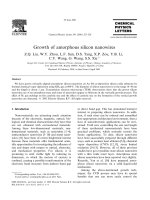
Fig. 1. a TEM image showing the morphology of Si nanowires synthesized by the evaporation method. b – d Nucleation stage of the Si
nanowires.
()
N. Wang et al.rChemical Physics Letters 299 1999 237–242 239
Ž.
Fig. 1 continued .
Ž.
nanowire product sponge-like, dark red in color
formed on the inside wall of the quartz tube. To
collect Si nanowire nuclei, a Mo grid was placed in
the region of the quartz tube where the nanowires
grew. Some Si nanowires nucleated and grew on the
grid. The Mo grid was directly observed in Philips
CM200FEG transmission electron microscope work-
ing under 200 kV. Raman measurements were car-
ried out using with a Renishaw 2000 micro-Raman
system.
Fig. 1a shows the typical morphology of as-grown
Ž
Si nanowires. The nanowires major component in
.Ž.
the product are extremely long ) 10 mm with
uniform diameters and smooth surfaces. Si nanopar-
ticles are found to coexist with the nanowires. A
striking feature is that Si nanoparticles appear in the
form of chain. Si nanowire nucleation on the Mo
grid is shown in Fig. 1b. In initial stage, Si nanopar-
ticles were formed as identified by electron diffrac-
tion. Most nanoparticles piled up on the substrate.
Ž
Notably, some favorable particles nuclei of
.
nanowires stood alone and underwent faster growth
since their preferable growth direction was normal to
Ž.
the surface of the substrate see Fig. 1b–d . There
was no detectable metal catalyst or impurity formed
on the tips of the nanowire nuclei. Each nucleus
simply consisted of a crystalline Si core and an
amorphous outer layer. The chemical composition of
the nuclei was determined by electron energy disper-
Ž.
sive spectroscopy EDS . Only silicon and oxygen
were detected which indicated that the amorphous
outer layer should have been silicon oxide. The Si
()
N. Wang et al.rChemical Physics Letters 299 1999 237–242240
crystalline core contained a high density of defects.
Most of the defects showed their contrast along the
growth axis of the nucleus. These defects were quite
Ž
similar to the planar defects stacking faults and
²:.
micro-twins along the axis of Si nanowire in 112
wx
observed in Si nanowires in our previous work 10 .
It is believed that silicon oxide plays an important
role in nanowire growth. We investigated the native
silicon oxide on single Si crystal surfaces. The oxide
thickness was only 2–3 monolayers. However, the
oxide shells of nanowires were quite thick. We
Ž.
observed that the shell thickness up to 3 nm gener-
wx
ally depended on the diameter of the nanowire 10 .
In the present experiment, the vapor materials gener-
ated from the mixture of silicon and SiO at 12008C
2
consisted mainly of SiO, with little silicon. This was
supported by the observation that the material con-
densed on the water-cooled Cu finger was Si O
xy
Ž.
xs0.51, ys 0.49 as determined by EDS. This
chemical composition was reliable since the vapor
phase was quenched on the cool finger. Silicon
Ž.
monoxide SiO is an amorphous semiconductor of
high resistivity which can easily be generated from
Ž.
powder mixtures especially in equimolar mixtures
wx
of silicon and SiO by heating 13–15 . TEM inves-
2
tigations confirmed the amorphous structure of the
SiO deposited on the Cu finger surface. By heating
the SiO sample in TEM, silicon precipitation was
Ž.
observed see Fig. 2a . Such precipitation of Si
nanoparticles from annealed SiO is quite well known
wx
15 .
According to the above observations, we propose
that the growth mechanism is silicon oxide assisted.
Ž.
The vapor phase of Si O x) 1 generated by ther-
x
mal evaporation is the key factor. The nucleation of
nanoparticles is assumed to occur at the substrate by
different decompositions of silicon oxide at the rela-
tively low temperature of 9308C as shown below.
Si O™ Si qSiO x)1
Ž.
xxy1
and
2SiO™ SiqSiO .
2
These decompositions result in the precipitation of
silicon nanoparticles, i.e. the nuclei of Si nanowires,
clad by shells of silicon oxide as observed in Fig. 1b.
The growth process may involve the following
factors. The relatively thick Si O on nanowire tips
x
wx
12 acts as a catalyst. The SiO component of the
2
shell, which could be formed during decomposition
of SiO in nanowire growth, retards the sideways
growth of the nanowire. Defects, such as stacking
faults in the nucleus tips, enhance the one-dimen-
Ä4
sional growth. The 111 surface, which has the
lowest surface energy among the surfaces in silicon,
plays an important role during nanowire growth.
Since surface energy is more important when the
crystal size is reduced to the nanometer scale, the
Ä4
appearance of 111 surfaces of the Si crystals paral-
lel to the axes of the nanowires reduces the system
energy. Combined, these factors determine the
²:
growth direction of Si nanowires to be 112 .
This proposed growth mechanism is supported by
the results of Raman study as shown in Fig. 3a. The
peak at 521 cm
y1
is broad and strongly asymmetric
compared to that from a single Si crystal. Such a
feature could be due to the small size effect of Si
wx
nanocrystals or defects 11,16 since there were many
nanoparticles in the product, as well as Si nanowires
wx
containing a high-density of defects 10,11 . In addi-
tion, the presence of SiO shells also contributes to
the asymmetry of the Raman peak. As shown in Fig.
Ž
3a, the spectrum taken from SiO deposited on the
.
Cu finger contains a broad peak located at about
480 cm
y1
. For comparison, Si nanowires which
Ž
were fully oxidized by annealing in the air white in
.
color were studied. No Raman scattering was de-
Ž.
tected see Fig. 3a . According to EDS measurement,
the fully oxidized nanowires consisted mainly of
SiO .
2
Ž.
Fig. 3b shows strong photoluminescence PL of
SiO at about 740 nm. The fully oxidized nanowire
gives a weak PL peak at about 600 nm. The PL from
Si nanowire product is weak and complicated. A
typical PL spectrum from Si nanowires covers the
range of 600–800 nm range. Clearly, the SiO and
SiO components of the nanowires are the main
2
contributors to this spectrum.
The proposed mechanism for nucleation and
growth can predict some of the morphology of
nanowires. For example, during the evaporation,
Si O vapor was continually generated and nucleation
x
could occur with different crystalline orientation ei-
ther on the side surfaces or tips of the nanowires.
The former resulted in the forking of the nanowires
Ž.
observed frequently and the latter caused re-nuclea-
()
N. Wang et al.rChemical Physics Letters 299 1999 237–242 241
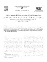
Ž. Ž.
Fig. 2. a Nanoparticles precipitated by heating the SiO thin film. b HRTEM image of the Si nanoparticle chain.
()
N. Wang et al.rChemical Physics Letters 299 1999 237–242242
Ž.
Fig. 3. a Raman spectra taken from the as-grown Si nanowires,
Ž.
SiO and fully oxidized Si nanowires. b PL spectra taken from
the as-grown Si nanowires, SiO and fully oxidized Si nanowires.
tion. The nuclei formed on the tips in an unfavorable
growth direction could not grow fast and re-nuclea-
tion occurred again. Such re-nucleation resulted in
Ž.
the formation of nanoparticle chains see Fig. 1 .
HRTEM image taken from one of the chains pro-
vided proof for this growth mechanism. As shown in
Fig. 2b, the silicon particles in the chain have differ-
ent orientations and most of the particles are not
wx
aligned with their 112 orientations parallel to the
growth direction.
In conclusion, bulk-quantity Si nanowires have
been synthesized by thermal evaporation of mixture
of silicon and SiO powder. Si oxide vapor gener-
2
ated from the powder mixture condensed on the
substrate and then decomposed, forming Si nanopar-
Ž.
ticles nuclei of nanowires . A Si nanowire nucleus
consisted of a polycrystalline Si core with a high
density of defects and a silicon oxide shell. The
growth mechanism was proposed to be closely re-
lated to the defect structure of Si crystal cores and
SiO.
Acknowledgements
This work was financially supported in part by the
Research Grants Council of Hong Kong and the
Strategic Research Grants of the City University of
Hong Kong.
References
wx Ž.
1 R.S. Wagner, W.C. Ellis, Appl. Phys. Lett. 4 1964 89.
wx Ž.
2 E.I. Givargizov, J. Cryst. Growth 32 1975 20.
wx
3 H.I. Liu, N.I. Maluf, R.F.W. Pease, J. Vac. Sci. Technol. B
Ž.
10 1992 2846.
wx
4 H. Namatsu, S. Horiguchi, M. Nagase, K. Kurihara, J. Vac.
Ž.
Sci. Technol. B 15 1997 1688.
wx
5 Y. Wada, T. Kure, T. Yoshimura, Y. Sudou, T. Kobayashi,
Ž.
Y. Gotou, S. Kondo, J. Vac. Sci. Technol. B 12 1994 48.
wx Ž.
6 T. Ono, H. Saitoh, M. Esashi, Appl. Phys. Lett. 70 1997
1852.
wx
7 R. Hasunuma, T. Komeda, H. Mukaida, H. Tokumoto, J.
Ž.
Vac. Sci. Technol. B 15 1997 1437.
wx
8 A.M. Morales, C.M. Lieber, ACS meeting 1997, Vol. 213,
pp651-INOR.
wx Ž.
9 A.M. Morales, C.M. Lieber, Science 279 1998 208.
wx
10 N. Wang, Y.H. Tang, Y.F. Zhang, D.P. Yu, C.S. Lee, I.
Ž.
Bello, S.T. Lee, Chem. Phys. Lett. 283 1998 368.
wx
11 Y.F. Zhang, Y.H. Zhang, N. Wang, D.P. Yu, C.S. Lee, I.
Ž.
Bello, S.T. Lee, Appl. Phys. Lett. 72 1998 1835.
wx
12 N. Wang, Y.H. Tang, Y.F. Zhang, C.S. Lee, S.T. Lee, not
published.
wx
13 S.W. Roberts, G.J. Parker, M. Hempstead, Opt. Mater. 6
Ž.
1996 99.
wx Ž.
14 U. Setiowati, S. Kimura, J. Am. Ceramic Soc. 80 1997 757.
wx Ž.
15 G. Hass, C.D. Salzberg, J. Opt. Soc. Am. 44 1954 181.
wx Ž.
16 G. Nolsson, G. Nelin, Phys. Rev. B 6 1972 3777.