Báo cáo khoa học: Protein engineering of pyruvate carboxylase Investigation on the function of acetyl-CoA and the quaternary structure doc
Bạn đang xem bản rút gọn của tài liệu. Xem và tải ngay bản đầy đủ của tài liệu tại đây (302.98 KB, 10 trang )
Protein engineering of pyruvate carboxylase
Investigation on the function of acetyl-CoA and the quaternary structure
Shinji Sueda, Md. Nurul Islam and Hiroki Kondo
Department of Biochemical Engineering and Science, Kyushu Institute of Technology, Japan
Pyruvate carboxylase (PC) from Bacillus thermodenitrificans
was engineered in such a way that the polypeptide chain was
divided into two, between the biotin carboxylase (BC) and
carboxyl transferase (CT) domains. The two proteins thus
formed, PC-(BC) and PC-(CT+BCCP), retained their
catalytic activity as assayed by biotin-dependent ATPase
and oxamate-dependent oxalacetate decarboxylation, for
the former and the latter, respectively. Neither activity was
dependent on acetyl-CoA, in sharp contrast to the complete
reaction of intact PC. When assessed by gel filtration
chromatography, PC-(BC) was found to exist either in
dimers or monomers, depending on the protein concentra-
tion, while PC-(CT + BCCP) occurred in dimers for the
most part. The two proteins do not associate spontaneously
or in the presence of acetyl-CoA. Based on these observa-
tions, this paper discusses how the tetrameric structure of
PC is built up and how acetyl-CoA modulates the protein
structure.
Keywords: acetyl-CoA; biotin; biotin-dependent carboxy-
lase; protein engineering; pyruvate carboxylase.
Pyruvate carboxylase (PC) is a biotin-dependent enzyme
and is involved in gluconeogenesis by converting pyruvate
to oxalacetate [1–3]. There are two forms of PC, single
polypeptide chain type and subunit type, but a large
majority belongs to the former class [1,4–7]. This form of
PC is made of about 1200 amino acids and is distributed
widely in both eukaryotes and some prokaryotes. The
reaction of PC is believed to proceed in two steps, just
likethose of other biotin-dependent carboxylases such as
acetyl-CoA carboxylase:
ATP þ HCO
À
3
þ enz-biotin Ð enz-biotin-CO
À
2
þ ADP þP
i
Scheme 1
enz-biotin-CO
À
2
þ pyruvate Ð enz-biotin þ oxalacetate
Scheme 2
In the first step (Scheme 1), the biotin moiety covalently
attached to the enzyme is carboxylated by bicarbonate
and ATP. In the second step (Scheme 2), the carboxyl
group is transferred from carboxybiotin to pyruvate.
Thus, PC carries at least three functional domains: a
biotin carboxyl carrier protein (BCCP) domain, a biotin
carboxylase (BC) domain which mediates the first partial
reaction and a carboxyl transferase (CT) domain which
catalyzes the second partial reaction. The BC domain is
located in the amino terminus of the single polypeptide
chainPC,followedbyCTwiththeBCCPdomaininthe
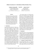
carboxyl terminus [Fig. 1]. The activity of PC is activa-
ted by acetyl-CoA and inhibited by aspartate [2,8–10].
Because of the lack of a three-dimensional structure, the
detailed mechanism of carboxylation and regulation of
PC remains obscure. Obviously, elucidation of the three-
dimensional structure of PC will unveil much of this
uncertainty and in fact such an undertaking is under way
in this laboratory. Additionally, a protein engineering
approach would be useful to examine the two partial
reactions individually. In this study, PC from Bacillus
thermodenitrificans (previously Bacillus stearothermophilus)
was engineered in such a way as to divide the protein
into two at the boundary of the BC and CT domains
(Fig. 1). The properties of the resulting two proteins,
PC-(BC) and PC-(CT + BCCP), were examined and
compared with those of the intact PC in order to gain
insight into the domain organization, the function of
acetyl-CoA and the reaction mechanism of PC.
Experimental procedures
Materials
Inorganic salts and common organic chemicals were
obtained from commercial sources. Acetyl-coenzyme A
was from Wako Pure Chemical (Osaka, Japan) and avidin
was from ProZyme (San Leandro, CA, USA). Reagents
for genetic engineering, such as restriction enzymes, were
purchased from Takara (Kyoto, Japan). Oligonucleotides
were custom synthesized by Hokkaido Science (Sapporo,
Japan). The TOPO TA cloning kit was the product of
Invitrogen.
Correspondence to S. Sueda, Department of Biochemical Engineering
and Science, Kyushu Institute of Technology, Kawazu 680-4, Iizuka
820-8502, Japan. Fax: + 81 948 29 7801, Tel.: + 81 948 29 7834,
E-mail:
Abbreviations: BC, biotin carboxylase; BCCP, biotin carboxyl
carrier protein; CT, carboxyl transferase; DTT, dithiothreitol;
KP
i
, potassium phosphate; PC, pyruvate carboxylase.
Enzymes: pyruvate carboxylase from Bacillus thermodenitrificans
(P94448) (EC 6.4.1.1); biotin carboxylase subunit of acetyl-CoA
carboxylase from Escherichia coli (P24182) (EC 6.4.1.2).
(Received 15 January 2004, revised 16 February 2004,
accepted 24 February 2004)
Eur. J. Biochem. 271, 1391–1400 (2004) Ó FEBS 2004 doi:10.1111/j.1432-1033.2004.04051.x
Construction of an overexpression plasmid
for intact PC
Previously, the B. thermodenitrificans PC gene was cloned
into pBluescript vector [11]. The resulting recombinant
plasmid (pPC) allowed Escherichia coli to express PC,albeit
at a relatively low level (data not shown). To enhance
expression, the promoter region was replaced with the high
expression promoter trc of pTrc99A vector. Thus, the
% 800 bp downstream region from the NcoI site containing
the trc promoter was amplified with pTrc99A as template
and using the following primers: Trc 1, 5¢-TTAGC
GG
GCCCATTAAGTTCTGTC-3¢ and Trc 2, 5¢-TTGCGA
ATTCGTCTTGTCTCCATGGTCTGTTTCCTGTGTG
AAAT-3¢ (restriction enzyme sites are underlined). The
EcoRI site, present at about 10 bp downstream from the
initiating ATG codon of the PC gene, and the ApaIsite,
present on pBluescript and pTrc99A, were exploited for
gene manipulation. A 19 bp segment of the PC gene (shown
above in italics) containing an EcoRI site was incorporated
into the reverse primer, Trc 2. The PCR reaction was
conducted under the following conditions: The reaction
mixture contained 5 units of Ex Taq
TM
(Takara), 1· Ex Taq
buffer, 200 l
M
each of the four dNTPs, 1 l
M
each of the
primers and 10 ng of pTrc99A in a final volume of 100 lL.
After denaturation at 94 °C for 5 min, the samples were
subjected to 30 cycles of denaturation (94 °C, 1 min),
annealing (58 °C, 1 min) and extension (72 °C, 1 min),
and subsequently subjected to additional extension (72 °C,
10 min). The PCR products were TA cloned and sequenced.
The plasmid thus prepared was digested with ApaIand
EcoRI, and the resulting fragment was ligated into the ApaI/
EcoRI sites of pPC. The second amino acid of native PC
is converted to glutamic acid from lysine because of the
introduction of the NcoI site into the start codon region of
this recombinant. This plasmid allowed E. coli to express
PC at a much higher level and the enzyme produced was
as active as native PC. Hence, this PC is called intact PC
despite the mutation of the second amino acid residue.
Construction of over-expression plasmids for PC-(BC)
and PC-(CT + BCCP)
The boundary of the BC and CT domains of B. thermo-
denitrificans PC was estimated to reside at residue 462 on the
basis of the reasoning described in the Results section. The
polypeptide chain was divided into two at this point by
placing a stop codon or an initiation codon for the expres-
sion of BC and CT plus BCCP, respectively. Expression
plasmids for PC-(BC) and PC-(CT + BCCP) were
constructed as follows: For the former, % 440 bp fragment
was amplified with pPC as the template using the
following primers: BC1, 5¢-ATT
GATATCGTCCAGTCG
CAAATTTTAATTGCT-3¢ and BC2, 5¢-ATA
GGATCC
TTAGAACACGAATAGTTCCGGCGTCGTATCGAT-3¢
(restriction enzyme sites are underlined). The forward
primer,BC1,harboredtheEcoRV site present on the PC
gene, and the reverse primer, BC2, harbored a stop codon
(denoted in bold). A BamHI site was introduced for
subsequent manipulation. PCR conditions were the same
as those for the amplification of the trc promoter, and the
PCR product was TA cloned and sequenced. The resulting
plasmid was digested with EcoRV and BamHI, and the
fragment formed was ligated into the EcoRV/BamHI sites
of pPC. The promoter of this plasmid was replaced with the
high expression promoter trc inexactlythesamewayasthat
of the intact PC. This plasmid, pPC-(BC), allowed E. coli
to express the BC domain of PC at a high level.
The PC-(CT + BCCP) expression plasmid was con-
structed as follows: an % 400 bp fragment was amplified
with pPC as template using the following primers: CT1,
5¢-ATAT
CCATGGCACGCCGGAAAGACGGAACGA
AAATG-3¢ and CT2, 5¢-CCGATCCCAC
GGATCCTCT
TTTAAAAAGCG-3¢ (restriction enzyme sites are under-
lined). The forward primer, CT1, harbored an NcoIsite
introduced for placing the start codon and cloning, and the
reverse primer, CT2, harbored a BamHI site present on the
PC gene. As a result of the engineering, the second amino
acid residue is converted from proline to alanine. PCR
conditions were the same as those described above, and
the PCR product was TA cloned and sequenced. Likewise,
a fragment representing the downstream region from the
BamHI site to the end of the open reading frame was
prepared (S. Sueda, unpublished observation). These two
fragments were cloned into pTrc99A through multiple steps
to yield a recombinant plasmid, pPC-(CT + BCCP), which
allowed E. coli to express the desired CT plus BCCP
domain of PC to a high level.
Purification of proteins
E. coli JM109 transformed with either one of the over-
expression plasmids prepared above was grown in Luria-
Bertani medium containing 50 lgÆmL
)1
ampicillin and
1 lgÆmL
)1
D
-biotin, where a biotin-binding domain was
present. Cells were harvested by centrifugation, suspended
in 0.12
M
potassium phosphate (KP
i
) buffer, pH 7.0,
containing 1 m
M
EDTA, 1 m
M
dithiothreitol (DTT) and
1m
M
phenylmethanesulfonyl fluoride, disrupted by soni-
cation and then centrifuged. The precipitate that formed
was removed by centrifugation, and ammonium sulfate was
added to the supernatant to 40–50% saturation for intact
PC and PC-(CT + BCCP), and 30–40% saturation for
PC-(BC). Again, the precipitate formed was collected by
centrifugation, dissolved in buffer A (20 m
M
KP
i
buffer,
pH 7.0, containing 0.1 m
M
EDTA and 0.1 m
M
DTT), and
dialyzed against the same buffer. The samples were subjec-
ted to anion exchange chromatography on diethylamino-
ethyl (DEAE)-cellulose (Whatman). Proteins were eluted by
a salt gradient from buffer A to buffer B (buffer A + 0.5
M
NaCl). The desired fractions, inspected by SDS/PAGE,
were collected and dialyzed against buffer A. The samples
were applied to gel filtration chromatography on Super-
Fig. 1. Schematic representation of the domain structures of intact PC
and engineered proteins, PC-(BC) and PC-(CT + BCCP).
1392 S. Sueda et al.(Eur. J. Biochem. 271) Ó FEBS 2004
dex
TM
200 (Amersham), eluted with 50 m
M
KP
i
buffer,
pH 7.0, containing 0.1
M
NaCl, 0.1 m
M
EDTA and 0.1 m
M
DTT, and the desired fractions collected. Intact PC and PC-
(CT+BCCP) were further purified by monomeric avidin-
Sepharose affinity chromatography [12–14] as reported
previously [15]. The samples were applied at a flow rate of
0.5 mLÆmin
)1
onto the monomeric avidin column equili-
brated with running buffer (50 m
M
KP
i
buffer, pH 7.0,
0.2
M
KCl, 1 m
M
EDTA, 5 m
M
2-mercaptoethanol). The
column was washed with several column volumes of running
buffer to remove unbound material. Proteins were eluted
with 1 mgÆmL
)1
biotin in running buffer at a flow rate of
0.2 mLÆmin
)1
. The eluted intact PC and PC-(CT+BCCP)
were dialyzed against 5 m
M
KP
i
buffer, pH 7.0, containing
0.1 m
M
EDTA and 0.1 m
M
DTT,andstoredat4°C. In the
meantime, PC-(BC) was further purified by anion exchange
chromatography on Mono Q
TM
HR 5/5 (Amersham).
Protein was eluted by a salt gradient from buffer C (20 m
M
Tris/HCl, pH 7.5) to buffer D (buffer C+0.35
M
NaCl).
The desired fractions were collected and dialyzed against
5m
M
KP
i
buffer, pH 7.0, with 0.1 m
M
EDTA and 0.1 m
M
DTT, and stored at 4 °C. The specific activity of PC,
determined below, was 9.5 UÆmg
)1
, where 1 U is defined as
the amount of enzyme to produce 1 lmol of oxalacetate per
min, and the protein concentration was determined from the
amino acid composition.
Pyruvate carboxylase assays
Pyruvate carboxylase activity was measured by monitoring
the oxalacetate formation using the coupled reaction with
malate dehydrogenase according to the methods described
previously [16–18]. Oxidation of NADH in the malate
dehydrogenase reaction was followed spectrophotometri-
cally at 340 nm. All assays were carried out at 30 °C, and
the reaction mixture contained the following components,
unless otherwise stated: 100 m
M
Tris/HCl (pH 8.0), 2 m
M
ATP, 5 m
M
MgCl
2
, 100 m
M
KCl, 5 m
M
pyruvate, 50 m
M
NaHCO
3
,0.1 m
M
acetyl-CoA, 0.15 m
M
NADH and 5 units
of malate dehydrogenase.
The K
m
(Michaelis constant) values for ATP, bicarbonate
and pyruvate were determined as follows: the K
m
for ATP
was obtained by varying its concentration from 0–5 m
M
at
fixed concentrations of bicarbonate (100 m
M
) and pyruvate
(5 m
M
), where the enzyme was 77% and 92% saturated with
them, respectively. In addition, free Mg
2+
concentration
was kept constant, with MgCl
2
,at3m
M
in excess of ATP;
free Mg
2+
concentration was approximated to be its ana-
lytical concentration minus that of ATP, as the true concen-
tration of free Mg
2+
calculated based on the dissociation
constant for MgATP of 0.0143 m
M
[19] was only 1%
different from the approximate value. At high ATP concen-
trations, substrate inhibition was evident, and thus two kinds
of analysis were applied for the data on ATP. First, the
simple Michaelis–Menten equation was fitted to the kinetic
data in the low concentration range (0–1 m
M
)usingthe
nonlinear regression analysis program,
ENZFITTER
(Biosoft,
Cambridge, UK). Then, the entire data (from 0–5 m
M
)were
analyzed by Eqn (1), which takes into account substrate
inhibition, where v, V
max
,[S]andK
I
represent observed
reaction rate, maximum rate, substrate concentration and
the substrate inhibition constant, respectively:
v ¼ V
max
Â
½S
½SþK
m
þ½S
2
½K
I
Eqn (1)
The K
m
value for bicarbonate was determined by varying
its concentration from 0.5–100 m
M
at fixed concentrations
of ATP (2 m
M
) and pyruvate (5 m
M
). As the endogenous
level of bicarbonate is known to be 0.5 m
M
at pH 8.0
[20], the concentration of bicarbonate was corrected for
this value. Likewise, the K
m
value for pyruvate was
determined by varying its concentration from 0–5 m
M
at
fixed concentrations of ATP (2 m
M
) and bicarbonate
(100 m
M
). The simple Michaelis–Menten equation was
used for the analysis of the data for bicarbonate and
pyruvate.
ATP cleavage assays
ATP cleavage activity of intact PC and PC-(BC) was
assayed according to the previously reported procedure [21].
The progress of the reaction was followed by monitoring the
formation of ADP in the presence of phosphoenolpyru-
vate and pyruvate kinase. The pyruvate formed was then
reduced to lactate by lactate dehydrogenase with the
concomitant oxidation of NADH, and this was measured
from a decrease in absorbance at 340 nm. All assays were
conducted at 30 °C, and the reaction mixture contained the
following components, unless otherwise stated: 100 m
M
Tris/HCl (pH 8.0), 2 m
M
ATP, 5 m
M
MgCl
2
, 100 m
M
KCl, 50 m
M
NaHCO
3
,0.1m
M
acetyl-CoA, 0.5 m
M
phos-
phoenol pyruvate, 0.15 m
M
NADH, 5 units of lactate
dehydrogenase and 5 units of pyruvate kinase. In the case of
the PC-(BC) assay, 50 m
M
free
D
-biotin was added to the
above reaction mixture.
The kinetic parameters, K
m
and V
max
,fortheATPase
reaction of PC-(BC) were determined as follows: the
kinetic parameters for ATP were obtained by varying its
concentration from 0–5 m
M
at fixed concentrations of
biotin (100 m
M
) and bicarbonate (100 m
M
), where the
enzyme is 68% and 62% saturated with them, respect-
ively. Obviously, this situation is not ideal for the accurate
estimation of kinetic parameters, but concentrations
higher than this will deviate too much from those of
physiological conditions. Accordingly, experiments were
carried out under these subsaturating conditions with
respect to biotin and bicarbonate. In the kinetics for ATP,
substrate inhibition was manifest at high concentrations
just like for intact PC, and thus two kinds of data
analysis were also made in this case. The kinetic
parameters for bicarbonate were determined by varying
its concentration from 0.5–100 m
M
at fixed concentrations
of ATP (2 m
M
) and biotin (100 m
M
); again, the concen-
tration of bicarbonate was corrected for the endogenous
bicarbonate at pH 8.0, 0.5 m
M
. The simple Michaelis–
Menten equation was used for the analysis of the data
obtained. The kinetic data for biotin were obtained by
varying its concentration from 0–100 m
M
at fixed con-
centrations of ATP (2 m
M
) and bicarbonate (100 m
M
). In
the ATPase reaction of PC-(BC), a weak activity (2% of
maximum) was observed in the absence of biotin, and
thus the data were analyzed by Eqn (2), which takes into
account this basal activity (v
0
):
Ó FEBS 2004 Protein engineering of pyruvate carboxylase (Eur. J. Biochem. 271) 1393
v ¼ V
max
Â
½S
K
m
þ½Sþv
0
Eqn ð2Þ
Oxalacetate decarboxylase assays
Oxalacetate decarboxylase activity of intact PC and PC-
(CT + BCCP) was measured with oxamate as the stimu-
lant, according to the procedures previously reported [22].
The reactions were monitored by measuring the formation
of pyruvate which was then reduced to lactate by lactate
dehydrogenase, and the concomitant oxidation of NADH
was monitored at 340 nm. All assays were performed at
30 °C, and the reaction mixture contained the following
components, unless otherwise stated: 100 m
M
Tris/HCl
(pH 8.0), 5 m
M
MgCl
2
,100m
M
KCl, 0.1 m
M
oxalacetate,
0.1 m
M
acetyl-CoA, 1 m
M
oxamate, 0.15 m
M
NADH, and
5 units of lactate dehydrogenase. The reactions were started
by the addition of intact PC or PC-(CT + BCCP), but
prior to the addition, a background rate of oxalacetate
decarboxylation was established, and this (2.4% of the
maximum) was subtracted from the rate in the presence
of enzyme.
Avidin-blot analysis and determination
of the N-terminal amino acid sequence
For avidin-blot analysis, electrophoresed samples were
electroblotted onto a nylon membrane (Pall Biosupport,
Portsmouth, UK) according to the conventional procedure
[23]. The membrane with blotted proteins was blocked with
skimmed milk in NaCl/Tris-Tween [20 m
M
Tris/HCl,
pH 7.6, 136 m
M
NaCl, 0.1% (v/v) Tween] for one hour.
The blocked membrane was washed three times with NaCl/
P
i
-Tween, and then immersed in NaCl/Tris buffer contain-
ing 0.4 UÆmL
)1
alkaline phosphatase-conjugated streptavi-
din (Boehringer Mannheim) for 20 min. The membrane
was then washed with NaCl/Tris-Tween three times, before
being developed by 0.78 m
M
4-nitroblue tetrazolium chlor-
ide and 0.40 m
M
5-bromo-4-chloro-3-indolylphosphate in
20 m
M
Tris/HCl, pH 9.5, containing 100 m
M
NaCl and
50 m
M
MgCl
2
.
For determining the amino-terminal sequence of the
proteins, electrophoresed samples were electroblotted onto
a poly(vinylidene difluoride) membrane (Atto, Tokyo,
Japan) according to the conventional procedure. Pieces
of the membrane containing the desired bands, as visualized
by ponceau S, were used for sequencing by Edman
degradation on a protein sequencer Model 491 (Applied
Biosystems).
Molecular size determination by HPLC gel-filtration
chromatography
High performance gel filtration chromatography was
carried out on a TSKgel G3000SWXL column
(7.8 mm · 30 cm) with TSK guard column SWXL
(6.0 mm · 4.0 cm) (Tosoh, Tokyo, Japan) using an HPLC
system (Hitachi, Tokyo, Japan). The samples were eluted at
a flow rate of 0.5 mLÆmin
)1
using a mobile phase of 100 m
M
KP
i
buffer (pH 7.0) containing 100 m
M
Na
2
SO
4
,andthe
eluted samples were monitored at 280 nm. The gel filtration
column was calibrated using a set of proteins (Amersham):
ribonuclease A (13.7 kDa), chymotrypsinogen A (23 kDa),
ovalbumin (43 kDa), albumin (67 kDa), aldolase
(158 kDa) and thyroglobulin (669 kDa). The apparent
molecular masses of the samples were estimated from the
calibration curve obtained. The samples were analyzed at
concentrations ranging from 1–100 l
M
,and20 lLeachwas
applied to the column.
Molecular mass determination by mass spectrometry
The molecular masses of PC-(BC) and PC-(CT + BCCP)
were determined by MALDI TOF mass spectrometry with
a Voyager DE-STR mass spectrometer (PerSeptive Bio-
systems, Framingham, MA, USA)
7
. 4-Hydroxyazobenzene-
2¢-carboxylic acid (10 mgÆmL
)1
in 0.1% (v/v) trifluoroacetic
acid in 70 : 30 water/acetonitrile) was used as the MALDI
matrix. Samples were prepared by mixing the protein
solution with the matrix solution. One microliter of this
mixture was deposited on the sample plate, dried at ambient
temperature and analyzed.
Results
Construction and purification of the engineered
proteins of PC
The boundary of the BC and CT domains of PC was
estimated as follows: the BC subunit of E. coli acetyl-CoA
carboxylase is catalytically active and the three-dimensional
structure is known [24,25]. Its C-terminus appeared to
correspond to residue 460 of B. thermodenitrificans PC by
sequence alignment [26]. Likewise, the amino acid sequences
of PCs from various sources, including those of subunit-
type PCs, were aligned to reveal that the N-terminus of CT
seemed to reside at residue 470 of B. thermodenitrificans PC.
Although there still remains some ambiguity concerning
the exact location of the boundary because the C- and
N-terminal regions of BC and CT domains, respectively, are
barely conserved, it seemed safe to divide the two domains
at residue 462 without impairing the two activities (Fig. 1).
Based on this assumption, over-expression plasmids for
PC-(BC) and PC-(CT + BCCP) which produce BC and
the rest of the molecule, respectively, were constructed as
detailed in Experimental procedures.
The engineered proteins of PC as well as intact PC were
purified by methods described under Experimental pro-
cedures. Monomeric avidin-Sepharose affinity chromato-
graphy was used for the purification of intact PC and
PC-(CT + BCCP) carrying the biotin prosthetic group
within their structures. Each purified protein was nearly
homogeneous as judged by visual inspection of SDS/PAGE
(Fig. 2A). The yields were typically 10, 15 and 20 mg, for
intact PC, PC-(BC) and PC-(CT + BCCP), respectively,
from a 2 L culture. Western blot analysis with alkaline
phosphatase-conjugated streptavidin for intact PC and
PC-(CT + BCCP) revealed that bands were observed at
the positions corresponding to those of SDS/PAGE
(Fig. 2B). The amino-terminal sequence of each protein
was analyzed by Edman degradation. The amino acid
sequences of intact PC and PC-(BC) were determined to be
1394 S. Sueda et al.(Eur. J. Biochem. 271) Ó FEBS 2004
METRRIRKVL, which was consistent with that deduced
from the DNA sequences. The correct mutation of the
second amino acid residue, arising from the introduction
of an NcoI site in the start codon region, to glutamate from
the original lysine, was confirmed. Likewise, the amino
acid sequence of PC-(CT + BCCP) was determined to be
ARRKDRGTKM, and this sequence was consistent with
that deduced from its DNA sequence except for the absence
of the first amino acid methionine. It was also confirmed that
the second residue was properly converted to alanine from
original proline, because of the design of the expression
plasmid.
In SDS/PAGE, the bands of intact PC and PC-(BC) were
observed at the positions corresponding to the molecular
masses deduced from their sequences, 128.5 and 51.4 kDa,
respectively, while that of PC-(CT + BCCP) was observed
at a position (65 kDa) considerably smaller than that
expected (77.1 kDa). To confirm the integrity of PC-
(CT + BCCP), this protein was analyzed by MALDI
TOF mass spectrometry together with PC-(BC). The mass
(m/z value) obtained was 77 047 ± 76 for PC-(CT +
BCCP) and 51 428 ± 53 for PC-(BC) (mean ± SD from
three determinations). These values are identical, within
experimental error, to the molecular masses deduced from
their sequences, 77 082 and 51 438 Da, respectively, prov-
ing that the two engineered proteins have the correct
structure.
Molecular properties of the engineered proteins of PC
Association states of the proteins were investigated by high
performance gel filtration chromatography. Apparent
molecular masses of the samples were estimated on the
basis of the calibration curve obtained by using a set of
standard proteins (Fig. 3). Typical elution profiles of intact
PC, PC-(BC) and PC-(CT + BCCP) are shown in Fig. 4.
For intact PC, two peaks were observed at 13.76 min and
17.02 min (Fig. 4A) and the apparent molecular masses
estimated from their retention times were 501.3 ±
11.5 kDa and 137.0 ± 4.8 kDa (mean ± SE from three
separate experiments), which were considered to be the
tetramer and monomer, respectively. The intensity of
the tetramer peak was about 10 times greater than that of
the monomer and this ratio did not change with protein
concentration over the range adopted (1–100 l
M
), verifying
that intact PC exists mainly as a tetramer, which is typical
for single polypeptide type PCs [4,27]. Also for PC-
(CT + BCCP), two peaks, major and minor, were
observed at 17.17 min and 18.82 min (Fig. 4B) and the
apparent molecular masses estimated from them were
128.2 ± 1.8 kDa and 66.2 ± 1.1 kDa, which appeared
to represent a dimer and monomer, respectively. Again, the
ratio of the intensity of the two peaks (10 : 1), did not
change with the protein concentration.
By contrast, the behavior of PC-(BC) on gel filtration
chromatography was different from those of the above
two proteins. Although two peaks were also observed for
PC-(BC), the ratio of the intensity of the peaks changed
markedly with the protein concentration. At a high
concentration (100 l
M
), a major peak was observed at
18.28 min (Fig. 4C) and the molecular mass estimated from
this peak was 81.9 ± 2.9 kDa, which appeared to represent
a dimer. On the other hand, at a low concentration (5 l
M
), a
major peak was observed at 19.41 min (Fig. 4D) and the
molecular mass estimated from it was 66.2 ± 1.1 kDa,
which appeared to represent the monomer.
Moreover, the mixtures of PC-(BC) and PC-(CT +
BCCP) at various ratios were analyzed to study their
interaction, but no new peak was observed other than those
derived from the constituent proteins, suggesting that
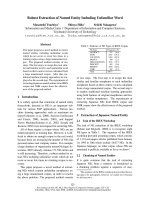
Fig. 3. Estimation of the molecular masses of intact and engineered PC
by gel filtration chromatography on the TSK G3000SWXL column. The
molecular masses of the proteins used for construction of the calib-
ration curve (s) were ribonuclease A (13.7 kDa), chymotrypsino-
gen A (23 kDa), ovalbumin (43 kDa), albumin (67 kDa), aldolase
(158 kDa) and thyroglobulin (669 kDa). Elution times of intact PC
and engineered proteins are represented by d, as labelled.
Fig. 2. SDS/PAGE (A) and avidin-blot analysis (B) of purified intact
PC and engineered proteins. (A) SDS/PAGE was run with 12.5%
polyacrylamide and 0.5 lg of proteins: M, marker; lane 1, intact PC;
lane 2, PC-(BC); lane 3, PC-(CT + BCCP). (B) SDS/PAGE was run
with 0.1 lg of proteins and electroblotted onto the membrane. The
proteins carrying biotin were detected by the reaction with alkaline
phosphatase-conjugated avidin: lane 1, intact PC; lane 2, PC-
(CT + BCCP).
Ó FEBS 2004 Protein engineering of pyruvate carboxylase (Eur. J. Biochem. 271) 1395
PC-(BC) and PC-(CT + BCCP) do not interact signifi-
cantly under the experimental conditions employed. Acetyl-
CoA had almost no effect on the association of the two
proteins, as the elution profile was hardly affected by
preincubation of the samples with 0.1 m
M
acetyl-CoA
followed by elution with buffer containing the same
concentration of acetyl-CoA.
Enzymic activity of intact PC
Pyruvate carboxylase activity of intact PC was assayed by
measuring the oxalacetate production in the presence of
malate dehydrogenase and NADH as described under
Experimental procedures. The K
m
values determined for
bicarbonate and pyruvate were 29.9 ± 1.4 m
M
and
0.31 ± 0.03 m
M
, respectively (estimate ± standard error
from the nonlinear regression analysis), which were virtually
identical to those of the literature, 28.6 m
M
for bicarbonate
and 0.33 m
M
for pyruvate [28]. In the kinetic analysis of
ATP, substrate inhibition was evident at high ATP
concentration, and thus two kinds of data analysis were
conducted as described in Experimental procedures. The K
m
obtained from the data where substrate inhibition is
insignificant was 0.46 ± 0.06 m
M
, while the value obtained
from the whole data based on Eqn (1) that takes into
account substrate inhibition was 0.87 ± 0.11 m
M
.TheK
m
for ATP of PC from the same source, obtained by simple
Michaelis–Menten analysis on the data where substrate
inhibition is not evident, was reported as 0.38 m
M
[28], close
to the corresponding value of the present work. Also, the
effect of acetyl-CoA on the pyruvate carboxylase reaction
was nearly the same among the present work and literature;
the activity was greatly increased upon addition of acetyl-
CoA and the activity in the absence of acetyl-CoA was
approximately 0.3% of the maximum (Table 1).
PC is known to catalyze the cleavage of ATP in the
absence of pyruvate [21]. It is hence possible to study the
reaction of BC (Scheme 1) independently of the CT reaction
(Scheme 2) by measuring this activity. This activity of PC
was determined under essentially the same conditions as the
complete reaction except for the omission of pyruvate
(Table 1). The rate of the ATP cleavage reaction is about
0.2% of that of the complete reaction of PC, which almost
coincides with that reported for chicken liver enzyme [21].
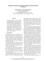
Fig. 4. Typical elution profiles for intact PC (A), PC-(CT + BCCP)
(B) and PC-(BC) (C and D) on the TSK G3000SWXL gel filtration
column. Proteins were chromatographed over a concentration range of
1–100 l
M
under the conditions described in Experimental procedures.
M, D and T denote monomer, dimer and tetramer, respectively.
(A) Two peaks were observed for intact PC at 13.76 and 17.02 min,
which correspond to the tetramer and monomer, respectively, and the
elution profile did not change over the concentration range examined.
(B) Two peaks were also observed for PC-(CT + BCCP) at 17.17 and
18.82 min, which correspond to the dimer and monomer, respectively,
and the elution profile was not dependent on the protein concentra-
tion. (C) and (D) The elution profile of PC-(BC) was markedly
dependent on the protein concentration: at the high concentration
[100 l
M
, (C)], the dimer predominated, while at the low concentration
[5 l
M
, (D)], the monomer predominated.
1396 S. Sueda et al.(Eur. J. Biochem. 271) Ó FEBS 2004
It was found that activity increased about 10-fold by the
addition of acetyl-CoA to 0.1 m
M
, thus this ATP cleavage
reaction is also dependent on acetyl-CoA. Similar depend-
ence on acetyl-CoA was also observed previously [21].
Enzymic activity of PC-(BC)
It was found that the truncated enzyme, PC-(BC), is as
capable of mediating ATP cleavage in the presence of free
D
-biotin as intact PC, suggesting that its three-dimensional
structure remains intact even in the absence of other
domains. The enzymic activity of PC-(BC) in the presence of
various concentrations of biotin, bicarbonate and ATP are
depicted in Fig. 5. As expected from the reaction mechan-
ism proposed for BC [2,29,30], this enzymic reaction was
completely dependent on three substrates; it is worthy of
noting that biotin is necessary for this reaction to proceed
(the activity in the absence of biotin is about 2% of
maximum in its presence). This subject is discussed in more
detail below. Kinetic parameters for the three substrates
were determined from the data shown in Fig. 5. The K
m
for
bicarbonate was 62.2 ± 5.3 m
M
, which was comparable
to that for the complete reaction of intact PC
(29.9±1.4m
M
). In the kinetics for ATP, substrate inhi-
bition was observed just like in intact PC, and the K
m
values
determined based on the simple Michaelis–Menton equa-
tion and Eqn (1), were 0.54 ± 0.04 m
M
and
1.03 ± 0.15 m
M
, which were close to those of intact PC
(0.46 ± 0.06 m
M
and 0.87 ± 0.11 m
M
). The K
m
value for
biotin of 50.9 ± 5.4 m
M
is considerably smaller than that
of the BC subunit of acetyl-CoA carboxylase from E. coli
(135 m
M
) [31]. To investigate the effect of acetyl-CoA on
this reaction, the assay was carried out under the standard
conditions but omitting acetyl-CoA, and the data obtained
are shown in Table 1. Unexpectedly, the activity of PC-(BC)
in the absence of acetyl-CoA was virtually unchanged from
that in its presence. In other words, the ATP cleavage
activity of PC-(BC) is not dependent on acetyl-CoA, in
sharp contrast to that of intact PC.
Enzymic activity of PC-(CT + BCCP)
It was reported that oxamate stimulated the decarboxyla-
tion of oxalacetate by PC [22]. It is hence possible to study
the CT reaction (Scheme 2) of PC separately from the BC
reaction (Scheme 1) with this assay [22]. The enzymic
activity of PC-(CT + BCCP), investigated by measuring
the oxalacetate decarboxylase activity in the presence of
oxamate, increased with an increase in oxamate concentra-
tion, as expected (Fig. 6A). The activity in the presence of a
saturating concentration of oxamate was about 40 times
higher than that in its absence (Table 2). The effect of
oxalacetate concentration on the decarboxylation reaction
at a fixed concentration of oxamate is shown in Fig. 6B. In
this case, substrate inhibition at high concentration of
oxalacetate is evident from the profile. Such a phenomenon
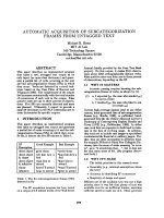
Fig. 5. Kinetic analysis for the ATP cleavage activity of PC-(BC). Activity of PC-(BC) (0.22 mg in 1 mL) was assayed with free biotin as the
substrate in 100 m
M
Tris/HCl (pH 8.0) containing 5 m
M
MgCl
2
,100m
M
KCl, 0.1 m
M
acetyl-CoA, 0.5 m
M
phosphoenol pyruvate, 0.15 m
M
NADH, 5 units of lactate dehydrogenase, 5 units of pyruvate kinase and variable concentrations of ATP, bicarbonate and biotin at 30 °C. (A)
Biotin was the variable substrate with 2 m
M
ATP and 100 m
M
bicarbonate; the K
m
for biotin was 50.9 ± 5.4 m
M
and the V
max
2.39 ± 0.12 UÆlmol
)1
. Kinetic parameters were determined by fitting Eqn (2) to the data. (B) Bicarbonate was the variable substrate with 2 m
M
ATP and 100 m
M
biotin; the K
m
for bicarbonate was 62.2 ± 5.3 m
M
and the V
max
2.58 ± 0.12 UÆlmol
)1
. Kinetic parameters were determined by
fitting the simple Michaelis–Menten equation to the data. (C) ATP was the variable substrate with 100 m
M
bicarbonate and 100 m
M
biotin. In the
kinetics for ATP, substrate inhibition was evident, and thus two different kinds of analysis were made for the obtained data. Kinetic parameters
determined with the data from 0–1.0 m
M
on the basis of simple Michaelis–Menten were as follows: K
m
0.54 ± 0.04 m
M
and V
max
2.39 ± 0.10 UÆlmol
)1
, while those determined with the data from 0–5.0 m
M
on the basis of Eqn (1) were as follows: K
m
1.03 ± 0.15 m
M
and V
max
3.91 ± 0.40 UÆlmol
)1
. The theoretical curve shown in this figure was drawn on the basis of Eqn (1). In each case, the standard errors in V
max
and
K
m
were determined from the nonlinear regression analysis.
Table 1. Effect of 0.1 m
M
acetyl-CoA on the pyruvate carboxylation of
intact PC and on the ATP cleavage reactions of intact PC and PC-(BC).
One unit of enzyme activity was defined as the amount of enzyme
required to catalyze the formation of 1 lmol of each product per min.
Values are the means ± SD from three separate experiments.
Protein Activity
Enzymic activity (UÆlmol
)1
)
With acetyl-CoA Without acetyl-CoA
Intact PC Overall 1220 ± 50 4.52 ± 0.36
Intact PC ATP cleavage 2.84 ± 0.21 0.31 ± 0.04
PC-(BC) ATP cleavage 1.02± 0.06 0.99 ± 0.07
Ó FEBS 2004 Protein engineering of pyruvate carboxylase (Eur. J. Biochem. 271) 1397
was observed also for PC from chicken liver and was
accounted for by competitive substrate inhibition [22]. The
decarboxylation activity of PC-(CT + BCCP) was found
to be of similar magnitude to that of PC (Table 2), and thus
PC-(CT + BCCP) retains the enzymic activity present in
the native structure despite lacking the BC domain. It
is noted that the decarboxylation activity of PC-
(CT + BCCP) and intact PC was virtually the same in
the presence and absence of acetyl-CoA under the standard
conditions used in this study. Therefore, the catalytic
reaction of the CT domain appears to be independent of
acetyl-CoA, just like the ATP-cleavage reaction of PC-(BC).
Discussion
In general, single polypeptide-type PCs exist in the tetra-
meric and subunit-type PCs in octameric form [1,2], but
little is known as to how these oligomeric structures are
formed. PC consists of three domains, BC, CT and BCCP,
but again little is known about how these domains are
organized three-dimensionally to generate active enzymes.
These are the subjects addressed in this article. It was found
that the separated BC and CT + BCCP domains of the
former type of PC from B. thermodenitrificans retain their
own catalytic activity, demonstrating that these two
domains are independent entities as a protein. Moreover,
from the elution profiles of gel filtration HPLC, PC-
(CT + BCCP) was found to exist mainly as a dimer, while
PC-(BC) was found to exist as a monomer or a dimer
depending on its concentration. In other words, both
engineered proteins associate with themselves to form
homodimers, and the association of PC-(CT + BCCP)
seems to be stronger than that of PC-(BC). In addition, the
association between PC-(BC) and PC-(CT + BCCP) was
not observed under the experimental conditions examined,
demonstrating that they do not possess strong affinity for
each other. Given that the same applies to intact PC as well,
it is deduced that the tetrameric form of PC is built up in the
following way: first, a dimer of PC is formed through the
association of each (CT + BCCP) domain of two proto-
mers of PC, and subsequently, individual BC domains of
the resulting two dimers associate to form a tetramer. In
other words, the tetrameric structure of PC appears to be
constructed through the interaction of the same domains,
namely BC with BC and (CT + BCCP) with (CT +
BCCP). This hypothesis awaits verification by X-ray
crystallographic analysis, which is under way in this
laboratory.
As for the reaction of BC, the following mechanism
involving the formation of an enzyme–carboxylphosphate
complex seems to be the most plausible one [2,29,30]:
ATP þ HCO
À
3
þenz-biotin Ðð
À2
O
3
POCO
À
2
Áenz-biotinÞ
þADP Scheme 3
ð
À2
O
3
POCO
À
2
Áenz-biotinÞÐenz-biotin-CO
À
2
þ P
i
Scheme 4
Apparently biotin is not required in the reaction of
bicarbonate with ATP (Scheme 3), but it is essential for
the putative carboxylphosphate intermediate to form.
Biotin appears to participate indirectly in this step by
inducing a conformational change so as to dispose the
active site residues in correct orientations
8
for bicarbonate
to undergo nucleophilic attack on the c-phosphate of
ATP. In the present work, ATP cleavage activity
(Scheme 3) of PC-(BC) was investigated with free biotin
Fig. 6. Oxalacetate decarboxylation reaction of PC-(CT + BCCP).
Activity of PC-(CT + BCCP) (0.39 mg in 1 mL) was assayed in
100 m
M
Tris/HCl (pH 8.0) containing 5 m
M
MgCl
2
,100m
M
KCl,
0.1 m
M
acetyl-CoA, 0.15 m
M
NADH, 5 units of lactate dehydro-
genase and variable concentrations of oxalacetate and oxamate at
30 °C. (A) Oxamate was the varied substrate with 0.1 m
M
oxalacetate.
(B) Oxalacetate was the varied substrate with 1.0 m
M
oxamate. Error
bars represent the standard deviations from the mean of three deter-
minations.
Table 2. Effect of 0.1 m
M
acetyl-CoA on the oxalacetate decarboxy-
lase activity (UÆlmol
)1
) of PC-(CT + BCCP) and intact PC. One unit
of enzyme activity was defined as the amount of enzyme required to
catalyze the formation of 1 lmol of pyruvate per min. Values are the
means ± SD from three separate experiments.
Acetyl-CoA
PC-(CT + BCCP) Intact PC
+Oxamate – Oxamate +Oxamate – Oxamate
Present 6.62 ± 0.48 0.18 ± 0.03 3.88 ± 0.32 0.43 ± 0.05
Absent 6.42 ± 0.54 0.19 ± 0.04 3.78 ± 0.37 0.41 ± 0.06
1398 S. Sueda et al.(Eur. J. Biochem. 271) Ó FEBS 2004
as substrate. As the activity of PC-(BC) was completely
dependent not only on bicarbonate but also on biotin, it
was confirmed that biotin is essential in the reaction of
bicarbonate with ATP.
Although a large number of studies has been devoted
to clarifying the role of acetyl-CoA in the PC reaction
[1,2,32–34], little is known about its activation mechanism.
It was found that acetyl-CoA did not affect the ATP
cleavage activity of PC-(BC), although it is essential in the
same reaction of the BC domain of intact PC. Likewise,
oxalacetate decarboxylation reactions of PC-(CT +
BCCP) and intact PC were not dependent on acetyl-
CoA. Taken together, it seems that acetyl-CoA partici-
pates in the reaction of BC but not of CT, and judging
from the disappearance of acetyl-CoA dependence in the
reaction of PC-(BC) with free biotin, acetyl-CoA may act
as a regulator in the interaction between the active site of
the BC domain and the biotin moiety of the BCCP
domain.
Based on these arguments, it is tempting to propose the
following hypothesis: in the absence of acetyl-CoA, the
active site of the BC domain cannot interact with biotin of
the BCCP domain due to the spatial separation between the
active site and biotin; however, upon binding of acetyl-CoA,
a conformational change is induced, so that biotin can reach
the active site to carry out the catalytic reaction. In the
reaction of PC-(BC) with free biotin, such a steric constraint
is absent; as a result, its acetyl-CoA dependence may be lost.
Conformation changes of PC induced by acetyl-CoA have
been observed by various means such as electron micros-
copy [35,36], ultracentrifugation [37] and others [38]. In
order to verify the above hypothesis, further investigation is
needed and studies using other engineered proteins as well
as X-ray crystallographic analysis are under way in this
laboratory.
Acknowledgements
The authors are grateful to Ms Tomoko Ishiguro and Ms Masayo
Nonaka for their assistance with construction of the recombinant
plasmids.
References
1. Jitrapakdee, S. & Wallace, J.C. (1999) Structure, function and
regulation of pyruvate carboxylase. Biochem. J. 340, 1–16.
2. Attwood, P.V. (1995) The structure and the mechanism of action
of pyruvate carboxylase. Int. J. Biochem. Cell. Biol. 27, 231–249.
3. Wallace, J.C., Jitrapakdee, S. & Chapman-Smith, S. (1998)
Pyruvate carboxylase. Int. J. Biochem. Cell. Biol. 30, 1–5.
4. Barden, R.E., Taylor, B.L., Isohashi, F., Frey, W.H., Zander, G.,
Lee, J.C. & Utter, M.F. (1975) Structural properties of pyruvate
carboxylases from chicken liver and other sources. Proc. Natl
Acad. Sci. USA 72, 4308–4312.
5.Lim,F.,Morris,P.,Occhiodoro,F.&Wallace,J.C.(1988)
Sequence and domain structure of yeast pyruvate carboxylase.
J. Biol. Chem. 263, 11493–11497.
6.Cohen,N.D.,Duc,J.A.,Beegen,H.&Utter,M.F.(1979)
Quaternary structure of pyruvate carboxylase from Pseudomonas
citronellolis. J. Biol. Chem. 254, 9262–9269.
7. Goss, J.A., Cohen, N.D. & Utter, M.F. (1981) Characterization of
the subunit structure of pyruvate carboxylase. J. Biol. Chem. 256,
11819–11825.
8. Cazzulo, J.J. & Stoppani, A.O.M. (1968) The regulation of yeast
pyruvate carboxylase by acetyl-coenzyme A and
L
-aspartate.
Arch. Biochem. Biophys. 127, 563–567.
9. Libor, S.M., Sundaram, T.K. & Scrutton, M.C. (1978) Pyruvate
carboxylase from a thermophilic Bacillus. Studies on the specificity
of activation by acyl derivatives of coenzyme A and on the
properties of catalysis in the absence of activator. Biochem. J. 169,
543–558.
10. Ashman, L.K., Keech, D.B., Wallace, J.C. & Nielsen, J. (1972)
Sheep kidney pyruvate carboxylase. Studies on its activation by
acetyl coenzyme A and characterization of its acetyl coenzyme A
independent reaction. J. Biol. Chem. 247, 5818–5824.
11. Kondo, H., Kazuta, Y., Saito, A. & Fuji, K. (1997) Cloning and
nucleotide sequence of Bacillus stearothermophilus pyruvate
carboxylase. Gene 191, 47–50.
12. Henrikson, K.P., Allen, S.H.G. & Maloy, W.L. (1979) An avidin
monomer affinity column for the purification of biotin-containing
enzymes. Anal. Biochem. 94, 366–370.
13. Kohanski, R.A. & Lane, M.D. (1990) Monovalent avidin affinity
column. Methods Enzymol. 184, 194–200.
14. Buckley, J.J., Libor, S. & Sundaram, T.K. (1979) Biotin subunits
of acetyl CoA carboxylase and pyruvate carboxylase from a
thermophilic Bacillus. Arch. Biochem. Biophys. 192, 396–404.
15. Jitrapakdee, S., Walker, M.E. & Wallace, J.C. (1999) Functional
expression, purification, and characterization of recombinant
human pyruvate carboxylase. Biochem. Biophys. Res. Commun.
266, 512–517.
16. Modak, H.V. & Kelly, D.J. (1995) Acetyl-CoA-dependent pyru-
vate carboxylase from the photosynthetic bacterium Rhodobacter
capsulatus: rapid and efficient purification using dye-ligand affinity
chromatography. Microbiol. 141, 2619–2628.
17. Mukhopadhyay, B., Stoddard, S.F. & Wolfe, R.S. (1998)
Purification, regulation, and molecular and biochemical charac-
terization of pyruvate carboxylase from Methanobacterium ther-
moautotrophicum strain DH. J. Biol. Chem. 273, 5155–5166.
18. Mukhopadhyay, B. & Purwantini, E. (2000) Pyruvate carboxylase
from Mycobacterium smegmatis: stabilization, rapid purification,
molecular and biochemical characterization and regulation of the
cellular level. Biochim. Biophys. Acta 1475, 191–206.
19. Morrison, J.F. (1979) Approaches to kinetic studies on metal-
activated enzymes. Methods Enzymol. 63, 257–294.
20. Levert, K.L., Lloyd, R.B. & Waldrop, G.L. (2000) Do cystein 230
and lysine 238 of biotin carboxylase play a role in the activation of
biotin? Biochemistry 39, 4122–4128.
21. Attwood, P.V. & Graneri, B.D.L.A. (1992) Bicarbonate-
dependent ATP cleavage catalysed by pyruvate carboxylase in the
absence of pyruvate. Biochem. J. 287, 1011–1017.
22. Attwood, P.V. & Cleland, W.W. (1986) Decarboxylation of
oxalacetate by pyruvate carboxylase. Biochemistry 25, 8181–8196.
23. Sambrook, J., Fritsch, E. F. & Maniatis, T. (1989) Transfer of
proteins from SDS-polyacrylamide gels to solid supports:
immunological detection of immobilized proteins (western blot-
ting). In Molecular Cloning: a Laboratory Manual, 2nd edn, pp.
18.60–18.75. Cold Spring Harbor Laboratory Press, Cold Spring
Harbor, NY.
24.Waldrop,G.L.,Rayment,I.&Holden,H.M.(1994)Three-
dimensional structure of the biotin carboxylase subunit of acetyl-
CoA carboxylase. Biochemistry 33, 10249–10256.
25. Thoden, J.B., Blanchard, C.Z., Holden, H.M. & Waldrop, G.L.
(2000) Movement of the biotin carboxylase B-domain as a result
of ATP binding. J. Biol. Chem. 275, 16183–16190.
26. Kondo, H., Shiratsuchi, K., Yoshimoto, T., Masuda, T., Kitaz-
ono, A., Tsuru, D., Anai, M., Sekiguchi, M. & Tanabe, T. (1991)
Acetyl-CoA carboxylase from Escherichia coli: Gene organization
and nucleotide sequence of the biotin carboxylase subunit. Proc.
NatlAcad.Sci.USA88, 9730–9733.
Ó FEBS 2004 Protein engineering of pyruvate carboxylase (Eur. J. Biochem. 271) 1399
27. Libor, S., Sundaram, T.K., Warwick, R., Chapman, J.A. &
Grundy, S.M.W. (1979) Pyruvate carboxylase from a thermo-
philic Bacillus: some molecular characteristics. Biochemistry 18,
3647–3653.
28. Cazzulo, J.J., Sundaram, T.K. & Kornberg, H.L. (1970) Proper-
ties and regulation of pyruvate carboxylase from Bacillus stearo-
thermophilus. Proc.R.Soc.Lond.,B,Biol.Sci.176, 1–19.
29. Knowles, J.R. (1989) The mechanism of biotin-dependent
enzymes. Annu. Rev. Biochem. 58, 195–221.
30. Lynen, F., Knappe, J., Lorch, E., Jutting, G. & Ringelmann, E.
(1959) Die biochemische Funktion des Biotins. Angew. Chem. 71,
481–486.
31. Polakis,S.E.,Guchhait,R.B.,Zwergel,E.E.&Lane,M.D.(1974)
Acetyl coenzyme A carboxylase system of Escherichia coli. J. Biol.
Chem. 249, 6657–6667.
32. Attwood, P.V. (1993) Locus of action of acetyl CoA in the biotin-
carboxylation reaction of pyruvate carboxylase. Biochemistry 32,
12736–12742.
33. Phillips, N.F.B., Sonswell, M.A., Chapman-Smith, A., Keech,
D.B. & Wallace, J.C. (1992) Isolation of a carboxylphosphate
intermediate and the locus of acetyl-CoA action in the pyruvate
carboxylase reaction. Biochemistry 31, 9445–9450.
34. Attwood, P.V. & Wallace, J.C. (1986) The carboxybiotin complex
of chicken liver pyruvate carboxylase. A kinetic analysis of
the effects of acetyl-CoA, Mg
2+
ions and temperature on its
stability and on its reaction with 2-oxobutyrate. Biochem. J. 235,
359–364.
35. Rohde, M., Lim, F. & Wallace, J.C. (1986) Pyruvate carboxylase
from Saccharomyces cerevisiae. Quaternary structure, effects of
allosteric ligands and binding of avidin. Eur. J. Biochem. 156,
15–22.
36. Attwood, P.V., Mayer, F. & Wallace, J.C. (1986) Avidin as a
probe of the conformational changes induced in pyruvate car-
boxylase by acetyl-CoA and pyruvate. FEBS Lett. 203, 191–196.
37. Taylor, B.L., Frey, W.H., Barden, R.E., Scrutton, M.C. & Utter,
M.F. (1978) The use of the ultracentrifuge to determine the cata-
lytically competent forms of enzymes with more than one oligo-
meric structure. J. Biol. Chem. 253, 3062–3069.
38. Frey, W.H. & Utter, M.F. (1977) Binding of acetyl-CoA to
chicken liver pyruvate carboxylase. J. Biol. Chem. 252, 51–56.
1400 S. Sueda et al.(Eur. J. Biochem. 271) Ó FEBS 2004