Báo cáo khoa học: A (1fi3)-b-D-glucan recognition protein from the sponge Suberites domuncula Mediated activation of fibrinogen-like protein and epidermal growth factor gene expression pot
Bạn đang xem bản rút gọn của tài liệu. Xem và tải ngay bản đầy đủ của tài liệu tại đây (1005.42 KB, 14 trang )
A(1fi3)-b-
D
-glucan recognition protein from the sponge
Suberites domuncula
Mediated activation of fibrinogen-like protein and epidermal growth factor gene
expression
Sanja Perovic
´
-Ottstadt
1
, Teresa Adell
1
, Peter Proksch
2
, Matthias Wiens
1
, Michael Korzhev
1
, Vera Gamulin
3
,
Isabel M. Mu¨ ller
1
and Werner E. G. Mu¨ ller
1
1
Institut fu
¨
r Physiologische Chemie, Abteilung Angewandte Molekularbiologie, Universita
¨
t, Mainz, Germany;
2
Institut fu
¨
r
Pharmazeutische Biologie, Heinrich-Heine-Universita
¨
t, Du
¨
sseldorf, Germany;
3
Institute Rudjer Boskovic, Department of
Molecular Biology, Zagreb, Croatia
Sponges (phylum Porifera) live in a symbiotic relationship
with microorganisms, primarily bacteria. Until now, mole-
cular proof for the capacity of sponges to recognize fungi in
the surrounding aqueous milieu has not been available. Here
we demonstrate, for the demosponge Suberites domuncula
(Porifera, Demospongiae, Hadromerida), a cell surface
receptor that recognizes (1fi3)-b-
D
-glucans, e.g. curdlan or
laminarin. This receptor, the (1fi3)-b-
D
-glucan-binding
protein, was identified and its cDNA analysed. The gene
coding for the 45 kDa protein was found to be upregulated
in tissue after incubation with carbohydrate. Simultaneously
with the increased expression of this gene, two further genes
showed an elevated steady state level of expression; one
codes for a fibrinogen-like protein and the other for the
epidermal growth factor precursor. Expression of the
(1fi3)-b-
D
-glucan-binding protein and the fibrinogen-like
protein occurred in cells on the sponge surface, in the pin-
acoderm. By Western blotting, the product of the fibrin-
ogen-like protein gene was identified, the recombinant
protein isolated, and antibodies raised to this protein. Their
application revealed that a 5 kDa factor is produced, which
is apparently processed from the 77 kDa epidermal growth
factor precursor. Finally, we provided evidence that a
tyrosine kinase pathway is initiated in response to exposure
to
D
-glucan; its phosphorylation activity could be blocked
by aeroplysinin. In turn, the increased expression of the
downstream genes was suppressed. We conclude that
sponges possess a molecular mechanism for recognizing
fungi via the
D
-glucan carbohydrates on their surfaces.
Keywords:
D
-glucan binding protein; epidermal growth
factor; fungi; sponges; symbiosis.
Sponges (phylum Porifera) are, among all metazoan taxa,
those animals which contain the widest range of specific and
very effective bioactive compounds [1,2]. It has been
assumed that most of these secondary metabolites are
produced by symbiotic microorganisms which are harbored
by the sponges [3]. Among these microorganisms, bacteria
[4] and fungi are the most potent producers of secondary
metabolites in sponges. Hence, sponges must be provided
with mechanisms to distinguish between harmful (perhaps
infectious) and symbiotic bacteria and fungi. At a molecular
level, most of the work carried out towards understanding
this host–microorganism symbiotic relationship has been
performed with the demosponge Suberites domuncula.
Sponges are provided with a very efficient immune
system, reminiscent of that found in higher metazoan phyla,
particularly deuterostomians [5]. In addition, sponges
produce the same proteinaceous defense molecules (e.g.
tachylectin) that are known to be induced in protostomians
as a defense against bacteria [6]. It has also been found that
S. domuncula recognizes the lipopolysaccharide (LPS)
molecule on the surface of bacteria and responds by
activation of the mitogen-activated protein kinase (MAPK)
pathway [7]. Until the present study was undertaken,
nothing was known, at a molecular level, about the system
by which sponges recognize fungi. In an approach to eluci-
date this mechanism, we activated sponge cells by selected
model glucan polymers, including the (1fi3)-b-
D
-glucans,
Correspondence to W. E. G. Mu
¨
ller, Institut fu
¨
r Physiologische
Chemie, Abteilung Angewandte Molekularbiologie, Universita
¨
t,
Duesbergweg 6, 55099 Mainz, Germany.
Fax: + 49 6131 39 25243, Tel.: + 49 6131 39 25910,
E-mail:
Abbreviations: EGF, epidermal growth factor; LPS, lipopolysaccha-
ride; MAPK, mitogen-actived protein kinase; PoAb, polyclonal
antibody.
Note: This article is dedicated to Professor Zeeck (University of
Go
¨
ttingen) on the occasion of his 65th birthday.
Note: The cDNA sequences from Suberites domuncula have been
deposited in EMBL/GenBank as follows: the (1fi3)-b-
D
-glucan-
binding protein (GLUBPp_SUBDO) under the accession number
AJ606470, the fibrinogen-like molecule (FIBl_SUBDO) under the
accession number AJ606471, and the epidermal growth factor
precursor (EGFl-PREC_SUBDO) under the accession number
AJ606469.
(Received 23 January 2004, revised 9 March 2004,
accepted 22 March 2004)
Eur. J. Biochem. 271, 1924–1937 (2004) Ó FEBS 2004 doi:10.1111/j.1432-1033.2004.04102.x
which have been isolated from cell walls of plants, but
also from bacteria and fungi [8]. Prominent purified glu-
can molecules of this group are (a) curdlan, a linear
polysaccharide from Alcaligenes faecalis [9,10] and (b) lami-
narin, a poly (1fi3)-b-
D
-glucan with some interstrand
(1fi6)-b-
D
-glucan branch points [11], isolated from the alga
Laminaria digitata.(1fi3)-b-
D
-Glucans induce immune res-
ponses in protostomians [12,13] and deuterostomians [14].
In the first series of experiments, using the model
compound curdlan, we demonstrated that sponges (S. do-
muncula) indeed react to incubation with (1fi3)-b-
D
-glucan.
First, we analysed whether curdlan influences the phos-
phorylation of MAPKs by S. domuncula; however, no
change in the phosphorylation level was seen (data not
shown). Subsequently, we determined whether treatment
with curdlan modulates the tyrosine kinase pathway of
sponges. Using an antibody specific for phosphotyrosine we
showed that at least one protein species underwent phos-
phorylation after incubation with this glucan. To determine
the specificity of this reaction, the known tyrosine kinase
inhibitor, aeroplysinin, isolated from the sponge Verongia
(syn: Aplysina) aerophoba [15,16] was used.
After proving that S. domuncula recognizes (1fi3)-b-
D
-
glucan, the respective (1fi3)-b-
D
-glucan-binding protein
had to be identified. Such a molecule has previously been
isolated and cloned from a number of protostomians – from
crustaceans [13,17], earthworm [12] and insects [18], as well
as from sea urchins [19]. After successfully cloning the
(1fi3)-b-
D
-glucan-binding protein from S. domuncula,we
continued our search for other potential binding proteins
that might be involved in recognizing the
D
-glucan. Prom-
ising candidates were molecules that display lectin proper-
ties, e.g. the horseshoe crab acetyl group-recognizing lectin
[20] or lectin molecules with fibrinogen domains, e.g. the
ficolins [21]. This rationale led to the isolation of a
fibrinogen-like molecule from S. domuncula.
It is known that cells from deuterostomians react to
fungal cell wall polysaccharides by producing cytokines [22].
The epidermal growth factor (EGF) domain occurs very
frequently in cytokines; for sponges this domain has already
been described [23]. Therefore, degenerate primers were
designed to identify genes which comprise this domain and
that are expressed by the stimulation of sponges with
D
-glucans. This approach resulted in the identification of a
cDNA whose deduced polypeptide, termed EGF precursor,
comprises three EGF domains.
Our data provide, for the first time, an insight into the
response of sponges to stimulation with (1fi3)-b-
D
-glucan.
We show that the polysaccharide binds to the (1fi3)-b-
D
-
glucan-binding protein; subsequently, a gene encoding a
fibrinogen-like protein, and also one for a cytokine, are
strongly expressed.
Materials and methods
Chemicals and enzymes
The sources of chemicals and enzymes used were as given
previously [24,25]. Laminarin from L. digitata,curdlan
from A. faecalis and LPS from Escherichia coli O55:B5, as
well as monoclonal antibody (mAb) against phosphotyro-
sine were purchased from Sigma-Aldrich (Deisenhofen,
Germany). Aeroplysinin was isolated from the sponge
Aplysina aerophoba, as described previously [15,16].
Curdlan was labeled with biotin according to Novotna
et al. [26]. The glucans were dissolved as described previ-
ously [12,27].
Sponges
Live specimens of S. domuncula (Porifera, Demospongiae,
Hadromerida) were collected near Rovinj (Croatia) and
maintained in aquaria in Mainz (Germany) for more than
10 months prior to use.
Exposure of tissue samples from
S. domuncula
to curdlan and Western blotting
Tissue samples (2 g) were maintained for 1–3 days in
seawater in the presence or absence of curdlan (10 lgÆmL
)1
)
and were then processed as described previously [28]. Where
indicated, the tissue was additionally treated with 1 lgÆmL
)1
of aeroplysinin. Samples were homogenized in lysis buffer
[1 · Tris-buffered saline (TBS), pH 7.5, 1 m
M
EDTA, 1%
Nonidet-P40, 10 m
M
NaF, protease inhibitor cocktail (one
tablet per 10 mL) and 1 m
M
sodium orthovanadate],
centrifuged and the supernatants analysed by Western blot.
To determine the phosphorylation of tyrosine, the tissue
samples were treated for 6 h with polysaccharide.
Total tissue extracts (20 lg per lane) were subjected to
electrophoresis in 8% polyacrylamide gels containing 0.1%
SDS, as described by Laemmli [29]. Western blotting
experiments were performed as described previously [30].
The membranes were incubated with mouse mAb-anti-
phosphotyrosine (mAb-aTyr) (1 : 2000 dilution). After
washing, the blots were incubated with peroxidase-coupled
goat antimouse IgG (1 : 2000 dilution). Detection of
the immunocomplex was carried out using the BM
Chemoluminescence Blotting Substrate kit from Roche
(Mannheim, Germany).
Ligand-binding blot
The assay was performed as described previously [12].
Extracts from tissue were incubated for 1 day with
10 lgÆmL
)1
curdlan, then treated with 0.2% SDS, but not
with 2-mercaptoethanol (the samples were not boiled prior
to separation). The samples were then size separated by
SDS-PAGE (12% gel). After separation, the proteins were
transferred to poly(vinylidene difluoride)-Immobilon. After
blocking with BSA (1%, w/v), the blots were incubated with
biotin-labeled curdlan (5 lgÆmL
)1
). Visualization was per-
formed with peroxidase-avidin, using 4-chloro-1-naphthol
as the substrate. In competition experiments, after transfer
of the proteins, the blots were first incubated with either
10 lgÆmL
)1
laminarin or 2 lgÆmL
)1
LPS. The blots were
then washed and incubated with biotin-labeled curdlan
followed by peroxidase-avidin/4-chloro-1-naphthol.
Isolation of cDNA for the (1fi3)-b-
D
-glucan-binding
protein
The cDNA encoding a potential (1fi3)-b-
D
-glucan binding
protein (GLUBPp_SUBDO) was isolated from the
Ó FEBS 2004 Activation of sponge cells by (1fi3)-b-
D
-glucan (Eur. J. Biochem. 271) 1925
S. domuncula cDNA library [24] by PCR. The primers
were designed against the highly conserved region within
the (1fi3)-b-
D
-glucan-binding proteins; in the b-1,3-glucan-
binding protein from the black tiger shrimp, Penaeus mon-
odon (accession number AF368168-1) the stretch reads
MLWPAIWM (amino acids 160–167). The degenerate
primer, 5¢-TGGCTITGGCCIGCIATA/C/GTGGATG-3¢,
was used in the PCR reaction, together with the vector
primer. The PCR was carried out as follows: initial
denaturation at 95 °C for 4 min, followed by 30 amplifi-
cation cycles at 94 °Cfor30s,62°C for 45 s and 70 °C
for 1.5 min, and a final extension at 70 °C for 10 min. The
reaction mixture was as described previously [31]. The
fragmentsobtainedwereusedtoisolatethecDNAfrom
the library [32] and identified one clone with a 1327
nucleotide insert [excluding the poly(A) tail]. The clone
was termed SDGLUBP; it was sequenced using an
automatic DNA sequencer (Li-Cor4200; MWG Biotech,
Ebersberg, Germany).
cDNA corresponding to the fibrinogen-like protein
Following the strategy described in the Introduction, a
conserved fibrinogen domain was selected for the design
of degenerate primers. Aligning different fibrinogen-
domain containing proteins, fibrinogens, fibroleukins/
techylectins, angiopoietins, ficolins and tenascins, the
following consensus was deduced: FSTxDNDND. It is
located in the human fibrinogen a/a-E chain precursor
(accession number P02671) between amino acids 785 and
793. The degenerate forward primer, 5¢-TTC/TTCIACI
TGGGAC/TACC/TGAC/TACC/TGAC/T-3¢,wasused
in the PCR reaction. The PCR conditions were as
described above, except that 65 °C were used during the
amplification cycles. Only one species of insert was
obtained, with a size of 1079 nucleotides. This clone
was termed SDFIBI.
cDNA encoding the putative EGF-like precursor,
EGFI-PREC_SUBDO
The EGF precursor, EGFI-PREC_SUBDO, was cloned
from the cDNA library using degenerate primers that
were designed against the conserved domain, including
the first Cys residue of the EGF domain from the
human pro-epidermal growth factor precursor (P01133),
DVNECAF; 5¢-GAC/TGAIAAC/TGAA/GTGC/TGCITTC/
T-3¢, was used in the PCR. The PCR conditions were as
described above, with the exception that a temperature of
57 °C was used during the amplification cycles. Only one
species of insert was obtained; it was 2446 nucleotides in
size. This clone was termed SDEGFI-PREC.
Sequence analysis
The sequences were analyzed using the computer programs
BLAST
[33] and
FASTA
[34]. Multiple alignments were
performed using
CLUSTAL W
, Version 1.6 [35]. Phylogenetic
trees were constructed on the basis of amino acid sequence
alignments by neighbour-joining, as implemented in the
NEIGHBOR
program from the
PHYLIP
package [36]. The
distance matrices were calculated using the
DAYHOFF PAM
matrix model, as described previously [37]. The degree of
support for internal branches was further assessed by
bootstrapping [36]. The graphic presentations were pre-
pared using
GENEDOC
[38].
Recombinant EGF precursor and production of antibodies
The sponge SDEGFI-PREC sequence was isolated by
PCR using the forward primer, f1 [5¢-
CCATGGAGA
AGATTCTAGCAACAGTCAATTCAAATGAC-3¢ (the
NcoI restriction site is underlined), nucleotides 1060–1098],
and the reverse primer, r1 [5¢-
GCGGCCGCTG
TATCTGAAGTTGGGGAATTACTGTGTTTGTTGTT-3¢
(the NotI restriction site is underlined); nucleotides 2206–
2241]. The full-length PCR product (1143 bp) was
expressed in E. coli. The cDNA was cloned into the
bacterial glutathione-S-transferase/oligohistidine/S expres-
sion vector, pET41a (Novagen, Madison WI, USA) via
the mentioned restriction sites. After transformation with
this plasmid, expression of the fusion protein was
induced in E. coli strain BL21 for 6 h at 37 °Cwith
1m
M
isopropyl thio-b-
D
-galactoside [32]. Bacterial pellets
were obtained from 500 mL cultures. The fusion protein
was extracted and purified first with the His-tag purifi-
cation kit (Novagen) and subsequently with the glutathi-
one-S-transferase-tag purification kit (Pharmacia,
Freiburg, Germany), as described by the manufacturer.
Finally, the fusion protein was cleaved with enterokinase
(5 U; Novagen), as recommended. The recombinant
EGF precursor, r-EGF_SUBDO, was obtained tag-free
through purification in a batch procedure using the
glutathione-S-transferase-tag purification kit; the recom-
binant protein remained in the supernatant. The purity
of the material was verified by electrophoresis through
10% polyacrylamide gels containing 0.1% SDS, accord-
ing to Laemmli [29]. The protein was dialyzed against
25 m
M
Tris/HCl buffer (pH 7.2), supplemented with
10 m
MDL
-dithiothreitol.
Polyclonal antibodies (PoAb) were raised against the
recombinant EGF protein in female rabbits (White New
Zealand), as previously described [39]. Animal experiments
were registered and performed according to German law.
After three booster immunizations, the serum was collected;
the PoAbs were termed PoAb-EGF protein. In control
experiments, 100 lL of the PoAb-EGF protein was adsor-
bedto20 lg of r-EGF_SUBDO (30 min; 4 °C) prior to use.
Western blotting of EGF
For the identification of EGF in extracts from sponge tissue,
extracts were prepared, as described above, and subjected to
electrophoresis through 15% polyacrylamide gels contain-
ing 0.1% SDS, as described previously [29]. The membranes
were incubated with rabbit PoAb-EGF precursor (1 : 500
dilution); the immune complexes were visualized by incu-
bation with alkaline phosphatase-conjugated antirabbit
IgG, followed by staining with 4-chloro-1-naphthol. To
quantify a given signal on the blots, scanning with the
GS-525 Molecular Imager (Bio-Rad) was performed. The
relative value, with respect to the signal seen in the
nontreated extract, is given for the signal seen in extract
from curdlan-treated tissue.
1926 S. Perovic
´
-Ottstadt et al. (Eur. J. Biochem. 271) Ó FEBS 2004
RNA preparation and Northern blot analysis
RNA was extracted from liquid-nitrogen pulverized tissue
using TRIzol reagent (GibcoBRL, Grand Island, NY,
USA), as described previously [40]. Then, 5 lgoftotal
RNA was electrophoresed and blotted onto Hybond-N
+
nylon membrane (Amersham, Little Chalfont, Bucks, UK).
Hybridization was performed with a 550 nucleotide region
of the SDGLUBP cDNA, a 220 nucleotide region of the
SDFIBI cDNA and a 200 nucleotide region of the
SDEGFL-PREC cDNA. Regions spanning the open read-
ing frames were selected. The housekeeping gene (b-tubulin)
of S. domuncula, SDTUB (accession number AJ550806),
was used as an internal standard. The probes were labeled
using the PCR-DIG-probe-synthesis kit (Roche). After
washing, DIG-labeled nucleic acid was detected with anti-
DIG Fab fragments and visualized by chemiluminescence
using CDP (Roche).
In situ
localization studies
The method applied was based on the procedure described
by Polak & McGee [41], with modifications described
recently [42]. Frozen sections of 8 lm were obtained, fixed
with paraformaldehyde, treated with Proteinase K and
subsequently fixed again with paraformaldehyde. To
remove the sponge color, the sections were washed with
increasing concentrations of ethanol and finally isopro-
panol. After rehydration, the sections were hybridized with
labeled probes, the 550 nucleotide SDGLUBP or the 200
nucleotide SDFIBL cDNA. After blocking, the sections
were incubated overnight, at 45 °C, with an alkaline
phosphatase-conjugated antidigoxigenin immunoglobulin.
The dye reagent, Nitro Blue tetrazolium/X-Phosphate,
was used for visualization of the signals. Antisense and
sense single stranded DNA digoxigenin-labeled probes were
synthesized by PCR using the PCR DIG Probe synthesis
Kit (Roche). Sense probes were used, in parallel, as negative
controls in the experiments.
Results
Effect of incubation with curdlan on the phosphorylation
of tyrosine in sponge tissue
It is known that (1fi3)-b-
D
-glucans are activators of gene
expression in mammalian cells [22]. Therefore, we investi-
gated whether sponges react to curdlan with an increased
phosphorylation of tyrosine. Tissue samples were incubated
in the presence or absence of 10 lgÆmL
)1
curdlan. Extracts
were prepared and the proteins were size separated by
SDS/PAGE. After transfer, the blot was incubated with
mAb-aTyr and then with a labeled secondary antibody. The
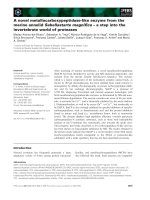
results show that in the absence of curdlan no bands were
detected on the blots (Fig. 1B; lane a); however, in the
extracts from curdlan-treated tissue a strongly staining band
of 32 kDa was observed (lane b). When the tissue was
treated with curdlan and the tyrosine kinase-inhibitor,
aeroplysinin (1 lgÆmL
)1
), no 32 kDa band was detected
(lane c). In parallel, the gels were stained with Coomassie
Brilliant Blue (Fig. 1A) and no change in the banding
pattern and their intensities occurred.
Detection of glucan-binding activity in extracts
from
S. domuncula
Tissue from S. domuncula was incubated for 1 day with
10 lgÆmL
)1
curdlan. Then, extracts were prepared and
subjected to PAGE in the presence of a low concentration of
SDS and in the absence of b-mercaptoethanol. After size
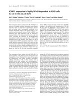
separation (Fig. 2; lane a), the proteins were transferred and
– after blocking – probed with labeled curdlan. A 43 kDa
polypeptide was observed in the extract (lane b). When the
blot was first preincubated with 10 lgÆmL
)1
laminarin and –
after washing – probed with the labeled curdlan, no band
was detected (lane c). However, when the blot was
preincubated with 2 lgÆmL
)1
LPS and subsequently with
labeled curdlan, the intensity of the band was only slightly
reduced (lane d). From these data we conclude that a
43 kDa protein is present in the extract from curdlan-
treated tissue, and that this protein comprises a specificity
for (1fi3)-b-
D
-glucans. In parallel, incubation experiments
with curdlan had been performed for only 6 h. Under these
conditions, the binding between labeled curdlan and the
43 kDa polypeptide was very low (data not shown).
Cloning of cDNA encoding the
S. domuncula
(1fi3)-
b-
D
-glucan-binding protein
Sequence. The insert with SDGLUBP comprises one
ORF, which ranges from nucleotides 46–48 to nucleo-
tides 1252–1254(stop); the cDNA is of full length, as
shown by Northern blot analysis (1.4 kb; see below). The
deduced protein shows high sequence similarity to the
Fig. 1. Phosphorylation of a 32 kDa protein after incubation of sponge
tissue with curdlan. Tissue samples were incubated for 6 h with or
without 10 lgÆmL
)1
curdlan. Protein extracts were then prepared and
size-separated by PAGE (8% gel). (A) The gel was stained with
Coomassie Brilliant Blue. (B) Proteins were blot transferred and
reacted with mouse antiphosphotyrosine mAb and then with labeled
goat anti-mouse IgG. Detection of the immunocomplex was carried
out as described in the Materials and methods. Protein extract from
tissue incubated in the absence (lane a, – cur), or in the presence (lane
b, + cur) of curdlan. In one series of experiments, the tissue was
additionally treated with 1 lgÆmL
)1
aeroplysinin (lane c, + aero). M,
protein size markers.
Ó FEBS 2004 Activation of sponge cells by (1fi3)-b-
D
-glucan (Eur. J. Biochem. 271) 1927
(1fi3)-b-
D
-glucan-binding proteins and was therefore
termed GLUBPp_SUBDO. The protein comprised 402
amino acid residues, with a calculated size of 45 040 Da,
and possessed, between amino acid 49 and amino acid 296,
one characteristic domain for Ôglycosyl hydrolases of the
family 16Õ (PFAM: PF00722) with a high significance value
(E-value) of 2e-05. Two transmembrane regions were
identified [43], which ranged from amino acids 2 to 23 and
from amino acids 361 to 401 (Fig. 3A). From these data we
conclude that the 43 kDa protein identified in the ligand-
binding blot probably corresponds to the 45 kDa (1fi3)-b-
D
-glucan-binding protein deduced from SDGLUBP.
Phylogenetic analysis. The sponge glucan-binding protein
shares highest sequence similarity with the (1fi3)-b-
D
-
glucan-binding proteins with average sizes 350–400 amino
acids. The highest similarity was calculated with the b-1,3-
glucan-binding protein from the black tiger shrimp,
P. monodon, having approximately 37% identical and
53% similar (with respect to the physico-chemical prop-
erties) amino acid residues to the sponge protein. The
similarity of the sponge glucan-binding protein to related
insect and crustacean proteins (35% identity/50% simi-
larity) was only slightly lower. No considerable similarity
was found to exist to the nonmetazoan and the
protostomian/nematode putative proteins present in the
database. After alignment of all similar sequences, a radial
tree was constructed which shows that the sponge glucan-
binding protein forms the basis for the insect molecule on
one side and the molecule from crabs on the other
(Fig. 3B).
cDNA encoding the fibrinogen-like protein
Sequence. One species of insert was identified – the ORF,
which spanned nucleotides 31–33 to nucleotides
877–879(stop). The full size cDNA (SDFIBI; 1.1 kb by
Northern blot analysis; see below) encoded the predicted
protein, termed FIBI_SUBDO, comprising 282 amino acid
residues (giving a calculated M
r
of 31 997). Domain searches
revealed that within the polypeptide, one fibrinogen domain
for b-andc-chains (PFAM: PF00147) exists between amino
acids 81–270. One conserved disulphide bond exists
connecting Cys225 to Cys239 and one eukaryotic secretory
signal sequence can be predicted [44] (Fig. 4A). The highest
similarity exists with vertebrate fibrinogens; therefore the
sequence was named fibrinogen-like protein.
Fibrinogens are the principal proteins of the vertebrate
clotting system and form hexamers, composed of the three
different chains: a, b and c [45]. As outlined by Spraggon
et al.[46],theb-andc-chains are homologous throughout
the complete sequence, while the a-chain comprises the
highest similarity only in the first 200 residues. Alignment
studies with the sponge and three mammalian fibrinogens
showed that the sponge fibrinogen-like protein, even though
the full-length sequence is available, shares similarity only
within the middle segment of the a-, b-andc-chains. Hence,
no further classification of the sponge protein to any of the
three vertebrate chains can be made. In the sponge
sequence, besides the first disulfide bridge mentioned, the
disulfide rings and the thrombin attack point (which exist in
the human sequence) are lacking. A potential arginine
residue in the sponge fibrinogen at amino acid position 18
cannot be recognized by thrombin owing to a negatively
charged glutamic acid residue at position P2 [47]. The
conserved central segments within the fibrinogen domain
[46] are present in the sponge protein.
Phylogenetic analysis. The analysis was performed with
the fibrinogen domain of the sponge fibrinogen-like protein.
The highest similarity, with approximately 35% identical
and 45% similar amino acid residues, was found to the
fibrinogens in the databases (Fig. 4B); the human fibrinogen
c-chain precursor (P02679) was used for the alignment
(Fig. 4A). The sequences were compiled and an unrooted
(slanted) tree was constructed (Fig. 4B). The trichotomous
tree shows that the families of the fibroleukins, with the
human member (Q14314) as an example, together with the
techylectins from the horseshoe crab, Tachypleus tridentatus
[20] (AB024737.1 and AB024738.1), and the angiopoietins
from mammals, e.g. humans (O15123), form the second
branch. The third branch is built by the ficolins, with the
mouse ficolin B as an example [48] (AF063217), and the
tenascins, including also the precursors from humans
(dJ1141O19.1), as members (Fig. 4B). The basis again is
the sponge-deduced protein.
cDNA of a potential EGF precursor
Sequence. One species of cDNA, which encodes a deduced
protein containing EGF domains, and was therefore termed
EGF precursor (SDEGFI-PREC) was isolated from the
library. The 2446 nucleotide contains an ORF, from
nucleotides 100–102 to nucleotides 2242–2244(stop); the
Fig. 2. Detection of a glucan-binding protein in extracts from Suber-
ites domuncula. A protein sample was prepared from tissue that had
been incubated for 1 day with 10 lgÆmL
)1
curdlan, as described in the
Materials and methods. Extract from curdlan-treated tissue was size
separated by SDS-PAGE (lane a). After separation, the protein ex-
tracts were transferred to poly(vinylidene difluoride)-Immobilon and
incubated with biotin-labeled curdlan (cur, 5 lgÆmL
)1
)(laneb).
Alternatively, the blots were first preincubated with 10 lgÆmL
)1
lam-
inarin (lam, lane c), or 2 lgÆmL
)1
lipopolysaccharide (LPS, lane d), for
5 h, and then washed and probed with biotin-curdlan (cur), as des-
cribed in the Materials and methods.
1928 S. Perovic
´
-Ottstadt et al. (Eur. J. Biochem. 271) Ó FEBS 2004
Fig. 3. The Suberites domuncula potential beta-1,3-glucan-binding protein (GLUBPp_SUBDO). (A) The deduced sponge sequence (GLU-
BPp_SUBDO) is aligned with the most related sequence, the b-1,3-glucan-binding protein from the black tiger shrimp Penaeus monodon
(GLUBP_PENMO, AF368168-1). Identical amino acids are shown in white on black. The positions of the two potential transmembrane regions
(TM) and the Ôglycosyl hydrolases-16Õ domain (glyco-hydr) are indicated. The segment towards which the degenerate primers were designed is
underlined by dashes. (B) Phylogenetic analysis of these two sequences with the GLUBP from the blue shrimp, Litopenaeus stylirostris
(GLUBP_LITSTY, AF473579-1), the putative Gram-negative bacteria-binding proteins from the Diptera Anopheles gambiae (ENSAN1_ANGA,
XP_312118.1), (ENSAN5_ANGA, XP_312116.1) and (BACBP_ANGA, CAA04496.1), as well as the GLUBP from the lobster Homarus gam-
marus (GLUBP_HOGAM, CAE47485.1) and the crayfish Pacifastacus leniusculus (GLUBP_PACLE, CAB65353.1). After alignment, the radial
tree was constructed.
Fig. 4. Suberites domuncula fibrinogen-like protein. (A) The deduced sponge protein, FIBl_SUBDO, is aligned with the related fibrinogen c-B chain
precursors from humans (FIBG_HUMAN; P02679). The conserved fibrinogen domain (FIBR) and one conserved disulfide bridge (C–C) are
present in the sponge protein, while the second disulfide bridge found in the human sequence is absent; this is marked ([C]–[C]). The predicted
eukaryotic secretory signal sequence terminates after amino acid 22 (SS). The thrombin attack point ({}) and the disulfide rings (underlined) in the
human sequence are indicated. The double underlined amino acids represent the regions towards which degenerate primers were designed at the
nucleotide level. The conserved central segments within the fibrinogen domain are marked (++). (B) A slanted cladogram was constructed using
the conserved fibrinogen domains of the two sequences mentioned above and of the following sequences. (i) Fibrinogens: fibrinogen a-2 chain
precursor from the sea lamprey Petromyzon marinus (FIB2_PETMA; P33573), fibrinogen a/a-E chain precursors from chicken (FIBA_CHICK;
P14448), human (FIBA_HUMAN; P02671) and rat fibrinogen (FIBA_RAT; P06399), and the fibrinogen c-B chain precursors from bovine
(FIBG_BOVIN; P12799), rat (FIBG_RAT; P02680), frog (FIBG_XENLA; P17634), and sea lamprey (FIBG_PETMA; P04115). (ii) Fibroleukins
and techylectins: the fibroleukin precursors from mouse (FGL2_MOUSE; P12804) and human (FGL2_HUMAN; Q14314), as well as the
techylectins from the horseshoe crab Tachypleus tridentatus (TECL5A-TACTR; AB024737.1 and TECL5B_TACTR; AB024738.1).
(iii) Angiopoietin: angiopoietin 1 and 2 precursors from mouse (AGP1_MOUSE; O08538 and AGP2_MOUSE; O35608), bovine (AGP1_BOVIN;
O18920 and AGP2_BOVIN; O77802), and human (AGP1_HUMAN; Q15389 and AGP2_HUMAN; O15123). (iv) Ficolins: ficolin A and B from
pig (FICOLA_PIG; L12344 and FICOLB_PIG; L12345), mouse (FICOLA_MOUSE; AB007813 and FICOLB_MOUSE; AF063217), rat
(FICOLA_RAT; AB026057), and the echinoderm Parastichopus parvimensis (FIBA_PARPA; P19477). (v) Tenascins: the tenascin precursors from
chicken (TENA_CHICK; P10039), human (TENA_HUMAN; P24821 – and – TENAl_HUMAN; dJ1141O19.1), fish Danio rerio
(TENAC_DARE; CAA61489.1), and pig (TENA_PIG; Q29116 – and – TENAX_PIG; CAA60686.1), as well as the the microfibril-associated
glycoprotein (MFA4_HUMAN; P55083). The numbers at the nodes are an indication of the level of confidence, given as a percentage, for the
branches as determined by bootstrap analysis (1000 bootstrap replicates).
Ó FEBS 2004 Activation of sponge cells by (1fi3)-b-
D
-glucan (Eur. J. Biochem. 271) 1929
size of the transcript, based on Northern blotting, is 2.6 kb.
The deduced 714 amino acids have a calculated M
r
of
77 901 (putative EGF precursor, EGFI-PREC_SUBDO).
By comparison with the Isrec-Server [49] domain database,
three EGF domains were identified in EGFI-PREC_SUB-
DO; they span the regions amino acids 331–368 (EGF1),
amino acids 364–410 (EGF2) and amino acids 407–455
(EGF3). Furthermore, three low-density lipoprotein
1930 S. Perovic
´
-Ottstadt et al. (Eur. J. Biochem. 271) Ó FEBS 2004
receptor repeats and one serine-rich segment were predicted
(Fig. 5A). One strong transmembrane region is present
between amino acids 624 and 663. The EGF domains are
characterized by three typical intramolecular disulfide
bonds [50], which are found in the sponge domains 2 and
3 with high similarity (Fig. 5A). The EGF domains from
Fig. 5. The putative epidermal growth factor (EGF) precursor, EGFl-PREC_SUBDO, from Suberites domuncula. (A) The 714 amino acid poly-
peptide comprises three potential low-density lipoprotein receptor repeats (LDL) and also three EGF-like domains (EGF). Within the EGF
domains 1 and 2, the three characteristic intramolecular disulfide bonds (C ¼¼ C) are marked. In addition, a Ser-rich segment (<Ser>) and a
transmembrane region () exists. The essential amino acid residues involved in binding to a receptor (#) and necessary for the biological function
of the factor (§) are marked in domain 2. (B) Unrooted tree constructed from the three sponge EGF domains (EGF_SUBDO) and the next similarity
domains present in the pro-EGF precursor from human (EGF_HOMO, P01133), the bovine fibrillin 1 precursor (MP340) (FBN1_BOVIN,
P98133; amino acids 2205–2229), the transforming growth factor-a precursor (TGF-a)fromOvis aries (sheep) (TGFA_SHEEP, P98135; amino
acids 7–49), fibrillin 1 from the Cnidaria Podocoryne carnea (FBN1_PODCA, AAA91336; amino acids 443–487), rat cubilin (CUBIL_RAT,
NP_445784; amino acids 2819–2864), frog (Xenopus laevis) neurogenic locus notch protein homolog precursor (NOTC_XENLA, P21783; amino
acids 1735–1780), and the hypothetical protein ZC116.3 from Caenorhabditis elegans (ZC116_CAEEL, CAA98952; amino acids 3000–3054).
Ó FEBS 2004 Activation of sponge cells by (1fi3)-b-
D
-glucan (Eur. J. Biochem. 271) 1931
S. domuncula can be grouped to the calcium-binding EGF-
like domains (NCBI:cd00054.2, EGF_CA); calcium is
crucial for protein–protein interactions. Comprehensive
studies have been performed with mammalian EGF to
elucidate the characteristic sites [51,52]. All amino acid
residues essential for ligand–receptor interaction and for
biological activity are present in domain 2 (Fig. 5A).
Phylogenetic analysis. The three EGF domains from
EGFI-PREC_SUBDO were aligned with the most closely
related EGF domains found in the proteins exclusively from
metazoans. The unrooted tree shows that the two sponge
EGF domains (2 and 3) share highest similarity to the
domains present in the human EGF (P01133), the bovine
fibrillin 1 (P98133) and the Podocoryne carnea (Cnidaria)
fibrillin 1 (AAA91336). More closely related to the sponge
EGF domain 3 are the domains present in the transforming
growth factor-a from sheep (P98135), the notch/xotch
protein from frog (P21783) and a hypothetical protein
(ZC116.3) from Caenorhabditis elegans.
Upregulation of gene expression for the described genes
in response to curdlan: Northern blotting
To assess the effect of curdlan and its subsequent binding to
the cell surface [probably to the (1fi3)-b-
D
-glucan-binding
protein] on the expression of the gene coding for this
receptor, Northern blot experiments were performed. The
results revealed that the expression level of the glucan-
binding protein at the beginning of the experiments is low.
However, after only 1 day of incubation in the presence of
10 lgÆmL
)1
curdlan, a strong upregulation of the expression
is seen, which increases during the following 2 days (Fig. 6).
Parallel experiments were performed to determine the
expression of the SDFIBI gene (fibrinogen-like protein).
Again, in the absence of curdlan, no transcripts can be
detected by this technique, while, in the presence of the
polysaccharide, a strong increase in the expression of
SDFIBI occurs. Likewise, a strong expression pattern was
determined for the SDEGFI-PREC gene (Fig. 6). The level
of expression of the housekeeping gene, tubulin, was not
altered by the presence of curdlan.
In view of the above finding, that curdlan causes
phosphorylation of Tyr residue(s) in polypeptides of
S. domuncula, which is prevented by aeroplysinin, it seemed
necessary to determine whether this protein tyrosine kinase
inhibitor also has an effect on the pronounced increase of
expression of the three genes under study. Therefore, the
tissue was incubated with 10 lgÆmL
)1
curdlan, together
with 1 lgÆmL
)1
aeroplysinin. In this co-incubation experi-
ment it was evident that the inhibitor, aeroplysinin,
completely prevented any upregulation of the expres-
sion of SDGLUBP [(1fi3)-b-
D
-glucan-binding protein],
SDFIBI (fibrinogen) or SDEGFI-PREC (EGF precursor)
(Fig. 6).
Identification of cells expressing glucan-binding protein
and fibrinogen
Data from the literature [12,13,22], as well as the binding
studies reported here, suggest that cells expressing the
(1fi3)-b-
D
-glucan-binding protein are located in areas
where the polysaccharide contacts the tissue, the pinaco-
derm. A similar localization, expression of the (1fi3)-b-
D
-
glucan-binding protein in the cells of the pinacoderm, can be
expected for fibrinogen or the related molecules, e.g. ficolins,
which are involved in host defense [48]. Therefore, in situ
hybridization studies were performed.
Our studies revealed that the cryosections from tissue
which had not been incubated with curdlan showed no cells
which hybridized with the antisense probes, either for the
sponge (1fi3)-b-
D
-glucan-binding protein (Fig. 7A, a) or
for fibrinogen (Fig. 7B, a). However, after only 1 day of
incubation with curdlan, the cells that are primarily located
around the canals react with the antisense probes (Fig. 7A,
b; Fig. 7B, b). After a longer incubation (for 3 days) the
density of the hybridizing cells increased considerably
(Fig. 7A, c; Fig. 7B, c). In contrast, no reaction was
observed if the cells were treated with both sense probes
(data not shown).
Level of low-molecular-weight EGF in tissue after
treatment with curdlan
As a first step, antibodies were required that could identify
the (mature) EGF product and the approximate level in
tissue by Western blotting. The recombinant protein was
prepared in E. coli using the cDNA (clone SDEGFI-PREC)
spanning the three EGF domains (corresponding to amino
acids 321–713). After induction with isopropyl thio-b-
D
-
galactoside, the bacterial extract was isolated and purified
by affinity chromatography (Fig. 8A; lanes a and b). The
68 kDa recombinant fusion protein (r-EGF_SUBDO) was
used to raise PoAbs, as described in the Materials and
methods. After cleavage with enterokinase, the sponge
Fig. 6. Steady-state expression of the SDGLUBP gene [(1fi3)-b-
D
-
glucan-binding protein], the SDFIBI gene (fibrinogen-like protein) and
the SDEGFI-PREC gene (epidermal growth factor EGF precursor) in
tissue from Suberites domuncula after exposure to 10 lgÆmL
)1
curdlan.
The housekeeping gene, b-tubulin, SDTUB (accession number
AJ550806), of S. domuncula was used as an internal standard. In one
series of experiments the tissue samples were co-incubated with
1 lgÆmL
)1
of aeroplysinin (aero). RNA extraction was performed 0, 1
or 3 days after incubation with curdlan. Equal amounts were loaded
onto the gel. The RNA was size separated, blot transferred and then
hybridized with the labeled probes, as described in the Materials and
methods.
1932 S. Perovic
´
-Ottstadt et al. (Eur. J. Biochem. 271) Ó FEBS 2004
recombinant protein, r-EGF_SUBDO, showed an expected
molecular weight of 41 kDa (data not shown).
This PoAb, PoAb-EGF precursor, was used for the
Western blot experiments. Tissue extract from S. domuncula
was prepared (Fig. 8B; lane a) and used for the blotting
studies. In nontreated, as well as in curdlan-treated
extracts from tissue, a strong band was detected using the
PoAb-EGF precursor, which corresponded to a molecular
weight of 5 kDa. This molecular weight predicts a poly-
peptide of approximately 45–50 amino acids, which matches
exactly a processed form of the EGF from the 78 kDa EGF
precursor (Fig. 8B; lanes b and c). No further major band,
including the 78 kDa EGF precursor, was detected. In the
control experiment, using PoAb-EGF precursor which had
been adsorbed with recombinant r-EGF_SUBDO, no band
was seen (Fig. 8B; lanes d), indicating that the immune
reaction with the 5 kDa protein is specific. The relative
expression value for the band corresponding to the 5 kDa
polypeptide was assessed. This approach revealed that in
curdlan-exposed tissue, a threefold higher level of the EGF
exists.
Discussion
Based on the known symbiotic relationship between spon-
ges and microorganisms, such as bacteria and fungi, it can
be deduced that these animals must have an efficient
recognition system which is able to discriminate between self
and symbiont. On the next level of specificity, the sponges
must be provided with pattern recognition molecules serving
as biosensors for the detection of invading pathogens
(parasitic), of commensalic (of benefit only for one partner)
or of mutualistic organisms (benefit for both partners). It
has been shown, in sponges, that some microbial secondary
metabolites, e.g. okadaic acid [4], are beneficial for the host.
However, the origin of most secondary metabolites identi-
fied in sponges is not clear. The understanding of the
pathways which result in the synthesis of these compounds
in sponges is crucial for their sustainable production/
exploitation for human benefit [53,54].
It is now established that sponges can recognize bacteria
and react to them with an increased phosphorylation of key
kinases of the MAPK pathway [7,28]. Furthermore, the first
genes encoding antibacterial proteins, such as perforin [55]
or the lectin tachylectin [6], have been identified in the
Fig. 8. Level of epidermal growth factor (EGF) in tissue after treatment
with curdlan. (A) Antibodies against the EGF precursor were prepared
from the recombinant protein expressed in Escherichia coli (A, a;
stained with Coomassie Brilliant Blue). The purified recombinant
41 kDa polypeptide (A, b; stained with Coomassie Blue) was used for
immunization. (B) The polyclonal antibody (PoAb)-EGF precursors
were used to identify the protein in extracts from sponge tissue. The
extracts were size separated and the gels stained with Coomassie
Brilliant Blue (B, a). Then, the proteins were blot transferred and
reacted with PoAb-EGF precursor. The level of cross-reacting proteins
was assessed by molecular imaging, as described in the Materials and
methods (B, b and c). In one series (B, d) the PoAb-EGF precursor was
adsorbed with recombinant r-EGF_SUBDO prior to the application.
The immunocomplex has been visualized by a labeled secondary
antibody. For further data see the Materials and methods.
Fig. 7. Spatial expression pattern of (1fi3)-b-
D
-glucan-binding protein and fibrinogen in sections of Suberites domuncula. Cryosections were per-
formed of S. domuncula tissues, which were then hybridized with DIG-labeled SDGLUBP antisense DNA (A), or SDFIBl antisense DNA (B).
Subsequently, the samples were incubated with antidigoxigenin/alkaline phosphatase and the signals detected with Nitro Blue tetrazolium/X-
Phosphate, as described in the Materials and methods. Tissue samples, which were untreated (a), or treated with 10 lgÆmL
)1
of curdlan for 1 day
(b), or 3 days (c), were analyzed. The canals (ca) of the aquiferous system are lined by an epithelial layer formed from pinacocytes. Magnifications:
A-a and B-a, · 25; A-b and B-b, · 50; A-c and B-c, · 100.
Ó FEBS 2004 Activation of sponge cells by (1fi3)-b-
D
-glucan (Eur. J. Biochem. 271) 1933
sponge S. domuncula. In addition, evidence has been
presented that the antibacterial alkyl-lipids, lyso-platelet-
activating factors, are produced in S. domuncula in response
to bacterial infection [56]. The molecular basis for the
interaction between fungi and sponges has not yet been
studied. It is well established that fungi present in sponges,
e.g. Penicillium sp. in Ircinia fasciculata, produce bioactive
products such as sorbicillactone [57]. In this study, we
present the first molecular data to demonstrate that sponges,
with S. domuncula as the model, have recognition receptors
for fungi on their cell surface. The fungal coat carbohydrate
–the(1fi3)-b-
D
-glucan, curdlan – served as a model
compound of choice. Fungi are known to bind, via this
carbohydrate, to the surface of insects [58], crustaceans [59],
and also to human cells [22]. The (1fi3)-b-
D
-glucan-binding
protein was first identified in crustacean blood [60] and then
cloned from the earthworm Eisenia foetida [12]. In plants,
the (1fi3)-b-
D
-glucan binds to a b-glucan elicitor-binding
protein of different structure [61].
Following a previously described approach [12], the
(1fi3)-b-
D
-glucan-binding protein was identified biochem-
ically. The binding of the linear carbohydrate, curdlan, was
abolished by the branched molecule, laminarin, indicating
that in S. domuncula a binding protein with specificity to
(1fi3)-b-
D
-glucan exists. Subsequent cloning studies re-
vealed a cDNA coding for a putative protein which shares
the characteristic feature of other metazoan (1fi3)-b-
D
-
glucan-binding proteins. The expression of the gene was
induced after exposure to (1fi3)-b-
D
-glucan. The sponge
polypeptide shares a high similarity to the related insect/
crustacean molecules, but shows no significant relationship
to protostomian/nematode, deuterostomian or non-meta-
zoan molecules in the database. In order to further support
the assumption that the sponge (1fi3)-b-
D
-glucan-binding
protein is involved in the recognition of potential fungi,
in situ hybridization studies were performed. They demon-
strated that the expression of this gene is especially high in
those regions of the sponge – the aquiferous canal system
(pinacoderm) – which are exposed to the carbohydrate
during the incubation period.
Next, it was attempted to identify downstream molecules,
potentially involved in the response of the sponge to (1fi3)-
b-
D
-glucan. A search for sequences in the S. domuncula EST
database, coding for potential clotting enzymes or coagulin,
known to be involved in the coagulation cascade in the
horseshoe crab [62], was unsuccessful. Therefore, the
S. domuncula cDNA library was screened for an alternative
scavenging system against carbohydrates, with lectins as the
first candidates. A C-type lectin has been isolated and
cloned from the hexactinellidian sponge, Aphrocallistes vas-
tus [63], which has been implicated in cell–cell interactions.
Furthermore, a galectin has been identified in S. domuncula,
which was also found to be an adhesion molecule causing
morphogenetic effects [64]. Hence, the next promising
lectins are those which are known, from higher metazoan
phyla, to be involved in innate immunity and comprise the
fibrinogen-like domain. Molecules with this domain exist
from the crown taxa to echinoderms [65] or ascidians [21] in
the deuterostomian branch, and to the horseshoe crabs [20]
in the protostomian line; this domain is thought to play a
role in carbohydrate binding [66]. PCR-based identification
and subsequent cloning of a full-length cDNA coding for a
fibrinogen-like protein was successful. The putative protein
comprises only one domain, which, however, has high
similarity to the fibrinogen domain; unexpectedly, no
further domain, e.g. a collagen domain as in the ficolins
[21], or lectin as in the horseshoe crab [20], or a coiled-coil
domain in angiopoietin [67], could be detected in the sponge
protein.
The sponge fibrinogen-like protein identified shares the
highest sequence similarity with the deuterostomian fibrin-
ogens, with the sea lamprey (Petromyzon marinus) sequence
as the most closely related. The lamprey fibrinogens [68]
and, to a smaller extent, also the echinoderm related
molecule [65], are known to be the ancestors for the three
homologous polypeptide chains (a
2
b
2
c
2
) in the vertebrate
blood clotting system. The a-, b-andc-subunits of the
fibrinogens are evolutionarily closely related; they differ
especially in the N- and/or C-terminal regions [45,46]. Only
one characteristic disulfide bridge exists in the predicted
fibrinogen-like protein from S. domuncula; in addition, the
thrombin cleavage site is lacking. However, the centrally
located conserved segments within the fibrinogen domain
[46] are present in the sponge protein. It is proposed that this
sponge protein represents the ancestor of all fibrinogen
domain-containing proteins found in Metazoa. In conse-
quence, the other vertebrate fibrinogens emerged from the
sponge protein, probably by gene duplication [65] and
domain shuffling [69]. The phylogenetic relationship (the
slanted cladogram) presented here, demonstrates that the
related molecules – the fibroleukins, secreted molecules from
T lymphocytes [70]; the techylectins/tachylectins from the
horseshoe crab, presumably proteins involved in non-self-
recognition [20]; the angiopoietins, which trigger the mech-
anism of blood vessel formation [67]; the ficolins, which are
plasma proteins with lectin activity and are thought to be
involved in host defense [71]; and, finally, the extracellular
matrix proteins, the tenascins [72] – are derived molecules
originating from the sponge fibrinogen-like protein.
In an earlier study it was shown that the deduced proteins
from sponges, as the basal metazoans, are more similar to
the related sequences from deuterostomians (humans), than
to those from protostomians (D. melanogaster and C. ele-
gans) [73]. In line with this view is the assumption that the
sponge fibrinogen-like protein gave rise to the blood clotting
system in deuterostomians/vertebrates and to lectins and the
extracellular matrix proteins, mentioned above. Sponges do
not contain a blood circulation system; however, in previous
studies it had been demonstrated that these animals
comprise several morphogens and cytokines, e.g. the
allograft inflammatory factor-1, the glutathione peroxidase,
the endothelial monocyte-activating polypeptide or the pre-
B-cell colony-enhancing factor [74], which, in higher meta-
zoan taxa, are transported in vascular systems.
Next, the level of expression of the fibrinogen-like protein
was analysed and its spatial distribution determined. It was
demonstrated that the expression is strongly upregulated
after exposure to curdlan. Even after only 1 day of
incubation, upregulation is seen. To strengthen the conclu-
sions, in situ hybridization studies were performed, which
again show that the expression of this gene is primarily seen
in the cells lining the canals – the pinacoderm.
From these two series of experiments it can be concluded
that curdlan initiates an activation circuit, from the surface
1934 S. Perovic
´
-Ottstadt et al. (Eur. J. Biochem. 271) Ó FEBS 2004
of the sponge cell to the gene expression level and back to a
further molecule potentially involved in the recognition of
the carbohydrate, the fibrinogen-like protein. In line with
previous data in human blood [66], it could be concluded
that the sponge ancestral protein, with its fibrinogen
domain, is also involved in binding to the carbohydrate.
The (1fi3)-b-
D
-glucans are known to activate a cell
metabolism/defense system, both in protostomians and in
deuterostomians. In the horseshoe crab, this carbohydrate
activates the prophenoloxidase activating system [75]. In
vertebrates, (1fi3)-b-
D
-glucans stimulate free radical pro-
duction in macrophages and expression of those genes
which are involved in inflammation [22]. At present, our
knowledge of sponge immune molecules, including cytok-
ines and enzymes involved in blood coagulation and the
complement system, is still at the very early stages. One
dominant domain, which is found in those molecules, and
whose existence had already been established in sponges, is
the EGF unit. By applying PCR identification and subse-
quent cloning, one gene coding for an EGF precursor was
identified. The deduced polypeptide comprised, besides
three low-density lipoprotein receptor repeats and one
transmembrane region, three EGF-like domains. A closer
classification groups the sponge domain to the calcium-
binding EGF-like domains, indicative that the molecule
undergoes protein–protein interactions [76]. The vertebrate
EGF protein is synthesized as a prepro-EGF, which is
subsequently processed to the active 53 amino acid EGF
(P01133). Three intramolecular disulfide bonds, which are
characteristic for EGF and required for biological activity
[77], are also found in the sponge and are highly conserved
in domain 2. Likewise, amino acids, which are involved in
ligand-receptor binding [52] and required for biological
activity [51], are present, especially in EGF domain-2. In
order to elucidate whether binding of (1fi3)-b-
D
-glucan to
sponge cells changes the steady-state expression level of the
sponge EGF precursor gene, Northern blot studies were
performed. They revealed that the expression level strongly
increased during the course of exposure to the carbohy-
drate.
The human prepro-EGF contains, like the sponge
polypeptide, a transmembrane region, which anchors the
molecule into the membrane [78]. During activation, the 53
amino acid EGF becomes released. In order to establish
whether the sponge putative EGF precursor molecule also
undergoes processing during incubation with curdlan, an
antibody was raised against the recombinant sponge
protein. This antibody was used to determine the size of
the mature peptide in the sponge tissue, before and after
exposure to curdlan. The data revealed that, almost
exclusively, a protein was identified with a size of 5 kDa.
The signal of this peptide was higher in Western blots of
extracts from curdlan-exposed sponge tissue. Based on this
finding, it is concluded that in the sponge an EGF molecule
of similar size to that found in mammals exists. Further-
more, the synthesis of this processed protein is upregulated
in the presence of curdlan.
As first proof that the (1fi3)-b-
D
-glucan-mediated chan-
ges in the expression level of the genes studied here might be
controlled by the same (or a coupled) pathway, inhibition
studies were performed. First, it was shown that at least one
protein undergoes phosphorylation at tyrosine residue(s) in
response to curdlan. Therefore, it was then studied whether
inhibition of tyrosine kinase-mediated phosphorylation
resulted also in a reduction of expression of the three genes
studied here. As a tool, the secondary metabolite, aeroplys-
inin, isolated from the sponge V. aerophoba [15,16], was
applied. Aeroplysinin has previously been suggested to be
an inhibitor of tyrosine kinase [79]. Inhibition studies, with
low concentrations of this bioactive compound, almost
completely blocked the expression of the genes coding for
the (1fi3)-b-
D
-glucan-binding protein, fibrinogen-like pro-
tein and the EGF precursor. This result is taken as a strong
indication that the curdlan-influenced expression of these
three genes, in response to the carbohydrate, are linked. No
toxicity of aeroplysinin on sponge cells in vitro could be
detected following application of the 3-(4,5-dimethylthiazol-
2-yl)-2,5-diphenyl-tetrazolium bromide viability assay
(R. Steffen, unpublished results).
Taken together, the data presented here demonstrate that
sponges, in this study using S. domuncula as the model,
comprise a (1fi3)-b-
D
-glucan-binding protein, which bind
to (1fi3)-b-
D
-glucans, e.g. curdlan or laminarin. After
binding of the carbohydrate to the receptor, the expression
of the gene coding for the binding protein increases. In turn,
the potential scavenger molecule, a fibrinogen-related
polypeptide, is synthesized. Finally, the production of a
low-molecular-weight EGF is accelerated, which might
result in a further stimulation of cell metabolism.
Acknowledgements
This work was supported by grants from the Deutsche Forschungsg-
emeinschaft (Mu
¨
/14–3), the Bundesministerium fu
¨
r Bildung und
Forschung Germany (project: Center of Excellence BIOTECmarin)
and the International Human Frontier Science Program (RG-333/
96-M).
References
1. Sarma, A.S., Daum, T. & Mu
¨
ller, W.E.G. (1993) Secondary
Metabolites from Marine Sponges. Akademie gemeinnu
¨
tziger
Wissenschaften zu Erfurt-Ullstein-Mosby Verlag, Berlin.
2. Fusetani, N. (2000) Drugs from the Sea.S.Karger,Basel.
3. Proksch, P., Edrada, R.A. & Ebel, R. (2002) Drugs from the seas –
current status and microbiological implications. Appl. Microbiol.
Biotechnol. 59, 125–134.
4. Wiens, M., Luckas, B., Bru
¨
mmer,F.,Ammar,M.S.A.,Steffen,R.,
Batel, R., Diehl-Seifert, B., Schro
¨
der, H.C. & Mu
¨
ller, W.E.G.
(2003) Okadaic acid: a potential defense molecule for the sponge
Suberites domuncula. Mar. Biol. 142, 213–223.
5. Mu
¨
ller, W.E.G. & Mu
¨
ller, I.M. (2003) Origin of the metazoan
immune system: identification of the molecules and their functions
in sponges. Integr. Comp. Biol. 43, 281–292.
6. Schro
¨
der, H.C., Ushijima, H., Krasko, A., Gamulin, V., Schu
¨
tze,
J., Mu
¨
ller, I.M. & Mu
¨
ller, W.E.G. (2003) Emergence and dis-
appearance of an immune molecule, an antimicrobial lectin,
in basal Metazoa: the tachylectin family. J. Biol. Chem. 278,
32810–32817.
7. Bo
¨
hm, M., Hentschel, U., Friedrich, A., Fieseler, L., Steffen, R.,
Gamulin, V., Mu
¨
ller, I.M. & Mu
¨
ller, W.E.G. (2001) Molecular
response of the sponge Suberites domuncula to bacterial infection.
Mar. Biol. 139, 1037–1045.
8. Riggi, S.J. & Luzio, N.R. (1961) Identification of a reticulo-
endothelial simulating agent in zymosan. Am.J.Physiol.200,297–
305.
Ó FEBS 2004 Activation of sponge cells by (1fi3)-b-
D
-glucan (Eur. J. Biochem. 271) 1935
9. Matsushita, M. (1990) Curdlan, (1fi3)-b-
D
-glucan from
Alcaligenes faecalis var. myxogenes IFO13140, activates the
alternative complement pathway by heat treatment. Immunol.
Lett. 26, 95–97.
10. Masihi, K.N., Madaj, K., Hintelmann, H., Gast, G. & Kaneko, Y.
(1997) Down-regulation of tumor necrosis factor-a, moderate
reduction of interferon-1b, but not interleukin-6 or interleukin-10,
by glucan immunmodulators curdlan sulfate and lentinan. Int. J.
Immunopharmacol. 19, 463–468.
11. Black, W.A.P., Cornhill, W.J., Dewar, E.T. & Woodward, F.N.
(1951) Manufacture of algal chemicals. III. Laboratory-scale iso-
lation of laminarin from brown marine algae. J. Appl. Chem. 1,
505–517.
12. Beschin, A., Bilej, M., Hanssens, F., Raymakers, J., Van Dyck, E.,
Revets,H.,Brys,L.,Gomez,J.,DeBaetselier,P.&Timmermans,
M. (1998) Identification and cloning of a glucan- and lipopoly-
saccharide-binding protein from Eisenia foetida earthworm
involved in the activation of prophenoloxidase cascade. J. Biol.
Chem. 273, 24948–24954.
13. Lee, S.Y., Wang, R. & So
¨
derhall, K. (2000) A lipopolysaccharide-
and beta-1,3-glucan-binding protein from hemocytes of the
freshwater crayfish Pacifastacus leniusculus. Purification, char-
acterization, and cDNA cloning. J. Biol. Chem. 275, 1337–1343.
14. Engstad, C.S., Engstad, R.E., Olsen, J.O. & Østerud, B. (2002)
The effect of soluble b-1,3-glucan and lipopolysaccharide on
cytokine production and coagulation activation in whole blood.
Int. Immunopharmacol. 2, 1585–1597.
15. Kreuter,M.H.,Robitzki,A.,Chang,S.,Steffen,R.,Michaelis,
M., Kljajic, Z., Bachmann, M., Schro
¨
der, H.C. & Mu
¨
ller, W.E.G.
(1992) Production of the cytostatic agent, aeroplysinin by the
sponge Verongia aerophoba in in vitro culture. Comp. Biochem.
Physiol. 101C, 183–187.
16.Ebel,R.,Brenzinger,M.,Kunze,A.,Gross,H.&Proksch,P.
(1997) Wound activation of prototoxins in the marine sponge
Aplysina aerophoba. J. Chem. Ecol. 23, 1451–1462.
17. Roux, M.M., Pain, A., Klimpel, K.R. & Dhar, A.K. (2002) The
lipopolysaccharide and beta-1,3-glucan binding protein gene is
upregulated in white spot virus-infected shrimp (Penaeus styliros-
tris). J. Virol. 76, 7140–7149.
18. Dimopoulos, G., Richman, A., Muller, H.M. & Kafatos, F.C.
(1997) Molecular immune responses of the mosquito Anopheles
gambiae to bacteria and malaria parasites. Proc. Natl Acad. Sci.
USA 94, 11508–11513.
19. Bachman, E.S. & McClay, D.R. (1996) Molecular cloning of the
first metazoan beta-1,3 glucanase from eggs of the sea urchin
Strongylocentrotus purpuratus. Proc. Natl Acad. Sci. USA 93,
6808–6813.
20. Gokudan, S., Muta, T., Tsuda, R., Koori, K., Kawahara, T., Seki,
N., Mizunoe, Y., Wai, S.N., Iwanaga, S. & Kawabata, S. (1999)
Horseshoe crab acetyl group-recognizing lectins involved in innate
immunity are structurally related to fibrinogen. Proc.NatlAcad.
Sci. USA 96, 10086–10091.
21. Kenjo, A., Takahashi, M., Matsushita, M., Endo, Y., Nakata, M.,
Mizuochi, T. & Fujita, T. (2001) Cloning and characterization of
novel ficolins from the solitary ascidian, Halocynthia roretzi.
J. Biol. Chem. 276, 19959–19965.
22. Kataoka, K., Muta, T., Yamazaki, S. & Takeshige, K. (2002)
Activation of macrophages by linear (1fi3)-b-
D
-glucans. J. Biol.
Chem. 277, 36825–36831.
23. Skorokhod, A., Gamulin, V., Gundacker, D., Kavsan, V., Mu
¨
ller,
I.M. & Mu
¨
ller, W.E.G. (1999) Origin of insulin receptor tyrosine
kinase: cloning of the cDNAs from marine sponges. Biol. Bull.
197, 198–206.
24. Kruse, M., Mu
¨
ller, I.M. & Mu
¨
ller, W.E.G. (1997) Early evolution
of metazoan serine/threonine- and tyrosine kinases: identification
of selected kinases in marine sponges. Mol. Biol. Evol. 14, 1326–1334.
25. Krasko, A., Batel, R., Schro
¨
der, H.C., Mu
¨
ller, I.M. & Mu
¨
ller,
W.E.G. (2000) Expression of silicatein and collagen genes in the
marine sponge Suberites domuncula is controlled by silicate and
myotrophin. Eur. J. Biochem. 267, 4878–4887.
26. Novotna, V., Mikes, L., Horak, P., Jonakova, V. & Ticha, M.
(1996) Preparation of water-soluble and water-insoluble
poly(acrylamide-allylamine) derivatives of polysaccharides. Int.
J. Bio-Chromatogr. 2, 37–47.
27. Castro,R.,Couso,N.,Obach,A.&Lamas,J.(1999)Effectof
different b-
D
-glucans on the respiratory burst of turbot (Psetta
maxima) and gilthead seabream (Sparus aurata) phagocytosis. Fish
Shellfish Immunol. 9, 529–541.
28. Bo
¨
hm, M., Mu
¨
ller, I.M., Mu
¨
ller, W.E.G. & Gamulin, V. (2000)
The mitogen-activated protein kinase p38 pathway is conserved in
metazoans: cloning and activation of p38 of the SAPK2 subfamily
from the sponge Suberites domuncula. Biol. Cell 29, 95–104.
29. Laemmli, U.K. (1970) Cleavage of structural proteins during
the assembly of the head of bacteriophage T4. Nature 227,
680–685.
30. Bachmann, M., Mayet, W.J., Schro
¨
der,H.C.,Pfeifer,K.,Meyer
zum Bu
¨
schenfelde, K H. & Mu
¨
ller, W.E.G. (1986) Association of
La and Ro antigen with intracellular structures in HEp-2 carci-
noma cells. Proc.NatlAcad.Sci.USA83, 7770–7774.
31. Wiens, M., Koziol, C., Hassanein, H.M.A., Batel, R. & Mu
¨
ller,
W.E.G. (1998) Expression of the chaperones 14–3)3andHSP70
induced by PCB 118 (2,3¢,4,4¢,5-pentachlorobiphenyl) in the
marine sponge Geodia cydonium. Mar. Ecol. Prog. Series 165,
247–257.
32. Ausubel,F.M.,Brent,R.,Kingston,R.E.,Moore,D.D.,Smith,
J.A., Seidmann, J.G. & Struhl, K. (1995) Current Protocols in
Molecular Biology. John Wiley and Sons, New York.
33. BLAST (2003) />34. FASTA (2003) />35. Thompson, J.D., Higgins, D.G. & Gibson, T.J. (1994) CLUSTAL
W: improving the sensitivity of progressive multiple sequence
alignment through sequence weighting, positions-specific gap
penalties and weight matrix choice. Nucleic Acids Res. 22, 4673–
4680.
36. Felsenstein, J. (1993) PHYLIP, Version 3.5. University of Wash-
ington, Seattle.
37. Dayhoff, M.O., Schwartz, R.M. & Orcutt, B.C. (1978) A model of
evolutionary change in protein. In Atlas of Protein Sequence and
Structure (Dayhoff, M.O., ed.), pp. 345–352. National Biomedical
Research Foundation, Washington, DC.
38. Nicholas, K.B. & Nicholas, H.B. Jr (1997) GENEDOC: a Tool for
Editing and Annotating Multiple Sequence Alignments,Version
1.1.004. Distributed by the author; />genedoc
39. Schu
¨
tze,J.,Krasko,A.,Diehl-Seifert,B.&Mu
¨
ller, W.E.G.
(2001) Cloning and expression of the putative aggregation factor
from the marine sponge Geodia cydonium. J. Cell Sci. 114, 3189–
3198.
40. Grebenjuk, V.A., Kuusksalu, A., Kelve, M., Schu
¨
tze, J., Schro
¨
der,
H.C. & Mu
¨
ller, W.E.G. (2002) Induction of (2¢)5¢) oligoadenylate
synthetase in the marine sponges Suberites domuncula and Geodia
cydonium by the bacterial endotoxin lipopolysaccharide. Eur. J.
Biochem. 269, 1382–1392.
41. Polak, J.M. & McGee, J.D. (1998) In Situ Hybridization.Oxford
University Press, Oxford.
42. Perovic
´
,S.,Schro
¨
der, H.C., Sudek, S., Grebenjuk, V.A., Batel, R.,
S
ˇ
tifaniæ, M., Mu
¨
ller,I.M.&Mu
¨
ller, W.E.G. (2003) Expression of
one sponge Iroquois homeobox gene in primmorphs from Sub-
erites domuncula during canal formation. Evol. Dev. 5, 240–250.
43. Rao, J. & Argos, P. (1986) A conformational preference para-
meter to predict helices in integral membrane proteins. Biochem.
Biophys. Acta 869, 197–214.
1936 S. Perovic
´
-Ottstadt et al. (Eur. J. Biochem. 271) Ó FEBS 2004
44. PC/GENE (1995) Data Banks CD-ROM; Release 14.0. Intellige-
netics, Inc., Mountain View, CA.
45. Doolittle, R.F. (1984) Fibrinogen and fibrin. Annu. Rev. Biochem.
53, 195–229.
46. Spraggon, G., Applegate, D., Everse, S.J., Zhang, J.Z., Veera-
pandian, L., Redman, C., Doolittle, R.F. & Grieninger, G. (1998)
Crystal structure of a recombinant alphaEC domain from human
fibrinogen-420. Proc. Natl Acad. Sci. USA 95, 9099–9104.
47. Barrett,A.J.,Rawlings,N.D.&Woessner,J.F.(2002)Handbook
of Proteolytic Enzymes. Academic Press, Amsterdam.
48. Ohashi, T. & Erickson, H.P. (1998) Oligomeric structure and tis-
sue distribution of ficolins from mouse, pig and human. Arch.
Biochem. Biophys. 360, 223–232.
49. Isrec-Server (2003) />50. Boonstra, J., Rijken, P., Humbel, B., Cremers, F., Verkleij, A. &
van Bergen en Henegouwen, P. (1995) The epidermal growth
factor. Cell Biol. Int. 19, 413–430.
51. Ray, P., Mey, F.J., Montelione, G.T., Liu, J.F., Narang, S.A.,
Scheraga, H.A. & Wu, R. (1988) Structure–function studies of
murine epidermal growth factor: expression and site-directed
mutagenesis of epidermal growth factor gene. Biochemistry 27,
7289–7295.
52. Engler, D.A., Montelione, G.T. & Niyogi, S.K. (1990) Human
epidermal growth factor. Distinct role of tyrosine 37 and arginine
41 in receptor binding as determined by site-directed mutagenesis
and nuclear magnetic resonance spectroscopy. FEBS Lett. 271,
47–50.
53. Cimino, G. & Ghiselin, M.T. (2001) Marine natural products
chemistry as an evolutionary narrative. In Marine Chemical
Ecology (McClintock, J.B. & Baker, B.J., eds), pp. 115–154. CRC
Press, Boca Raton.
54. Mu
¨
ller, W.E.G., Bru
¨
mmer, F., Batel, R., Mu
¨
ller, I.M. & Schro
¨
der,
H.C. (2003) Molecular biodiversity. Case study: Porifera (spon-
ges). Naturwissenschaften 90, 103–120.
55. Thakur,N.L.,Hentschel,U.,Krasko,A.,Pabel,C.T.,Anil,A.C.
&Mu
¨
ller, W.E.G. (2003) Antibacterial activity of the sponge
Suberites domuncula and its primmorphs: potential basis for epi-
bacterial chemical defense. Aquat. Microbiol. Ecol. 31, 77–83.
56. Mu
¨
ller, W.E.G., Klemt, M., Thakur, N.L., Schro
¨
der, H.C., Aiello,
A.,D’Esposito,M.,Menna,M.&Fattorusso,E.(2003)Molecular/
chemical ecology in sponges: evidence for an adaptive antibacterial
response in Suberites domuncula. Mar. Biol. 144, 19–29.
57. Bringmann, G., Lang, G., Mu
¨
hlbacher, J., Schaumann, K.,
Steffens, S., Rytik, P.G., Hentschel, U., Morschha
¨
user, J., Brun,
R. & Mu
¨
ller, W.E.G. (2003) Sorbicillactone A, a structurally un-
precedented bioactive novel-type alkaloid from a sponge-derived
fungus. In Marine Molecular Biotechnology (Mu
¨
ller, W.E.G., ed.),
pp. 231–253. Springer-Press, Berlin.
58. Ma, C. & Kanost, M.R. (2000) A b1,3-glucan recognition protein
from an insect, Manduca sexta, agglutinates microorganisms and
activates the phenoloxidase cascade. J. Biol. Chem. 277, 36825–36831.
59. Sritunyalucksana, K., Lee, S.Y. & So
¨
derha
¨
ll, K. (2002) The beta-
1,3-glucan binding protein from the black tiger shrimp, Penaeus
monodon. Dev. Comp. Immunol. 26, 237–245.
60. Cerenius, L., Liang, Z., Duvic, B., Keyser, P., Hellman, U., Palva,
E.T., Iwanaga, S. & So
¨
derha
¨
ll, K. (1994) A b-
D
-glucan binding
protein in crustacean blood. Structure and biological activity of a
fungal recognition cascade. J. Biol. Chem. 269, 29462–29467.
61. Umemoto, N., Kakitani, M., Iwamatsu, A., Yoshikawa, M.,
Yamaoka, N. & Ishida, I. (1997) The structure and function of a
soybean b-glucan-elicitor-binding protein. Proc. Natl Acad. Sci.
USA 94, 1029–1034.
62. Kawabata, S.I., Osaki, T. & Iwanaga, S. (2003) Innate immunity
in the horseshoe crab. In Innate Immunity (Ezekowitz, R.A.B. &
Hoffmann, J.A., eds), pp. 109–125. Humana Press, Totowa.
63. Gundacker, D., Leys, S.P., Schro
¨
der, H.C., Mu
¨
ller, I.M. & Mu
¨
l-
ler, W.E.G. (2001) Isolation and cloning of the C-type lectin from
the hexactinellidian sponge Aphrocallistes vastus: a putative ag-
gregation factor. Glycobiology 11, 21–29.
64. Wiens, M., Mangoni, A., D’Esposito, M., Fattorusso, E.,
Korchagina, N., Schro
¨
der, H.C., Grebenjuk, V.A., Krasko, A.,
Batel, R., Mu
¨
ller, I.M. & Mu
¨
ller, W.E.G. (2003) The molecular
basis for the evolution of the metazoan bodyplan: extracellular
matrix-mediated morphogenesis in marine demosponges. J. Mol.
Evol. 57, S60–S75.
65. Xu, X. & Doolittle, R.F. (1990) Presence of a vertebrate fibrino-
gen-like sequence in an echinoderm. Proc. Natl Acad. Sci. USA 87,
2097–2101.
66. Le,Y.,Lee,S.H.,Kon,O.L.&Lu,J.(1998)Human
L
-ficolin:
plasma levels, sugar specificity and assignment of its lectin activity
to the fbrinogen-like (FBG) domain. FEBS Lett. 425, 367–370.
67. Davis, S., Aldrich, T.H., Jones, P.F., Acheson, A., Compton,
D.L., Jain, V., Ryan, T.E., Bruno, J., Radziejewski, C., Maison-
pierre, P.C. & Yancopoulos, G.D. (1996) Isolation of angiopoie-
tin-1, a ligand for the TIE2 receptor, by secretion-trap expression
cloning. Cell 87, 1161–1169.
68. Pan, Y. & Doolittle, R.F. (1992) cDNA sequence of a second
fibrinogen a chain in lamprey: an archetypal version alignable with
full length b and c chains. Proc. Natl Acad. Sci. USA 89, 2066–
2070.
69. Patthy, L. (1999) Protein Evolution. Blackwell Science, Oxford.
70. Marazzi, S., Blum, S., Hartmann, R., Gundersen, D., Schreyer,
M.,Argraves,S.,vonFliedner,V.,Pytela,R.&Ruegg,C.(1998)
Characterization of human fibroleukin, a fibrinogen-like protein
secreted by T lymphocytes. J. Immunol. 161, 138–147.
71. Ichijo, H., Hellman, U., Wernstedt, C., Gonez, L.J., Claesson-
Welsh, L., Heldin, C.H. & Miyazono, K. (1993) Molecular
cloning and characterization of ficolin, a multimeric protein
with fibrinogen- and collagen-like domains. J. Biol. Chem. 268,
14505–14513.
72. Gulcher, J.R., Nies, D.E., Alexakos, M.J., Ravikant, N.A., Stur-
gill, M.E., Marton, L.S. & Stefansson, K. (1991) Structure of the
human hexabrachion (tenascin) gene. Proc. Natl Acad. Sci. USA
88, 9438–9442.
73. Gamulin, V., Mu
¨
ller, I.M. & Mu
¨
ller, W.E.G. (2000) Sponge
proteins are more similar to those of Homo sapiens than to Cae-
norhabditis elegans. Biol.J.LinneanSoc.71, 821–828.
74. Mu
¨
ller, W.E.G., Blumbach, B. & Mu
¨
ller, I.M. (1999) Evolution of
the innate and adaptive immune systems: relationships between
potential immune molecules in the lowest metazoan phylum
(Porifera) and those in vertebrates. Transplantation 68, 1215–1227.
75. So
¨
derha
¨
ll, K. & Cerenius, L. (1998) Role of the prophenoloxidase
activating system in invertebrate immunity. Curr. Opin. Immunol.
10, 23–28.
76. Downing, A.K., Knott, V., Werner, J.M., Cardy, C.M., Camp-
bell, I.D. & Handford, P.A. (1996) Solution structure of a pair of
calcium-binding epidermal growth factor-like domain: implication
for the Marfan syndrome and other genetic disorders. Cell 85,
597–605.
77. Taylor, J.M., Mitchell, W.M. & Cohen, S. (1972) Epidermal
growth factor: physical and chemical properties. J. Biol. Chem.
247, 5928–5934.
78. Rall,L.B.,Scott,J.,Bell,G.I.,Crawford,R.J.,Penshow,J.D.,
Niall, H.D. & Coghlan, J.P. (1985) Mouse prepro-epidermal
growth factor synthesis by the kidney and other tissues. Nature
313, 228–231.
79. Rodrigues-Nieto, S., Goza
´
les-Iriarte, M., Carmona, R., Munoz-
Cha
´
puli, R., Medina, M.A. & Quesada, A.R. (2001) Anti-angio-
genic activity of aeroplysinin-1, a brominated compound isolated
from a marine sponge. FASEB J doi: 10.1096.fj.01-0427fje
Ó FEBS 2004 Activation of sponge cells by (1fi3)-b-
D
-glucan (Eur. J. Biochem. 271) 1937