Báo cáo khoa học: Structure and potential C-terminal dimerization of a recombinant mutant of surfactant-associated protein C in chloroform/methanol ppt
Bạn đang xem bản rút gọn của tài liệu. Xem và tải ngay bản đầy đủ của tài liệu tại đây (345.14 KB, 10 trang )
Structure and potential C-terminal dimerization of a recombinant
mutant of surfactant-associated protein C in chloroform/methanol
Burkhard Luy
1
, Alexander Diener
2
, Rolf-Peter Hummel
3
, Ernst Sturm
3
, Wolf-Ru¨ diger Ulrich
4
and Christian Griesinger
5
1
Institut fu
¨
r Organische Chemie und Biochemie, Technische Universita
¨
tMu
¨
nchen, Garching, Germany;
2
Institut fu
¨
r Organische
Chemie, Johann Wolfgang Goethe-Universita
¨
t Frankfurt, Germany;
3
Department of Physical Organic Chemistry and
4
Department of
Chemical Research, Altana Pharma AG, Konstanz, Germany;
5
Max Planck Institut fu
¨
r Biophysikalische Chemie, Go
¨
ttingen, Germany
The solution structure of a recombinant mutant [rSP-
C (FFI)] of the human surfactant-associated protein C
(hSP-C) in a mixture of chloroform and methanol was
determined by high-resolution NMR spectroscopy. rSP-
C (FFI) contains a helix from Phe5 to the C-terminal Leu34
and is thus longer by two residues than the helix of porcine
SP-C (pSP-C), which is reported to start at Val7 in the same
solvent. Two sets of resonances at the C-terminus of the
peptide were observed, which are explained by low-order
oligomerization, probably dimerization of rSP-C (FFI) in
its a-helical form. The dimerization may be induced by
hydrogen bonding of the C-terminal carboxylic groups or
by the strictly conserved C-terminal heptapeptide segment
with a motif similar to the GxxxG dimerization motif of
glycophorin A. Dimerization at the heptapeptide segment
would be consistent with findings based on electrospray
ionization MS data, chemical cross-linking studies, and
CNBr cleavage data.
Keywords: dimerization; NMR spectroscopy; surfactant;
surfactant protein C (SP-C).
Surfactant-associated protein C (SP-C) is a 34–35-amino-
acid peptide which is highly conserved among species
(Table 1). It is part of the protein–phospholipid complex
that is secreted into the alveolar space [1] and is
responsible for lowering of the alveolar surface tension.
Recombinant (r)SP-C (FFI) surfactant (Venticute) has
proved to be highly effective in animal experiments [2,3]
as well as in pilot clinical trials [4,5]. The structure of
porcine SP-C (pSP-C) has been solved in CDCl
3
/CD
3
OH/
0.1
M
HCl (32 : 64 : 5, v/v/v), and it has been found
that the peptide forms an a-helix from residue 7 to the
C-terminal residue 34 [6]. The N-terminal structure as well
as the hydrophobic a-helix seems to be conserved in the
micellar environment as shown for the N-terminal 17
residues of pSP-C in fully deuterated dodecylphospho-
choline micelles [7]. A second set of resonances was found
for the full-length pSP-C peptide in chloroform/methanol
at the C-terminus, which was explained by partial
oxidation of the methionine residue M32. In general,
samples of the lipophilic pSP-C are not completely stable
in chloroform/methanol mixtures and form a gel-like
b-sheet aggregate after several days at 10 °C[8].Amutant
of the human SP-C (hSP-C) has been produced recom-
binantly by omitting the residue [Phe() 1)] that is only
partially present and performing the following substitu-
tions: C4F, C5F and M32I. The rationale behind the
substitutions is that the two cysteine residues are naturally
palmitoylated, which would have been difficult to achieve
for a bacterially expressed protein. The mutation of
residue 32 was to prevent the undesired putative oxidation
of methionine. In this article, we present the structure of
the rSP-C (FFI) mutant in CDCl
3
/CD
3
OH (1 : 1, v/v)
with a comparison with the structure of pSP-C. A second
set of C-terminal signals is explained by the coexistence
of monomeric and oligomeric (probably dimeric)
rSP-C (FFI).
Materials and methods
Preparation of the sample
For the studies on rSP-C (FFI) (Altana Pharma AG,
Konstanz, Germany; WO patent no. 95/32992), we used the
solid substrate consisting of the peptide (90%), HCl (4%),
propan-2-ol (3%), water (2%), and methyl ester (1%).
Samples of rSP-C (FFI) were prepared by dissolving
3–12 mg of the powder in 600 lLCDCl
3
/CD
3
OH (1 : 1,
v/v) or CDCl
3
/CD
3
OD (1 : 1, v/v). The resulting rSP-
C (FFI) concentration was 1.1–4.4 m
M
, respectively. The
solid peptide was stored at )20 °C, and the prepared
samples were stored in liquid nitrogen between NMR
measurements. Dissolved samples had a lifetime of 72 h
at 10 °C. Over time, the dissolved peptide maintained
identical NMR chemical shifts, but strongly reduced
intensity, indicating similar aggregation to b-sheet-like
Correspondence to C. Griesinger, Max Planck Institut fu
¨
r Biophysi-
kalische Chemie, Abt. NMR based Structural Biology,
Am Fassberg 11, 37077 Go
¨
ttingen, Germany.
Fax: + 49 551201 2202, Tel.: + 49 551201 2201,
E-mail:
Abbreviations: SP-C, surfactant-associated protein C; hSP-C, human
SP-C; pSP-C, porcine SP-C; rSP-C, recombinant human SP-C; rSP-C
(FFI), FFI variant of recombinant human SP-C; TACSY, taylored
correlation spectroscopy.
(Received 17 December 2003, revised 1 March 2004,
accepted 23 March 2004)
Eur. J. Biochem. 271, 2076–2085 (2004) Ó FEBS 2004 doi:10.1111/j.1432-1033.2004.04106.x
structures as observed for natural pSP-C in the solvent used.
Because of the limited lifetime, samples were prepared
immediately before NMR measurements.
NMR measurements
2D
1
H-NMR spectra were recorded on Bruker DRX 800,
DMX 600, AMX 600 and AMX 400 spectrometers in the
pure-phase absorption mode using the States-TPPI method
[9]. All spectra were recorded at 10 °C, and processing
and baseline corrections were performed using the standard
Bruker software
XWINNMR
. The complete set of experiments
recorded is given in Table 2.
The
1
H-NMR chemical shifts were calibrated relative to
trimethylsilane. The residual water signal and the signal
of the hydroxy proton of CD
3
OH are degenerate at
4.8 p.p.m. and were reduced using presaturation [10].
Before Fourier transformation, the time domain data were
multiplied with shifted squared sinebell window functions.
The vicinal scalar coupling constants
3
J
NHa
were deter-
mined using the SIAM-TACSY and Keeler–Titman
approaches [11,12] using macros written by T. Prasch for
the program
FELIX
(Felix 95; MSI, San Diego, CA, USA).
Signal overlap in the 800-MHz NOESY made peak
integration unreliable. So, instead, signal height of the
cross-peaks was used for a conservative estimation of the
maximum distances and classification of cross-peaks as
weak, medium and strong. For the calibration of the
intensities of the NOE peaks, a statistical analysis of the
d
aN
(i,i+3) signals of residues 11–30 was performed using
typical values for an ideal a-helix [13]. The a-helical
structure of this part of the peptide is clearly evident from
H
a
chemical shifts [14,15].
Results
NMR assignment
Sequence-specific
1
H-NMR assignment was achieved by
standard procedures for small proteins [13] using the
computer program
NDEE
(Spin Up, Lu
¨
nen, Germany).
Owing to the high abundance of the amino acids valine,
leucine and isoleucine in the sequence of rSP-C (FFI), there
was extensive overlap in the homonuclear
1
H-NMR spectra.
Nevertheless, almost all spin systems (vide infra) could be
assigned from the TOCSY spectra (Fig. 1A) and the
DQF-COSY spectra (not shown) collected under identical
conditions (Table 3).
The unique spin systems His8, Lys10, Arg11 and Ala29,
and the pairs of Phe and Pro residues and Gly28 and Gly33
were unambiguously identified, as well as 10 of the 11
valines. The N-terminal Gly1 shows a single very broad
H
N
/H
a
cross-peak. Although all 34 amino acids were found,
the spin systems of seven leucines, five isoleucines and the
residual valine could only be unambiguously identified
using sequential NOE information.
The high dispersion of the 800-MHz NOESY spectrum
made it possible to obtain the complete assignment of rSP-
C (FFI) (Fig. 1B,C). Starting from the unambiguously
identified residues, we were able to carry out the sequential
assignment for residues 1–17 and 24–34 by d
aN
and d
NN
cross-peaks. As an a-helical secondary structure was
assumed from chemical-shift arguments, d
aN
(i,i+3) and
d
aN
(i,i+4) NOE cross-peaks were used, leading to the
assignment of the residual amino acids 18–23.
We encountered special difficulties in identifying the
following connectivities: the chemical shifts of the amide
Table 2. NMR experiments.
Sample Experiment
Spectrometer
frequency (MHz)
Data
matrix
Processed
matrix
Mixing
time (ms)
Total
time (h)
1.1 m
M
rSP-C (FFI) in CDCl
3
/CD
3
OH (1 : 1) TOCSY 600 4096 · 768 4096 · 1024 70 11
NOESY 600 4096 · 768 4096 · 1024 50 8
NOESY 600 4096 · 768 4096 · 1024 100 8
NOESY 600 4096 · 768 4096 · 1024 200 8
DQF-COSY 600 4096 · 1024 4096 · 1024 – 12
4.4 m
M
rSP-C (FFI) in CDCl
3
/CD
3
OH (1 : 1) NOESY 800 8192 · 1024 8192 · 1024 50 24
1.1 m
M
rSP-C (FFI) in CDCl
3
/CD
3
OH (1 : 1) SIAM-TACSY 600 4096 · 400 4096 · 1024 70 12
1.1 m
M
rSP-C (FFI) in CDCl
3
/CD
3
OH (1 : 1) NOESY 400 4096 · 1024 4096 · 1024 50 12
Table 1. Amino-acid sequences of several SP-C polypeptides, including human, porcine and recombinant human SPC with FFI substitution [rSP-
C (FFI)].
Species Amino-acid sequence
Numbering 1 11 21 31
hSP-C
(F) GIPCCPVHLK RLLIVVVVVV LIVVVIVGAL LMGL
rSP-C (FFI) GIPFFPVHLK RLLIVVVVVV LIVVVIVGAL LIGL
pSP-C L RIPCCPVNLK RLLVVVVVVV LVVVVIVGAL LMGL
Cow SP-C LIPCCPVNIK RLLIVVVVVV VLVVVIVGAL LMGL
Rat SP-C RIPCCPVHLK RLLIVVVVVV LVVVVIVGAL LMGLH
Canine SP-C GIPCFPSSLK RLLIIVVVIV LVVVVIVGAL LMGLH
Ó FEBS 2004 Recombinant mutant of surfactant protein C (Eur. J. Biochem. 271) 2077
protons of Leu21, Val23 and Leu31 are degenerate so there
was a large overlap in the d
NN
cross-peaks. As the amide
protons of Ile22 and Leu30 also overlapped, the assignment
was even more difficult. The identical H
a
chemical shifts of
Leu12, Leu13 and Leu21 caused further problems in the
sequential assignment. The same occurred for the d
NN
connectivities to Val27 because Ile26 and Gly28 have almost
identical amide proton chemical shifts. Except for some side
chain protons of Ile14, Ile22 and Ile26, all
1
H resonances of
rSP-C (FFI) were assigned. Stereochemical assignments for
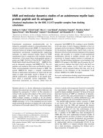
Fig. 1. NMR assignment. (A) Assignment of the spin systems of 32 nonproline residues out of the 34 amino acids of rSP-C (FFI) illustrated in the
TOCSY experiment with a mixing time of 70 ms. Shown is the so-called fingerprint region where the well-dispersed H
N
protons are correlated to
the H
a
and side chain protons. The spin systems of Lys10 and Arg11 are indicated by rectangles as both contain a second H
N
in the side chain. The
N-terminal Gly1 appears as a weak and very broad peak. All H
a
chemical shifts of residues 5–31 show an upfield shift compared with random-coil
data indicating an a-helical structure in an empirical pattern-recognition approach [13,16]. (B) H
N
-H
N
region of the 800-MHz NOESY experiment.
Sequential d
NN
(i,i+1) connectivities can be found for all nonproline amino acids. For the C-terminal residues 31–34, a second set of resonances can
be sequentially assigned indicated by the prime in the annotation of the corresponding NOE connectivity. (C) H
N
-H
a
region of the 800-MHz
NOESY experiment. All resolved interresidual NOE connectivities are annotated. In particular, the d
Na
(i,i+3) and d
Na
(i,i+4) connectivities are
indicators of an a-helical secondary structure. Intraresidual signals are not annotated. (D) Summation of the experimental NMR data. Shown are
all resolved NOE connectivities, where thin bars indicate distances > 4.0 A
˚
, medium bars distances of 3.0–4.0 A
˚
, and thick bars distances < 3.0 A
˚
.
The d
Na
(i,i+3), d
Na
(i,i+4) as well as the d
NN
(i,i+2) and the strong d
NN
(i,i+1) connectivities clearly show the a-helical structure of rSP-C (FFI). In
addition,
3
J
NHa
coupling constants are summarized, with small circles indicating couplings < 5.0 Hz and large circles for constants > 6.0 Hz.
Pentagons classify the exchange properties of amide protons in weak exchange (filled pentagons), medium exchange (open pentagons) and strong
exchange (no pentagon) as described in the text.
2078 B. Luy et al.(Eur. J. Biochem. 271) Ó FEBS 2004
the diastereotopic groups were inferred from NOEs
through floating chirality calculations.
Second set of resonances
Closer inspection of the spectra revealed two sets of
resonances for Gly28, Leu30, Ile32, Gly33 and Leu34,
which differ mainly in the chemical shifts of the amide
protons and the c protons of Ile32 and Leu34. A compar-
ison of the spectra showed different relative intensities of the
two sets of resonances with respect to the concentration of
rSP-C (FFI) in CDCl
3
/CD
3
OH (1 : 1, v/v) and the age of
the sample. For a systematic analysis, freshly prepared
samples with concentrations of 0.7–3.5 m
M
were used in
NOESY experiments with a mixing time of 50 ms. At low
concentration, the two sets of signals were almost equally
strong, whereas at higher concentrations of rSP-C, one of
the signal sets was more predominant. Attempts to fit the
relative intensities of the two sets of resonances to a
quantitative monomer–dimer equilibrium model failed
(data not shown). However, the concentration dependence
shown in Fig. 5 can be considered an indication of
intermolecular interaction. The comparable linewidths of
the signals of the two sets of resonances still suggest that
monomeric and dimeric units are involved.
Amide proton exchange
The exchange properties of the amide protons were
obtained from a 400-MHz NOESY spectrum of
rSP-C (FFI) in CDCl
3
/CD
3
OD (1 : 1, v/v) with the sample
freshly prepared about 1 h before the experiment. All
measurable H
a
-H
N
cross-peaks were integrated and com-
pared with the integrals of the 800-MHz NOESY spectrum.
The most intense signals were taken as 100% relative
intensity, making the assumption that no significant
exchange occurred in the given time frame within the center
of the well-ordered a-helix. The relative intensities of the
H
a
-H
N
cross-peaks of residues His8, Ala29 and Leu31 were
about 50% of those recorded in the 800-MHz NOESY
spectrum in CDCl
3
/CD
3
OH (1 : 1, v/v), and the intensities
of residues 9–28 and 30 were 80% or higher. From these
estimates of the relative intensities, hydrogen bonds for the
structure calculations were assumed for His8 to Leu31. The
amide protons of residues 1–7 and 32–34 could not be
detected in the fully deuterated solvent.
Structure of rSP-C (FFI)
Using the empirical pattern-recognition approach [16], the
combination of strong sequential d
NN
connectivities, obser-
vation of a significant number of d
aN
(i,i+3), d
ab
(i,i+3),
and d
aN
(i,i+4) connectivities,
3
J
NHa
coupling constants of
less than 5 Hz for all non-Gly residues in the polypeptide
segment Phe5, Val7–Leu30, and retarded amide proton
exchange for residues 8–31 indicate that rSP-C (FFI) forms
alonga-helix comprising approximately residues 5–34.
For a more precise definition of the structure of rSP-
C (FFI), a set of 203 intraresidual, 201 interresidual and
seven ambiguous NOE-derived upper distances were used
together with 23 / angles derived from
3
J(H
N
,H
a
) coupling
constants as input data for a structure calculation using the
program
XPLOR
[17]. In addition, we introduced 24 hydro-
gen bonds derived from the slow exchange rate of the amide
protons. No stereospecific assignments were used in the
floating chirality simulated annealing protocol. For residues
28–34, we used only the set of resonances with the stronger
intensities because identical relative NOEs were observed
for the two species.
For the structure calculations, we used a standard
simulated annealing protocol designed for proteins [18].
After an initial energy minimization involving 50 optimiza-
tion steps with conjugated gradients, a high temperature
phase with 2000 K was simulated for 32.5 ps in which all
upper limits built the active constraints. The following step
was the first cooling phase from 2000 K to 1000 K in 25 ps
with the dihedral angles as additional constraints. After the
Table 3. Chemical shifts of rSP-C (FFI).
Residue H
N
H
a
H
b
Others
Gly1 8.23 3.73
Ile2 8.61 4.45 1.90 c1.66, 1.00; d1.23,0.95
Pro3 4.38 2.15, 1.99 c2.10; d3.95, 3.72
Phe4 8.08 4.49 3.18, 3.09 d7.17; e7.27; f7.19
Phe5 8.46 4.68 3.29 d7.28; e7.42; f7.36
Pro6 4.25 2.34, 2.00 c2.14; d3.65
Val7 7.62 3.69 2.28 c1.13, 1.01
His8 8.05 4.47 3.35, 3.29 d7.22; e8.74
Leu9 8.13 3.97 1.70, 1.60 c1.65; d1.03, 0.98
Lys10 7.95 3.91 2.03 c1.64, 1.50; d1.79;
e2.93; f2.92
Arg11 7.89 3.94 2.02, 1.99 c1.70; d3.30, 3.24; e7.50;
1g7.18; 2g6.68
Leu12 7.82 4.01 1.69 c1.81; d0.94
Leu13 8.04 4.01 1.89 c1.71; d0.95
Ile14 7.77 3.64 2.08 c1.94, 1.20; d0.97, 0.93
Val15 7.67 3.52 2.40 c1.17, 1.01
Val16 8.01 3.51 2.33 c1.15, 1.00
Val17 8.03 3.52 2.33 c1.16, 1.02
Val18 8.15 3.57 2.32 c1.14, 1.03
Val19 8.36 3.57 2.32 c1.15, 1.00
Val20 8.35 3.49 2.31 c1.15, 1.00
Leu21 8.25 4.01 1.99, 1.93 c1.75; d1.02, 0.94
Ile22 8.30 3.60 2.16 c1.17; d0.99
Val23 8.25 3.53 2.40 c1.16, 0.99
Val24 8.59 3.57 2.42 c1.17, 1.02
Val25 8.28 3.71 2.39 c1.17, 1.03
Ile26 8.45 3.70 2.09 c1.95, 0.98; d1.17
Val27 8.93 3.59 2.23 c1.14, 1.02
Gly28 8.45 3.88, 3.77
8.42 3.86, 3.77
Ala29 8.21 4.09 1.62
8.19 4.08 1.62
Leu30 8.25 4.18 2.12, 2.03 c1.60; d0.99
Leu31 8.30 4.14 2.07 c1.60; d0.99, 0.96
8.28 4.13 2.07 g1.60; d0.99,0.96
Ile32 7.70 4.35 2.18 c1.64,1.51; d1.02
7.64 4.38 2.18 g1.61,1.55; d1.02
Gly33 7.95 4.09, 3.87
7.91 4.09, 3,85
Leu34 8.02 4.51 1.73 c1.78; d1.00
8.08 4.55 1.64 g1.74; d1.00
Ó FEBS 2004 Recombinant mutant of surfactant protein C (Eur. J. Biochem. 271) 2079
second cooling phase from 1000 K to 100 K in 10 ps, a
second energy minimization was performed with 200 steps
of conjugated gradients for each structure. The rmsd values
and the distance and dihedral angle violations for the best 10
out of 60 structures are given in Table 4. The final structures
shown in Figs 3 and 4 were determined by an additional
refinement in vacuo including the experimental restraints,
full charges, and a dielectric constant set to e ¼ 4r
ij
using a
heating and cooling protocol.
Figure 2 shows
MOLMOL
stereographic projections [19] of
the heavy atoms of rSP-C (FFI). The structure of rSP-
C (FFI) is a well-defined a-helix ranging from Phe5 to
Leu34. Note that the distribution of the / and w angles
indicates an a-helical structure up to Phe5, although residue
6 is a proline. Strong evidence for this comes from the
unambiguously identified d
aN
(i,i+3) and d
aN
(i,i+4) cross-
signals for Phe5 and Pro6 (cf. Fig. 1D).
Discussion
Comparison of rSP-C (FFI) with pSP-C
The 34-residue peptide rSP-C (FFI) contains mainly
apolar amino acids, i.e. 11 valines, seven leucines and
five isoleucines, and forms a well-defined a-helix along
residues 5–34 dissolved in CDCl
3
/CD
3
OH (1 : 1, v/v). The
solution structure of pSP-C with 76% sequence identity
(Table 1) in CDCl
3
/CD
3
OH/0.1
M
HCl (32 : 64 : 5, v/v/v)
was investigated by Johansson et al. [6]. To compare the
structure of pSP-C with rSP-C (FFI), we show in Fig. 3
the differences in chemical shifts of the H
N
and H
a
signals
of the corresponding residues. It can be seen that the
chemical shifts for residues 10–29 are almost identical,
with slightly greater variations at nonidentical amino
acids. Only the N-terminal nine residues show significant
chemical-shift differences mainly introduced by the
sequence deviations at residues 4, 5 and 8. This difference
at the N-terminus can also be seen when the two resulting
structures shown in Fig. 4 are compared. Whereas the
backbone of the central a-helix is very well defined in
both structures, the N-terminal variability for the pSP-C is
greater than that of rSP-C (FFI). This reflects the NOE-
data-based fact that rSP-C (FFI) has a defined a-helix
comprising residues 5–34, whereas for pSP-C an a-helical
region at residues 7–34 has been reported [6].
However, the slow deuterium exchange for Leu9 and
small distances d
Na
(i,i+3) and d
ab
(i,i+3) for Pro6 and Val7
suggest that even pSP-C adopts an a-helix starting with
capping at residue Cys5 [8]. Substitution of acylated Cys
with Phe in the polypeptide seems to influence the
N-terminal a-helix formation including Pro6 in
rSP-C (FFI). A possible explanation is the occurrence of
aromatic interactions between Phe5 and His8 which may
lead to stabilization of the extended a-helix. The structures
of both pSP-C and rSP-C (FFI) were determined in
chloroform/methanol, an environment in which hydropho-
bic elements can move freely. Membranous environments
such as the surfactant, however, have a directional effect on
the hydrophobic palmitoylated Cys and Phe residues and on
the charged Lys and Arg residues at positions 10 and 11,
which probably results in slightly different N-terminal
structures for the SP-C variants in their biologically
active form.
The central helix of pSP-C has a slightly lower rmsd value
than that of rSP-C, probably because of the longer stretch of
Val residues, leading to extremely stable stacking. In rSP-
C (FFI) this homogeneous stacking is interrupted by Ile14
and Ile22, which may introduce slight mobility into the
hydrophobic a-helix. However, this increased mobility still
leaves the central helix quite rigid and does not seem to be
important, as it was shown in mutation studies that SP-C
retains its function even after the replacement of all valines
by leucines or other a-helical amino-acid sequences [20,21].
Two sets of resonances
Two sets of resonances were found for rSP-C (FFI) at the
C-terminal residues Gly28, Leu30, Ile32, Gly33 and Leu34.
Similar duplication of resonances has been reported for
pSP-C, affecting residues Val27, Ala29, Leu30, Leu31 and
Met32 [6]. In the case of pSP-C, the additional signals were
explained by partial oxidation of Met32 to methionine
sulfoxide. In the case of rSP-C (FFI), a different explan-
ation must be found for the second set of resonances
because Met32 is substituted by Ile32. The careful studies on
pSP-C show a variation of 20–50% of the minor populated
Table 4. Analysis of the 10 best calculated structures before and after the refinement.
Before refinement After refinement
E
tot
(kcalÆmol
)1
) 165.9 ± 12.7 (142.4.181.6) ) 265.4 ± 7.5 ()266.9 … )246.1)
Distance violations
Number > 0.5 A
˚
1.1 ± 0.6 (0 … 2) 0
Sum (A
˚
) > 0.1 A
˚
0.68 ± 0.36 (0… 1.14) 0
Maximum (A
˚
) 0.51 ± 0.18 (0 … 0.57) 0
Torsion-angle violations
Number > 0.5 A
˚
0 1.2 ± 0.9 (0 … 3)
Sum (°) 0 13.0 ± 11.9 (0 … 34.9)
Maximum (°) 0 9.1 ± 6.0 (0. 16.7)
Rmsds (A
˚
)
Backbone (8–33) 0.59 ± 0.19 (0.36.0.99) 0.34 ± 0.06 (0.24.0.41)
Heavy atoms (8–33) 1.05 ± 0.18 (0.90.1.44) 0.82 ± 0.13 (0.67.1.00)
Backbone (18–28) 0.23 ± 0.08 (0.14.0.41) 0.07 ± 0.02 (0.04.0.10)
Heavy atoms (18–28) 0.61 ± 0.08 (0.51.0.77) 0.45 ± 0.12 (0.35.0.68)
2080 B. Luy et al.(Eur. J. Biochem. 271) Ó FEBS 2004
set of resonances among samples prepared from different
batches. We observed the same variation even in samples
prepared from the same batch. A closer look at the acquired
spectra indicates a dependence of the relative population
of the signals on the overall SP-C concentration. As a
consequence, we acquired a set of 2D NOESY spectra with
identical mixing times but different concentrations of rSP-
C (FFI) in CDCl
3
/CD
3
OH (1 : 1, v/v). The relative popu-
lations of the two sets of resonances in these spectra with
respect to the overall SP-C concentration are shown in
Fig. 5. The dependence observed is a clear indication of
intermolecular interaction. The relatively narrow linewidths
of the observed signals led to the conclusion that oligomers
of low order are present, probably monomeric and dimeric
units, but trimeric or tetrameric units may also be possible;
larger oligomers can be excluded because the linewidths
would have to be significantly broader than observed. The
linewidths of the two sets of resonances do not differ
significantly, therefore the two oligomers must be of
comparable size, and a monomer/tetramer equilibrium,
for example, cannot explain the observed signals. The
absence of further resonances implies that we are observing
specific oligomers. Finally, chemical shifts of the Ha
resonances are a clear indication that both oligomers are
mainly a-helical and that their structures differ only slightly.
The NMR data therefore point to the coexistence of
a monomeric and homodimeric a-helical form of
rSP-C (FFI).
Fig. 2. Stereographic projection of the best 10
out of 60 structures of rSP-C (FFI). (A) Side
view of the heavy atoms of the full-length
peptide. (B) View from the bottom along
residues 15–27 of the tightly packed a-helix.
Ó FEBS 2004 Recombinant mutant of surfactant protein C (Eur. J. Biochem. 271) 2081
The literature on SP-C describes many oligomerization
processes, most of which are either aggregates with mainly
b-sheet-like or undetermined structure. Specific oligomeri-
zation, i.e. dimerization, is only reported in a few cases:
MS data provide evidence for dimeric SP-C [22,23], and
chemical cross-linking studies also show mainly a specific
dimer of mature SP-C (Fig. 8C in [24]). Yet unpublished
high-resolution Fourier-transform ion-cyclotron-resonance
MS, light-scattering and CD experiments reveal the exist-
ence of an a-helical dimer at acidic pH ([25]; A. Seidl,
G. Maccarone, N. Youhnovski, K. P. Schaefer and
M. Przybylski, unpublished data). CNBr cleavage data
even put the dimerization site near Met32 at the C-terminus,
i.e. at the site at which the dual resonances are observed [23].
The coexistence of monomeric and homodimeric rSP-
C(FFI) as derived from the NMR data therefore corres-
ponds well to other reported experimental observations.
Fibril formation
The data from Fig. 5 could not be fitted to a simple
monomer–dimer equilibrium model, but this is not surpri-
sing considering that rSP-C (FFI), like pSP-C, shows a
complete transition to b-sheet fibrils over time [8,26,27].
Immediately after rSP-C (FFI) is dissolved in chloroform/
methanol, short, fiber-like impurities of up to 1 mm length
are observed in solution and on the glass walls of the NMR
tube on visual inspection. This indication of already formed
fibrils makes it necessary to describe rSP-C (FFI) by at least
a three-state model with two a-helical states, probably
monomer and dimer, and b-sheet fibrils that cannot be
observed by high-resolution NMR because of their high
molecular mass. A three-state model with monomeric,
nonhelical and b-fibril states has already been presented [8].
Interestingly, the existence of an a-helical transition state
(SPC
#
in [8]) was proposed in that publication, which would
Fig. 4. Comparison of the 10 best structures of rSP-C (FFI) (left) and
pSP-C (right). The backbone of the a-helix is shown. Clearly visible
is the better defined secondary structure of rSP-C (FFI) near the
N-terminus.
Fig. 5. Concentration dependence of the relative integrals of the two sets
of resonances observed at the C-terminus. Ratios are given for well-
resolved residues Ile32, Gly33 and Leu34.
Fig. 3. Differences in the chemical shifts of rSP-C (FFI) compared with
pSP-C [6] for the H
N
(A) and the H
a
protons (B). Whereas residues
10–29 show almost identical chemical shifts, residues at the N-terminus
and C-terminus differ more strongly.
2082 B. Luy et al.(Eur. J. Biochem. 271) Ó FEBS 2004
match the potential a-helical dimer found here. The
interpretation of the potential dimeric state as the transition
state to b-fibril formation would also match recent solid-
state and liquid-state NMR results, which suggest that the
smallest fibril diameter in b-amyloid fibrils is due to a
parallel b-sheet dimer [28,29] and also that the minimum
unit needed for fibril growth of a-synuclein is a dimer [30].
The disappearance of high-resolution NMR signals after
several days at 10 °C in chloroform/methanol shows that
the equilibrium state of rSP-C (FFI) is the b-sheet-like
multimer. The a-helical states are therefore not equilibrated,
and, in addition to the observed concentration dependence,
a dependence on the age of the prepared samples can be
predicted in the given solvent. It should be noted that
neither rSP-C (FFI) nor pSP-C [8] show any transition to
b-fibrils in dodecylphosphocholine micelles even after
several weeks at room temperature.
Sample handling and the situation
in vivo
SP-C is very difficult to handle. In general, basic conditions
should be avoided and properties of the molecule depend
strongly on the conditions for synthesis, the kind of
purification used, and the aggregation states it was trans-
ferred to. In this study, we relied on the elaborate procedure
developed by Altana Pharma and only suspended the
powder provided directly in chloroform/methanol. The
NMR spectra yielded good results and therefore there
appeared to be no need to change the method. Whether
oligomerization can be avoided by different sample treat-
ment remains to be proven.
The local environment of the molecule also has a large
impact on its behavior. Wild-type SP-C, like rSP-C (FFI),
is solely monomeric at micromolar concentrations in pure
organic solvents but has a strong tendency to aggregate in
more hydrophilic environments. Relatively high concentra-
tions can be obtained in dodecylphosphocholine micelles in
which SP-C is stable for months in its a-helical form [8]. The
surfactant consists of 1% by weight of SP-C [31]. The
concentration of rSP-C (FFI) and extracted pSP-C in
the NMR studies is therefore similar to the concentration
of SP-C in its natural environment, although it shows a slow
transition to b-sheet fibrils. However, whether the homo-
dimer in chloroform/methanol is representative of the
biologically active SP-C in the surfactant cannot be judged
from the experiments presented. A hint may be gained from
chemical cross-linking data on mature SP-C in cytosolic
vesicles of A549 cells (Fig. 8C in [24]), which provide
evidence of dimer formation during trafficking.
Potential dimerization site
The evidence suggests dimerization of SP-C, and it might be
allowed to speculate on the potential dimerization site. The
C-terminus of rSP-C (FFI) only contains apolar side chains
and it can be assumed that it is situated at the hydrophobic
palmitoyl chains of the surfactant phospholipids. In this
environment, hydrogen-bonding interactions and strong
hydrophobic associations are most likely to be the source of
intermolecular attraction. A minor dimerization motif can
be found in the C-terminal carboxylic group. Similar to the
dimer formation of acetic acid, SP-C may form a dimer via
hydrogen bonding (Fig. 6A). The acidic conditions of the
NMR sample as well as the natural environment of SP-C
would allow such a dimer formation. However, in the acidic
NMR sample, relatively fast hydrogen exchange rates are
expected which do not match the slow exchange regime
observed for the two sets of resonances. Therefore, hydro-
gen bonding of the carboxylic group is unlikely to be
the cause of the observed dimerization, but we cannot
exclude it.
An alternative dimerization motif can be found in the
strictly conserved C-terminal heptapeptide segment-span-
ning residues Gly28 to Leu34: the heptapeptide segment
of rSP-C (FFI), as well as all other SP-C variants, has an
AxxxG pattern that perfectly matches the requirements for
helix–helix association as described in [32]. Interestingly, the
residues for which double resonances are observed are all
within the strictly conserved heptapeptide segment with the
AxxxG motif (Fig. 6B). Attempts to model two distinct
structures for the two sets of resonances failed because of
massive overlap of the side chain resonances in the region
of interest in particular. However, as mentioned above, we
can conclude from chemical-shift arguments that the two
structures should be very similar and are a-helical in
character. For the same reasons, it was impossible to obtain
a structure of the potential dimer based on intermonomeric
NOEs. A theoretical model based on the monomeric
structure presented in this paper and computational dock-
ing studies is derived in the following paper [33].
Conclusion
We have derived by NMR spectroscopy the high-resolution
3D structure of rSP-C (FFI) dissolved in CDCl
3
/CD
3
OH
(1 : 1, v/v). The lipophilic peptide forms a tight a-helix for
residues 5–34 which is two residues longer than the a-helix
Fig. 6. Potential dimerization motifs for rSP-C (FFI). (A) Hydrogen
bonding at the C-terminal carboxy group may lead to dimerization.
(B) Comparison of the amino-acid sequences of glycophorin A and
rSP-C (FFI) shows a potential AxxxG dimerization motif similar to
the van der Waals dimer of glycophorin A [33–35] at the strictly
conserved heptapeptide segment where two sets of resonances are
observed.
Ó FEBS 2004 Recombinant mutant of surfactant protein C (Eur. J. Biochem. 271) 2083
observed in pSP-C, with 76% sequence identity in the same
solvent. Both peptides show two sets of resonances for a
number of C-terminal residues. Because of the lack of Met
we can exclude oxidation to methionine sulfoxide as the
cause of the second set of resonances for rSP-C (FFI),
which was previously assumed in the case of pSP-C [6].
Studies on the concentration dependence of the dual
resonances together with the narrow linewidth of the
NMR signals suggest the coexistence of a monomeric and
dimeric a-helical structure in the given solvent. There are
two potential dimerization sites in SP-C: the C-terminal
carboxylic group may form a dimer via hydrogen bonding;
the C-terminal heptapeptide segment, which is conserved in
all known SP-C species, contains an AxxxG motif that
closely resembles the GxxxG helix–helix dimer motif of
glycophorin A. Even though the latter dimerization motif is
consistent with other experimental results and therefore
highly likely, additional studies such as point mutations at
the potential dimerization site are necessary to unambigu-
ously determine the origin of the intermolecular interaction
that leads to the second set of resonances.
Acknowledgements
C.G. gratefully acknowledges support from the DFG, the MPG, and
the Fonds der Chemischen Industrie. B.L and A.D. were supported by
the Fonds der Chemischen Industrie. B.L. is also supported by the DFG
(Emmy Noether LU 835/1–1). We thank Bettina Elshorst for help with
NDEE
, Michael Nilges for help with the
XPLOR
protocols, and Michael
Przybylski (University of Konstanz) for providing his results before
publication. Special thanks go to Michael K. Gilson (CARB, Rockville,
MD, USA) for many detailed scientific discussions.
References
1. Goerke, J. (1998) Pulmonary surfactant: functions and molecular
composition. Biochim. Biophys. Acta 1408, 79–89.
2. Ha
¨
fner, D., Germann, P.G. & Hauschke, D. (1998) Effects of rSP-
C surfactant on oxygenation and histology in a rat-lung-lavage
model of acute lung injury. Am.J.Respir.Crit.CareMed.158,
270–278.
3. Audrey, J.D., Alan, H.J., Ha
¨
fner, D. & Ikegami, M. (1998) Lung
function in premature lambs and rabbits treated with a recom-
binant SP-C surfactant. Am.J.Respir.Crit.CareMed.157,
553–559.
4. Spragg, R.G., Lewis, J., Wurst, W. & Rathgeb, F. (2000) Treat-
ment of ARDS with rSP-C surfactant. Am.J.Respir.Crit.Care
Med. 161, A47.
5. Walmrath, D., De Vaal, J.B., Bruining, H.A., Kilian, J.G.,
Papazian, L., Hohlfeld, J., Vogelmaier, C., Wurst, W., Schaffer,
P., Rathgeb, F., Grimminger, F. & Seeger, W. (2000) Treatment of
ARDS with recombinant SP-C (rSP-C) based synthetic surfactant.
Am. J. Respir. Crit. Care Med. 161, A379.
6. Johansson, J., Szyperski, T., Curstedt, T. & Wu
¨
thrich, K. (1994)
The NMR structure of the pulmonary surfactant-associated
polypeptide Sp-C in an apolar solvent contains a valyl-rich alpha-
helix. Biochemistry 33, 6015–6023.
7. Johansson,J.,Szyperski,T.&Wu
¨
thrich, K. (1995) Pulmonary
surfactant-associated polypeptide SP-C in lipid micelles: CD stu-
dies of intact SP-C and NMR secondary structure determination
of depalmitoyl-SP-C (1–17). FEBS Lett. 362, 261–265.
8. Szyperski, T., Vandenbussche, G., Curstedt, T., Ruysschaert,
J.M., Wu
¨
thrich, K. & Johansson, J. (1998) Pulmonary surfactant-
associated polypeptide C in a mixed organic solvent transforms
from a monomeric alpha-helical state into insoluble beta-sheet
aggregates. Protein Sci. 7, 2533–2540.
9. Marion, D., Ikura, M., Tschudin, R. & Bax, A. (1989) Rapid
recording of 2D NMR-spectra without phase cycling: application
to the study of hydrogen exchange in proteins. J. Magn. Reson. 85,
393–399.
10. Wider, G., Hosur, R.V. & Wu
¨
thrich, K. (1983) Suppression of the
solvent resonance in 2D NMR-spectra of proteins in H
2
Osolu-
tion. J. Magn. Reson. 52, 130–135.
11. Prasch, T., Gro
¨
schke, P. & Glaser, S.J. (1998) SIAM, a novel
NMR experiment for the determination of homonuclear coupling
constants. Angew. Chem. Int. Ed. 37, 802–806.
12. Titman, J.J. & Keeler, J. (1990) Measurement of homonuclear
coupling-constants from NMR correlation spectra. J. Magn.
Reson. 89, 640–646.
13. Wu
¨
thrich, K. (1986) NMR of Proteins and Nucleic Acids. Wiley,
New York.
14. Wishart, D.S., Sykes, B.D. & Richards, F.M. (1991) Relationship
between nuclear-magnetic-resonance chemical-shift and protein
secondary structure. J. Mol. Biol. 222, 311–333.
15. Wishart, D.S., Sykes, B.D. & Richards, F.M. (1992) The chemical-
shift index: a fast and simple method for the assignment of protein
secondary structure through NMR- spectroscopy. Biochemistry
31, 1647–1651.
16. Wu
¨
thrich, K., Billeter, M. & Braun, W. (1984) Polypeptide sec-
ondary structure determination by nuclear magnetic-resonance
observation of short proton–proton distances. J. Mol. Biol. 180,
715–740.
17. Bru
¨
nger, A.T. (1992) X-PLOR: a System for X-Ray Crystallo-
graphy and NMR. Yale University Press, New Haven, CT.
18. Nilges, M. & O’Donoghue, I.S. (1998) Ambiguous NOEs and
automated NOE assignment. Prog. NMR Spectrosc. 32, 107–139.
19. Koradi, R., Billeter, M. & Wu
¨
thrich, K. (1996) MOLMOL: a
program for display and analysis of macromolecular structures.
J. Mol. Graph. 14, 51–55.
20. Nilsson, G., Gustafsson, M., Vandenbussche, G., Veldhuizen,
E., Griffiths, W.J., Sjovall, J., Haagsman, H.P., Ruysschaert,
J.M.,Robertson,B.,Curstedt,T.&Johansson,J.(1998)
Synthetic peptide-containing surfactants: evaluation of trans-
membrane versus amphipathic helices and surfactant protein C
poly-valyl to poly-leucyl substitution. Eur. J. Biochem. 255,
116–124.
21. Clercx, A., Vandenbussche, G., Curstedt, T., Johansson, J.,
Jornvall, H. & Ruysschaert, J.F. (1995) Structural and functional
importance of the C-terminal part of the pulmonary surfactant
polypeptide Sp-C. Eur. J. Biochem. 229, 465–472.
22. Mayer-Fligge, P., Volz, J., Kru
¨
ger,U.,Sturm,E.,Gernandt,W.,
Scha
¨
fer, K.P. & Przybylski, M. (1998) Synthesis and structural
characterization of human-identical lung surfactant SP-C protein.
J. Pept. Sci. 4, 355–363.
23. Przybylski, M., Maier, C., Ha
¨
gele, K., Bauer, E., Hannappel, E.,
Nave, R., Melchers, K., Kru
¨
ger, U. & Scha
¨
fer, K.P. (1994) Pri-
mary structure elucidation, surfactant function and specific for-
mation of supramolecular dimer structures of lung surfactant
associated SP-C proteins. In Hodges, R.S. & Smith, J.A., eds.
Peptides, Chemistry, Structure and Biology, pp. 338–340. Escom
Science Publishers, Leiden.
24. Wang, W.J., Russo, S.J., Mulugeta, S. & Beers, M.F. (2002)
Biosynthesis of surfactant protein C (SP-C). J. Biol. Chem. 277,
19929–19937.
25. Seidl, A. (2003) Massenspektrometrische Analyse: Chemische
Modifizierung und Synthese Von Lipoproteinen.PhDThesis,
Gorre-Verlag, Konstanz, Germany.
26. Johansson, J. (2001) Membrane properties and amyloid fibril
formation of lung surfactant protein. Biochem. Soc. Trans. 29,
601–606.
2084 B. Luy et al.(Eur. J. Biochem. 271) Ó FEBS 2004
27. Johansson, J. (2003) Molecular determinants for amyloid fibril
formation: lessons from lung surfactant protein C. Swiss Medical
Weekly 133, 275–282.
28. Petkova, A.T., Ishii, Y., Balbach, J.J., Antzutkin, O.N., Leapman,
R.D., Delaglio, F. & Tycko, R. (2002) A structural model for
Alzheimer’s beta-amyloid fibrils based on experimental con-
straints from solid state NMR. Proc. Natl Acad. Sci. USA 99,
16742–16747.
29. Tycko, R. (2003) Applications of solid state NMR to the struc-
tural characterization of amyloid fibrils: methods and results.
Prog. Nucl. Magn. Reson. Spectrosc. 42, 53–68.
30. Fernandez, C.O., Hoyer, W., Zweckstetter, M., Jares-Erijman,
E.A., Subramaniam, V., Griesinger, C., Jovin, T.M. (2004) NMR
of alpha-synuclein complexes with polyamines elucidates the
mechanism and kinetics of induced aggregation. EMBO J.
in press.
31. Kru
¨
ger,P.,Schalke,M.,Wang,Z.,Notter,R.H.,Dluhy,R.A.&
Lo
¨
sche, M. (1999) Effect of hydrophobic surfactant peptides SP-B
and SP-C on binary phospholipid monolayers. I. fluorescence and
dark-field microscopy. Biophys. J. 77, 903–914.
32. Eilers, M., Patel, A.B., Liu, W. & Smith, S.O. (2002) Comparison
of helix interactions in membrane and soluble alpha-bundle pro-
teins. Biophys. J. 82, 2720–2736.
33. Kairys, V., Gilson, M.K., Luy, B. (2004) Structural model for an
AxxxG-mediated dimer of surfactant-associated protein C. Eur. J.
Biochem. 271, 2086–2092.
34. MacKenzie, K.R., Prestegard, J.H. & Engelman, D.M. (1997) A
transmembrane helix dimer: structure and implications. Science
276, 131–133.
35. Smith, S.O., Song, D., Shekar, S., Groesbeek, M., Ziliox, M. &
Aimoto, S. (2001) Structure of the transmembrane dimer inter-
face of glycophorin A in membrane bilayers. Biochemistry 40,
6553–6558.
Ó FEBS 2004 Recombinant mutant of surfactant protein C (Eur. J. Biochem. 271) 2085