Báo cáo khoa học: Protection of chylomicron remnants from oxidation by incorporation of probucol into the particles enhances their uptake by human macrophages and increases lipid accumulation in the cells ppt
Bạn đang xem bản rút gọn của tài liệu. Xem và tải ngay bản đầy đủ của tài liệu tại đây (398.45 KB, 11 trang )
Protection of chylomicron remnants from oxidation by incorporation
of probucol into the particles enhances their uptake by human
macrophages and increases lipid accumulation in the cells
Elizabeth H. Moore
1
, Mariarosaria Napolitano
2
, Michael Avella
1
, Fatos Bejta
1
, Keith E. Suckling
3
,
Elena Bravo
2
and Kathleen M. Botham
1
1
Department of Veterinary Basic Sciences, The Royal Veterinary College, London, UK;
2
Istituto Superiore di Sanita,
Department of Hematology, Oncology and Molecular Medicine, Viale Regina Elena, Rome, Italy;
3
Glaxo SmithKline,
Medicines Research Centre, Stevenage, Herts., UK
The effects of protection of chylomicron remnants from
oxidation on their uptake and induction of lipid accumula-
tion in macrophages were investigated using chylomicron
remnant-like particles (CRLPs) containing the lipophilic
antioxidant drug, probucol, and macrophages derived from
the human monocyte cell line, THP-1. The total lipid content
of THP-1 macrophages was markedly higher (·2.2) after
48 h of incubation of THP-1 macrophages with CRLPs
containing probucol (pCRLPs) when compared to CRLPs
without probucol, and this was because of increases in tri-
acylglycerol (·2.3) and cholesterol (·1.8) levels, while cho-
lesteryl ester concentrations were not significantly changed.
Determination of the uptake of CRLPs and pCRLPs by the
cells using particles labelled with the fluorescent probe 1,1¢-
dioctadecyl-3,3,3¢3¢-tetramethylindo-carbocyanine perchlo-
rate showed that pCRLPs are taken up at a faster rate than
CRLPs. The synthesis of triacylglycerol, as measured by
the incorporation of [
3
H]oleate and [
3
H]glycerol, was also
increased in macrophages incubated with pCRLPs as
compared to CRLPs without probucol, but phospholipid
and cholesteryl ester formation from [
3
H]oleate was unaf-
fected. In addition, no differences between the effects of
CRLPs and pCRLPs on the expression of mRNA for a
range of genes believed to be involved in lipoprotein uptake,
intracellular lipid metabolism and the efflux of cholesterol
from macrophages was detected. These results suggest that
antioxidants carried in chylomicron remnants enhance lipid
accumulation in macrophages by increasing the rate of
uptake of the particles and raising the intracellular synthesis
of triacylglycerol, but not cholesteryl ester, and that these
effects are brought about by changes at the post-transcrip-
tional level. Antioxidants carried in chylomicron remnants
therefore may promote the development of atherosclerosis,
and this is likely to be particularly important in conditions
where clearance of remnants from the circulation is delayed.
Keywords: chylomicron remnants; probucol; macrophages;
lipid accumulation; antioxidants.
Atherosclerotic lesions are intiated by the formation of fatty
streaks in the artery, which form when macrophages in the
vessel wall take up lipoproteins from the subendothelial
space and eventually become so engorged with lipid that
they take on a foamy appearance and are termed foam cells
[1,2]. Evidence from a large number of previous studies has
indicated that low density lipoprotein (LDL) has a major
role in the generation of foam cells, but it is also clear that
oxidation of the lipoprotein particles, a process which can
occur within the artery wall, is necessary before extensive
lipid accumulation is induced [3], and this has led to the
development of the hypothesis that antioxidants have a
beneficial effect in the prevention of atherosclerosis and
related cardiovascular disease.
There is considerable epidemiological evidence to indicate
that diets rich in fruits and vegetables (for example, the
Mediterranean diet), which contain relatively high levels
of natural antioxidants such as vitamin E and carotenoids,
reduce the risk of heart disease [4–6]. A number of
prospective studies have also suggested that consumption
of antioxidant vitamins, such as vitamin E and b-carotene,
may be beneficial [7,8], and randomized clinical trials with
vitamin E supplementation have supported this view [9,10].
Correspondence to K.M.Botham,DepartmentofVeterinaryBasic
Sciences, The Royal Veterinary College, Royal College St., London
NW1 0TU, UK. Fax: + 44 20 7388 1027, Tel.: + 44 20 7468 5274,
E-mail:
Abbreviations: ABCA1, ATP-binding cassette transporter A1;
ACAT1, acyl coenzyme A : cholesterol acyltransferase 1; acLDL,
acetylated low-density lipoprotein; apoE, apolipoprotein E; CRLPs,
chylomicron remnant-like particles; DGAT1, acyl coenzyme A:
diacylglycerol acyl transferase 1; DiI, 1,1¢-dioctadecyl-3,3,3¢3¢-tetra-
methylindo-carbocyanine perchlorate; HMDM, human monocyte-
derived macrophages; HNE, 4-hydroxy-2(E)-nonenal; LDL, low-
density lipoprotein; LDLr, low-density lipoprotein receptor; LRP,
low-density lipoprotein receptor-related protein; lycCRLPs, chylo-
micron remnant-like particles containing lycopene; MDA,
malondialdehyde; oxLDL, oxidized low-density lipoprotein; pCRLPs,
chylomicron remnant-like particles containing probucol; PMA,
4b-phorbol 12-myristate 13-acetate; SR-A, scavenger receptor A;
SR-B1, scavenger receptor B1; TBARS, thiobarbituric acid reactive
substances; VLDL, very-low-density lipoprotein.
Enzymes: acyl coenzyme A: cholesterol acyltransferase (EC 2.3.1.26);
acyl coenzyme A: diacylglycerol acyl transferase (EC 2.3.1.20).
(Received 27 February 2004, revised 7 April 2004,
accepted 16 April 2004)
Eur. J. Biochem. 271, 2417–2427 (2004) Ó FEBS 2004 doi:10.1111/j.1432-1033.2004.04164.x
Despite these strong indications of potential benefits,
however, a recent review of large-scale trials of dietary
supplementation with b-carotene or vitamin E in healthy
human populations has concluded that the results fail to
confirm any protective effects of these compounds against
cardiovascular disease [11].
Dietary lipids, including fats, cholesterol and lipid-soluble
antioxidants, are absorbed in the intestine and secreted into
lymph in chylomicrons, large triacylglycerol-rich lipopro-
teins which pass into the blood via the thoracic duct and are
then rapidly metabolized by lipoprotein lipase in extra-
hepatic capillary beds, removing some of the triacylglycerol
and leaving smaller remnant particles. The chylomicron
remnants retain all the cholesterol and minor lipid compo-
nents, such as antioxidants, and deliver them to the liver for
processing [12]. There is now a large and growing body of
evidence indicating that chylomicron remnants are strongly
atherogenic. They have been shown to be taken up into the
artery wall [13–15] as efficiently as LDL [16], and remnant-
like particles containing apolipoprotein E (apoE) have been
isolated from human aortic intima and atherosclerotic
plaque [17,18]. In addition, delayed clearance of remnants
from the circulation is correlated with the development of
atherosclerotic lesions [19,20]. Previous work in our labor-
atory and by others has demonstrated that chylomicron
remnants are able to induce foam cell formation in human
monocyte-derived macrophages (HMDM) [21] and in
human and murine macrophage cell lines [22–24], and that,
in striking contrast to LDL, the remnant particles do not
require prior oxidation to bring about this effect.
Information about the effects of antioxidants on macro-
phage foam cell formation has come mainly from studies
with vitamin E and the synthetic lipophilic antioxidant drug,
probucol, and the results have been inconsistent. Vitamin E
has been reported to decrease lipid accumulation and/or
the uptake of oxidized LDL (oxLDL) or acetylated LDL
(acLDL) in HMDM and the murine macrophage cell line
J774, both when added to the medium [25,26], or incorpor-
ated into the LDL particles [27], but in other studies no
effects of pretreatment with the vitamin could be detected
in experiments with HMDM [28], or mouse macrophages
[29,30]. Similarly, Yamamoto et al. [31] found that probu-
col, either in the free form or bound to the lipoprotein,
suppressed lipid accumulation and the uptake of acLDL by
human macrophage cell lines, while other workers have
found either no effect [32,33] or increased lipid accumula-
tion [30,34] with probucol pretreatment in rabbit or mouse
peritoneal macrophages.
Although it is clear that transport in the blood in
chylomicron remnants provides dietary lipid-soluble anti-
oxidants with the opportunity to interact directly with the
artery wall to influence atheroma development, little is
known about how the incorporation of antioxidants into
the remnant particles influences their effects on macro-
phages. In the first study in this area, we have demonstrated
recently that chylomicron remnant-like particles (CRLPs)
containing lycopene cause markedly increased lipid accu-
mulation in macrophages derived from the human mono-
cyte cell line, THP-1, suggesting that, contrary to what
might be expected, protection of chylomicron remnants
from oxidation enhances, rather than inhibits, their induc-
tion of foam cell formation [35]. Our experiments, however,
could not rule out the possibility that the findings were a
result of properties specific to the lycopene molecule, rather
than its antioxidant effects. In the present work therefore we
investigated the effects of the incorporation of the phenolic
lipophilic drug, probucol, which is structurally unrelated to
lycopene [36], into CRLPs on lipid accumulation in THP-1
macrophages. After confirming that the induction of lipid
accumulation in the cells by the particles is enhanced when
they are protected from oxidation, we investigated the
mechanism of this effect by comparing the influence of
CRLPs and CRLPs containing probucol (pCRLPs) on the
uptake of the particles by the cells, intracellular lipid
synthesis, and the expression of mRNA for genes believed
to play a part in foam cell formation, including those
regulating lipoprotein uptake [such as the LDL receptor
(LDLr), the low-density lipoprotein receptor-related protein
(LRP), scavenger receptor A (SR-A), and CD36], intracel-
lular lipid metabolism [acyl coenzyme A: cholesterol acyl-
transferase 1 (ACAT1), acyl coenzyme A: diacylglycerol
acyltransferase 1 (DGAT1)], the efflux of cholesterol from
the cells [scavenger receptor B1 (SR-B1)], and the ATP-
binding cassette transporter A1 (ABCA1).
Materials and methods
RPMI-1640, fetal bovine serum,
L
-alanyl-
L
-glutamine
(glutamax), penicillin/streptomycin and 2-mercaptoethanol
were obtained from Gibco. Fetal bovine serum was heat
inactivated by incubation at 56 °C for 30 min before use.
Trypan blue, fatty acid-free BSA, phospholipids, choles-
terol, cholesteryl oleate, probucol, lycopene and 4b-phorbol
12-myristate 13-acetate (PMA) were supplied by Sigma. The
fluorescent probe 1,1¢-dioctadecyl-3,3,3¢3¢-tetramethylindo-
carbocyanine perchlorate (DiI) was purchased from
Cambridge Bioscience. The radioisotopes L-3 phosphatidyl-
choline 1-palmitoyl-2-[1
14
C]linoleoyl and [1(3)-
3
H]glycerol
were purchased NEN Life Science Products Inc., and
[9,10(n)-
3
H]oleate was from Amersham International.
Preparation of CRLPs
CRLPs were prepared by sonication of a lipid mixture
containing 70% trilinolein, 2% cholesterol, 5% cholesteryl
ester and 25% phospholipids, in Tricine buffer (20 m
M
,
pH 7.4) containing 0.9% NaCl, followed by density-gradi-
ent centrifugation and binding to human apoE, as described
previously [37]. Sonication was performed at 22–24 lmfor
20 min at 56 °C, and the resulting emulsion was adjusted to
adensityof1.21 gÆmL
)1
with KBr, layered under a stepwise
density gradient, and centrifuged at 17 000 g for 20 min at
20 °C. The upper layer of grossly emulsified lipids was then
removed and replaced with an equal volume of 0.9% NaCl
(d ¼ 1.006 gÆmL
)1
) and the tubes were centrifuged at
70 000 g for 1 h (20 °C). Lipid particles harvested from
the top layer were incubated with the dialysed (18 h, 4 °C),
d > 1.063 gÆmL
)1
, fraction of human plasma (obtained
from the National Blood Service, North London Centre,
London, UK) at 37 °C with shaking for 5 h [lipid particles/
plasma, 1 : 1, (v/v)]. CRLPs were then isolated by ultra-
centrifugation (120 000 g,16h,12°C), harvested from the
top layer and stored at 4 °C under argon until required.
Analysis by SDS–PAGE showed that the particles
2418 E. H. Moore et al. (Eur. J. Biochem. 271) Ó FEBS 2004
contained apoE and no other lipoproteins. For the prepar-
ation of CRLPs containing antioxidants and/or the DiI
fluorescent label, probucol, lycopene and/or DiI was added
to the lipid mixture prior to sonication. Taking into account
the amount of probucol or lycopene added to the lipid
mixture for sonication and the percentage recovery of the
starting lipids in the CRLPs, we estimate that the concen-
trations of probucol or lycopene used in our experiments
did not exceed 10 l
M
.
Culture of THP-1 cells
THP-1 monocytes were maintained in suspension in RPMI-
1640 containing 10% fetal bovine serum, 2 m
M
glutamine,
100 UÆmL
)1
penicillin, 100 mgÆmL
)1
streptomycin and
50 m
M
2-mercaptoethanol (culture medium), at a density
of 3–9 · 10
5
cells per mL at 37 °Cin5%air/95%CO
2
.The
cells were induced to differentiate into macrophages by
incubation for 72 h in the presence of PMA (200 ngÆmL
)1
).
Then, cells adhering to the culture plates, and the medium
containing the PMA and any remaining monocytes, were
removed. Viability of THP-1 macrophages, as assessed by
Trypan blue exclusion, was > 95%.
For studies on lipid accumulation, CRLPs or pCRLPs
(30 lgÆmL
)1
cholesterol), were added to the macrophages
and the incubation was continued for a further 48 h before
the cells were harvested and their lipid content determined.
For uptake studies, DiI-labelled CRLPs or pCRLPs
(30 lgÆmL
)1
cholesterol) were incubated with THP-1
macrophages for various time-periods and the cells were
then viewed with a Zeiss LMS 510 laser-scanning confocal
microscope.
To determine the incorporation of [
3
H]oleate into cellular
lipids, THP-1 macrophages were incubated with CRLPs or
pCRLPs (30 lgÆmL
)1
cholesterol) for 48 h. The medium
was then removed and, after washing the cells three times
with 2 mL of warm phosphate-buffered saline (NaCl/P
i
),
replaced with culture medium (1 mL) containing
[9,10-
3
H]oleic acid (37 KBqÆmL
)1
, 55 000 d.p.m.Ælmol
)1
)
and 2% fat-free BSA. The incubation was continued for
1 h, the cells were washed three times with 2 mL of NaCl/P
i
,
and the lipids were extracted with isopropanol/hexane
(3:2,v/v)(2mL· 30 min, then 1 mL · 20 min repeated
four times), separated by TLC (hexane/diethyl ether/formic
acid, 80 : 20 : 2, v/v/v) and the bands corresponding
to triacylglycerol, diacylglycerol, phospholipid, cholesteryl
ester, and nonesterified fatty acids (visualized with iodine
vapour) were scraped into vials for determination of the
radioactivity by liquid scintillation counting using Lumagel-
safe scintillant (Perkin Elmer Life Sciences). Preliminary
experiments showed that [
3
H]oleate incorporation into
lipids was linear over a period of 2 h under the conditions
used. Incorporation of [
3
H]glycerol into triacylglycerol was
determined using a modification of the method described by
Davis et al. [38]. Cells were incubated for 6 h at 37 °Cin
95% air/5% CO
2
in serum-free medium containing CRLPs
or pCRLPs (20 lgÆmL
)1
cholesterol) in the presence of
[
3
H]glycerol (148 KBqÆmL
)1
,20l
M
). After incubation, the
cells were washed twice with NaCl/P
i
.[
14
C]Phosphatidyl-
choline (900 d.p.m.) was added to each tube as an internal
standard and the lipids were extracted and separated by
TLC, as described previously [22]. Proteins were harvested
from the plates, after extraction of the lipids, by the addition
of 1 mL of NaOH (1
M
).
mRNA analysis
The relative abundance of transcripts for the LDLr, LRP,
SR-A, SR-B1, CD36, ACAT1, DGAT1, and ABCA1 was
determined by RT-PCR. Total mRNA was extracted from
the cells using a kit from Promega UK, and first-strand
synthesis was carried out using Avian myeloblastosis virus
reverse transcriptase. The primers used and the product
sizes for the genes tested are shown in Table 1. Amplifica-
tion conditions were: initial denaturation at 95 °Cfor
15 min, followed by 30 cycles (LDLr, ACAT1, DGAT1), 33
cycles (SR-A, SR-B1, CD36, ABCA1) or 34 cycles (LRP)
consisting of denaturation at 94 °C for 30 s, annealing at
58 °C for 1 min and extension at 72 °C for 1 min, with the
final extension at 72 °C for 10 min. The products were
analysed by electrophoresis on an agarose gel [1.2%, (w/v)]
containing ethidium bromide (0.5 lgÆmL
)1
) and the bands
were quantified by absorbance volume analysis using values
for glyceraldehyde-3-phosphate dehydrogenase (GAPDH)
obtained simultaneously in the same system. The linearity
of the assay for each gene was established in preliminary
experiments.
Analytical methods
The total cholesterol (cholesterol + cholesteryl ester),
cholesterol, triacylglycerol and phospholipid content of
Table 1. Primer sequences and product sizes for RT-PCR. ABCA1, ATP-binding cassette transporter A1; ACAT1, acyl coenzyme A: cholesterol
acyltransferase 1; DGAT1, acyl coenzyme A: diacylglycerol acyl transferase 1; GAPDH, glyceraldehyde-3-phosphate dehydrogenase; LDLr, low-
density lipoprotein receptor; LRP, low-density lipoprotein receptor-related protein; SR-A, scavenger receptor A; SR-B1, scavenger receptor B1.
Gene Forward primer Reverse primer Product size (bp)
LDLr
AGTTGGCTGCGTTAATGTGAC TTCCTCACACTGGCACTTGTA 343
LRP
CCCAGGTGTCTACCATCACAC GGGGTTGTAGAGTTCCAGGTC 326
SR-A
ATTGCCCTTTACCTCCTCGT ATGAGGTTGGCTTCCATGTC 248
CD36
AGATGCAGCCTCATTTCCAC TGGGTTTTCAACTGGAGAGG 175
SR-B1
GAAACTGCAGCTGAGCCTCT ACCTACTTGGCTCCGGATTT 250
ACAT1
CTACAAGGCAGGCAGTATTGG TAAGCGTCCTGTTCATTTCGT 334
DGAT1
CCTGTGTTGAGGGAGTACCTG GGGCGAAACCAATGTATTTCT 328
ABCA1
AACAGTTTGTGGCCCTTTTG AGTTCCAGGCTGGGGTACTT 157
GAPDH
AATGACCCCTTCATTGACCTC GTTCACACCCATGACGAACAT 309
Ó FEBS 2004 Foam cell induction by chylomicron remnants (Eur. J. Biochem. 271) 2419
CRLPs and cell samples was determined by enzymatic
analysis using kits supplied by Sigma. Cholesteryl ester
levels were calculated by subtracting the values for choles-
terol from those for total cholesterol. The thiobarbituric
acid reactive substances (TBARS) content of the prepara-
tions was determined as described by Steinbrecher et al.
[39]. Tetraethoxypropane, which yields malondialdehyde
(MDA), was used as a standard. The levels of MDA
and 4-hydroxy-2(E)-nonenal (4-HNE) were assayed using
Bioxytech LPO-586 (Oxis International Inc, Portland, OR,
USA). For the determination of conjugated diene formation,
CRLPs or pCRLPs (50 nmolÆmL
)1
triacylglycerol) were
incubated at 30 °C in the presence or absence of CuSO
4
(50 l
M
) and the change in absorbance at 234 nm was
measured.
Fluorescence per cell in macrophages treated with DiI-
labelled CRLPs or pCRLPs was quantified by absorbance
volume analysis. The fluorescence for the entire field was
assessed, the background deducted and the value divided by
the number of cells in the field. Two or three fields with
approximately equivalent numbers of cells were analysed
in each experiment. Values were normalized for variations
in the fluorescence of different preparations using the
fluorescence units per lmol of cholesterol, measured in a
fluorimeter.
Significance limits were calculated using the Student’s
t-test, Student’s paired t-test or
ANOVA
, as indicated.
Results
Characteristics of CRLPs and pCRLPs
The lipid content of the CRLPs and pCRLPs used is shown
in Table 2. The total cholesterol, triacylglycerol and
phospholipid content, and the triacylglycerol/total choles-
terol ratio, were not significantly different in the two types of
particles. TBARS values, however, were significantly lower
in pCRLPs than in CRLPs. The lipid and TBARS content
of the CRLPs and pCRLPs was not affected by the
incorporation of the DiI fluorescent label. After incubation
with CuSO
4
(10 l
M
, 18 h), the levels of the lipid peroxida-
tion products MDA and 4-HNE (pmolÆnmol
)1
triacylglyc-
erol) in pCRLPs (7.2 ± 1.7) were also markedly lower
(P<0.05) than those in CRLPs (62.1 ± 15.4). Little
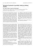
conjugated diene formation, as measured by the increase
in absorbance at 234 nm, was observed when CRLPs or
pCRLPs were incubated at 30 °C for periods of up to
420 min, but on incubation with CuSO
4
(50 l
M
), CRLPs
showed a marked rise in diene formation, which was not
seen with pCRLPs (Fig. 1).
Effect of CRLPs containing probucol on lipid
accumulation in THP-1 macrophages
Exposure of THP-1 macrophages to either CRLPs or
pCRLPs caused a considerable increase in the total lipid
accumulated in the cells after 48 h (Table 3). Comparison of
the effects of the two types of particles, however, showed
that pCRLPs had a markedly greater effect. The total lipid
contentinpCRLP-treatedcellswas221%thatofCRLP-
treated macrophages (Table 3), and this was a result of
increases in both triacylglycerol (+132%) and total choles-
terol (+73%) levels. Cholesteryl ester levels, however, were
not significantly changed; thus the increase in the total
cholesterol fraction was entirely the result of higher
cholesterol concentrations (+84%) (Table 3).
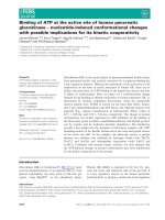
Uptake of CRLPs and pCRLPs by THP-1 macrophages
THP-1 macrophages were incubated with DiI-labelled
CRLPs or pCRLPs for periods between 1 and 24 h, and
the cells were then viewed by confocal microscopy. The
amount of fluorescence associated with the cells increased
markedly, with time, in experiments with both types of
particles, but there was clearly more in pCRLP-treated
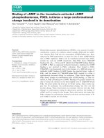
macrophages, even at early time-points (Fig. 2). Quantifi-
cation of the cell-associated fluorescence (Fig. 3) confirmed
that the rate of uptake of pCRLPs was considerably higher
than that of CRLPs.
Table 2. Lipid and thiobarbituric acid reactive substances (TBARS)
content of chylomicron remnant-like particles (CRLPs). Standard
CRLPs, and CRLPs containing probucol (pCRLPs), were prepared as
described in the Materials and methods and the triacylglycerol (TG),
total cholesterol (TC), phospholipid (PL) and TBARS levels were
determined. Data shown represent the mean ± SEM from eight
preparations. MDA, malondialdehyde.
Parameter CRLPs pCRLPs
TC (lmolÆmL
)1
) 0.97 ± 0.23 0.93 ± 0.19
TG (lmolÆmL
)1
) 3.74 ± 0.88 3.93 ± 0.76
PL (lmolÆmL
)1
) 0.53 ± 0.12 0.61 ± 0.11
TG : TC 3.98 ± 0.23 4.33 ± 0.29
TBARS (nmol of MDA
per lmol of TG)
2.54 ± 1.1 0.65 ± 0.57
a
a
P < 0.01 vs. CRLPs (Student’s t-test).
Fig. 1. Conjugated diene formation after incubation of CRLPs or
pCRLPs in the presence or absence of CuSO
4
. Chylomicron remnant-
like particles (CRLPs) or CRLPs containing probucol (pCRLPs)
(50 nmolÆmL
21
triacylglycerol) were incubated in the presence or
absence of CuSO
4
(50 m
M
)at30°C and the absorbance at 234 nm
was measured, at the intervals indicated, for 420 min. n,CRLPs;m,
CRLPs + CuSO
4
; s,pCRLPs;d,pCRLPs+CuSO
4
.Dataare
expressed as the change in absorbance with time. Each point represents
the mean of four experiments performed with separate CRLP prepa-
rations, and error bars show the SEM.
2420 E. H. Moore et al. (Eur. J. Biochem. 271) Ó FEBS 2004
Effect of pCRLPs on lipid synthesis in THP-1 macrophages
The effects of CRLPs and pCRLPs on lipid synthesis in
THP-1 macrophages were investigated by measuring the
incorporation of [
3
H]oleate into triacylglycerol, diacylglyc-
erol, phospholipid and cholesteryl ester after incubation of
the cells with the particles for 48 h (Table 4). The amount of
triacylglycerol formed from [
3
H]oleate was increased by
50% in macrophages exposed to pCRLPs as compared to
CRLPs, while the radioactivity found in diacylglycerol
was decreased by 40%. Incorporation of [
3
H]oleate into
phospholipid and cholesteryl ester, and the amount of
radioactivity found in nonesterified fatty acids, however,
was not significantly different in experiments with the two
types of particles.
Similar experiments using [
3
H]glycerol (n ¼ 4) also
showed that triacylglycerol formation was increased in
macrophages treated with pCRLPs (12.02 ± 1.95 pmol of
triacylglycerol formed per minute per mg of cell protein) as
compared to CRLPs (9.79 ± 0.94 pmol of triacylglycerol
formed per minute per mg of cell protein, P < 0.05).
Effect of CRLPs containing probucol or lycopene
on mRNA expression in THP-1 macrophages
THP-1 macrophages were incubated with CRLPs or
pCRLPs for 48 h and the levels of mRNA for nine genes
believed to play a role in foam cell formation was measured
by RT-PCR. The results are shown in Table 5. The relative
abundance of transcripts for LDLr, CD36, ACAT1 and
ABCA1 was not significantly changed by either type of
particle. There was a significant decrease, however, in
mRNA levels for the LRP ()71 to )78%), SR-A ()33%)
and SR-B1 ()20 to )34%) in experiments with both CRLPs
and pCRLPs. In addition, DGAT1 mRNA concentrations
were increased by both types of lipoproteins, but because of
the large variation in individual samples, these changes did
not reach significance. However, all six values from CRLP-
treated cells (three for CRLPs and three for pCRLPs) were
higher than those from control cells incubated without
lipoproteins, with the increases ranging from 35 to 350%.
No differences were observed between the effects of CRLPs
and pCRLPs on any of the genes investigated.
We have reported the influence of CRLPs containing
lycopene (lycCRLPs) on the expression of mRNA for
LDLr, LRP, ACAT1 and DGAT1 in THP-1 macrophages
in a previous study [35]. In order to compare the effects of
pCRLPs and lycCRLPs on all the genes tested in the present
work, we therefore measured the effects of lycCRLPs on
levels of mRNA for SR-A, CD36, SR-B1, adipophilin
and ABCA1. The results were generally similar to those
obtained with pCRLPs (Table 6), with mRNA levels for
CD36 and ABCA1 levels not significantly changed, and
decreases of a similar extent observed in those for SR-A
()39%) and SR-B1 ()21%), although, in this case, the
change in SR-B1 mRNA did not reach significance.
Discussion
The aim of this study was to investigate the effects of
lipophilic antioxidants carried in chylomicron remnants on
the induction of lipid accumulation in macrophages.
However, as it is difficult to obtain chylomicron remnants
from human blood uncontaminated with lipoproteins of
similar density, such as chylomicrons and very-low-density
lipoprotein (VLDL), it was necessary to use model CRLPs
to mimic the action of the remnant lipoproteins. The size,
density and lipid composition of the CRLPs used was
similar to that of physiological remnants [40,41], and they
also contained human apoE. Extensive previous studies in
both humans and experimental animals have shown that
model particles of this type are cleared from the blood and
metabolized in a similar way to the corresponding physio-
logical lipoproteins [42–45], and CRLPs containing apoE
from the appropriate species have also been found to have
effects which mimic those of physiological remnants in rat
hepatocytes and pig endothelial cells [40,46–48]. As lipo-
philic antioxidants can easily be incorporated into the model
particles, CRLPs provide a suitable and convenient model
for our experiments.
In previous work, we have demonstrated that CRLPs
containing lycopene, a carotenoid with antioxidant proper-
ties [6], markedly enhance lipid accumulation in THP-1
cells, suggesting that dietary antioxidants carried in chylo-
micron remnants may promote, rather than inhibit,
macrophage foam cell formation [35]. In order to test the
hypothesis that this effect was caused by the protection of
the remnants from oxidation, rather than a specific effect of
lycopene, and to further investigate the mechanisms
involved, we used CRLPs containing probucol, a phenolic
antioxidant lipophilic drug that is structurally unrelated to
lycopene [36]. Probucol has been used extensively to study
the effects of antioxidants on atherosclerosis [49], and has
been shown to bind to VLDL and LDL, and to provide the
particles with greater antioxidant protection than vitamin E
and many other antioxidants [50]. The concentration of
Table 3. Effect of chylomicron remnant-like particles (CRLPs) and CRLPs containing probucol (pCRLPs) on the cholesterol (C), cholesteryl ester
(CE) and triacylglycerol (TG) content of THP-1 macrophages. THP-1 macrophages were incubated with CRLPs or pCRLPs (30 lgÆmL
)1
of total
cholesterol) for 48 h and the TG, C, CE, and total cholesterol (TC) (C + CE) content of the cells was determined. Data are expressed as nmolÆmg
)1
of cell protein and represent the mean ± SEM from five separate experiments.
Lipid Control CRLPs pCRLPs pCRLPs/CRLPs (%)
Total lipid (TG + TC) 164.4 ± 38.3 556.7 ± 75.0 1291.5 ± 311.2
a
221.1 ± 29.7
TG 90.6 ± 22.7 453.7 ± 60.6 1117.4 ± 308.5
a
232.4 ± 42.5
TC 73.8 ± 15.9 103.0 ± 17.0 174.1 ± 33.3
a
172.9 ± 27.3
C 72.0 ± 16.4 83.6 ± 12.2 150.6 ± 25.3
a
183.7 ± 24.6
CE 1.8 ± 1.0 19.4 ± 5.5 23.5 ± 10.7 111.1 ± 52.8
a
P<0.05 vs. CRLPs (Student’s paired t-test).
Ó FEBS 2004 Foam cell induction by chylomicron remnants (Eur. J. Biochem. 271) 2421
Fig. 2. THP-1 macrophages were incubated with 1,1¢-dioctadecyl-3,3,3¢3¢-tetramethylindo-carbocyanine perchlorate (DiI)-labelled chylomicron rem-
nant-like particles (CRLPs) or pCRLPs (30 lgÆmL
21
cholesterol) for 1 h (A, CRLPs; B, pCRLPs), 6 h (C, CRLPs; D, pCRLPs) or 24 h (E, CRLPs;
F, pCRLPs), and viewed by confocal microscopy. In each set of three panels, the top left shows the fluorescence, the top right the cells, and the bottom
left the two merged. Images shown are from a typical experiment of three performed.
2422 E. H. Moore et al. (Eur. J. Biochem. 271) Ó FEBS 2004
probucol added to the macrophages (not exceeding 10 l
M
)
in our experiments was comparable to [32,34,51] or lower
than [31,33,52,53] the levels used in previous work to study
the effects of the drug on foam cell formation in vitro.
Furthermore, the pCRLPs were significantly more resistant
to oxidation than CRLPs, as indicated by the lower
concentration of TBARS in the particles (Table 2), the
markedly lower levels of MDA and 4-HNE after their
exposure to CuSO
4
, and their resistance to copper-induced
conjugated diene formation (Fig. 1).
In agreement with our previous work on physiological
chylomicron remnants and CRLPs [21–23], incubation of
macrophages with CRLPs caused a considerable increase in
intracellular total lipid accumulation without prior oxida-
tion of the particles (Table 3). When probucol was incor-
porated into the CRLPs, however, this effect was enhanced
by more than twofold, and this was caused by an increase in
triacylglycerol (·2.3) and cholesterol (·1.8) levels, while the
cholesteryl ester content was unaffected. These results are
strikingly similar to those obtained in our previous work
with CRLPs containing lycopene [35], and thus strongly
suggest that the protection of chylomicron remnants from
oxidation enhances, rather than inhibits, their induction of
lipid accumulation in macrophages.
The enhancement of lipid accumulation in macrophages
by CRLPs containing probucol and lycopene contrasts
sharply with the effects of antioxidants on the induction of
foam cell formation by acLDL or oxLDL. Yamamoto et al.
[31,52] have found that foam cell formation in response
to acLDL in the human cell lines UE-12 and THP-1 is
suppressed by probucol, when added to the medium either
in its free form or in acLDL prepared from patients given
the drug, and vitamin E has also been reported to decrease
the induction of lipid accumulation by oxLDL or acLDL in
HMDM and in J774 cells in both these conditions [25–27].
In other studies, however, no effect on the induction of foam
cell formation by acLDL or oxLDL in the presence of
probucol or vitamin E was detected [28,29,32], and two
investigations have shown an increase in macrophage
cholesteryl ester content after incubation with acLDL in
the presence of probucol [30,34]. Despite this inconsistency,
there have been no reports of antioxidants causing massive
increases in the induction of lipid accumulation in macro-
phages by acLDL or oxLDL comparable to those found in
the present study with CRLPs containing probucol or
lycopene. These findings suggest that the effects of dietary
lipophilic antioxidants on macrophages differ markedly,
depending on the lipoprotein in which they are carried, so
that during their transport from the gut to the liver in
chylomicron remnants they promote foam cell formation,
and their beneficial effects are only apparent after their
incorporation into LDL, as oxidation of these particles
greatly enhances their atherogenic effects [3].
One possible explanation for the raised lipid content of
macrophages treated with pCRLPs, as compared to
CRLPs, is that protection of the particles from oxidation
increases their uptake by the cells. We investigated this
hypothesis using CRLPs and pCRLPs labelled with the DiI
fluorescent probe, and the results clearly demonstrate that
the presence of probucol in CRLPs markedly increases their
rate of uptake by THP-1 macrophages (Figs 2 and 3). These
findings are again in contrast to those on the effects of
antioxidants on the uptake of acLDL or oxLDL, which has
been reported (in experiments using DiI fluorescence-
labelled or radiolabelled lipoproteins) to be decreased
after incubation of macrophages pretreated with or in the
presence of probucol [31,53], or vitamin E [25,26,53],
although Ku et al. [33] found no effect of probucol on
acLDL uptake in rabbit peritoneal macrophages.
The mechanisms mediating the uptake of chylomicron
remnants in macrophages are not yet clearly defined. The
LDLr appears to play a role, but other receptors (such as
the LRP) and various scavenger receptors (such as SR-A
and CD36) may also be involved [54]. The faster rate of
uptake of pCRLPs suggests that probucol may promote
interaction with the receptor protein, or possibly that
different receptors mediate their uptake. Investigation of the
effects of CRLPs and pCRLPs on the expression of mRNA
for the LDLr and the LRP in the present work showed that
both types of particles suppressed mRNA levels for the
LRP, but had no effect on those for the LDLr (Table 5),
and this is generally consistent with the results obtained in
our previous study with CRLPs containing lycopene [35]. In
addition, we found that the expression of mRNA for SR-A
wasdecreasedbybothpCRLPsandlycCRLPs,whilethat
for the class B scavenger receptor, CD36, was essentially
unaffected (Table 5). There were no significant differences,
however, in mRNA levels for any of the receptors tested in
macrophages treated with CRLPs, with or without anti-
oxidants. We conclude therefore that the increased rate of
uptake of pCRLPs, as compared to CRLPs, unequivocally
Fig. 3. THP-1 macrophages were incubated with 1,1¢-dioctadecyl-
3,3,3939-tetramethylindo-carbocyanine perchlorate (DiI)-labelled chyl-
omicron remnant-like particles (CRLPs) (m) or pCRLPs (d)
(30 lgÆmL
21
cholesterol) for the times indicated. The amount of
fluorescence associated with the cells was determined by optical vol-
ume density analysis and normalized for variations in the fluorescence
in different preparations using the fluorescence units per lmol of
cholesterol. Each point shows the mean of three separate experiments,
and error bars show the SEM. The difference between the two curves
was highly significant (P<0.001;
ANOVA
repeated measures).
Ó FEBS 2004 Foam cell induction by chylomicron remnants (Eur. J. Biochem. 271) 2423
demonstrated in the experiments with fluorescent-labelled
particles, is not caused by regulation of these proteins at the
transcriptional level.
Another mechanism by which antioxidants may influence
lipid accumulation in macrophages is by altering intracel-
lular lipid metabolism. Pretreatment of J774 macrophages
with vitamin E, as well as incorporation of vitamin E into
LDL, has been found to decrease cholesteryl ester synthesis
from radiolabelled oleate in the presence of oxLDL or
acLDL [25,27], although Asmis et al. [29] did not detect any
effect of the vitamin on cholesteryl ester formation in the
murine macrophage cell line, PD388D1. Takemura et al.
Table 4. Effect of chylomicron remnant-like particles (CRLPs) and CRLPs containing probucol (pCRLPs) on the incorporation of [
3
H]oleate into
lipids in THP-1 macrophages. THP-1 macrophages were incubated with CRLPs or pCRLPs (30 lgÆmL
)1
of cholesterol) for 48 h. The medium
containing lipoproteins was then removed, the cells were incubated with [
3
H]oleate (37 KBqÆmL
)1
) for 1 h and the incorporation of radioactivity
into triacylglycerol (TG), diacylglycerol (DG), cholesteryl ester (CE) and phospholipid (PL) during a 1 h incubation was determined. Data are
expressed as pmol lipid formed min
)1
Æmg
)1
of cell protein and represent the mean ± SEM from three separate experiments. NEFA, nonesterified
fatty acids.
Lipid CRLPs pCRLPs pCRLPs/CRLPs (%)
TG 8.73 ± 0.55 12.99 ± 0.61
b
149.1 ± 4.6
DG 2.35 ± 0.12 1.35 ± 0.08
a
58.4 ± 5.7
PL 14.54 ± 0.97 17.29 ± 4.13 116.4 ± 22.8
CE 0.84 ± 0.15 0.72 ± 0.02 92.7 ± 20.0
NEFA 1.67 ± 0.28 1.08 ± 0.34 87.6 ± 11.5
a
P<0.05,
b
P<0.01 vs. CRLPs (Student’s paired t-test).
Table 5. Effects of chylomicron remnant-like particles (CRLPs) and CRLPs containing probucol (pCRLPs) on mRNA levels for genes involved in foam
cell formation. THP-1 macrophages were incubated with or without CRLPs or pCRLPs (30 lgÆmL
)1
of cholesterol) for 48 h, and the levels of
mRNA for the genes indicated were determined by RT-PCR. The bands were quantified by absorbance (OD) analysis and the values were
normalized using those obtained for glyceraldehyde-3-phosphate dehydrogenase (GAPDH) in the same system. Data are expressed as OD units
and as the percentage of the values found with untreated (control) macrophages, and represent the mean ± SEM from three experiments. ABCA1,
ATP-binding cassette transporter A1; ACAT1, acyl coenzyme A : cholesterol acyltransferase 1; DGAT1, acyl coenzyme A : diacylglycerol acyl
transferase 1; LDLr, low-density lipoprotein receptor; LRP, low-density lipoprotein receptor-related protein; SR-A, scavenger receptor A; SR-B1,
scavenger receptor B1.
Gene
Control CRLPs pCRLPs
OD units OD units % Control OD units % Control
LDLr 0.41 ± 0.19 0.47 ± 0.16 168.1 ± 63.5 0.48 ± 0.25 86.3 ± 48.7
LRP 1.06 ± 0.19 0.33 ± 0.11 28.9 ± 6.1
a
0.25 ± 0.14 21.8 ± 10.2
b
SR-A 1.59 ± 0.44 1.12 ± 0.37 67.9 ± 5.9
a
1.17 ± 0.49 67.2 ± 11.9
a
CD36 1.75 ± 0.67 1.69 ± 0.54 101.3 ± 7.5 1.89 ± 0.45 122.6 ± 27.8
SR-B1 2.45 ± 0.23 1.55 ± 0.14 63.6 ± 2.2
b
1.94 ± 0.19 80.2 ± 7.3
a
ACAT1 0.43 ± 0.15 0.41 ± 0.06 126.2 ± 49.1 0.55 ± 0.08 151.3 ± 34.5
DGAT1 0.34 ± 0.11 0.86 ± 0.06 305.9 ± 87.1 0.90 ± 0.21 290.3 ± 85.1
ABCA1 1.31 ± 0.46 1.35 ± 0.35 108.5 ± 10.7 1.39 ± 0.60 101.5 ± 15.3
a
P < 0.05,
b
P<0.01 vs. control macrophages (
ANOVA
).
Table 6. Effects of chylomicron remnant-like particles (CRLPs) and CRLPs containing lycopene (lycCRLPs) on mRNA levels of genes involved in
foam cell formation. THP-1 macrophages were incubated with or without CRLPs or lycCRLPs (30 lgÆmL
)1
cholesterol) for 48 h, and mRNA levels
of the genes indicated were determined by RT-PCR. The bands were quantified by absorbance (OD) analysis and the values were normalized using
those obtained for glyceraldehyde-3-phosphate dehydrogenase (GAPDH) in the same system. Data are expressed as OD units and as the percentage
of the values found with untreated (control) macrophages, and represent the mean ± SEM from three experiments. ABCA1, ATP-binding cassette
transporter A1; SR-A, scavenger receptor A, SR-B1, scavenger receptor B1.
Gene
Control CRLPs pCRLPs
OD units OD units % Control OD units % Control
SR-A 1.61 ± 0.18 1.03 ± 0.27 61.6 ± 9.4
a
1.01 ± 0.25 61.3 ± 11.5
a
CD36 2.19 ± 0.13 1.35 ± 0.19 61.6 ± 8.6 2.13 ± 0.53 96.5 ± 23.4
SR-B1 2.78 ± 0.23 2.05 ± 0.24 73.2 ± 3.0 2.15 ± 0.58 79.4 ± 25.8
ABCA1 1.74 ± 0.20 1.01 ± 0.19 59.1 ± 11.6 1.32 ± 0.33 80.6 ± 27.6
a
P < 0.05, vs. control macrophages (
ANOVA
).
2424 E. H. Moore et al. (Eur. J. Biochem. 271) Ó FEBS 2004
[34], on the other hand, have reported increased cholesteryl
ester production and the activity of ACAT, the enzyme
responsible for cholesterol esterification, in mouse perito-
neal macrophages exposed to probucol in the presence and
absence of acLDL. In our experiments, the presence of
probucol in CRLPs did not change the rate of formation
of cholesteryl ester in THP-1 macrophages (Table 4). In
addition, there was no significant effect on the expression of
mRNA for ACAT1, the isoform of the enzyme found in
macrophages (Table 5), and this is consistent with our
previous findings with CRLPs containing lycopene [35].
In contrast to the lack of any effect of pCRLPs, as
compared to CRLPs, on cholesteryl ester synthesis in
macrophages, triacylglycerol synthesis was increased signi-
ficantly, as demonstrated in experiments with both [
3
H]ole-
ate (Table 5) and [
3
H]glycerol. The accompanying decrease
in the amount of radioactivity from [
3
H]oleate found in
diacylglycerol suggests that the activity of DGAT1, the
enzyme which controls the final and only committed step in
triacylglycerol synthesis in macrophages, using diacylglyc-
erol as the substrate [55], may be increased. The expression
of DGAT1 mRNA, however, was raised by both pCRLPs
and CRLPs, suggesting that any effect of probucol occurs at
a post-transcriptional level. The increase in DGAT1 mRNA
levels found here differs from our previous study, where we
found that CRLPs containing lycopene caused a significant
decrease, which was not observed with control CRLPs [35].
This is the only major difference we have found, to date,
between the effects of CRLPs containing probucol or
lycopene, and thus may be related to specific effects of the
molecules, rather than their antioxidant properties. In
general, our findings on intracellular lipid synthesis show
that increased triacylglycerol synthesis, but not cholesteryl
ester formation, contributes to the enhancement of lipid
accumulation by antioxidants carried in chylomicron
remnants.
Cholesterol efflux from macrophages is mediated by the
ABCA1 [56], and the scavenger receptor SR-B1, which
binds the high-density lipoprotein cholesterol acceptor [57].
The expression of mRNA for ABCA1 was not changed by
CRLPs or CRLPs containing probucol or lycopene, while
that for the SR-B1 was decreased by all three types of
particles to a similar extent (Tables 5 and 6). We found no
evidence therefore that antioxidants carried in chylomicron
remnants influence the transcription of genes involved in the
efflux of cholesterol from macrophages.
Probucol has been used extensively to investigate the
effects of antioxidants on atherosclerosis development, and
the results generally have provided strong support for the
beneficial effects of such compounds. A number of studies,
however, have shown consistently that probucol promotes
atherogenesis in apoE- or LDLr-deficient mice [49]. Our
findings, that lipid accumulation in macrophages is
enhanced by probucol carried in chylomicron remnants,
provide a possible explanation for this apparently para-
doxical effect, as both apoE and the LDLr have an
important role in the removal of chylomicron remnants
from the blood, and studies have demonstrated that
remnant levels in plasma are raised and clearance is delayed
in mice deficient in either of these two proteins [58–60].
In conclusion, the experiments reported here demonstrate
that antioxidants carried in chylomicron remnants enhance
lipid accumulation in macrophages, and that this is caused
by a markedly increased rate of uptake of the particles and
by a raised intracellular synthesis of triacylglycerol, but not
of cholesteryl ester. Furthermore, the effect does not appear
to be caused by changes in the transcription of genes
involved in the regulation of the uptake of the lipoprotein
particles, cholesteryl ester or triacylglycerol synthesis, or the
efflux of cholesterol from the cells. These findings suggest
that the type of lipoprotein carrier of dietary antioxidants is
crucial for their effects on macrophages. Thus, when they
are carried in LDL, oxidation and the subsequent detri-
mental effects of the particles are inhibited, but lipid
accumulation is promoted during their transport postpran-
dially in chylomicron remnants. This may be particularly
important in conditions where the clearance of remnants
from the circulation is delayed, and may also provide part of
the explanation for the failure to demonstrate beneficial
effects of dietary lipophilic antioxidants in large-scale
intervention studies [11].
Acknowledgements
This work was supported by grants from the Istituto Superiore di
Sanita
`
(ISS Art.524; fasc 2147/RI and C3BP). E. H. M. and F. B. were
supported by BBSRC CASE studentships sponsored by Glaxo
SmithKline.
References
1. Ross, R. (1993) The pathogenesis of atherosclerosis: a perspective
for the 1990s. Nature 362, 801–809.
2. Libby, P., Geng, Y.J., Aikawa, M., Schoenbeck, U., Mach, F.,
Clinton, S.K., Sukhova, G.K. & Lee, R.T. (1996) Macrophages
and atherosclerotic plaque stability. Curr. Opin. Lipidol. 7,
330–335.
3. Steinberg, D. (1997) Low density lipoprotein oxidation and its
pathobiological significance. J. Biol. Chem. 272, 20963–20966.
4. Trichopoulou, A. & Vasilopoulou, E. (2000) Mediterranean diet
and longevity. Br. J. Nutr. 84, 205–209.
5. de Lorgeril, M. (1998) Mediterranean diet in the prevention of
coronary heart disease. Nutrition 14, 55–57.
6. Rao, A.V. (2002) Lycopene, tomatoes and the prevention of
coronary heart disease. Exp. Biol. Med. 227, 908–913.
7. Kohlmeier, L., Kark, J.D., Gomez-Garcia, E., Matin, B.C., Steck,
S.E., Kardinaal, A.F.M., Ringstad, J., Thamm, M., Masaev, V.,
Riemersma, R., Martin-Moreno, J.M., Huttunen, J.K. & Kok, F.
(1997) Lycopene and myocardial infarction risk in the EURAMIC
study. Am. J. Epidemiol. 146, 618–626.
8. Agarwal, S. & Rao, A.V. (2000) Carotenoids and chronic diseases.
Drug Metab. Drug Interact. 17, 189–209.
9. Virtamo, J., Rapola, J.M., Ripatti, S., Heinonen, O.P., Taylor,
P.R., Albanes, D. & Huttenen, O.P. (1998) Effect of vitamin E and
beta-carotene on the incidence of primary nonfatal myocardial
infarction and fatal coronary heart disease. Arch. Intern. Med. 158,
668–675.
10. Stephens, N.G., Parsons, A., Schodiel, P.M., Kelly, F., Cheese-
man, K. & Mitchison, M.J. (1996) Radomised controlled trial of
vitamin E in patients with coronary disease: Cambridge heart
antioxidant study (CHAOS). Lancet 347, 781–786.
11. Clarke, R. & Armitage, J. (2002) Antioxidant vitamins and risk of
cardiovascular disease. Review of large scale randomised trials.
Cardiovasc. Drugs Ther. 16, 411–415.
12. Redgrave, T.G. (1983) Formation and metabolism of chylo-
microns. Int. Rev. Physiol. 28, 103–130.
Ó FEBS 2004 Foam cell induction by chylomicron remnants (Eur. J. Biochem. 271) 2425
13. Mamo, J.C., Proctor, S.D. & Smith, D. (1998) Retention of chy-
lomicron remnants by arterial tissue; importance of an efficient
clearance mechanism from plasma. Atherosclerosis 141,S63–
S69.
14. Proctor, S.D. & Mamo, J.C.L. (1998) Retention of fluorescent-
labelled chylomicron remnants within the intima of the artery wall
– evidence that plaque cholesterol may be derived from post-
prandial lipoproteins. EurJ.Clin.Invest.28, 497–503.
15. Grieve, D.J., Avella, M.A., Elliott, J. & Botham, K.M. (1998)
Influence of chylomicron remnants on endothelial cell function in
the isolated perfused rat aorta. Atherosclerosis 139, 273–281.
16. Mamo, J.C.L. & Wheeler, J.R. (1994) Chylomicrons or their
remnants penetrate rabbit thoracic aorta as efficiently as do
smaller macromolecules, including low density lipoprotein, high
density lipoprotein and albumin. CoronaryArteryDis.5, 695–705.
17. Yla-Herttuala, S., Jaakkola, O., Enholm, C., Tikkanen, M.J.,
Solakivi, T., Sarkioja, T. & Nikkari, T. (1988) Characterisation of
two lipoproteins containing apolipoproteins B and E from lesion-
free human aortic intima. J. Lipid Res. 29, 563–572.
18. Rapp, J.H., Lespine, A., Hamilton, R.L., Colyvas, N., Chaume-
ton, A.H., Tweedie-Harman, J., Kotite, L., Kunitake, S.T., Havel,
R.J. & Kane, J.P. (1994) Triglyceride-rich lipoproteins isolated
by selective affinity anti-apolipoprotein B immunosorption
from human atherosclerotic plaque. Arterioscler. Thromb. 14,
1767–1774.
19. Benlian, P., De Gennes, P.L., Foubert, L., Zhang, H., Gagne, S.E.
& Hayden, M. (1996) Premature atherosclerosis in patients with
familial chylomicronemia caused by mutations in the lipoprotein
lipase gene. N Engl. J. Med. 335, 848–854.
20. Groot, P.H.E., van Stiphout, W.A.H., Krauss, X.H., Jansen, H.,
vanTol,A.,vanRamshorst,E.,Chin-On,S.,Hofmann,A.,
Cresswell, S.R. & Havekes, L. (1991) Postprandial lipoprotein
metabolism in normolipidemic men with and without coronary
heart disease. Arterioscler. Thromb. 11, 653–662.
21. YUK, C. & Mamo, J.C. (2000) Chylomicron remnant-induced
foam cell formation and cytotoxicity: a possible mechanism of cell
deathinatherosclerosis.Clin. Sci. (London) 98, 183–192.
22. Napolitano, M., Rivabene, R., Avella, M., Botham, K.M. &
Bravo, E. (2001) The internal redox balance of cells influences the
metabolism of lipids of dietary origin by J774 macrophages:
implications for foam cell formation. J. Vasc. Res. 38, 350–360.
23. Batt, K.V., Botham, K.M., Jackson, B. & Suckling, K.E. (2001)
Comparison of the effects of low density lipoprotein and chylo-
micron remnants on foam cell formation in the human monocytic
cell line THP-1. Atherosclerosis Suppl. 2, 109.
24. Napolitano, M., Avella, M., Botham. K.M. & Bravo, E. (2003)
Chylomicron remnant induction of lipid accumulation in J774
macrophages is associated with up-regulation of triacylglycerol
synthesis which is not dependent on oxidation of the particles.
Biochim. Biophys. Acta 1631, 255–264.
25. Shige, H., Ishikawa, T., Suzukawa, M., Nishiwaki, M., Yamash-
ita, T., Nakajima, K., Ito, T., Higashi, K., Ayaori, M., Yonemura,
A., Nestel, P. & Nakamura, H. (1998) Vitamin E reduces cho-
lesterol esterification and uptake of acetylated low density lipo-
protein in macrophages. Lipids 33, 1169–1175.
26. Devaraj, S., Hugou, I. & Jialal, I. (2001) Alpha tocopherol
decreases CD36 expression in human monocyte-derived macro-
phages. J. Lipid Res. 42, 521–527.
27. Suzukawa, M., Abbey, M., Clifton, P. & Nestel, P.J. (1994) Effects
of supplementing with vitamin E on the uptake of low density
lipoprotein and the stimulation of cholesteryl ester formation in
macrophages. Atherosclerosis 110, 77–86.
28. Asmis, R. & Jelk, J. (2000) Vitamin E supplementation of human
macrophages prevents neither foam cell formation nor increased
susceptibility of foam cells to lysis by oxidized LDL. Arterioscler.
Thromb. Vasc. Biol. 20, 2078–2086.
29. Asmis, R., Llorente, V.C. & Gey, K.F. (1995) Prevention of
cholesteryl ester accumulation in P388D1 macrophage-like cells by
increased cellular vitamin E depends on species of extracellular
cholesterol. Conventional heterologous non-human cell cultures
are poor models of human atherosclerotic foam cell formation.
Eur. J. Biochem. 233, 171–178.
30. Trach. C.C., Wulfroth. P.M., Severs, N.J. & Robenek, H. (1996)
Influence of native and modified lipoproteins on migration of
mouse peritoneal macrophages and the effect of the antioxidants
vitamin E and probucol. Eur. J. Cell Biol. 71, 199–205.
31. Yamamoto, A., Hara, H., Takaichi, S., Wakasugi, S. & Tomi-
kawa, M. (1988) Effect of probucol on macrophages, leading to
regression of xanthomas and atheromatous vascular lesions.
Am. J. Cardiol. 62, 31B–36B.
32. Nagano,Y.,Kita,T.,Yokode,M.,Ishii,K.,Kume,N.,Otani,H.,
Arai, H. & Kawai, C. (1989) Probucol does not affect lipoprotein
metabolism in macrophages of Watanabe heritable hyperlipidemic
rabbits. Arteriosclerosis 9, 453–461.
33. Ku, G., Schroeder, K., Schmidt, L.F., Jackson, R.L. & Doherty,
N.S. (1990) Probucol does not alter acetylated low density lipo-
protein uptake by murine peritoneal macrophages. Atherosclerosis
80, 191–197.
34. Takemura, T., Sakai, M., Matsuda, H., Matsumura, T., Biwa, T.,
Anami, Y., Nishikawa, T., Sasahara, T. & Shichiri, M. (2000)
Effects of probucol on cholesterol metabolism in mouse peritoneal
macrophages: inhibition of HDL mediated cholesterol efflux.
Atherosclerosis 152, 347–357.
35. Moore, E.H., Napolitano, M., Prosperi, A., Avella, M., Suckling,
K.E.,Bravo,E.&Botham,K.M.(2003)Incorporationoflyco-
pene into chylomicron remnant-like particles enhances their
induction of lipid accumulation in macrophages. Biochem.
Biophys. Res. Commun. 312, 1216–1219.
36. Niguchi, N. & Niki, E. (2000) Phenolic antioxidants: a rationale
for design and evaluation of novel antioxidant drugs for athero-
sclerosis. Free Rad. Biol. Med. 28, 1538–1546.
37. Napolitano, M., Batt, K.V., Avella, M., Bravo, E. & Botham,
K.M. (2001) Lipid synthesis in macrophages derived from the
human cell line THP-1: modulation of the effects of native and
oxidised chylomicron remnant-like particles by estrogen. Clin. Sci.
(London) 101, 403–413.
38. Davis, R.A., Engelhorn, S.C., Pangburn, S.H., Weinstein, D.B. &
Steinberg, D. (1979) Very low density lipoprotein synthesis and
secretion by cultured rat hepatocytes. J. Biol. Chem. 254, 2010–
2016.
39. Steinbrecher, U.P., Parthasarathy, S., Leake, D.S., Witzum, J.L.
& Steinberg, D. (1984) Modification of low density lipoprotein by
endothelial cells involves lipid peroxidation and degradation of
low density lipoprotein phospholipids. Proc. Natl Acad. Sci. USA
81, 3883–3722.
40. Diard, P., Malewiak, M I., Lagrange, D. & Griglio, S. (1994)
Hepatic lipase may act as a ligand in the uptake of artificial
chylomicron remnant-like particles by isolated rat hepatocytes.
Biochem. J. 299, 889–894.
41. Redgrave, T.G., Fidge, N.H. & Yin, J. (1982) Specific, saturable
binding and uptake of rat chylomicron remnants by rat skin
fibroblasts. J. Lipid Res. 23, 638–644.
42. Redgrave, T.G., Ly, H.L., Quintao, C.R., Ramberg, C.F. &
Boston, R. (1993) Clearance from plasma of triacylglycerol and
cholesteryl ester after intravenous injection of chylomicron-like
lipid emulsions in rats and man. Biochem. J. 290, 843–847.
43. Maranhao, R.C., Fers, M.C., Martins, M.T., Mesquita, C.H.,
Toffoletto, O., Vinagre, C.G.C., Gianinni, S.D. & Pileggi, F.
(1996) Plasma kinetics of a chylomicron-like emulsion in patients
with coronary artery disease. Atherosclerosis 126, 15–25.
44. Oliveira, H.C., Hirata, M.H., Redgrave, T.G. & Maranhao, R.C.
(1988) Competition between chylomicrons and their remnants for
2426 E. H. Moore et al. (Eur. J. Biochem. 271) Ó FEBS 2004
plasma removal: a study with artificial emulsion models of chy-
lomicrons. Biochim. Biophys. Acta 958, 211–217.
45. Martins, I.J., Vermeulen, R. & Redgrave, T.G. (2002) Relative
roles of mitochondrial and peroxisomal fatty acid oxidation in the
metabolism of chylomicron remnants in rats and mice as assessed
by a stable-isotope breath test. Atherosclerosis 150, 13–20.
46. Sultan, F., Lagrange, D., Le Liepvre, X. & Griglio, S. (1989)
Chylomicron remnant uptake by freshly isolated hepatocytes.
Effects of heparin and of hepatic triacylglycerol lipase. Biochem. J.
258, 587–594.
47. Goulter, A.B., Avella, M.A., Elliott, J. & Botham, K.M. (2002)
Chylomicron remnant-like particles inhibit receptor-mediated
endothelium-dependent vasorelaxation in porcine coronary
arteries. Clin. Sci. (London) 103, 450–460.
48. Thuren, T., Sisson, P. & Waite, M. (1991) Hydrolysis of lipid
mixtures by rat hepatic lipase. Biochim. Biophys. Acta 1083,
217–220.
49. Stocker, R. (1999) Dietary and pharmacological antioxidants in
atherosclerosis. Curr. Opin. Lipidol. 10, 589–597.
50. Pfuetze, K.D. & Dujovne, C.A. (2000) Probucol. Curr. Ather-
oscler. Report 2, 47–57.
51. Liu, G.X., Ou, D.M., Liu, J.H., Huang, H.L. & Liao, D.F. (2000)
Probucol inhibits lipid peroxidation of macrophage and affects its
secretory properties. Acta Pharmacol. Sin. 21, 637–640.
52. Yamamoto, A., Takaichi, S., Hara, H., Nishikawa, O., Yoko-
yama, S., Yamamura, T. & Yamaguchi, T. (1986) Probucol
prevents lipid storage in macrophages. Atherosclerosis 62, 209–
217.
53. Selmer, D., Senekowitsch-Schmidtke, R., Schneider, W. & Elstner,
E.F. (1997) Binding and uptake of
125
iodine-labelled, oxidized low
density lipoprotein by macrophages: comparison of the effects of
alpha tocopherol, probucol, pyridoxal-5¢phosphate and magnes-
ium-pyridoxal-5¢-phosphate glutamate. Z. Naturforsch. [C] 52,
97–104.
54. Yu, K.C. & Cooper, A.D. (2001) Postprandial lipoproteins and
atherosclerosis. Front. Biosci. 6, D332–D354.
55. Cases, S., Smith, S.J., Zheng, Y.W., Myers, H.M., Lear, S.R.,
Sande, E., Novak, S., Collins, C., Welch, C.B., Lusis, A.J.,
Erickson, S.K. & Farese, R.V. (1998) Identification of a gene
encoding an acyl CoA: diacylglycerol acyltransferase, a key
enzyme in triacylglycerol synthesis. Proc. Natl Acad. Sci. USA 95,
13018–13023.
56. Oram, J.F. (2002) ATP-binding cassette transporter A1 and cho-
lesterol trafficking. Curr. Opin. Lipidol. 13, 373–381.
57. Chinetti, G., Gbaguidi, F.G., Griglio, S., Mallat, Z., Antonucci,
M., Poulain, P., Chapman, J., Fruchart, J C., Tedgui, A., Najib-
Fruchart, J. & Staels, B. (2000) CLA-1/SR-B1 is expressed in
atherogenic lesion macrophages and regulated by activators of
peroxisome proliferator-activated receptors. Circulation 101,
2411–2417.
58. Ishibashi, S., Perrey, S., Chen, Z., Shimada, M., Ohashi, K.,
Harada,K.,Yazaki,Y.&Yamada,N.(1996)Roleofthelow
density lipoprotein (LDL) receptor pathway in the metabolism of
chylomicron remnants. A quantitative study in knock-out mice
lacking the LDL receptor, apolipoprotein E, or both. J. Biol.
Chem. 271, 22422–22427.
59. Mortimer, B.C., Martins, I., Zeng, B.J. & Redgrave, T. (1997) Use
of gene manipulated models to study the physiology of lipid
transport. Clin. Exp. Pharmacol. Physiol. 24, 281–285.
60. Zsigmond, E., Fuke, Y., Li, L., Kobayashi, K. & Chan, L. (1998)
Resistance of chylomicron and VLDL remnants to post heparin
lipolysis in apoE deficient mice: the role of apoE in lipoprotein
lipase mediated lipolysis in vivo and in vitro. J. Lipid Res. 39,
1852–1861.
Ó FEBS 2004 Foam cell induction by chylomicron remnants (Eur. J. Biochem. 271) 2427