Báo cáo khoa học: Effect of valine 106 on structure–function relation of cytosolic human thymidine kinase Kinetic properties and oligomerization pattern of nine substitution mutants of V106 ppt
Bạn đang xem bản rút gọn của tài liệu. Xem và tải ngay bản đầy đủ của tài liệu tại đây (410.6 KB, 9 trang )
Effect of valine 106 on structure–function relation of cytosolic human
thymidine kinase
Kinetic properties and oligomerization pattern of nine substitution mutants of V106
Hanne Frederiksen*†, Dvora Berenstein† and Birgitte Munch-Petersen
Department of Life Sciences and Chemistry, Roskilde University, Denmark
Information on the regulation and structure–function rela-
tion of enzymes involved in DNA precursor synthesis is
pivotal, as defects in several of these enzymes have been
found to cause depletion or deletion of mitochondrial DNA
resulting in severe diseases. Here, the effect of amino acid 106
on the enzymatic properties of the cell-cycle-regulated
human cytosolic thymidine kinase 1 (TK1) is investigated.
On the basis of the previously observed profound differences
between recombinant TK1 with Val106 (V106WT) and
Met106 (V106M) in catalytic activity and oligomerization
pattern, we designed and characterized nine mutants of
amino acid 106 differing in size, conformation and polarity.
According to their oligomerization pattern and thymidine
kinetics, the TK1 mutants can be divided into two groups.
Group I (V106A, V106I and V106T) behaves like V106WT,
in that pre-assay exposure to ATP induces reversible
transition from a dimer with low catalytic activity to a tetr-
amer with high catalytic activity. Group II (V106G, V106H,
V106K, V106L and V106Q) behaves like V106M in that they
are permanently high activity tetramers, irrespective of ATP
exposure. We conclude that size and conformation of amino
acid 106 are more important than polarity for the catalytic
activity and oligomerization of TK1. The role of amino acid
106 and the sequence surrounding it for dimer–tetramer
transition was confirmed by cloning the putative interface
fragment of human TK1 and investigating its oligomeri-
zation pattern.
Keywords: dimer–tetramer formation; enzyme kinetics;
enzyme mutants; structure–function relation; thymidine
kinase.
Enzymes involved in salvage and metabolism of deoxy-
nucleosides have an important role in the regulation of
DNA precursors for DNA synthesis and repair. Recently,
severe syndromes, such as mitochondrial neurogastrointes-
tinal encephalomyopathy and mitochondrial DNA deple-
tion syndrome which lead to multiple mitochondrial DNA
abnormalities, were found to be caused by defects in the
cytoplasmic thymidine phosphorylase [1,2] or the two
mitochondrial deoxynucleoside kinases: deoxyguanosine
kinase (dGK) and thymidine kinase 2 (TK2) respectively
[3,4]. In contrast with earlier work suggesting spatial and
metabolic separation of thymidine phosphate pools between
the cytosol and mitochondria [5,6], recent evidence suggests
the two compartments are connected by a rapid and
dynamic exchange [7]. These findings may explain why
defects in deoxynucleotide metabolic enzymes, mitochond-
rial as well as cytoplasmic, lead to severe mitochondrial
DNA abnormality syndromes. Therefore, it is of great
importance to acquire detailed knowledge about the prop-
erties of the enzymes involved in balancing the cellular and
mitochondrial dNTP pools.
Human cytosolic thymidine kinase (TK1; EC 2.7.1.21) is
a salvage pathway enzyme in the synthesis of the DNA
precursor dTTP. It catalyzes the first step of this pathway, in
which thymidine is phosphorylated to dTMP [8]. In turn,
intracellular dTMP is rapidly phosphorylated to dTTP, an
allosteric effector of ribonucleotide reductase [9]. Imbal-
ances in the dTTP pool are thus followed by an imbalanced
supply of the four deoxyribonucleoside triphosphates for
DNA synthesis and repair, and result in increased rates of
mutation and the probability of carcinogenesis [10]. TK1 is
cell-cycle regulated and its activity fluctuates with DNA
synthesis [11,12]. The subunit size of TK1 is 24 kDa [13,14],
and the native enzymes purified from human lymphocytes
[14] and HeLa cells [15] were found to be tetramers in the
presence of ATP. In the presence of thymidine instead of
Correspondence to B. Munch-Petersen, Department of Life Sciences
and Chemistry, Roskilde University, PO Box 260, DK-4000 Roskilde,
Denmark. Fax: + 45 46743011, Tel.: + 45 46742418,
E-mail:
Abbreviations: dCK, deoxycytidine kinase; dGK, deoxyguanosine
kinase; dNK, multisubstrate nucleoside kinase from Drosophila mel-
anogaster; GST, glutathione S-transferase; HSV1-TK, Herpes simplex
type-1 thymidine kinase; TK1, human cytosolic thymidine kinase;
rLy-TK1
Val106
, recombinant TK1 expressed from cDNA derived from
human lymphocytes, the same as rLy-TK1 (V106WT); rLy-TK1
66)136
,
the putative interface fragment of TK1 corresponding to residues
66–136; TK1+ATP, rLy-TK1 incubated and stored with 2.5 m
M
ATP/MgCl
2
;TK1)ATP, rLy-TK1 incubated and stored without
ATP/MgCl
2
; TK2, human mitochondrial thymidine kinase.
Enzyme: Human cytosolic thymidine kinase (TK1; EC, 2.7.1.21).
*Present address: Institute of Food and Veterinary Research,
Department of Toxicology and Risk Assessment, Mørkhøj Bygade 19,
DK-2860 Søborg, Denmark.
These authors contributed equally to this publication.
Note: A website is available at
(Received 1 March 2004, revised 6 April 2004,
accepted 16 April 2004)
Eur. J. Biochem. 271, 2248–2256 (2004) Ó FEBS 2004 doi:10.1111/j.1432-1033.2004.04166.x
ATP or without substrates present, TK1 appears as a dimer
[14]. Human TK1 has 234 amino acids, and, in the originally
published primary sequence, amino acid 106 was methio-
nine [16,17]. Our group has recently analysed TK1 cDNA
and genomic DNA from 22 normal or transformed cell
lines, and in all cases we found a valine at amino acid
position 106 [18]. Also, alignment of mammalian TK1 and
TK from vaccinia virus (Fig. 1) demonstrates the presence
of valine at the site corresponding to amino acid 106 in
human TK1, which is located in a highly conserved area
thought to encompass the magnesium-binding and thymi-
dine-binding sites [19,20]. We have found a remarkable
difference in catalytic activity between recombinant TK1
expressed from human lymphocyte cDNA, rLy-TK1
Val106
(V106WT) and its mutant rLy-TK1
Met106
(V106M) [18].
V106WT was a dimer with low catalytic activity (K
0.5
for
thymidine about 15 l
M
), but pre-assay exposure to ATP
induced an enzyme concentration-dependent reversible
transition from a dimer to a tetramer with an % 30-fold
higher catalytic activity (K
0.5
for thymidine % 0.5 l
M
)
[14,18,21]. The maximal velocities for the ATP exposed
and unexposed forms were the same. In contrast, irrespect-
ive of pre-assay exposure to ATP, V106M was a permanent
tetramer with low K
0.5
for thymidine (% 0.5 l
M
) and similar
maximal velocities, which were % 2–3-fold lower than that
of V106WT [18,21].
Until recently, the only deoxyribonucleoside kinase with
a known 3D structure solved by X-ray crystallography was
the Herpes simplex virus type-1 thymidine kinase (HSV1-
TK) [22–26]. In 2001, the X-ray crystallographic structure
was reported for two cellular deoxynucleoside kinases – the
Drosophila melanogaster multisubstrate deoxynucleoside
kinase (dNK) and the human deoxyguanosine kinase
(dGK [27]) – and in 2003 the X-ray crystallographic
structure of the human deoxycytidine kinase (dCK) was
solved [28]. The amino acid sequence identity is 34%
between dNK and dGK [29], and 47% between dGK and
dCK [28], and the structures of dNK, dGK and dCK
appeared to be very similar [27,28]. Despite the very low
sequence identity of the cellular kinases with the Herpes
virus TK (% 10%),thecorestructureshaveasimilarfold
and there is also a close resemblance to the human and yeast
thymidylate kinases [8,27]. Therefore, although the sequence
identity of TK1 with HSV1-TK and the other cellular
kinases belonging to the dNK group is too low for a reliable
homology model (% 10%), TK1 may have the same overall
structure as the other nucleoside kinases. Furthermore, a
prediction of the secondary structure of TK1 [30–32] places
Val106 in the middle of an a-helix which aligns in
CLUSTAL
W
[33] with one of the interface helices (a-helix 4) of HSV1-
TK. This may indicate that the area surrounding Val106 is
integrated into the oligomerization interface.
To obtain more information about this putative interface
region of TK1, we sought to clarify the importance of amino
acid 106 for the structure and function of the enzyme by
mutating Val106 to amino acids differing in polarity, size
and conformation, and subsequently investigating their
effect on the quaternary structure and kinetics. Further-
more, we confirmed that amino acid 106 and the neigh-
bouring residues are involved in dimer–tetramer transition
by cloning the putative interface fragment of human TK1,
rLy-TK1
66)136
, and investigating the influence of the
V106M mutation on the oligomerization properties of this
fragment.
Materials and methods
Bacterial strains and plasmids
The thymidine kinase-deficient strain of Escherichia coli,
KY895 [34], and E.colistrain BL21 were used to propagate
bacterial plasmids. BL21 was used for expression of
recombinant TK1 enzymes. We have previously cloned
the entire TK1 coding sequence into the BamHI–EcoRI
restriction sites of the glutathione S-transferase (GST)
fusion vector pGEX-2T, as described in [18]. This vector
encodes a thrombin cleavage site between the GST gene and
the multiple cloning site.
Construction of pGEX-2T-LyTK1
Val106X
mutants
The plasmid pGEX-2T-LyTK1
Val106
[18] was used as
template DNA for PCR, and mutations in the GTG codon
coding for Val106 were introduced with the Quick
Change
TM
site-directed mutagenesis kit from Stratagene
(according to the instructions of the manufacturer). The
sense [5¢-TTTTTCCCTGACATCGTGGAGTTCTGCGA
GGCC(358–390)-3¢] and antisense [5¢-GGCCTCGCAGA
ACTCCACGATGTCAGGGAAAAA(390–358)-3¢]muta-
genic primers were substituted as follows in the target codon
for Val106 (bold): G
CG/CGC for Ala106, CAG/CTG for
Gln106, G
GT/ACC for Gly106, CAC/GTG for His106,
ATC/GAT for Ile106, CTG/CAG for Leu106, AAA/TTT
for Lys106,
ATG/CAT for Met106 and ACC/GGT for
Thr106. The altered bases are underlined and the codons are
given in the sense/antisense primer, respectively. The base
Fig. 1. Amino-acid sequence alignment of the putative interface region of human TK1 with related enzymes. Val106 is in bold and italics, the putative
Mg
2+
-binding motif VIGID
97
[19,20] and the putative thymidine-binding motif FQRK
131
[20] are in italics in all sequences. In the human sequence,
the b-branching amino acids and the a-helix breaking glycines and prolines are in bold and underlined. The sequences have the following GenBank
identifier numbers: gi/23503074, human; gi/6678357, mouse; gi/125428, Chinese hamster; gi/125427, chicken; gi/9791018, vaccinia virus. Identical
amino acids are indicated by asterisks.
Ó FEBS 2004 Effect of amino acid 106 on human TK1 (Eur. J. Biochem. 271) 2249
numbering is as described in [16] where the translation
initiation is at position 58, and therefore the codon for
amino acid 106 starts at base number 373.
Construction of pGEX-2T-LyTK1
66)136(Val106)
Plasmid pGEX-2T-LyTK1
Val106
[18]wasusedastemplate
for PCR with a sense primer: 5¢- GGG
GGATCCTGCA
CACATGACCGGAACACC(247–273)-3¢ designed to
contain a GGG overhang and an antisense primer:
5¢-CGGCACC
GAATTCTAGATGGCCCCAAATGGC
TTCCT(480–445)-3¢. The numbering is as described in [16].
The underlined bases are changed in comparison with the
original sequence to introduce a BamHI site (in bold) and
the coding sequence for thrombin cleavage in the sense
primer, and an EcoRI site (in bold) in the antisense primer.
Thus, the N-terminal amino acids of the expressed fragment
will be GS
66
CTHD instead of
66
CTHD. The PCR condi-
tions were: 4 lgÆmL
)1
template DNA, 3 m
M
MgCl
2
,
0.2 m
M
each dNTP and 0.36 l
M
each primer in 10 m
M
Tris/HCl buffer (pH 8.3) and 1 unit of Thermus aquaticus
DNA polymerase (Stratagene) in a total volume of 25 lL;
30 cycles; 1 min at 94 °C, 1 min at 55 °C,and1minat
72 °C. The purified PCR product was ligated into the
BamH1–EcoR1 restriction sites of the pGEX-2T vector and
transformed into competent E.colicells. Codon CTG(466–
468) was mutated to a UAG stop signal by site-directed
mutagenesis performed with the QuickChange
TM
site-
directed mutagenesis kit according to the manufacturer’s
instructions. The following mutagenic primers were used:
sense, 5¢-CCATTTGGGGCCATC
TAGAACCTGGTGC
CGCTG(451–483)-3¢;antisense,5¢-CAGCGGCACCAG
GTTC
TAGATGGCCCCAAATGG(483–451)-3¢. The
underlined bases were changed in comparison with the
original sequence; the stop codon is in bold.
Construction of pGEX-2T-LyTK1
66)136(Met106)
GTG at positions 373–375 (bold), corresponding to amino
acid 106 (numbers as described in [16]), was mutated to
ATG with the QuickChange
TM
site-directed mutagenesis kit
with the following primers: sense primer, 5¢-CAGTTTT
TCCCTGACATC
ATGGAGTTCTGCGAGGCCATG
(355–393)-3¢; antisense primer, 5¢-CATGGCCTCGCAGA
ACTCCATGATGTCAGGGAAAAACTG(393–355)-3¢.
The changed bases are underlined.
DNA sequencing
pGEX-2T-LyTK1
Val106
, pGEX-2T-LyTK1
Val106X
mutant
plasmids and pGEX-2T-LyTK1
66)136(Met106)
plasmid were
sequenced on both strands using the Thermo Sequenase
sequencing kit (Amersham Biosciences), and pGEX-2T-
LyTK1
66)136(Val106)
plasmid was sequenced on both strands
with Sequenase
TM
version 2.0 DNA Sequencing Kit
(Amersham Biosciences).
Expression and purification of rLy-TK1 recombinant
enzymes and rLy-TK1
66)136
proteins
Expression and purification of the GST-TK1 fusion
proteins have been described in detail previously [18].
Briefly, induction was performed at 25 °C by the addition of
0.1 m
M
isopropyl thio-b-
D
-galactopyranoside, the bacterial
lysate was filtered and applied to a glutathione–Sepharose
4B column (Amersham Biosciences), and TK1 was cleaved
from the GST part with thrombin (Amersham Biosciences).
After addition of glycerol, dithiothreitol, MgCl
2
and Triton
X-100 to final concentrations of 10%, 5 m
M
,5 m
M
and 1%,
respectively, the thrombin cleavage fractions were stored at
)80 °C. The yield of enzyme protein from 300 mL bacterial
culture was 1–3 mg in the thrombin cleavage fractions, and
the purification fold, calculated as the ratio between the
specific activity in the pooled cleavage fractions and in
the crude bacterial extract, was % 20. The yield of rLy-
TK1
66)136
proteins in the cleavage fractions was 3–6 mg per
litre bacterial culture. The purity of the preparations was
estimated to be over 90% by SDS/PAGE (not shown).
ATP incubation and storage of the rLy-TK1 recombinant
enzymes for kinetic experiments and gel filtration
The thrombin cleavage fractions were diluted to 5 lgÆmL
)1
in dilution buffer A (50 m
M
Tris/HCl, pH 7.5, 5 m
M
MgCl
2
,0.1
M
KCl, 2 m
M
Chaps, 10% glycerol and 5 m
M
dithiothreitol) with and without 2.5 m
M
ATP, and incuba-
tedonicefor2hbeforestorageat)80 °C. The enzymes
incubated and stored with and without ATP are referred to
as the +ATP and –ATP forms, respectively.
Estimation of subunit molecular size of rLy-TK1
66)136
by tricine/ethylene glycol/SDS/PAGE
Because the standard SDS/PAGE methods resulted in
diffuse protein bands and insufficient resolution of the
relatively small rLy-TK1
66)136
peptide (< 8 kDa), a
method of Scha
¨
gger & von Jagow [35] modified according
to Separation Technique File no. 112 from Pharmacia (now
Amersham Biosciences) was used. The upper and lower gel
was made 4.5% and 13% with polyacrylamide, respectively,
and the gel buffer was 30% ethylene glycol/0.112
M
acetate/
0.112
M
Tris/HCl, pH 6.5. The electrode buffer consisted of
0.2
M
Tris, 0.2
M
tricine (instead of glycine) and 0.55%
SDS, pH 8.1. The Peptide marker kit, molecular mass
2512–16 949 Da, from Amersham Biosciences was used as
the molecular mass standard.
Native molecular size
The apparent molecular size of recombinant enzymes was
determined by gel filtration on a Superdex 200 column
(10 · 300 mm) connected to a Gradifrac automatic sampler
(Amersham Biosciences) as described previously [14,18].
A 200-lL portion of thrombin cleavage fraction stored at
)80 °C at a protein concentration of 5 lgÆmL
)1
was mixed
with 100 lL of the equilibration and elution buffer B
(50 m
M
imidazole/HCl, pH 7.5, 5 m
M
MgCl
2
,0.1
M
KCl,
2m
M
Chaps and 5 m
M
dithiothreitol), containing
0.17 mgÆmL
)1
Blue Dextran 2000 as internal marker for
determination of column void volume. Then 200 lLofthis
mixture (protein concentration 3.25 lgÆmL
)1
) was applied
to the column. The +ATP enzyme samples contained
2.5 m
M
ATP,andwereelutedinbufferBwith2.5 m
M
ATP.
Fractions of 200 lL were collected and mixed with 100 lL
2250 H. Frederiksen et al.(Eur. J. Biochem. 271) Ó FEBS 2004
buffer B containing 30% glycerol and 2 m
M
ATP for
enzyme stabilization, and assayed for thymidine kinase
activity at standard assay conditions with 100 l
M
thymidine.
The native molecular size of the rLy-TK1
66)136
proteins
was estimated on a Superose 12 column (10 · 300 mm)
connected to a Gradifrac automatic sampler (Amersham
Biosciences) as described previously [14,18]. Protein from
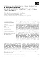
Fig. 2. Gel filtration of rLy-TK1(V106WT)
and rLy-TK1(V106X) enzymes. Approxi-
mately 0.65 lgproteinin200lLwasinjected
into a Superdex 200 column. (A) Dimeric
enzymes: V106WT, V106A, V106I, and
V106T; (B) tetrameric enzymes: V106G,
V106H, V106K, V106L, V106M, and V106Q.
The molecular mass markers (|) are (from left
to right): b-amylase (200 kDa), BSA
(66 kDa), ovalbumin (45 kDa), carbonic
anhydrase (29 kDa) and cytochrome
c (12.4 kDa). V
e
is the elution volume, and V
0
is the void volume estimated with blue dextran
2000. The horizontal bars indicate the range of
duplicate determinations.
Ó FEBS 2004 Effect of amino acid 106 on human TK1 (Eur. J. Biochem. 271) 2251
the thrombin cleavage fraction (200 lL; protein concentra-
tion 0.5 mgÆmL
)1
), containing 0.17 mgÆmL
)1
Blue Dextran
2000 as internal marker for determination of the column
void volume, was applied. The column was equilibrated and
eluted with buffer B without Chaps. Five hundred microliter
fractions were collected for estimation of protein concen-
tration by the method of Bradford [36].
Thymidine kinase assay
TK1 activity was assayed by measuring the initial velocities
using the DE-81 filter paper method as described previously
[14,18]. Standard assay conditions were 5 ngÆmL
)1
enzyme,
50 m
M
Tris/HCl, pH 7.5, 2.5 m
M
MgCl
2
,10m
M
dithio-
threitol, 2.5 m
M
ATP, 0.5 m
M
Chaps, 3 mgÆmL
)1
BSA,
3m
M
NaF and the indicated concentrations of
[methyl-
3
H]thymidine (Amersham Biosciences) in a final
volume of 50 lL. For each velocity, four time samples were
taken. The enzymes, stored without ATP at 5 lgÆmL
)1
,
were diluted immediately before the start of the reaction
with ice-cold enzyme dilution buffer C (50 m
M
Tris/HCl,
pH 7.5, 1 m
M
Chaps and 3 mgÆmL
)1
BSA). For dilution of
the TK1 +ATP form, 2.5 m
M
ATP and 2.5 m
M
MgCl
2
were included in the dilution buffer.
Enzyme kinetics
The kinetic parameters and the degree of co-operativity
were determined as previously described [18]. The experi-
mental data were fitted to the Hill equation
v ¼
VÁs
n
K
n
0:5
þ s
n
and the kinetic parameters determined with the nonlinear
regression software from Graphpad PrismÒ. V is the
maximal velocity, n is the Hill constant, and K
0.5
, like K
m
in the Michaelis–Menten equation, defines the substrate
concentration S where v ¼ 0.5 V
max
[37].
Results
Subunit and native molecular size
In a previous study we have shown that replacement of
Val106 with methionine affected the dimer–tetramer ratio
and kinetic properties of recombinant TK1 from human
lymphocytes [18]. To identify the functional group of amino
acid 106 responsible for this dimer–tetramer transition and
change in thymidine K
0.5
, we introduced the following nine
mutations: V106A, V106G, V106H, V106I, V106K, V106L,
V106M, V106Q and V106T. We then characterized the
enzymatic properties of the mutant enzymes.
Theapparentnativesizesofthe–ATPformsofV106WT
and of the mutant recombinant enzymes (subunit size
24 kDa, in agreement with previous results [13,14]) were
determined by gel filtration, and the profiles are shown in
Fig. 2. The applied volume was 200 lLwithanenzyme
concentration of 3.25 lgÆmL
)1
,becausewewishedto
operate at the supposed physiological concentration of
TK1 protein calculated to be % 4 lgÆmL
)1
in S-phase cells
[21]. V106A, V106I and V106T were eluted essentially as
V106WT: a substantial part of each of these enzymes was
eluted from the Superdex column with an approximate size
of 50 kDa, i.e. as dimers (Fig. 2A). In contrast, when the
same concentrations of V106G, V106H, V106K, V106L
and V106Q were applied, they were eluted similarly to
V106M with an approximate size of 100 kDa, i.e. as
tetramers (Fig. 2B). Accordingly, we named the first group
of enzymes Ôthe dimeric enzymesÕ, and the second group Ôthe
tetrameric enzymesÕ.
The oligomerization pattern of the peptides rLy-
TK1
66)136(Val106)
and rLy-TK1
66)136(Met106)
is shown in
Fig. 3. rLy-TK1
66)136(Val106)
was eluted as two separate
peaks with molecular sizes of % 29 kDa and 12 kDa,
whereas rLy-TK1
66)136(Met106)
was eluted as a single sharp
peak of % 29 kDa. According to the calculated (and
verified by SDS/PAGE) subunit size of 7.7 kDa, rLy-
TK1
66)136(Val106)
appeared to be eluted as a mixture of a
tetramer and a dimer, whereas rLy-TK1
66)136(Met106)
was
eluted as a tetramer only. This oligomerization pattern
strongly supports our assumption that the peptide rLy-
TK1
66)136
is an integral part of the TK1 oligomerization
interface, and that amino acid 106 is indeed of significance
for the subunit arrangement of the enzyme molecule.
Kinetic properties
We have previously shown that replacement of Val106 with
methionine results in a rLy-TK1 with high catalytic activity
(K
0.5
¼ 0.5 l
M
), irrespective of pre-assay exposure to ATP,
and associated with the tetrameric state of the enzyme
[18,21]. Figure 4 shows the relation between the initial
velocity and the thymidine concentration for both the –ATP
and +ATP form of the V106 mutant enzymes at saturating
concentration of ATP. The calculated kinetic parameters
are given in Table 1. The substrate kinetics of the dimeric
enzymes V106A, V106I and V106T (Fig. 4A) is essentially
the same as that previously observed for V106WT [18] and
for the endogenous TK1 purified from human lymphocytes
Fig. 3. Gel filtration of rLy-TK1
66)136
proteins. About 100 lgofrLy-
TK1
66)136(Val106)
(d)andrLy-TK1
66)136(Met106)
(r) were injected into
a Superose 12 column. The molecular mass markers (|) are (from left to
right): b-amylase (200 kDa), BSA (66 kDa), ovalbumin (45 kDa),
carbonic anhydrase (29 kDa) and cytochrome c (12.4 kDa). V
e
is the
elution volume, and V
0
is the void volume estimated with blue dextran
2000.
2252 H. Frederiksen et al.(Eur. J. Biochem. 271) Ó FEBS 2004
[14]: The –ATP form of these enzymes displays nonhyper-
bolic, ÔcreepingÕ binding curves, with high K
0.5
values and
n (Hill coefficient) values < 1 (Table 1), whereas their
corresponding +ATP forms have low K
0.5
and n values
slightly above 1. The substrate kinetics of the permanently
tetrameric mutants V106G, V106H, V106K, V106L and
V106Q (Fig. 4B) is essentially the same as that previously
described for V106M [18], as both the –ATP and +ATP
form of these enzymes have low K
0.5
and n values above 1
(Table 1). Although the –ATP form of V106K does not
gain the V
max
value of its +ATP form, both the +ATP
and –ATP forms have low K
0.5
values of 0.7 and 1.2 l
M
and
n values of 1.4, similar to the other enzymes in the tetrameric
group.
The ratios between the K
0.5
values for the –ATP and
+ATP forms clearly justify the above proposed grouping
as dimeric and tetrameric enzymes. The K
0.5
(–ATP)
to K
0.5
(+ATP) ratios for V106WT, V106A, V106I, and
V106T are 30 or higher (Table 1), in agreement with the
previous observations for V106WT [14,18]. In contrast,
both the –ATP form and +ATP form of the tetrameric
enzymes, V106G, V106H, V106K, V106L, V106M, and
V106Q have the same low K
0.5
values (0.3–1.2 l
M
), and
their K
0.5
(–ATP) to K
0.5
(+ATP) ratios are %1. Despite the
low K
0.5
values, the phosphorylating capacity of the
tetrameric enzymes seems to be compromised, as the V
max
values of both the +ATP and –ATP forms are 2–3-fold
lower than those of the dimeric enzymes (Table 1).
Discussion
There is no known 3D structure for the group of enzymes to
which TK1 belongs. The only available 3D structures of the
deoxynucleoside kinases are for the thymidine kinase from
Herpes virus [22–26] and for the TK2-like enzymes, i.e.
the multisubstrate deoxynucleoside kinase from Drosophila
melanogaster, dNK, the human deoxyguanosine kinase,
dGK [27], and the human deoxycytidine kinase, dCK [28].
Despite the very low overall amino-acid sequence homology
(% 10%), the region of mammalian TK1 enzymes with
amino acid 106 aligns with the dimerization region of
HSV1-TK.Aminoacid106ispositionedinanareaofTK1
that is (a) highly conserved among vertebrates and viruses of
the pox family and may be important for the regulation and
substrate affinity of the enzyme and (b) predicted to form an
amphipathic helix facilitating subunit interaction [8]. Con-
sequently, we cloned, expressed and purified the putative
interface domain of rLy-TK1, rLy-TK1
66)136
, and investi-
gated the oligomerization properties of rLy-TK1
66)136
with
valine or methionine as amino acid 106. Our results
confirmed the importance of amino acid 106 for the subunit
arrangement of the enzyme molecule, because in gel-
filtration experiments, the Met106 rLy-TK1 interface frag-
ment was eluted as a tetramer, whereas the Val106 rLy-TK1
fragment was eluted as a mixture of a dimer and a tetramer.
For further investigation of the role of size, conformation
and polarity of amino acid 106 for the function and
structure of human TK1, we created nine mutant enzymes
at amino acid site 106 by site-directed mutagenesis of the
recombinant human lymphocyte TK1, rLy-TK1
Val106
(V106WT). After expression and purification, the effect of
the mutated amino acids on the oligomerization pattern and
kinetic properties was examined.
Our results suggested that the recombinant enzymes
could be divided into two groups. Group I, the dimeric
enzymes, containing V106A, V106I and V106T, shared their
oligomerization and kinetic properties with V106WT, i.e.
their –ATP form had high K
0.5
values for thymidine, % 27–
43 l
M
for valine, isoleucine and threonine, and 13 l
M
for
alanine. The thymidine substrate kinetic pattern was
nonhyperbolic, with ÔcreepingÕ velocity vs. substrate curves,
and the Hill coefficient was determined to be 0.8, indica-
ting a negative co-operative reaction mechanism. At the
Fig. 4. Relation between the initial velocity of dTMP formation and
thymidine concentration. Open symbols, +ATP forms; closed sym-
bols, –ATP forms. (A) Dimeric enzymes: V106WT, V106A, V106I and
V106T; (B) tetrameric enzymes: V106G, V106H, V106K, V106L,
V106M and V106Q. v is the initial velocity.
Ó FEBS 2004 Effect of amino acid 106 on human TK1 (Eur. J. Biochem. 271) 2253
investigated concentrations in gel-filtration experiments,
they appeared as both dimers and tetramers with native
molecular sizes of about 50 and 100 kDa, respectively.
The +ATP form of the group I enzymes had low K
0.5
values for thymidine, % 0.3–0.9 l
M
, and Hill coefficients
slightly above 1. Except for alanine, the amino acids at site
106 in the dimeric group are of similar size and conforma-
tion, but different polarities, and the hydroxyl moiety of
threonine does not seem to cause any disturbances. Hence,
the hydrophobicity of the residue at site 106 is not critical
for the function and conformation of rLy-TK1.
Group II, the tetrameric enzymes, containing V106G,
V106H, V106K, V106L and V106Q, have properties similar
to V106M, i.e. in both the absence and presence of ATP
they have low K
0.5
values for thymidine, % 0.3–1.2 l
M
,and
Hill coefficients between 1.4 and 2, indicating positive
co-operativity, and they are eluted as tetramers in gel-
filtration experiments. Dilution experiments have shown
that the stability of the –ATP form of group II enzymes is
strikingly low compared with the group I enzymes. If diluted
to 25 ngÆmL
)1
and incubated at 4 °C for 30 min, the
dimeric group I enzymes retained 80–100% of their
enzymatic activity, while the tetrameric group II enzymes
retained only 0.5–3%. As the amino acids at site 106 in
group II differ in polarity as well as in size and conforma-
tion, these properties do not appear to explain the decreased
K
0.5
values, the decreased stability, or the conformational
changes.
Although it is clear that group I comprises enzymes that
have a substantial portion eluted as a dimer and biphasic
kinetics with a high K
0.5
value, the correlation between the
K
0.5
value and the dimer–tetramer ratio is less clear. This
may rely on the fact that the mutation not only interferes
with the dimer–tetramer transition but also the interaction
with the substrate.
Because valine, isoleucine and leucine are nonpolar
amino acids with similar hydrophobicity, size and side
chain conformation, grouping of V106I with the V106WT
in the dimeric group I was expected, but the absence of
V106L was not. The presence of the polar V106T in group I
was also unexpected. However, the side chains of valine,
isoleucine and threonine have one property in common, i.e.
branching at the b-carbon atom classically considered to
destabilize a-helices because of steric clashes. The side chain
of leucine differs from valine, isoleucine and threonine by
having a branch at its c-carbon atom, and, although the
a-helical propensity of leucine is nearly as high as that of
alanine [38], the long hydrophobic side chain of leucine
resembles the side chain of methionine in its length and
the absence of the branched b-carbon. This may explain
why the oligomerization and kinetic properties of V106L
are the same as those of V106M [18] and other enzymes
of the tetrameric group II (Table 1).
Dimer–tetramer transition, which is dependent on
enzyme concentration and pre-exposure to ATP [14,18,21],
would require an enzyme with substantial conformational
flexibility. b-Branching amino acids may have a regulatory
role in such conformation-dependent transitions, as they are
known to increase the strain within an a-helix, and so to
destabilize helix–helix interaction [39–41]. In fact, Val106 in
human TK1 is preceded by another b-branched residue,
Ile105, and among the 71 conserved residues in the segment
66–136, there are six a-helix-breaking glycines, three prolines
and 15 b-branching amino acids (Fig. 1).
The activity of TK1 correlates with the DNA synthesis
[11,12], and we have previously proposed a model in which
fluctuation of TK1 activity during the cell cycle is due to a
shift from a low activity dimer dominating at low TK1
concentrations in G1 to a high activity tetramer dominating
in the S phase with high TK1 concentrations [21].
Perturbation in transition pattern from a low thymidine
affinity dimer to a high thymidine affinity tetramer has
recently been reported for a recombinant TK1 (V106WT)
enzyme, in which Ser13 was substituted with aspartate [42].
The S13D substitution mimics phosphorylation of Ser13,
shown to be the site of heavy mitotic phosphorylation in
HeLa cells [43–45]. Thymidine kinetics and gel-filtration
experiments show that the S13D mutation causes an
equilibrium shift from a tetramer to a dimer paralleled by
an % 10-fold increase in K
m
[42]. These results explain the
previously observed downregulated activity of phosphoryl-
ated TK1 at G2/M phases in proliferating cells [43–45].
Table 1. Kinetic parameters of rLy-TK1(V106WT) and the mutant enzymes. V
max
, K
0.5
and the Hill constant n were determined as described in
Materials and Methods. The best fit ± SE to all data is given.
Enzyme
V
max
(lmolÆmin
)1
Æmg
)1
) K
0.5
(l
M
) n
K
À ATP
0:5
/K
þ ATP
0:5
)ATP +ATP )ATP +ATP –ATP +ATP
Group I – dimeric enzymes
V106WT 11.0 ± 1.3 9.4 ± 0.2 27.1 ± 8.5 0.6 ± 0.05 0.8 ± 0.07 1.2 ± 0.05 45
V106A 8.0 ± 0.8 6.6 ± 0.2 12.7 ± 3.2 0.3 ± 0.03 0.8 ± 0.06 1.3 ± 0.1 42
V106I 10.3 ± 1.8 8.3 ± 0.3 29.4 ± 16.4 0.9 ± 0.1 0.8 ± 0.1 1.2 ± 0.1 33
V106T 8.4 ± 1.9 7.1 ± 0.2 43 ± 28 0.4 ± 0.04 0.8 ± 0.1 1.2 ± 0.1 108
Group II – tetrameric enzymes
V106G 3.6 ± 0.2 3.0 ± 0.1 0.4 ± 0.1 0.3 ± 0.1 1.9 ± 0.4 1.7 ± 0.4 1.3
V106H 3.7 ± 0.2 3.1 ± 0.1 0.3 ± 0.2 0.4 ± 0.1 1.6 ± 0.7 2.0 ± 0.4 0.8
V106K 2.2 ± 0.06 4.1 ± 0.1 0.7 ± 0.1 1.2 ± 0.1 1.4 ± 0.1 1.4 ± 0.1 0.6
V106L 4.1 ± 0.1 3.9 ± 0.1 0.4 ± 0.1 0.7 ± 0.1 1.9 ± 0.2 1.8 ± 0.1 0.6
V106M 2.9 ± 0.06 2.9 ± 0.06 0.6 ± 0.08 0.6 ± 0.1 1.5 ± 0.1 1.5 ± 0.1 1.0
V106Q 4.9 ± 0.2 4.2 ± 0.1 0.8 ± 0.1 0.8 ± 0.1 1.5 ± 0.2 1.6 ± 0.1 1.0
2254 H. Frederiksen et al.(Eur. J. Biochem. 271) Ó FEBS 2004
The observations described above [42] extend our model
[21] by showing that phosphorylation of TK1 is involved in
the dimer–tetramer transition, as well. Taken together, these
observations imply that the shift in TK1 between a low
activity dimer with apparently negative co-operativity and a
high activity tetramer with apparently hyperbolic reaction
mechanism plays a significant physiological role in the
regulation of TK1 activity and hence the biosynthesis of
dTTP.
The vital importance of enzyme regulation by co-
operative mechanisms has recently been underlined by the
H121N mutant of the mitochondrial TK2, found in some
patients with the mitochondrial DNA depletion syndrome,
combined with severe myopathy and early death [46]. It is
therefore of great importance to obtain as much informa-
tion as possible about regulation and enzymatic properties
of enzymes in DNA precursor metabolism.
Acknowledgements
We are indebted to Marianne Lauridsen for excellent technical
assistance. This work was supported by the Danish Research Council
and the NOVO research foundation.
References
1. Nishino, I., Spinazzola, A. & Hirano, M. (1999) Thymidine
phosphorylase gene mutations in MNGIE, a human mitochon-
drial disorder. Science 283, 689–692.
2. Spinazzola, A., Marti, R., Nishino, I., Andreu, A.L., Naini, A.,
Tadesse, S., Pela, I., Zammarchi, E., Donati, M.A., Oliver, J.A. &
Hirano, M. (2002) Altered thymidine metabolism due to defects of
thymidine phosphorylase. J. Biol. Chem. 277, 4128–4133.
3. Mandel, H., Szargel, R., Labay, V., Elpeleg, O., Saada, A., Sha-
lata,A.,Anbinder,Y.,Berkowitz,D.,Hartman,C.,Barak,M.,
Eriksson, S. & Cohen, N. (2001) The deoxyguanosine kinase gene
is mutated in individuals with depleted hepatocerebral mito-
chondrial DNA. Nat. Genet. 29, 337–341.
4. Saada, A., Shaag, A., Mandel, H., Nevo, Y., Eriksson, S. &Elpeleg,
O. (2001) Mutant mitochondrial thymidine kinase in mitochon-
drial DNA depletion myopathy. Nat. Genet. 29, 342–344.
5. Berk, A.J. & Clayton, D.A. (1973) A genetically distinct thymidine
kinase in mammalian mitochondria. Exclusive labeling of mito-
chondrial deoxyribonucleic acid. J. Biol. Chem. 248, 2722–2729.
6. Bogenhagen, D. & Clayton, D.A. (1976) Thymidylate nucleotide
supply for mitochondrial DNA synthesis in mouse 1-cells. Effect
of 5-fluorodeoxyuridine and methotrexate in thymidine kinase
plus and thymidine kinase minus cells. J. Biol. Chem. 251, 2938–
2944.
7. Pontarin, G., Gallinaro, L., Ferraro, P., Reichard, P. & Bianchi,
V. (2003) Origins of mitochondrial thymidine triphosphate:
dynamic relations to cytosolic pools. Proc. Natl Acad. Sci. USA
100, 12159–12164.
8. Eriksson, S., Munch-Petersen, B., Johansson, K. & Eklund, H.
(2002) Structure and function of cellular deoxyribonucleoside
kinases. Cell. Mol. Life Sci. 59, 1327–1346.
9. Thelander, L. & Reichard, P. (1979) Reduction of ribonucleotides.
Annu. Rev. Biochem. 48, 133–158.
10. Kunz, B.A., Kohalmi, S.E., Kunkel, T.A., Mathews, C.K.,
McIntosh, E.M. & Reidy, J.A. (1994) International Commission
for Protection Against Environmental Mutagens and
Carcinogens. Deoxyribonucleoside triphosphate levels: a critical
factor in the maintenance of genetic stability. Mutat. Res. 318,1–
64.
11. Sherley, J.L. & Kelly, T.J. (1988) Regulation of human thymidine
kinase during the cell cycle. J. Biol. Chem. 263, 8350–8358.
12. Kristensen, T., Jensen, H.K. & Munch-Petersen, B. (1994) Over-
expression of human thymidine kinase mRNA without corre-
sponding enzymatic activity in patients with chronic lymphatic
leukemia. Leuk. Res. 18, 861–866.
13. Munch-Petersen, B., Cloos, L., Tyrsted, G. & Eriksson, S. (1991)
Diverging substrate specificity of pure human thymidine kinases 1
and 2 against antiviral dideoxynucleosides. J. Biol. Chem. 266,
9032–9038.
14. Munch-Petersen, B., Tyrsted, G. & Cloos, L. (1993) Reversible
ATP-dependent transition between two forms of human cytosolic
thymidine kinase with different enzymatic properties. J. Biol.
Chem. 268, 15621–15625.
15. Sherley, J.L. & Kelly, T.J. (1988) Human cytosolic thymidine
kinase. Purification and physical characterization of the enzyme
from HeLa cells. J. Biol. Chem. 263, 375–382.
16. Bradshaw, H.D. Jr & Deininger, P.L. (1984) Human thymidine
kinase gene: molecular cloning and nucleotide sequence of a
cDNA expressible in mammalian cells. Mol. Cell. Biol. 4, 2316–
2320.
17. Flemington, E., Bradshaw, H.D. Jr, Traina-Dorge, V., Slagel, V.
& Deininger, P.L. (1987) Sequence, structure and promoter
characterization of the human thymidine kinase gene. Gene 52,
267–277.
18. Berenstein, D., Christensen, J.F., Kristensen, T., Hofbauer, R. &
Munch-Petersen, B. (2000) Valine, not methionine, is amino acid
106 in human cytosolic thymidine kinase (TK1). Impact on
oligomerization, stability, and kinetic properties. J. Biol. Chem.
275, 32187–32192.
19. Black, M.E. & Hruby, D.E. (1992) Site-directed mutagenesis of a
conserved domain in vaccinia virus thymidine kinase. Evidence for
a potential role in magnesium binding. J. Biol. Chem. 267, 6801–
6806.
20. Folkers, G., Trumpp-Kallmeyer, S., Gutbrod, O., Krickl, S.,
Fetzer, J. & Keil, G.M. (1991) Computer-aided active-site-directed
modeling of the herpes simplex virus 1 and human thymidine
kinase. J. Comput. Aided Mol. Des. 5, 385–404.
21. Munch-Petersen, B., Cloos, L., Jensen, H.K. & Tyrsted, G. (1995)
Human thymidine kinase 1. Regulation in normal and malignant
cells. Adv. Enzyme Regul. 35, 69–89.
22.Wild,K.,Bohner,T.,Aubry,A.,Folkers,G.&Schulz,G.E.
(1995) The three-dimensional structure of thymidine kinase from
Herpes simplex virus type 1. FEBS Lett. 368, 289–292.
23. Brown, D.G., Visse, R., Sandhu, G., Davies, A., Rizkallah, P.J.,
Melitz, C., Summers, W.C. & Sanderson, M.R. (1995) Crystal
structures of the thymidine kinase from herpes simplex virus type-I
in complex with deoxythymidine and Ganciclovir. Nat. Struct.
Biol. 2, 876–881.
24. Wild, K., Bohner, T., Folkers, G. & Schulz, G.E. (1997) The
structures of thymidine kinase from herpes simplex virus type 1 in
complex with substrates and a substrate analogue. Protein Sci. 6,
2097–2106.
25. Champness, J.N., Bennett, M.S., Wien, F., Visse, R., Summers,
W.C., Herdewijn, P., Declercq, E., Ostrowski, T., Jarvest, R.L. &
Sanderson, M.R. (1998) Exploring the active site of herpes simplex
virus type-1 thymidine kinase by X-ray crystallography of com-
plexes with acyclovir and other ligands. Protein Struct. Funct.
Genet. 32, 350–361.
26. Bennett, M.S., Wien, F., Champness, J.N., Batuwangala, T.,
Rutherford, T., Summers, W.C., Sun, H., Wright, G. & Sander-
son, M.R. (1999) Structure to 1.9 A
˚
resolution of a complex with
herpes simplex virus type-1 thymidine kinase of a novel, non-
substrate inhibitor: X-ray crystallographic comparison with
binding of acyclovir. FEBS Lett. 443, 121–125.
Ó FEBS 2004 Effect of amino acid 106 on human TK1 (Eur. J. Biochem. 271) 2255
27. Johansson, K., Ramaswamy, S., Ljungcrantz, C., Knecht, W.,
Piskur, J., Munch-Petersen, B., Eriksson, S. & Eklund, H. (2001)
Structural basis for substrate specificities of cellular
deoxyribonucleoside kinases. Nat. Struct. Biol. 8, 616–620.
28. Sabini, E., Ort, S., Monnerjahn, C., Konrad, M. & Lavie, A.
(2003) Structure of human dCK suggests strategies to improve
anticancer and antiviral therapy. Nat. Struct. Biol. 10, 513–519.
29. Munch-Petersen, B., Knecht, W., Lenz, C., Sondergaard, L. &
Piskur, J. (2000) Functional expression of a multisubstrate
deoxyribonucleoside kinase from Drosophila melanogaster and its
C-terminal deletion mutants. J. Biol. Chem. 275, 6673–6679.
30. Cuff, J.A., Clamp, M.E., Siddiqui, A.S., Finlay, M. & Barton, G.J.
(1998) JPred: a consensus secondary structure prediction server.
Bioinformatics 14, 892–893.
31. Cuff, J.A. & Barton, G.J. (1999) Evaluation and improvement of
multiple sequence methods for protein secondary structure pre-
diction. Proteins 34, 508–519.
32. Cuff, J.A. & Barton, G.J. (2000) Application of multiple sequence
alignment profiles to improve protein secondary structure pre-
diction. Proteins 40, 502–511.
33. Higgins, D.G. & Thompson, J.D. (1996) Using CLUSTAL for
multiple sequence alignments. Methods Enzymol. 266, 383–402.
34. Igarashi, K., Hiraga, S. & Yura, T. (1967) A deoxythymidine
kinase deficient mutant of Escherichia coli. Mapping and trans-
duction studies with phage phi-80. Genetics 57, 643–654.
35. Scha
¨
gger, H. & von Jagow, G. (1987) Tricine-sodium dodecyl
sulfate-polyacrylamide gel electrophoresis for the separation of
proteins in the range from 1 to 100 kDa. Anal. Biochem. 166,368–
379.
36. Bradford, M.M. (1976) A rapid and sensitive method for the
quantitation of microgram quantities of protein utilizing the
principle of protein-dye binding. Anal. Biochem. 72, 248–254.
37. Cornish-Bowden, A. (ed.) (1995) Control of enzyme activity. In
Fundamentals of Enzyme Kinetics, pp. 203–237. Portland Press
Ltd, London.
38. Creighton, T.E. (1993) Proteins. Structures and Molecular Prop-
erties. 2nd edn. Freeman and Company, New York.
39. Dao-pin, S., Baase, W.A. & Matthews, B.W. (1990) A mutant T4
lysozyme (Val131–Ala) designed to increase thermostability by
the reduction of strain within an alpha-helix. Proteins 7,198–
204.
40. Deber,C.M.,Li,Z.,Joensson,C.,Glibowicka,M.&Xu,G.Y.
(1992) Transmembrane region of wild-type and mutant M13 coat
proteins. Conformational role of beta-branched residues. J. Biol.
Chem. 267, 5296–5300.
41. Deber, C.M., Khan, A.R., Li, Z., Joensson, C., Glibowicka, M. &
Wang, J. (1993) Val fi Ala mutations selectively alter helix–helix
packing in the transmembrane segment of phage M13 coat pro-
tein. Proc. Natl Acad. Sci. USA 90, 11648–11652.
42. Li, C.L., Lu, C.Y., Ke, P.Y. & Chang, Z.F. (2004) Perturbation
of ATP-induced tetramerization of human cytosolic thymidine
kinase by substitution of serine-13 with aspartic acid at the mitotic
phosphorylation site. Biochem. Biophys. Res. Commun. 313, 587–
593.
43. Chang, Z.F. & Huang, D.Y. (1993) The regulation of thymidine
kinase in HL-60 human promyeloleukemia cells. J. Biol. Chem.
268, 1266–1271.
44. Chang, Z.F., Huang, D.Y. & Hsue, N.C. (1994) Differential
phosphorylation of human thymidine kinase in proliferating and
M phase-arrested human cells. J. Biol. Chem. 269, 21249–
21254.
45. Chang, Z.F., Huang, D.Y. & Chi, L.M. (1998) Serine 13 is the site
of mitotic phosphorylation of human thymidine kinase. J. Biol.
Chem. 273, 12095–12100.
46. Wang, L., Saada, A. & Eriksson, S. (2003) Kinetic properties of
mutant human thymidine kinase 2 suggest a mechanism for
mitochondrial DNA depletion myopathy. J. Biol. Chem. 278,
6963–6968.
2256 H. Frederiksen et al.(Eur. J. Biochem. 271) Ó FEBS 2004