Báo cáo khoa học: Modeling the Qo site of crop pathogens in Saccharomyces cerevisiae cytochrome b ppt
Bạn đang xem bản rút gọn của tài liệu. Xem và tải ngay bản đầy đủ của tài liệu tại đây (444.86 KB, 8 trang )
Modeling the Q
o
site of crop pathogens in
Saccharomyces cerevisiae
cytochrome
b
Nicholas Fisher
1
, Amanda C. Brown
1
, Graham Sexton
2
, Alison Cook
2
, John Windass
2
and Brigitte Meunier
1
1
The Wolfson Institute for Biomedical Research, UCL, London, UK;
2
Syngenta, Jealott’s Hill International Research Centre,
Bracknell, Berkshire, UK
Saccharomyces cerevisiae has been used as a model system
to characterize the effect of cytochrome b mutations found
in fungal and oomycete plant pathogens resistant to Q
o
inhibitors (QoIs), including the strobilurins, now widely
employed in agriculture to control such diseases. Specific
residues in the Q
o
site of yeast cytochrome b were modified
to obtain four new forms mimicking the Q
o
binding site of
Erysiphe graminis, Venturia inaequalis, Sphaerotheca fuligi-
nea and Phytophthora megasperma. These modified versions
of cytochrome b were then used to study the impact of the
introduction of the G143A mutation on bc
1
complex activ-
ity. In addition, the effects of two other mutations F129L
and L275F, which also confer levels of QoI insensitivity,
were also studied. The G143A mutation caused a high
level of resistance to QoI compounds such as myxothiazol,
axoxystrobin and pyraclostrobin, but not to stigmatellin.
The pattern of resistance conferred by F129L and L275F
was different. Interestingly G143A had a slightly deleterious
effect on the bc
1
function in V. inaequalis, S. fuliginea
and P. megasperma Q
o
site mimics but not in that for
E. graminis. Thus small variations in the Q
o
site seem to
affect the impact of the G143A mutation on bc
1
activity.
Based on this observation in the yeast model, it might be
anticipated that the G143A mutation might affect the fitness
of pathogens differentially. If so, this could contribute to
observed differences in the rates of evolution of QoI resist-
ance in fungal and oomycete pathogens.
Keywords:Q
o
inhibitors; bc
1
complex; cytochrome b;
resistance; plant pathogens.
The mitochondrial bc
1
complex is a membrane-bound
enzyme that catalyzes the transfer of electrons from
ubiquinol to cytochrome c and couples this electron
transfer to the vectorial translocation of protons across
the inner mitochondrial membrane. In eukaryotes it is
comprised of 10 or 11 different polypeptides, and addi-
tionally operates as a structural and functional dimer.
Cytochrome b, cytochrome c
1
and the Rieske iron–sulfur
protein (ISP) form the catalytic core of the enzyme. The
catalytic mechanism, called the Q-cycle, requires two
distinct quinone-binding sites (Q
o
, quinol oxidation site,
and Q
i
, quinone reduction site), which are located on
opposite sides of the membrane and linked by a trans-
membrane electron-transfer pathway. The mitochondrially
encoded cytochrome b subunit provides both the quinol
and quinone binding pockets and the transmembrane
electron pathway (via hemes b
l
and b
h
).
A number of quinol antagonists are known that inhibit
bc
1
activity. These are either specific for the Q
i
site, such
as antimycin, or for the Q
o
site, such as myxothiazol,
stigmatellin, natural and synthetic strobilurins. Some of
the latter Q
o
inhibitor compounds (QoIs) are now widely
used in agriculture to control fungal and oomycete plant
pathogens. Resistance to these inhibitors has, however,
emerged in field populations of some such plant patho-
gens. Two target site mutations in cytochrome b in
particular appear to play a central role in the mechanism
of resistance: G143A which has been reported in resistant
isolates from various important pathogens ([1] and
references within) and F129L which has been found in
pathogens of turf grass, vines and potatoes. A143 is also
found in the strobilurin-producing basidiomycete Mycena
galopoda [2]. In E. graminis, the mutation G143A has
spread widely and is without any apparent fitness penalty.
In other pathogens, such as V. inaequalis, G143A has thus
far been detected only in a localized geographical area.
Still other pathogens have, however, not yet shown QoI
resistance despite their exposure to Q
o
I fungicides ([1]).
Several mechanisms might explain differences in the
emergence of such resistance. One factor may be subtle
variations in the structure and function of the Q
o
binding
domain of the pathogens.
In this work, the resistance mutations, in particular
G143A were investigated in the context of yeast bc
1
structures. Yeast was used as a model system to construct
several forms of the Q
o
domain, mimicking distinctive
plant pathogen derived forms of this region based on both
primary and tertiary structure comparisons, and to study
the effect of the introduction of the QoI resistance
mutation G143A on enzyme activity. Some of these
distinctive changes in the Q
o
domain have been found to
affect the impact of the resistance mutation on enzyme
activity.
Correspondence to B. Meunier, the Wolfson Institute for Biomedical
Research, UCL, Gower Street, London, WC1E 6BT, UK.
E-mail:
Abbreviations: ISP, iron–sulfur protein; PMSF, phenylmethylsulfonyl
fluoride; QoI, Q
o
inhibitor.
(Received 27 February 2004, revised 5 April 2004,
accepted 16 April 2004)
Eur. J. Biochem. 271, 2264–2271 (2004) Ó FEBS 2004 doi:10.1111/j.1432-1033.2004.04169.x
Experimental procedures
Media and chemicals
The following media were used for the growth of yeast:
YPD [1% (w/v) yeast extract, 2% (w/v) peptone, 3% (w/v)
glucose], YPG [1% (w/v) yeast extract, 2% (w/v) peptone,
3% (w/v) glycerol], transformation medium [0.7% (w/v)
yeast nitrogen base, 3% (w/v) glucose, 2% (w/v) agar, 1
M
sorbitol, and 0.8 gÆL
)1
of a complete supplement mixture
minus uracil; Anachem]. Decyl ubiquinone and myxo-
thiazol were purchased from Sigma. Stigmatellin was
purchased from Fluka.
Generation of the yeast mutant strains
Plasmid pBM5, carrying the wild-type intron-free version of
the CYTB gene, was constructed by blunt end cloning of a
PCR product of CYTB into the pCRscript vector (Strata-
gene). Site directed mutageneses were performed using the
Quickchange Site-Directed Mutagenesis Kit (Stratagene)
according to the manufacturer’s recommendations. After
verification of the sequence, plasmids carrying the intended
mutant genes were used for microprojectile bombardment
mediated mitochondrial transformation of yeast as des-
cribed in [3].
Preparation of decylubiquinol
Ten milligrams of 2,3-dimethoxy-5-methyl n-decyl-1,4-ben-
zoquinone (decylubiquinone, Sigma), an analogue of ubi-
quinone was dissolved in 0.4 mL nitrogen-saturated hexane.
An equal volume of aqueous 1.15
M
sodium dithionite was
added, and the mixture shaken vigorously until colorless.
The upper, organic phase was collected, and the decyl-
ubiquinol recovered by evaporating off the hexane under
nitrogen. The decylubiquinol was dissolved in 100 lL 96%
(v/v) EtOH (acidified with 10 m
M
HCl)andstoredin
aliquots at )80 °C. The concentration of decylubiquinol
was determined spectrophotometrically from absolute
spectra, using e
288)320
¼ 4.14 m
M
)1
Æcm
)1
.
Preparation of crude mitochondrial membranes and
measurement of cytochrome
c
reductase activity
Wild-type and mutant yeast strains were grown to stationary
phase (48 h) in 200 mL YPD cultures at 28 °C. The cells
(approximately 2 g wet weight per culture) were then
harvested by centrifugation at 4000 g for 10 min. Cell pellets
were then washed by resuspension in 40 mL 50 m
M
potas-
sium phosphate, 2 m
M
EDTA (pH 7.5) and centrifuged as
before. The harvested cells were resuspended in 10 mL
50 m
M
potassium phosphate, 2 m
M
EDTA (pH 7.5) sup-
plemented with 0.2 m
M
phenylmethylsulfonyl fluoride
(PMSF) and 0.05% (w/v) bovine serum albumin prior to
disruption in a Retsch MM300 glass bead mill operating at
30 Hz for 10 min at 4 °C. Membranes were separated from
cell debris by centrifugation at 10 000 g for 20 min. The
supernatant was centrifuged at 100 000 g for 90 min and the
pelleted membranes resuspended in 1 mL of 50 m
M
potas-
sium phosphate (pH 7.5), 2 m
M
EDTA containing 10%
(v/v) glycerol. Resuspended membranes were stored in
0.1 mL aliquots at )80 °C. Cytochrome c reductase activity
measurements were made in 50 m
M
potassium phosphate,
pH 7.5, 2 m
M
EDTA, 10 m
M
KCN, 0.025% (w/v) lauryl
maltoside and 30 l
M
equine cytochrome c at room tem-
perature. Membranes were dilutedto 2.5 n
M
cytochrome bc
1
complex (determined from the reduced minus oxidized
difference spectra, using e ¼ 28.5 m
M
)1
Æcm
)1
at 562–575 nm
[4]. Cytochrome c reductase activity was initiated by the
addition of decylubiquinol (5–100 l
M
). Reduction of cyto-
chrome c was monitored in a Cary 4000 spectrophotometer
at 550 vs. 542 nm over a 4 min time-course. Initial rates
(computer-fitted as zero-order kinetics) were measured as a
function of decylubiquinol concentration, and V
m
and K
m
values derived from Eadie–Hofstee (v vs. v/[S]) plots [5]. All
rate measurements were performed in triplicate.
Spectroscopic analysis of cytochromes in whole cells
Spectra were generated by scanning cell suspensions with a
single beam spectrophotometer built in-house and operating
at room temperature. The cells, grown on YPD plates for
48 h, were resuspended at a concentration of 200 mg cells
per milliliter and reduced by dithionite. The cytochrome
concentration was estimated from the reduced spectra as
described in [3].
Results and discussion
Construction of yeast mutants with modified
cytochrome
b
Q
o
sites
The sequence of cytochrome b is highly conserved between
species, especially in catalytic domains such as the Q
o
region. This site is actually a relatively large domain formed
from components encompassing amino acid residues
120–150 and 260–280 of cytochrome b. The cavity consists
of two lobes, a heme b
l
ÔproximalÕ lobe and a ÔdistalÕ lobe.
The distal lobe is close to the surface region of cytochrome b
and is involved in interactions with the peripheral domain of
the iron–sulphur protein. The stigmatellin head-group binds
in this distal lobe of the Q
o
site and is positioned in a pocket
formed by amino acid tracts 122–131 (transmembrane helix
C), 142–152 (helix cd1 and the cd1-cd2 linker), 268–280
(helix ef). The methoxyacrylamide moiety of myxothiazol,
and methoxyacrylate moiety of strobilurin-related inhibi-
tors, occupy the proximal domain, and are closely associ-
ated (< 5 A
˚
separation) with the sidechains of residues
F129 (transmembrane helix C), Y132 (ibid), G143 (helix
cd1) and F275 (helix ef) [1,6].
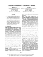
Comparison of cytochrome b sequences around residues
129 and 143, involved in QoI resistance, showed some
variations between pathogen species (Fig. 1). Firstly,
S. cerevisiae, used as a model system in this work, has
a unique feature: the CCV(133–135) sequence which,
although also found in related yeast (Fig. 1A), is replaced
by the sequence VLP(133–135) in most other organisms,
including all plant pathogens we have analyzed and more
distantly related species including mammals. To address the
question whether the ÔCCVÕ sequence is essential to yeast bc
1
complex function or assembly, this sequence was replaced
bythemorecommonÔVLPÕ sequence and the respiratory
growth competence, the cytochrome b level and bc
1
activity
Ó FEBS 2004 Modeling the Q
o
site of crop pathogens (Eur. J. Biochem. 271) 2265
were monitored (Tables 1 and 2). No effect was observed,
suggesting that the yeast enzyme can accommodate the VLP
sequence without loss of function. This new form of Q
o
domain, with the common VLP(133–135) sequence, has
therefore been used throughout the other studies reported
here.
The effect of other variations in the Q
o
binding domain
on bc
1
function and inhibitor resistance was then investi-
gated. Four plant pathogens were chosen for this study,
E. graminis (Ascomycete, pathogen of wheat), V. inaequalis
(Ascomycete, pathogen of apple), S. fuliginea (Ascomycete,
pathogen of cucumber) and P. megasperma (Oomycete,
causing root rot disease) based on comparison of their
primary sequences. The cytochrome b sequences of these
plant pathogens, either obtained from public databases or
by targeted PCR amplification and sequencing of field
isolates, showed only small but distinctive changes in the Q
o
site (Fig. 1). Three permutations at position 136: tyrosine,
phenylalanine and tryptophan, and three permutations at
position 141: histidine, leucine and phenylalanine were
observed in the four pathogens. In addition, a change of
residue 275 from leucine to phenylalanine is seen P. mega-
sperma cytochrome b. This latter change has been also
reported in Pneumocystis carinii resistant to atovaquone
treatment [7] and is naturally present in the corresponding
mammalian enzyme [8]. Appropriate changes in the yeast
cytochrome b sequence were introduced in order to obtain
four new forms of cytochrome b: E. graminis-like (AB1),
Fig. 1. Comparison of cytochrome b sequences in a region comprising the Q
o
domain. (A) Aligned sequences from yeasts and, as a representative
mammal, humans (residues 121–155, S. cerevisiae numbering). (B) Corresponding sequence comparison of S. cerevisiae with the four plant
pathogens employed in this study. (C) The sequence of the 15 yeast variants constructed and analyzed in this work. The mutated residues are in
bold. The sequences of E. graminis, V. inaequalis and P. megasperma are available from the EMBL database. The sequence of S. fuliginea was
determined by targeted RT-PCR amplification as described in [11].
2266 N. Fisher et al. (Eur. J. Biochem. 271) Ó FEBS 2004
V. inaequalis-like (AB4), S. fuliginea-like (AB7) and
P. megasperma-like (AB9) mutants (Fig. 1C). These new
forms of cytochrome b were also used to compare the
impact of the introduction of the mutations G143A,
F129L and L275F on bc
1
complex activity. To this
end, we introduced these additional mutations into the
Table 1. Respiratory growth competence, cytochrome b content and resistance to Q
o
inhibitors. To determine the doubling time, cells were inoculated
in respiratory medium (YPG) and the optical density was monitored periodically at 600 nm. The cytochrome b (cyt b)contentwasdeterminedin
whole cells by spectrophotometry as described in experimental procedures, using e ¼ 25 m
M
-1
.cm
-1
at 562–575 nm. The cyt b concentration in the
wild type cells was 5.7 nmol per gram of cells. The respiratory growth in presence of inhibitor was monitored on respiratory media (YPG) plus 1 or
10 l
M
inhibitor as described in Fig. 3. +++ indicates vigorous growth; ++ and +, weaker growth; – , no growth.
Strains Mutations
Doubling
Time (hrs)
Cyt b content
(%)
Growth on
Myxothiazol Stigmatellin Azoxystrobin Pyraclostrobin
10 10 1 10 1 10
WT 4 100 – + – – – –
Erysiphe graminis-like
AB1 VLP 4 95 – – – – – –
AB2 G143A 5 100 +++ – +++ +++ +++ +++
AB3 F129L 5 100 +++ + + - - -
AB17 F129L, G143A 5 90 +++ + +++ +++ +++ +++
Venturia inaequalis-like
AB4 H141L 4 100 – – – – – –
AB13 H141L, G143A 5 100 +++ – +++ ++ +++ +++
AB5 F129L, H141L 6 100 + – – – – –
AB18 F129L, H141L, G143A 10 100 ++ + ++ ++ ++ ++
Sphaerotheca fuliginea -like
AB7 Y136F, H141L 4 100 – – – – – –
AB8 Y136F, H141L, G143A 5 95 ++ – +++ ++ +++ +++
Phytophthora megasperma-like
AB9 Y136W, H141F 4.5 85 – – – – – –
AB10 Y136W, H141F, G143A 5 75 +++ – +++ +++ +++ +++
AB16 F129L, Y136W, H141F 5 90 +++ ++ ++ – – –
AB11 Y136W, H141F, L275F 5 60 – + – – – –
AB12 F129L, Y136W, H141F, L275F 5 90 +++ ++ – – ++ +
Table 2. QH
2
cytochrome c reductase activities. QH
2
cytochrome c reductase activity was assayed as described in experimental procedures.
Strains Mutations
bc
1
Complex activity
Rates (s
)1
)
at 50 l
M
QH
2
V
m
(s
)1
)
K
m
(QH
2
l
M
)
WT 40 +/– 1.8 (100%) 80 18
Erysiphe graminis-like
AB1 VLP 40 +/– 1.1 (100%) 82 17
AB2 G143A 35 +/– 1.8 (87%) 74 12
AB3 F129L 35 +/– 2.5 (87%) – –
AB17 F129L, G143A 32 +/– 2.2 (80%) – –
Venturia inaequalis-like
AB4 H141L 28 +/– 1.1 (100%) 42 12
AB13 H141L, G143A 14 +/– 1.0 (50%) 25 6
AB5 F129L, H141L 26 +/– 0.5 (93%) – –
AB18 F129L, H141L, G143A 12 +/– 1.7 (43%) – –
Sphaerotheca fuliginea-like
AB7 Y136F, H141L 39 +/– 2.1 (100%) 68 17
AB8 Y136F, H141L, G143A 26 +/– 1.2 (67%) 36 10
Phytophthora megasperma-like
AB9 Y136W, H141F 27 +/– 0.6 (100%) 38 12
AB10 Y136W, H141F, G143A 11 +/– 1.0 (41%) 23 8
AB16 F129L, Y136W, H141F 12 +/– 1.1 (44%) – –
AB11 Y136W, H141F, L275F 12 +/– 1.8 (44%) 27 12
AB12 F129L, Y136W, H141F, L275F 18 +/– 1.8 (67%) – –
Ó FEBS 2004 Modeling the Q
o
site of crop pathogens (Eur. J. Biochem. 271) 2267
pathogen-like mutants. In total, 15 variants were constructed
(Figs 1C and 2). These were generated by a biolistic trans-
formation procedure, which produces homoplasmic yeast
strains carrying only the variant cytochrome b sequence [3],
andthenusedtomonitorrespiratoryfunctioninvariousways.
Effects of mutations on respiratory growth and
cytochrome
b
content
All the variant cytochrome b yeast strains constructed were
respiration competent. Their doubling times in nonferment-
able medium (YPG) were 4–5 h, with the exception of
strains AB5 and AB13 which showed doubling times of
6 and 10 h, respectively. This phenotype was not investi-
gated further. In order to assess the effect of mutations on
the assembly of the bc
1
complex, we also monitored the
concentration of cytochromes in whole cells, as described in
experimental procedures: changes introduced in the Q
o
domain had little effect on cytochrome b assembly. Cyto-
chrome b content was between 90 and 100% of that of the
wild-type, in the E. graminis-, V. inaequalis-andS. fuligi-
nea-like constructs; though the changes introduced in the
P. megasperma-like constructs seemed to hinder enzyme
assembly slightly as judged by the decrease in cytochrome b
content (Table 1). Lowest cytochrome b levels were
observed in the strain harboring the three mutations
Y136W, H141F and L275F (60% of the wild type).
Interestingly, these three changes are naturally present in
mammals. The introduction of a fourth mutation, F129L
restored the cytochrome b content to near wild-type level
(Table 1). It seems likely that the introduction of three bulky
residues, Y136W, H141F and L275F, sterically hinders the
folding of cytochrome b and the assembly of the complex.
The replacement of phenylalanine at position 129 by a
smaller residue leucine may then alleviate the hindrance and
restore the proper folding of cytochrome b.
Resistance to Q
o
inhibitors
As mutations G143A and F129L had been found in plant
pathogen isolates resistant to QoIs, we monitored the
respiratory growth competence of the different constructs in
the presence of stigmatellin, which binds in the distal lobe
of the Q
o
site, and myxothiazol, azoxystrobin and pyra-
clostrobin, which bind at the proximal lobe of the Q
o
site
(Fig. 3 and Table 1).
The control strains, AB1, AB4, AB7 and AB9 were all
sensitive to myxothiazol, stigmatellin, azoxystrobin and
pyraclostrobin. Introduction of G143A in all four Q
o
forms
led to strong resistance to myxothiazol, azoxystrobin and
pyraclostrobin: strains AB2, AB13, AB8 and AB10 grew on
nonfermentable medium in presence of 10 l
M
of each of
these compounds but were still sensitive to stigmatellin.
Interestingly structural studies suggest that the Ca hydrogen
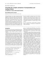
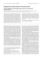
Fig. 2. Structure of the Q
o
site. The cyto-
chrome b a-carbon backbone is shown in
orange. The location of residues altered to
model the Q
o
-sites from the pathogenic fungi
discussed in the text are shown in green. The
VLP(133-135) region of cytochrome b is indi-
catedinwhite.Q
o
-bound stigmatellin and
hemes b
l
/b
h
are represented in cyan and red,
respectively. This figure was prepared from the
yeast bc
1
crystal structure coordinates
1KYO.pdb [12] using
VISUAL MOLECULAR
DYNAMICS
software [13].
2268 N. Fisher et al. (Eur. J. Biochem. 271) Ó FEBS 2004
atom of G143 approaches within 3.5 A
˚
of the methoxy-
acrylamide moiety of myxothiazol and hence mutation to
the bulkier residue alanine is likely to abolish the binding of
this class of Q
o
antagonist [1,6]. A similarly close interaction
with the benzene ring ÔlinkerÕ region of azoxystrobin and
pyraclostrobin could explain resistance to these compounds.
The pattern of resistance induced by F129L was different.
Strains AB3 and AB16 were resistant to myxothiazol and
stigmatellin. They also show limited cross-resistance to
azoxystrobin as growth was observed at 1 l
M
azoxystrobin
but not at 10 l
M
. Yeast cells carrying this mutation were
rather more sensitive to pyraclostrobin: no growth was
observed at 1 l
M
pyraclostrobin. The sidechain of F129
approaches within 3 A
˚
of the myxothiazol methoxyacryl-
amide moiety. By contrast, F129 has a closest approach of
4A
˚
with the hydrophobic tail of stigmatellin. The likely
mechanism of F129L stigmatellin resistance is therefore not
clear, but it could be due to a subtle alteration of the
backbone fold at Q
o
, or a change in accessibility for the
antagonist to the Q
o
site. The slight variance in sensitivity
to azoxystrobin and pyraclostrobin is likely to be due to the
difference in pharmacophore structure between these two
compounds, as discussed in more detail below. As men-
tioned above, strain AB5 showed a weaker growth that
could explain the apparent sensitivity.
Interestingly AB12, which combined F129L with L275F,
was sensitive to azoxystrobin but resistant to pyraclostro-
bin. In this case it is likely that the two changes have slightly
modified the structure of the Q
o
site, which can now
accommodate azoxystrobin but not pyraclostrobin. The
sidechain of F275 in chicken bc
1
complex is involved in a
stabilizing ring–stacking hydrophobic interaction with the
phenyl group of MOA-stilbene [6], a Q
o
inhibitor closely
related to strobilurin. This may explain why strain AB11
(Y136W, H141F, L275F) retains sensitivity to the strobilu-
rin-related inhibitor azoxystrobin. Strain AB12 (Y136W,
H141F, L275F + F129L) demonstrated resistance to both
myxothiazol and pyraclostrobin, but remained sensitive to
azoxystrobin. Pyraclostrobin and Azoxystrobin differ in
pharmacophore structure; the former contains an alkoxy-
amino moiety, whereas the latter is methoxyacrylate based
(Fig. 4). Significantly, the pharmacophore of pyraclostrobin
occupies a smaller volume than that of azoxystrobin, and
might have a greater degree of rotational freedom due to the
Fig. 4. Structure of Q
o
inhibitors azoxystrobin and pyraclostrobin [1].
Pharmacophore groups are indicated by boxes.
Fig. 3. Sensitivity to Q
o
inhibitor exposure.
The name and position of the strains are
shown in the right-hand panel. A drop of each
strain was inoculated on a nonfermentable
medium plate (YPG) with or without 10 l
M
inhibitor and incubated for 3–4 days.
Ó FEBS 2004 Modeling the Q
o
site of crop pathogens (Eur. J. Biochem. 271) 2269
lack of methoxyacrylate p-bonded structure. Mutation of
both F129 and L275 to leucine and phenylalanine, respect-
ively, are required to inhibit pyraclostrobin binding.
As expected, strains AB17 and AB18 harboring both
G143A and F129L combined resistance to myxothiazol,
azoxystrobin and pyraclostrobin with resistance to stig-
matellin.
In order to quantify the level of resistance induced by
G143A, bc
1
complex sensitivity to myxothiazol and stig-
matellin was monitored in membranes from strain AB2 and
its control AB1. QH
2
cytochrome c reductase activity (using
2.5 n
M
bc
1
complex), as in Table 2, was measured in
presence of increasing concentration of inhibitors. The
concentration of stigmatellin required for 50% decrease of
activity (I
50
) was around 2.5 n
M
for AB1 and AB2, whereas
the I
50
for myxothiazol was 2.5 n
M
for AB1 and 18 l
M
for
AB2: a 7500-fold increase. This is in good agreement with
previous results. The G143A mutation was first reported in
mammalian cells after selection in presence of myxothiazol,
conferring > 7000-fold resistance to the inhibitor [9].
Effect of mutations on bc
1
complex activity
In order to study possible effects of the mutations on bc
1
function, mitochondrial membranes were prepared from the
different strains and cytochrome c reductase activity was
monitored spectrophotometrically as described in experi-
mental procedures. As shown in Table 1, the replacement
of the yeast sequence CCV(133-135) by the much more
common sequence VLP, in the E. graminis-like strain had
no effect on enzyme activity. In the V. inaequalis-like strain
(AB4), histidine 141 was replaced by leucine. The activity of
the resultant enzyme was then decreased by 30% compared
to the wild-type yeast. Activity was however, restored to
near wild-type levels by the introduction of a second change,
Y136F, in the S. fuliginea-like strain (AB7). The P. mega-
sperma-like enzyme (in strain AB9), which harbored
Y136W and H141F also showed a 30% decrease in bc
1
activity.
The introduction of G143A, F129L or both changes
together (though this has not been seen in any natural
isolate to our knowledge) in the E. graminis-like Q
o
site had
little effect on bc
1
activity (80–87% of wild-type rate). Thus
this Q
o
site can accommodate the G143A and F129L
mutations without loss of function. This is consistent with
previous observations with E. graminis itself, which showed
that the isolates carrying the G143A mutation did not suffer
any fitness penalty [10]. In the V. inaequalis-like strains, the
situation was different. As mentioned above, the control
strain (AB4) harboring the change H141L showed a lower
activity than the wild-type yeast strain (turnover number
28 s
)1
vs. 40 s
)1
). Interestingly the introduction of the
G143A mutation in this Q
o
site further decreased the bc
1
activity to 14 s
)1
(50% of the control AB4). In contrast,
F129L had no effect. In AB18, which combined G143A and
F129F, the enzyme activity was 43% of the control. It seems
therefore that the V. inaequalis-likeenzymecannotaccom-
modate the G143A mutation without reduction of function.
Similar results were obtained with the P. megasperma-and
the S. fuliginea-like Q
o
sites. The introduction of G143A
caused, respectively, a 60% and 33% decrease of the bc
1
activity compared to the controls. We have also used the
P. megasperma-like form to monitor the effect of F129L
and L275F. The mutation L275F is naturally occurring in
Phytophothora sp. The introduction of these mutations
decreased the bc
1
activity to 44% of the control AB9. Their
combination in AB12 restored the activity to 67% of the
control AB9. Thus the introduction of L275F in the
P. megasperma-like Q
o
caused a decrease both in bc
1
content and activity, while F129L partially compensated
the defect.
To gain further information on the effect of the mutation
G143A, steady-state cytochrome c reductase activity was
monitored as a function of decylubiquinol (QH
2
) concen-
tration. The apparent V
m
and K
m
for QH
2
were calculated
from initial rate measurements using derived Eadie–Hofstee
plots (Table 1). The mutation G143A appeared to decrease
both the V
m
and the K
m
for quinol in AB13, AB8 and
AB10. It might therefore be that this mutation slightly
affects the structure of the Q
o
site which, as a result,
becomes saturated with substrate more rapidly than the
control due to lower electron transfer, or alternatively it may
reflect a decreased ÔonÕ rate for quinol binding. The
replacement of glycine by alanine is a relatively conservative
structural change, and unlikely to disrupt the fold of the cd1
helix. The introduced methyl group may sterically hinder
interactions with the quinol headgroup, or unfavorably alter
the conformation of bound quinol such that electron
transfer or deprotonation rates are decreased.
Thus variations in the Q
o
domain seem to affect the
impact of the QoI resistance mutation G143A on cyto-
chrome bc
1
activity. In some cytochrome b forms, the
introduction of G143A decreases the QH
2
cytochrome c
activity of the complex. Under standard laboratory condi-
tions in S. cerevisiae, this decrease has no effect on cell
growth as little as 20% of bc
1
complex activity is enough to
support respiratory growth. Therefore a decline in respir-
atory growth will only be seen when the complex is severely
inhibited. However in other organisms, such as plant
pathogens, when the energetic demands are higher, this
decrease might affect the fitness of the cells. In combination
with other factors, this could explain the differences in the
evolution of QoI resistance in fungal and oomycete
pathogens. Interestingly the characteristic Q
o
site features
of E. graminis, one of the pathogens which showed field
resistance to Q
o
I fungicides particularly quickly, seem to be
most functionally accommodating of the resistance-associ-
ated G143A mutation.
Acknowledgements
This work was supported by Syngenta. The authors acknowledge the
contributions made by our colleagues, Ms Carole Stanger and Ms.
Judith Burbidge, to the analysis of cytochrome b gene and/or mRNA
sequences from plant pathogen isolates. We would also particularly
wish to recognize the interest, enthusiasm and insight in initiating these
studies shown by our late colleague Steve Heaney, and this paper is
dedicated to his memory.
References
1. Gisi, U., Sierotzki, H., Cook, A. & McCaffery, A. (2002)
Mechanisms influencing the evolution of resistance to Qo inhibitor
fungicides. Pest Manag. Sci. 58, 859–867.
2270 N. Fisher et al. (Eur. J. Biochem. 271) Ó FEBS 2004
2. Kraiczy, P., Haase, U., Gencic, S., Flindt, S., Anke, T., Brandt, U.
& von Jagow, G. (1996) The molecular basis for the natural
resistance of the cytochrome bc
1
complex from strobilurin-pro-
ducing basidiomycetes to center Q
P
inhibitors. Eur. J. Biochem.
235, 54–63.
3. Hill, P., Kessl, J.J., Meshnick, S.R., Trumpower, B.L. & Meunier,
B. (2003) Recapitulation in Saccharomyces cerevisiae of cyto-
chrome b mutations conferring resistance to atovaquone in
Pneumocystis jiroveci. Antimicrob. Agents Chemother. 47, 2725–
2731.
4. Vanneste, W.H. (1966) Molecular proportion of the fixed cyto-
chrome components of the respiratory chain of Keilin-Hartree
particles and beef heart mitochondria. Biochim. Biophys. Acta 113,
175–178.
5. Cornish-Bowden, A. (1995) Fundamentals of Enzyme Kinetics.
Portland Press, London.
6. Zhang,Z.,Huang,L.,Shulmeister,V.M.,Chi,Y I.,Kim,K.K.,
Hung, L W., Crofts, A.R., Berry, E.A. & Kim, S H. (1998)
Electron transfer by domain movement in cytochrome bc
1
. Nature
392, 677–684.
7. Kazanjian, P., Armstrong, W., Hossler, P.A., Huang, L., Beard,
C.B.,Carter,J.,Crane,L.,Duchin,J.,Burman,W.,Richardson,J.
& Meshnick, S.R. (2001) Pneumocystis carinii cytochrome b
mutations are associated with atovaquone exposure in patients
with AIDS. J. Infect. Dis. 183, 819–822.
8. Kessl, J.J., Lange, B.B., Merbitz-Zahradnik, T., Zwicker, K., Hill,
P.,Meunier,B.,Palsdottir,H.,Hunte,C.,Meshnick,S.&
Trumpower, B.L. (2003) Molecular basis for atovaquone binding
to cytochrome bc
1
complex. J. Biol. Chem. 278, 31312–31318.
9. Howell, N. & Gilbert, K. (1988) Mutational analysis of the mouse
mitochondrial cytochrome b gene. J. Mol. Biol. 203, 607–618.
10. Chin, K.M., Chavaillaz, D., Kaesbohrer, M., Staub, T. &
Felsenstein, F.G. (2001) Characterizing resistance risk of Erysiphe
graminis f.sp. tritici to strobilurins. Crop Protection 20, 87–96.
11. Sierotzki, H., Parisi, S., Steinfeld, U., Tenzer, I., Poirey, S. &
Gisi, U. (2000) Mode of resistance to respiration inhibitors at the
cytochrome bc
1
enzyme comples of Mycosphaerella fijiensis field
isolates. Pest Manag. Sci. 56, 833–841.
12. Hunte, C., Koepke, J., Lange, C., Rossmanith, T. & Michel, H.
(2000) Structure at 2.3 angstrom resolution of the cytochrome bc
1
complex from the yeast Saccharomyces cerevisiae co-crystallized
with an antibody Fv fragment. Structure Fold. Des. 8, 669–684.
13. Humphrey, W., Dalke, A. & Schulten, K. (1996) VMD – visual
molecular dynamics. J. Mol. Graph. 14, 33–38.
Ó FEBS 2004 Modeling the Q
o
site of crop pathogens (Eur. J. Biochem. 271) 2271