Báo cáo khoa học: Ras oncogene induces b-galactoside a2,6-sialyltransferase (ST6Gal I) via a RalGEF-mediated signal to its housekeeping promoter pptx
Bạn đang xem bản rút gọn của tài liệu. Xem và tải ngay bản đầy đủ của tài liệu tại đây (418.84 KB, 12 trang )
Ras oncogene induces b-galactoside a2,6-sialyltransferase (ST6Gal I)
via a RalGEF-mediated signal to its housekeeping promoter
Martin Dalziel
1
, Fabio Dall’Olio
2
, Arron Mungul
3
,Ve
´
ronique Piller
1
and Friedrich Piller
1
1
Centre de Biophysique Mole
´
culaire, CNRS UPR 4301 affiliated with the University of Orle
´
ans and INSERM, Orle
´
ans, France;
2
Dipartimento di Patologia Sperimentale, Universita
`
di Bologna, Italy;
3
Cancer Research UK, Breast Cancer Biology Group,
Guy’s Hospital, London, UK
Several oncogenic proteins are known to influence cellular
glycosylation. In particular, transfection o f codon 12 point
mutated H-Ras increases CMP-Neu5Ac: Galb1,4GlcNAc
a2,6-sialyltransferase I (ST6Gal I) activity in rodent
fibroblasts. Given that Ras mediates its effects through at
least three secondary effector pa thways (Raf, RalGEFs and
PI3K) a nd that transcriptional c ontrol of m ouse ST6Gal I is
achieved by the selective use of multiple promoters, we
attempted t o i dentify which of these parameters a re i nvolved
in linking the R as signal to ST6Gal I gene transcription in
mouse fibroblasts. Transformation by human K-Ras o r
H-Ras ( S12 a nd V12 point mutations, respectively) results in
a 1 0-fold increase in ST6Gal I mRNA, but no alter ation in
the expression of related sialyltran sferases. Using an indu-
cible H-Ras
V12
expression system, a direct causal link be-
tween activated H-Ras expression and elevated ST6Gal I
mRNA was demonstrated. The accumulation of the
ST6Gal I transcript in response to activated Ras was
accompanied b y a n i ncrease o f a2 ,6-sialylt ransferase activity
and of Neu5Aca2,6Gal at the cell s urface. Results obtained
with H-Ras
V12
partial loss of function mutants H -Ras
V12S35
(Raf signal only), H-Ras
V12C40
(PI3-kinase s ignal only) and
H-Ras
V12G37
(RalGEFs signal only) suggest that the H-Ras
induction of the m ouse ST6Gal I gene (Siat1) transcription
is primarily routed through RalGEFs. 5¢-Rapid amplifica-
tion of cDNA ends analysis demonstrated that the increase
in ST6Gal I mRNA upon H-Ras
V12
or K-Ras
S12
transfec-
tion is mediated by the Siat1 housekeeping promoter
P3-associated 5¢ untranslated exons.
Keywords: oncogenic Ras; sialyltransferase; RalGEF;
housekeeping p romoter.
The human Ras gene family is composed of H-Ras,
K-Ras and N-Ras [1] encoding three related p21 Ras
proteins that function as small GTPases bound to the
plasma membrane through lipid anchors. They effectually
link extracellular, ligand-generated s ignals to cytoplasmic
signalling cascades through their ability to bind in a GTP-
dependent manner various effector proteins and thus
altering their localization, protein–protein interaction and
activity. Mutations in the Ras genes at codons 12, 13 and
61 render the Ras proteins constitutively active in their
GTP-bound form [2]. Those mutations lead to oncogenic
Ras and are found in approximately 30% of all human
cancers [3], though the frequency varies with different
cancer types.
The presence of constitutively activated H-Ras
V12
in
primary cell cultures o f human tumours has been linked t o
an increase in N-glycan branching a nd sialylation [4,5].
Subsequent studies with H-Ras
V12
-transfected rat fibro-
blasts identified the enh anced activities of the CMP-
Neu5Ac:Galb1,4GlcNAc a2,6-sialyltransferase (ST6Gal I,
EC 2.4.99.1) [6–9], a nd N-acetylglucosaminyl transferase V
[10] as the two likeliest effectors of these glycosylation
changes.
Increased levels of ST6Gal I have been identified in
breast [11], c olon [12–14], cervical [15] and prostate cancer
[16]. Elevated ST6Gal I activity has also been linked to
markers of poor prognosis in breast cancer patients [11], to
the d ifferentiation state of t he tumour in prostate [16] and
colon [17] cancer, to secondary local colon tumour
reoccurrence [18] and finally to metastasis in both cervical
[19] and colon cancer [20]. Moreover, ST6Gal I
over-expression and inhibition experiments have shown
Correspondence to F. Piller, Centre de Biophysique Mole
´
culaire,
rue Charles Sadron, F45071 Orle
´
ans Ce
´
dex 02, France.
Fax: +33 238 631517, Tel.: +33 238 257643,
E-mail:
Abbreviations: FACS, fluorescence activated cel l so rt er; FB S, fe tal
bovine serum; FITC, fluorescein isothiocyanate; Gal, galactose; Glc-
NAc, N-acetylglucosamine; GalNAc, N-acetylgalactosamine; MAA,
Maackia amurensis agglut i nin; M ES, 2 -morpholino ethanesulfonic
acid; Neu5Ac, N-acetylneuraminic acid; RACE, rapid amplification of
cDNA ends; SNA, Sambucus nigra agglutinin; Siat1, Mouse ST6Gal I
gene; ST3Gal I, CMP-Neu5Ac:Galb1,3GalNAc a2,3-sialyltransferase
I; ST3Gal II, CMP-Neu5Ac:Galb1,3GalNAc a2,3-
sialyltransferase II; ST3Gal III, CMP-Neu5Ac:Galb1,(3)4GlcNAc
a2,3-sialyltransferase; ST3Gal IV, CMP-Neu5Ac:Galb1,3GalNAc/
Galb1,4GlcNAc a2,3-sialyltransferase; ST6Gal I, CMP-
Neu5Ac:Galb1,4GlcNAc a2,6-sialyltransferase I; ST6Gal II, CMP-
Neu5Ac:Galb1,4GlcNAc a2,6-sialyltransferase II; ST6GalNAc I,
CMP-Neu5Ac:GalNAc a2,6-sialyltransferase I; Tc, tetracycline;
UT, untranslated.
Enzymes: CMP-Neu5Ac:Galb1,4Glc NAc a2,6-si alyltran sfera se
(ST6GalI, EC 2.4.99.1); CMP-Neu5Ac:GalNAc a2,6-sialyltrans ferase
I, (ST6GalNAcI, EC 2.4.99.3); CMP-Neu5Ac:Galb1,3GalNAc a2,3-
sialyltransferase I (ST3Gal I , EC 2.4.99.6).
(Received 1 2 May 20 04, revised 2 July 2004, accepted 12 J uly 2004)
Eur. J. Biochem. 271, 3623–3634 (2004) Ó FEBS 2004 doi:10.1111/j.1432-1033.2004.04284.x
that ST6Gal I exhibits a profound influence on the meta-
static potential of tumour cells in vitro [21,22]. Interestingly,
increased cell surface a2,6-sialylation, identical t o those
reported to be the result o f activated Ras expression, also
influences parameters thought to be important for c ellular
metastatic ability, such as motility [9] and b1- integrin
activity [23].
ST6Gal I catalyses the biosynthetic transfer of Neu5Ac
from CMP-Neu5Ac to the nonreducing end of type II
N-acetyllactosamine structures to form Neu5Aca2,6Gal-
b1,4GlcNAc-R on glycoproteins and glycolipids [24].
Although a second member of the ST6Gal family,
ST6Gal II, has recently been identified [25], the combina-
tion of weak and t issue restrictive expression, primarily in
adult b rain [26], leaves S T6Gal I as the dominant ST6Gal
enzyme in all adult tissues. This is consistent with
gene kno ckout experiments in mice, where loss of
ST6Gal I results in an almost complete absence of
Neu5Aca2,6Galb1,4GlcNAc-R in adult and fetal tissue
[27]. Transcriptional control of the mouse ST6Gal I gene
Siat1 is regulated during development a nd differentiation by
the s elective usage of multiple promoter regions. Differential
utilization of these promoters results in mature transcripts
that are identical except for their untranslated 5¢ (5¢UT)
leader se quences. A t l east four Siat1 promoters a re known:
P1 controls ST6Gal I expression in live r [28], P2 i n
B-lymphocytes [29], P3 is used to achieve multi-tissue
housekeeping expression [29] and finally P4 which is active
in the mammary gland during lactation [30].
In this report, we have studied the molecular events that
take place between codon 12 mutated H-Ras or K-Ras
expression and elevated ST6Gal I activity in NIH3T3 cells.
We present data i ndicating that the R as signal, mediated b y
RalGEFs, leads directly to an accumulation of ST6Gal I
mRNA, transcribed from the Siat1 h ousekeeping promoter
P3, ultimately resulting in enhanced ST6Gal I enzyme
activity and cell surface a2,6-sialylation.
Experimental procedures
Materials
[
32
P]dCTP[aP] (3000 CiÆmmol
)1
), CMP- [
14
C]Neu5Ac
(286 mCiÆmmol
)1
), Random primer based radiolabelling
Mega prime kit, MicroSpin G-50 DNA purification
columns and positively charged nylon membrane were
purchased from Amersham (Saclay, France). Plasmid
pCR2.1, pcDNA3.1, Escherichia coli TOP10F strain,
Trizol and M13 reverse primer were from Invitrogen
(Cergy-Pontoise, France). Marathon rapid amplification
of cDNA ends (RACE) kit and human leukaemia cell
line K562 poly(A)
+
RNA were from Clontech (Palo
Alto,CA,USA).TheDNAgelextractionkitwasfrom
Qiagen (Coutaboeuf, France). Wizard DNA miniprep kits
were from Promega (Charbonnie
`
res-les-Bains, France).
Biotinylated Sambucus nigra agglutinin (SNA) and
Maackia amurensis (MAA) lectins were from Vector
Laboratories (Burlingame, CA, USA). Galb1,4GlcNA cb-
OCH
2
Ph was a generous gift of C. LeNarvor and
C. Auge
´
,Universite
´
Paris-Sud, O rsay, France. DMEM
and fetal bovine serum (FBS) were from BioWest (Paris,
France).
cDNA probes and DNA constructs
Mouse ST6Gal I exon II (750 bp PstI fragment) genomic
DNA and ST3Gal I, II, III and IV PCR fragments were
provided by J. Lau (Roswell Park Cancer Institute, Buffalo,
NY, USA). Mouse ST3Gal I, II, III and IV probes were
amplified using the following primer pairs. ST3Gal I
(product size 241 bp): p125 (sense), 5 ¢-ACCTCACCTTCT
TCCTGCTCTTC-3¢ and p128 (antisense), 5¢-AGCGTTG
TGGACTGTCAGCA-3¢;ST3GalII(921bp):pPE1
(sense), 5¢-GGCTATTCAGAATTCCAGCGCCTCGGC
AAGGA-3¢ and pPE2 (anti sense), 5¢-TGCCAGAC
CCTCGAGTGACTGGTTCTGAAGGCGCTCAGG-3¢;
ST3Gal III (633 bp): p138 (sense), 5¢-CCCTCTGCCT
CTTCCTGGTC-3¢ and p139 (antisense), 5¢-TCGTTCAT
ATTGCTCAGGTCG-3¢; S T3Gal IV (449 bp): p136
(sense), 5¢-CCTGGCTCTGGTCCTTGTTGT-3 ¢ and
p137 (antisense), 5¢-AGCCCACATCTCCCTCGTAGC -3¢.
ST3Gal I, II, III and IV PCR products were cloned into
pCR2.1, propagated in E. coli TOP10F, confirmed using
M13 reverse primer (M13r) sequencing (MWG Biotech,
Courtaboeuf, France) and released by EcoRI digestion for
use as northern probes. Mouse ST6GalNAc I cDNA was
obtained from S . Tsuji, The Glycoscience I nstitute, T okyo,
Japan. A 1.1 kb ST6GalNAc I probe was r eleased by
HindIII/EcoRV digestion. A 604 nt ST6Gal II cDNA
probe, spanning the first predicted coding exon in the
mouse chromosome 17 sequence E NSMUSG00000024172
(22918721–23318720), downloaded from the Wellcome
Trust Sanger Institute mouse genome server (http://
www.ensembl.org/Mus_musculus), was amplified by PCR
from mouse genomic DNA using the primer pair mst172
(5¢-GGATGGCACCGGCAGACATG-3¢) and mst171
(5¢-CACAGAAATGGGATCAGGCC-3¢). Both human
H-Ras and K-Ras cDNA were obtained from Cancer
Research UK. Human H-Ras cDNA probe was isolated
from an EcoRI digestion of H-Ras
V12
cDNA cloned into
the EcoRI site of pcDNA3.1. The 0.4 kb human K-Ras
cDNA probe (encoding the 3¢ region of the ORF) was
isolated by EcoRI digestion of a 1.1 k b human K-Ras
S12
cDNA cloned into the EcoRI site of pcEXV-3. The human
V12 H -Ras partial loss of function mutants H-Ras
V12S35
,
H-Ras
V12C40
(bothinpcDNA3.1)andH-Ras
V12G37
(in
pSG5) were obtained from A. Scibetta (Cancer Research
UK, Guy’s Hospital, London, UK) [31,32]. The
H-Ras
V12G37
cDNA was r ecloned into p cDNA3.1 using
EcoRI. Finally, mouse 18S cDNA was purchased from
Ambion (Huntingdon, UK).
Identification and cloning of mouse ST6Gal II
Using the Wellcome Trust Sanger Institute mouse
genome server database a second ST6Gal family member,
ST6Gal II was identified on chromosome 17 (ENS-
MUSG00000024172) as presenting significant homology
to the ST6Gal I gene (Siat1) on chromosome 16.
Subsequently, a 604 nt genomic probe spanning the first
coding exon was generated by PCR. Of the five potential
ST6Gal II coding exons, exon I contains the least
homology to Siat1 . When t his probe was hybridized to
multiple tissue total RNA extracted from a single male
C57Bl6 mouse (stomach, whole brain, spleen, kidney,
3624 M. Dalziel et al. (Eur. J. Biochem. 271) Ó FEBS 2004
testis, large intestine, small intestine and liver), only the
whole brain sample showed any d etectable signal (data
not shown). A full-length cDNA ( 1.6 kb) was then
amplified by PCR using Pfu polymerase from whole
mouse brain cDNA, cloned into pCR2.1 and sequenced.
The sequence was consistent with the predicted gene
structure contained within the ENSMUSG00000024172
genomic sequence. In brief, five exons encoding a 524
amino acid sialyltransferase (containing L, S and VS
motifs) with 32% overall primary sequence homology to
mouse ST6Gal I (48% within the catalytic C-terminal
and 18% within the N-terminal halves of t he protein).
This cDNA was re-cloned into pcDNA3.1-Flag and
transiently expressed in Chinese hamster ovary cells,
which then exhibit high amounts of a2,6-linked Neu5Ac
on cell surface N -glycans, as revealed by SNA staining
(data not shown). Although the exact acceptor substrate
specificity was not determined, these observations were
deemed suffi cient for the validation of both the identity
of the ENSMUSG00000024172 sequence as the mouse
ST6Gal II sequence (thus named Siat2) and the u se of
the 604 nt PCR f ragment as a specific ST6Gal II probe
in subsequent experiments. While this work was in
progress, the sequences of the human and mouse
ST6Gal II cDNAs were reported and confirmed our
results [25,26].
Cell lines
All cells were grown at 3 7 °C in a humidified atmosphere
of 5% (v/v) CO
2
, in DMEM (Gibco)/10% (v/v) FBS
containing 100 UÆmL
)1
penicillin, 100 lgÆmL
)1
glutamate,
100 lgÆmL
)1
streptomycin and 1.25 lgÆmL
)1
amphoteri-
cin B. Mouse cell lines 3T3 and K-Ras
S12
,whichare
NIH3T3 parental and NIH3T3 transfected with activated
human Ras cDNA, respectively, were obtained from
Cancer Research UK. H-Ras
V12
, which is NIH3T3
transfected with codon 12 position 2 point mutation
GGC to GTC (Gly fi Val), activated mutant of human
H-Ras, and mock transfected control line 3T3pB322 were
obtained from E. He
´
bert (CBM, Orle
´
ans, France) [33].
The presence of oncogenic Ras and the R as variant as
well as nature of the mutation were controlled by RT-
PCR and nucleotid e sequencing. The 410.4 cell line
(mouse mammary gland carcinoma cell line) was provi-
ded by B. Miller (Michigan Cancer Foundation, Detroit,
MI, USA) [34]. Th e NIH3T3 cell line s transfected w ith
tetracycline (Tc) repressed H-Ras
V12
construct (mib125),
constitutive H-Ras
V12
(mib128) and the parental NIH3T3
line (mib35) [35], were obtained from B. Willumsen
(University of Copenhagen, Denmark).
Stable transfections
Plasmid DNA (V12-S35, V12-C40 and V12-S35, all in
pcDNA3.1) used for transfections was purified using a
Wizard Plus DNA purification system (Promega) and
linearized with PvuI. Transfection of mouse NIH3T3 cells
was performed by electroporation using a G ene P ulser
electroporator (Bio-Rad, Hercules, CA, U SA) a t 960 lFD,
100 W, 0.25 V . Stable transfectants were selected with
0.5–0.75 mgÆmL
)1
geneticin (G418).
Extraction of total RNA from cell lines
All buffers and solutions used in the preparation and
analysis of RNA were prepared using DEPC treated water.
Total RNA was extracted from cell pellets collected at
80–90% confluence by the Trizol method according to the
manufacturer’s instructions. RNA pellets were air dried
before dissolving in 20–50 lL DEPC-treated sterile water.
RNA was then quantified using spectrophotometric meas-
urement a t 260/280 nm and quality checked on a 1% ( w/v)
agarose gel in Tris/Borate/EDTA buffer stained with
ethidium bromide.
5¢-RACE analysis
Twenty-five micrograms of total RNA were annealed to
the primer mST1-p1 (5¢-GATGATGGCAAAC AGGAG
AA-3¢) and reverse transcribed. The primer mST1-p1 is
complementary to a region in exon II between nucleotides
+50 (5¢)to+69(3¢) relative to the adenosine of the first
ATG codon. Thus, mST1-p1 will only bind Siat1 transcripts
that contain the exon II ATG translation start site and
authentic reverse transcription events of Siat1 mRNA must
span at least the exon I–exon II boundary. The resultant
cDNA was then ligated overnight at 16 °Ctothe50
nucleotide Marathon adaptor sequence (Clontech) as per
instructions and s ubjected to PCR amplification, using the
TOUCHDOWN
program recommended by Clontech (94 °C
for 1 min, five cycles of 94 °Cfor30s,72°Cfor4min,five
cycles of 94 °Cfor30s,70°C for 4 min and finally 25
cycles of 94 °C for 20 s, 68 °C for 4 m in), using the
Marathon adaptor anchor sense primer AP1 (5¢-CCATCC
TAATACGACTCACTATAGGGC-3¢) and either the
Siat1 exon I antisense primer md11 (5¢-CTGCTTCTG
GCTAATCTTCTGGGGTTGG -3¢)ortheexonOanti-
sense primer O2 (5¢-CTCAGCATCCGGCTGGAAAGTG
GGTACCACG-3¢). PCR amplification of ST6Gal I
sequence from contaminating genomic DNA is not possible
as the Siat1 gene does not contain sequences that will
specifically anneal the anchor primer. The PCR products
were isolated from a 3% (w/v) agarose gel (0.5 lgÆmL
)1
ethidium bromide), purified and then ligated into the
plasmid vector pCR2.1, cloned in TOP10 competent cells,
isolated by miniprep, digested with EcoRI to check for
insert, and finally sequenced, using an M13rev primer.
RT-PCR of H-Ras and K-Ras mRNA
Using 30 lg of total RNA, c DNA was synthesized as
described in the 5¢-RACE section save that the initial
primer used was the poly(A)
+
primer supplied in the
Marathon RACE kit. Using a 1 : 100 dilution of cDNA,
PCR was then performed using primers located in exon 1
and 2 to ensure that only cDNA derived from mRNA
could be amplified (at the expected size of 250 bp). PCR
conditions used: 94 °Cfor1min,50°Cfor1min,72°C
for 1 min, 25 cycles. P rime rs for human H-Ras and K-Ras
were as described [36]. H-Ras exon 1 sense: 5 ¢-CTGAG
GAGCGATGACGGAAT-3¢, H-Ras exon 2 antisense:
5¢-ACACACACAGGAAGCCCTCC-3¢,K-Rasexon1
sense: 5¢-CCTGCTGAAAATGACTGAAT-3 ¢,K-Ras
exon 2 antisense: 5¢-ATACACAAAGAAAGCCCTCC-3 ¢.
Ó FEBS 2004 Oncogenic Ras signalling and ST6Gal I activation (Eur. J. Biochem. 271) 3625
The PCR products were isolated from a 3% (w/v) agarose
gel (0.5 lgÆmL
)1
ethidium bromide), purified on Qiaex resin
(Qiagen) and t hen ligated into the plasmid vector pCR2.1,
cloned in TOP10F c ompetent cells, isolated on m ini-Wizard
columns (Promega), digested with EcoRI to check for
insert, and finally sequenced using m 13rev.
Northern analysis
Total RNA was run on 1% (w/v) agarose/formaldehyde
gels and transferred onto positively charged nylon filter
(Amersham) using 20 · NaCl/Cit (diethyl pyrocarbonate-
treated) capillary transfer. Filters were then prehybridized
in a hybridization oven with 10 mL of hybridization
buffer [ 0.5
M
sodium phosphate (pH 7 .0)/1 m
M
EDTA/
7% (w/v) SDS] for 1 h at 65 °C, then hybridized in
5 m L of hybridization buffer containing
32
P-labelled
DNA probes [25 ng of DNA labelled with a random
prime DNA labelling kit (Promega) and [
32
P]dCTP[aP],
overnight at 65 °C. Next day, blots were washed for 1 h
in buffer A [0.04
M
sodium phosphate, pH 7.0/1 m
M
EDTA/5% (w/v) BSA/5% (w/v) SDS], then twice with
buffer B [0.04
M
sodium phosphate, pH 7.0/1 m
M
EDTA/1% (w/v) SDS] at 65 °C and exposed to a
Kodak phosphor screen and visualized using a Molecular
Dynamics (STORM) scanner. The bands were quantified
by the
IMAGEQUANT
software (Molecular Dynamics,
Sunnyvale, CA, USA).
Sialyltransferase enzyme assay
ST6Gal I was assayed as previously described [37]. The
reaction mixture contained in a total volume o f 25 lL
10 lL of total cell lysate ( 100 lgprotein),2m
M
Galb1,4GlcNAcb-OCH
2
Ph, 50 l
M
CMP-[
14
C]Neu5Ac
(90 000 cpmÆnmol
)1
), 50 m
M
Mes pH 6.0, 5 m
M
MgCl
2
and 0.2% (v/v) Triton CF-54. After 1 h at 37 °Cthe
reaction was stopped with ice-cold 0.1
M
NH
4
HCO
3,
the
radiolabelled product isolated on reverse phase
C-18 cartridges and quantified by liquid scintillation
counting.
Fluorescence activated cell sorter (FACS) analysis
Cells were trypsinized, washed with N aCl/P
i
and incubated
at 2 · 10
7
cellsÆmL
)1
in 25 lL of biotinylated SNA or MAA
(10 lgÆmL
)1
)inNaCl/P
i
containing 5% (v/v) FBS and
0.1% (w /v) s odium azide (NaCl/P
i
/FBS). Cells were left on
ice for 30 min, washed three times an d streptavidin-FITC
(10 lgÆmL
)1
) was added to resuspended cells (25 lLÆwell
)1
).
After 30 min on ice, the cells were washed with NaCl/P
i
/
FBS, resuspended in 200 lLofNaCl/P
i
/FBS, fixed in 1.5%
(v/v) f ormaldehyde and analyzed on a B ecton Dickson
FACscan flow cytometer.
Genomic sequence of
Siat1
Genomic sequence covering Siat1 on chromosome 16
(ENSMUSG00000022885) was downloaded from the
Wellcome Trust Sanger Institute mouse genome server
spanning chromosome 16 region 22918721–23318720
(400 000 nucleotides).
Results
Effect of activated human Ras on the expression of
different members of the mouse sialyltransferase family
in 3T3 cells
Using the 0.75 kb Siat1 exon II probe, ST6Gal I mRNA
was found to be approximately 10-fold increased in the
3T3K-Ras
S12
and 3T3H-Ras
V12
cell lines relati ve to 3T3
and 3T3pB322 (Fig. 1). In contrast, ST6Gal II transcripts
were not detected in any of the cell lines, whilst ST6Gal-
NAc I was present only as a weak band in the 3T3H-Ras
V12
sample (Fig. 1). Strong ST6Gal II and ST6GalNAc I
signals w ere observed in t he positive controls, m ouse whole
brain and cell line 410.4 RNA, respectively. ST3Gal I
mRNA was detected i n all four lines, and was slightly
decreased in the 3T3K-Ras
S12
cells. Expression of ST3-
Gal I I was weak in all cell lines, with little difference
between Ras transfected an d control lines. ST3Gal III le vels
were equally high in both 3T3 and 3T3K-Ras
S12
lines and
no difference between the two cell lines could be observed
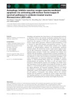
Fig. 1. Altered ST6Gal I transcript expression is the predominant
change within the sialyltransferase family in response to a ctivated Ras.
Northern hybridization with cDNA probes o f ST6Gal I , ST6Gal II,
ST6GalNAc I, ST3Gal I, S T3Gal II, ST3Gal III, ST3Gal IV sialyl-
transferases and H-Ras
V12
or K-Ras
S12
(indicated on the side). For all
panels, 30 lg of total RNA were loaded in the following order: lane 1:
3T3; lane 2: 3T3K-Ras
S12
; l ane 3: 3T3pB322; lane 4: 3T3H-Ras
V12
;
and lane 5: positive controls (whole mouse brain or mouse breast
tumour cell line 4 01.4 RNA for ST6Gal II and ST6GalNAc I,
respectively). Loading c on trol: 18S RNA.
3626 M. Dalziel et al. (Eur. J. Biochem. 271) Ó FEBS 2004
also at shorter exposure times. The mRNA levels were
somewhat lower in the 3T3pB322 and in the 3T3H-Ras
V12
lines. ST3Gal IV, however, was expressed a t s lightly higher
levels in both Ras transformed cell lines than in the parental
controls. These data demonstrate that activated Ras has a
high positive effect only on the expression of ST6Gal I a nd
no or very little effect on other members of the sialyl-
transferase family.
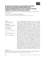
Expression of oncogenic Ras induces an increase of both
cellular ST6Gal I activity and cell surface a2,6- sialylation
FACS analysis (Fig. 2) w ith t he Neu5 Aca2,3Gal-specific
lectin from MAA found no significant differences between
Ras transformed and control fibroblasts. Furthermore,
MAA staining was high in both cell lines, consistent with the
observation of relative high amounts of ST3Gal I, III and
IV mRNA already in t he nontransformed cells (Fig. 1) and
only slightly stronger in the Ras transformed cell line.
However, we were not able to correlate the higher amounts
of cell surface Neu5Aca2,3Gal in the transformed cells to
increased a2,3-sialyltransferase activity ([7] a nd data not
shown), and they may therefore be due not to an
augmentation in transferase activity but to an increase in
precursor s tructures a s would be expected in Ras trans-
formed cells where branching of N-glycans has been shown
to be more abundant [10,38]. On the other hand, a2,6-
sialyltransferase activity toward the acceptor Galb1,4Glc-
NAcb-OCH
2
Ph was elevated approximately sixfold in
H-Ras
V12
and K -Ras
S12
expressing cell lines relative to
mock transfected 3T3 cells (Table 1 ). In addition, FACS
analysis using the Neu5Aca2,6Gal-specific lectin SNA
found a sixfold higher mean fluorescence intensity on the
3T3K-Ras
S12
cells than on the parental line 3T3 (Fig. 2).
These data confirm e arlier work linking increased ST6Gal I
activity and cell surface SNA staining to the expression of
oncogenic Ras in rodent fibroblasts [6–9].
Conditional transient expression of activated Ras induces
ST6Gal I mRNA accumulation
Both cell lines 3T3K-Ras
S12
and 3T3H-Ras
V12
had been
selected on the basis of colony formation in s oft a gar a s a n
indicator for a malignant phenotype. However, during the
lengthy selection process o ther genetic changes may o ccur
and may contribute to t he increased expression o f the
ST6Gal I gene. Therefore we obtained a cell line transfected
with a plasmid carrying the neomycine resistance gene
under the control of a strong constitutive promoter and
the H -Ras
V12
gene under the control of t he tetracycline
repressor. Stable transfectants were selected with G418
whilst H-Ras
V12
was repressed with tetracycline during the
selection process [35]. T hese cells (mib125
+Tc
) show the
same low l evel of ST6Gal I mRNA (Fig. 3, lanes 1 and 3 )
and corresponding enzyme activity (not shown) as the
nontransfected parent cells. Upon de-rep ression of R as
expression by removing tetracycline from the medium
(mib125
–Tc
) both H-Ras
V12
and ST6Gal I mRNAs
increased to the same levels as those observed in the cell
line constitutively expressing H-Ras
V12
(Fig. 3, lanes 2 and
Fig. 2. Increas ed cell surface a2,6-sialylation in cells expressing oncogenic Ras. FACS analysis of lectin-labelled c ells: left panels, SNA staining; right
panels, MAA staining; upper panels contro l 3T3 cells, lower panels 3T3K -Ras
S12
transformed cells as indicated. Narrow lines FITC-streptavidin
controls, bold lines over grey background biotinylated le ctin/FITC-streptavidin staining.
Ó FEBS 2004 Oncogenic Ras signalling and ST6Gal I activation (Eur. J. Biochem. 271) 3627
4). T he increase of mRNA was concomitant t o the increase
in enzyme activity and SNA staining (not shown). When the
Ras gene was again repressed, the ST6Gal I mRNA
decreased to normal levels within 72 h of tetracycline
treatment (Fig. 3, lanes 5 and 6). Again, enzyme activity and
SNA staining followed the same trend. These results
demonstrate that, like malignant phenotype and focus
formation [35], the increase i n ST6Gal I is directly depend-
ent on the expression of activated Ras.
H-Ras signals to Siat1 primarily through the RalGEF
pathway
To investigate t he contribution o f i ndividual Ras signalling
pathways on the expression of ST6Gal I, th ree effector
domain mutants of oncogenic H-Ras were transfected into
3T3 fibroblasts and after selection of stable transfectants the
total RNA w as extracted from several clones for each
transfection and analyzed by northern hybridization (Fig. 4
shows one represen tative northern f or each transfection
experiment). Only clones from cells transfected with the
H-Ras
V12G37
which allows binding of only the RalGEFs
exhibited the same high levels of ST6Gal I mRNA and
ST6Gal I activity toward the disaccharide Galb1,4Glc-
NAcb-OCH
2
Ph as H-Ras
V12
transformed cells (Table 1).
The mutant H-Ras
V12S35
activating only the Raf k inase
pathway h ad no effect on ST6Gal I expression whereas the
Ras mutants H-Ras
V12C40
which bind only to PI3-kinase
could, in some of the c lones analyzed, produce an increase
of ST6Gal I mRNA and ST6Gal I enzyme activity. Only
the results from the clone with the highest ST6Gal I mRNA
level are shown (Fig. 4, Table 1). Oncogene expression
was confirmed by H-Ras specific RT-PCR as p reviously
described for 3T3H-Ras
V12
.
H-Ras and K-Ras induce
Siat1
transcription via
the housekeeping promoter P3
To obtain information on the nature o f t he 5 ¢-UTRs of the
ST6Gal I transcripts expressed by Ras transfected and
control 3T3 cells , RNA from each of the cell lines 3T3,
3T3K-Ras
S12
,3T3H-Ras
V12
, mib125
–Tc
and mib128 was
subjected to 5 ¢-RACE analysis (md11/AP1 primer pair).
The re sults are summarized in Table 2. In all the cell line s
studied, the ST6Gal I 5¢UT sequences ob tained were
composed of three k inds of sequences: (a) the sequence
usually transcribed from the P3 promoter (exon Q and O
immediately 5¢ of the untranslated c onserved exon I); (b) a
previously unidentified 5¢UT sequence v ery close to that
obtained f rom t he P3 promoter (a novel exon, n amed exon
R, 5¢ of exons O and I), and (c) a probably t runcated
sequence containing only part o f the sequence transcribed
from the P 3 promoter ( partial exon O immediately 5¢ of the
untranslated con served exon I). The dominant form in every
Fig. 3. Transient expr ession of H-Ras
V12
coincides with ST6Gal I
expression. Northern hybridization analysis of NIH3T3 line mib125 in
which H-Ras
V12
expression is repressed by Tc. For all panels, 30 lgof
total RNA were loaded in the following order: lane 1: mi b35 parental
3T3 cells; lane 2: mib128 expressi ng H-Ras
V12
constitutively; lane 3:
mib 125 + Tc; lane 4: mib125 –Tc f or 72 h; lane 5: mib125 –Tc for
120 h ; lane 6: mib125 kept w/o Tc for 72 h then Tc was added for 72 h.
Labelled cDNA prob es were as indicated to the left. Loading control:
18S R NA.
Fig. 4. H-Ras
V12
signals to Siat1 primarily through the RalGEFs signal
transduction pa thway. Northern analysis of total RNA from NIH3T3
cells transfected w ith p artial lo ss of function H-Ras
V12
mutants S 35,
G37 and C40 with cDNA probes of ST6Gal I and H -Ras as indicate d
to the left. For all pan els, 30 lg of total RNA were loaded in the
following order: lane 1: 3T3; lane 2: mock transfected 3T3; lane 3 :
3T3H-Ras
V12C40
;lane4:3T3H-Ras
V12S35
;lane5:3T3H-Ras
V12G37
;
and lane 6: 3T3H-Ras
V12
. Loading co ntrol: 18S RNA.
Table 1. ST6Gal I activities in Ras transformed and control NIH3T3
cells. Enzyme activities were measured in total cell lysates as described
under Experimental procedures. The values are the mean of three
independent experiments.
Cell lines
ST6Gal I activity
(pmolÆh
)1
Æmg protein
)1
)
3T3 4.8 ± 0.4
3T3pB322 11.7 ± 1.8
3T3K-Ras
S12
74.2 ± 2.6
3T3H-Ras
V12
77.4 ± 4.7
3T3Neo 11.1 ± 0.9
3T3H-Ras
V12–S35
8.1 ± 0.4
3T3H-Ras
V12–C40
32.3 ± 1.7
3T3H-Ras
V12–G37
70.1 ± 3.1
3628 M. Dalziel et al. (Eur. J. Biochem. 271) Ó FEBS 2004
sample studied was the ÔtruncatedÕ P3 form (ranging from 74
to 95% of the total number of clones). As these truncated P3
sequences could be representative of either partial 5 ¢-RACE
cDNA synthesis or actual t ranscription initiation within
exon O, a second 5¢-RACE experiment w as carried out
using an antisense primer located at the extreme 3¢ tip of
exon O (primer O2) a nd the M arathon AP1. When the
3T3pB322 and 3 T3H-Ras
V12
samples were subjected to this
modified 5¢-RACE, only clones representing the common
(Q-O-I) and novel (R-O-I) P3 isoforms were obtained
(Table 2). These data indicate that the truncated P3
sequences generated from the AP1/md11 5¢-RACE experi-
ment are derived from incomplete cDNA synthesis within
exon O itself, probably as the result of seco ndary mRNA
structure. Furthermore, we can eliminate the possibility that
R is merely an unprocessed sequence between Q and O as
the O2 RACE could cover the entire Q form, whilst still
giving rise to clones containing the shorter R-O sequence,
strongly suggesting that both Q and R are separate and
distinct 5¢-termini.
Contribution of Q and R forms of P3 to Ras signal
Although no obvious association with either the classical o r
alternative P3 f orms could b e seen w ith any of the R as
expressing cell lines when 5¢-RACE was carried out using
AP1/md11, t here was a bias toward the Q form in 3T3H-
Ras
V12
and the R f orm in 3T3pB322 cells using AP1/O 2, as
seen by ethidium bromide-stained gel analysis of the
products (not shown) and subsequent sequencing (Table 2),
although the number of clones analyzed was small. Thus
PCR probes for exons Q and R were amplified from 3T3H-
Ras
V12
cDNA and used as probes to detect the expression of
each isoform in the three activated H-Ras expressing cell
lines 3T3H-Ras
V12
,mib125
–Tc
and mib128, relative to the
control lines, 3T3pB322, mib125
+Tc
and mib35, r espect-
ively. The exon Q probe gave a detectable hybridization
signal in all three of the Ras cell lines but very little or no
signal in the respective controls (Fig. 5) Similarly, the e xon
R p robe resulted in a detectable signal in all t hree Ras lines
but none in the respective c ontrols (Fig. 5). High ST6Gal I
mRNA levels in the three Ras samples was confirmed using
the exon II probe as before (Fig. 5). The signal obtained
with the Q probe was much greater than that of R in the
3T3H-Ras
V12
sample (consistent with the O2 RACE results)
whereas the R signal was greater than Q in the m ib125
–Tc
and mib128 samples. T he data presented in Fig. 5 confirm
the presence o f two independent transcription start sites in
the P3 promoter region and indicate that transcription from
both sites is up-regulated in response to transformation with
oncogenic Ras.
Mapping of exon R relative to exons Q and O within
the mouse
Siat1
gene
Making use o f the online public access E nsembl mouse
genome server ( />the entire Siat1 gene was mapped (Ensembl gene ID:
ENSMUSG00000022885, chromosome 16 nucleotides
22918721–23318720), and exon R subsequently located
between exons Q and O, with an 820 bp intron between Q
and R (Fig. 6B). The common splice sequence s een in the
RACE clones allowed the exact definition of the 3¢
termination of both exons Q and R within the Siat1
genomic sequence, as well as the 5 ¢ of exon O (Fig. 6A). All
three exons contain the splice donor sequence GT imme-
diately 3¢ of the exon. Further, exon O has a splice acceptor
AG immediately 5¢ of it. Using this information, a complete
schematic representation o f the complete mouse Siat1 gene,
including the Ras induced P3 mRNA isoforms, was
constructed (Fig. 7). A nalysis of the 5¢ sequences upstream
of exons Q and R b y the MatInspector database [39] failed
to find TATA or CAAT boxes and identified several
Table 2. 5¢UT sequences o f S T6Gal I mRNA in Ras t ransformed and
control cell lines. The n um bers o f clo nes with 5 ¢ sequences begin ning
within exons Q, R or O are given. For e xon nomenclature a nd the
overall o rganization of t he Siat1 gene see Fig. 7 .
Cell lines
Number of clones starting in exons
QRO
5¢-RACE results using primer pair md11 (Siat1 exon I) and AP1
(marathon adaptor)
3T3 5 1 41
3T3K-Ras
S12
5734
3T3H-Ras
V12
4250
mib125
–Tc
1231
mib128 0 2 37
5¢-RACE results using primer pair O2 (extreme 3¢ of exon O) and
AP1 (marathon adaptor)
3T3pB322 0 3 0
3T3H-Ras
V12
91 0
Fig. 5. Contribution of e xons Q and R to the 5¢UT region of ST6Gal I
mRNA from Ra s tr ansfe cted an d c ontr ol 3T 3 ce lls. Northern hybrid-
ization of 50 lg total RNA from Ras transfected and control lines
using the PCR generated probes for 5¢UTexonsQandRandthefirst
coding exon II as indicated to the left. Lane 1: 3T3pB322, lane 2:
3T3H-Ras
V12
; l ane 3: mib35; lane 4: mib125
+Tc
; l ane 5: mib125
–Tc
;
lane 6: m ib128. Loading control: 18S R NA.
Ó FEBS 2004 Oncogenic Ras signalling and ST6Gal I activation (Eur. J. Biochem. 271) 3629
transcription factor binding sites t ypical of housekeeping
promoters. Among these, double GC boxes and Ras-
responsive element binding protein-1 sites are located
immediately 5¢ of bo th exons Q and R (Fig. 6B).
Discussion
Aberrant glycosylation occurs in essentially all types of
experimental and human cancers [40]. A long-standing
debate is how aberrant glycosylation is related to cancer and
whether it is the result of initial oncogenic transformation.
Studies on R as transformed r odent fibroblasts indicated
that the expression of oncogenic H-Ras
V12
leads to changes
in the N-glycan structure of cell surface g lycoproteins. The
principal modifications on N-glycans observed were
increased complexity of N-glycan branching and changes
in N-glycan sialylation from a3- to a6-linked Neu5Ac
sialylation [4–10].
However, these studies were carr ied out on single clones
of H-Ras
V12
transformed fibroblasts which had been
selected over prolonged periods of time for an increased
growth rate and for the a bility to form colonies in soft agar.
During this lengthy selection process unidentified genetic
changes may h ave occurred which could have contributed
to the m odification i n N-glycan b iosynthesis. Such changes
did occur as a few clones could be selected which did not
show altered N-glycan structures or increased sialylation [9].
In order to address t he q uestion whether H-Ras
V12
was
directly or indirectly involved, via ST6Gal I, in the
augmentation of N-glycan sialylation, we measured ST6Ga-
l I in NIH3T3 fibroblasts that express activated Ras
conditionally. In these cells the transformed phenotype
and the ability to form foci in soft agar are directly
dependent on the expression of Ras and are c ompletely
reversible [35]. In the absence of H-Ras
V12
these cells exhibit
the same low levels of ST6Gal I as the non transformed
Fig. 6. The P 3 promoter region of Siat1: 5¢-
RACE data deri ved from Q and R containing
clones allowed the precise definition of b oth the
3¢ ends of exons Q and R as we ll as the 5¢ end
of exon O. (A) Proposed conserved splice
acceptor lo cation at th e 5¢ endofexonO,
utilized by both Q and R. (B) Exon sequences
Q and R in u pper case, introns in lower case,
exon–intron boundaries underlined. The d is-
tance between Q a nd R is l ess than 820
nucleotides. Putative tran scription factor
binding sites: GC box (shaded); Ras-respon-
sive element b inding protein-1 (double
underlined).
3630 M. Dalziel et al. (Eur. J. Biochem. 271) Ó FEBS 2004
fibroblasts and only when t he expression of H-Ras
V12
is
induced do these cells show the same high levels of ST6Gal I
mRNA as the constitutively H-Ras
V12
transformed cell
lines. This enhancement of ST6Gal I expression is reversible
at the level of mRNA as well as at the level of cell s urface
expression of the Neu5Aca2,6Galb4GlcNAc epitope (not
shown). These results clearly indicate that the presence of
activated Ras alone and n o other genetic or e pigenetic
events are responsible for the elevated expression of
ST6Gal I i n Ras transformed murine fibroblasts.
Among the members of the Ras gene family we found
that both K-Ras
S12
and H-Ras
V12
promote the same large
increase in ST6Gal I mRNA in fibroblasts. The former is of
particular relevance as it is the predominantly mutated RAS
gene in human cancer [41]. Although N-Ras was not
included in our study, it i s known that expression of normal
N-Ras has a positive influence on both cellular sialylation
and ST6Gal I activity [42]. The influence o f H- a nd K-Ras
transformation appears t o be restricted t o the Sia t1 gene, at
least amongst its immediate family members, a s none of the
other sialyltransferases a nalyzed showed notable changes in
their transcript levels. In several human cancers where
activating Ras mutations a re common, it may b e significant
that high ST6Gal I activity is the most frequent alteration to
the expression pattern of the sialyltransferase family.
As Ras signals directly to the Siat 1 gene we wanted to
know through which pathway the signal may be delivered.
Ras signals mainly through t hree pathways: t he Raf-MEK-
ERK signalling cascade which promotes proliferation
through the activation of transcription f actors; the PI3
kinase pathway where lipid kinases generate second mes-
sengers which have diverse effects o n cellular physiology
and t he RalGEF signalling cascade whic h i nvolves a whole
family of RalGTPases but most of the downstream
activators are still not identified. For each of the three
pathways, partial loss of function mutants of activated Ras
proteins have been created [31,32] which can selectively bind
to one of the e ffectors and thus signal through one pathway
only. H-Ras
V12S35
binds only to R af and is unable to
activate the two other signalling cascades whereas
H-Ras
V12C40
binds exclusively to PI3 kinases and the
H-Ras
V12G37
mutant specifically activates the RalGEF
pathway. When the t hree constructs coding the mutant
Ras proteins were transfected into 3T3 fibroblasts only the
H-Ras
V12G37
mutant was able to induce the increased
expression of ST6Gal I similar to the wild type oncogenic
H-Ras
V12
. Interestingly, the PI3 kinase pathway may also
contribute to the activation of the ST6Gal I gene but at a
much lower level and not all of t he clones with high levels
of H-Ras
V12SC40
showed increased amounts o f ST6Gal I
mRNA. These results indicate that Ras signals to the
ST6Gal I gene principally through the RalGEF signalling
pathway. The rise in ST6Gal I mRNA was a lways accom-
panied by a concomitant increase i n ST6Gal I enzyme
activity.
The RalGEF pathway is the least well documen ted of the
three major sign alling pathways and mo st of the physiolo-
gical consequences of RalGEF activation are still outstand-
ing issues. H owever its i mportance has recently come into
focus with the mounting evidence that it is the principal
pathway used by Ras to transform human cells [43]. One
recent study links RalGEF activation i n rodent fibroblasts
to the development of highly invasive metastases when those
cells are administered subcutaneously to nude mice [44]. The
formation of aggressive tumours may be correlated to the
increased expression of ST6Gal I as clones of Ras trans-
formed rat fibroblasts which had lost the e xpression of
the Neu5Aca2,6Galb4GlcNAc epitope synthesized by
ST6Gal I were f ound to be much less metastatic than the
clones which still possessed this glycan structure [9].
Tissue-specific expression levels of ST6Gal I are regulated
by the use o f tissue specific splice forms of its mRNA
derived f rom selective t ranscription of multiple promoters.
In order t o localize the promoter region targeted by Ras we
wanted to identify the 5¢UT isoform, which is ind uced by
the R alGEF s ignal. In all cell lines studied that expre ss
activated K-Ras
S12
or H-Ras
V12
, the ST6Gal I transcripts
found represent the isoform transcribed from the P3
Fig. 7. Mapping o f P3 w ithin the com plete
Siat1 genomic structure. Schematic r epresen-
tation of the mouse ST6Gal I gene Siat1.This
gene spans over 130 kb on chromosome 16.
The transcription start s ites at the four major
promotors are indicated by a rrows. The open
reading frame is encoded by exons II through
VI. Exon I is an invariant 5¢UT exon found in
all Siat1 m RNA. The two l oc ations of the
presumed transcriptional start sites used by
Siat1 in Ras expressing cells are indicated by
big arrows (P3a and P3b). The resulting
mature transcripts both contain the 5¢UT
exons O and I preceded by either exon Q or
exon R. Transcription start sites from tissue
specific promoters are indicated by s mall
arrows:P1,liver;P2a-c,Bcells,P4,lactating
mammary gland.
Ó FEBS 2004 Oncogenic Ras signalling and ST6Gal I activation (Eur. J. Biochem. 271) 3631
housekeeping p romoter. Although enhanced steady state
transcription is the most obvious explanation for the
accumulation of ST6Gal I mRNA in the presence of
K-Ras
S12
or H-Ras
V12
it cannot be excluded that increased
mRNA stability may also contribute to the high levels of
ST6Gal I mRNA in Ras transformed cells. However,
previous work has shown that quantitative c hanges of a
particular class o f ST6Gal I 5¢UT transcripts ( including P3)
are primarily the result of t ranscriptional a ctivity a t the
matching promoter [45–47].
In the adult mouse, P3 is normally active in most tissues
and gives rise to a mature ST6Gal I mRNA leading with the
5¢UT sequences encoded b y exons Q, O a nd I (Fig. 7). We
detected these same transcripts in ST6Gal I mRNA derived
from Ras transformed cells, but alongside a previously
unreported variant where exon Q is replaced by a s equence
named exon R. As both exons Q and R make use of a
conserved splice junction at exon O, are located within the
same region on the Siat1 gene (less t han 8 20 bp apart) and
are c oexpressed, there are two possible explanations for the
presence of these a lternative 5¢UT leader s equences. Both Q
and R isoforms could be derived from a single promoter
with a certain degree of initiation site variability. Although it
is quite common for housekeeping promoters to have several
transcription initiation sites, these are u sually foun d much
closer together than those f or exons Q and R, normally less
than a hundred and usually within 30 nucleotides of each
other [48]. O n t he oth er h and, Q and R may represent
transcription initiation sites of two r elated and overlapping
housekeeping promoter regions within the Siat1 gene.
However, neither Q nor R appears to be favoured by the
activation through th e RalGEF pathway although s ome
differences could be observed b etween cell lines.
A preliminary analysis of the sequences directly upstream
of the two tr anscription start sites i dentified several putative
consensus sequences for transcription factors t ypical of
housekeeping promoters. As both of these regions are
equally responsive to Ras, shared consensus sequences for
transcription f actors could be a key to understand their
regulation. It is therefore interesting to note that two
sequences recognized by Ras- responsive element b inding
protein-1 are present within a few hundred nucleotides of
each transcription start site.
An additional ob servation suggests that the transcription
is initiated a t a true housekeeping promoter. Although t he
amount of ST6Gal I m RNA p roduced in Ras t ransformed
cells is close to the levels found in liver [49] the s pecific
enzymatic activity of ST6Gal I in the transformed fibro-
blasts is much lower [27,37]. This is consistent with
observations that some transcripts derived from housekeep-
ing promoters have a low translation rate i n large part due
to stable secondary structures at their 5¢UT region [50]. This
strong secondary structure could possibly account for the
high frequency of truncated sequences in the 5¢-R ACE
experiments described in this study.
The key effect of Ras on growth is to overcome c ontact
inhibition between cells [35]. T he rise of a6-linked sialic acid
on N-glycans of cell surface glycoproteins as a direct result of
oncogenic Ras expression may contribute to the repression of
contact inhibition. Contact-mediated inhibition of cell
migration and cell proliferation is co-ordinately regulated
by integrins and their receptors. Recently it has been reported
that b1 integrin activity is dependent on the sialylation of its
N-glycans a nd that the R as induced change from a3-linked
to a6-linked sialic acid alters the binding to some of its ligands
[23]. In addition, RalGEF activation through Ras induces
aggressive behaviour in tumours that may als o be related to
the increase in ST6Gal I activity. Together these two
examples indicate that shifts in sialyltransferase expression
patterns may be an important contribution of oncogenic Ras
to the m etastatic potential of tumours.
Acknowledgements
MD is the recip ient of a postdoctoral fellowship from Le STU DIUMÒ
(Orle
´
ans, France). This wo rk was supported by grants f rom the Ligue
Nationale contre le Cancer (comite
´
sde
´
partementaux du L oiret et du
Loir et Ch er), the Centre National d e la Recherche Scientifique:
Prote
´
omique et Ge
´
nie des Prote
´
ines, by the Groupement de Recherche:
Ge
´
nomique et Ge
´
nie des Glycosyltransfe
´
rases and by t he Orle
´
ans
chapter of the Lions Club. FD acknowledges grants from MURST and
the Universita
`
di Bologna. We are gra teful to Dr J. La u (Ros well Park
Cancer Institute, Bu ffalo, NY, USA), P rof E. H e
´
bert (CBM, Orle
´
ans,
France), Dr B. Miller (Michigan Cancer Foundation, Detroit, MI,
USA), Dr A. Scibetta (Cancer Research UK, Guy’s Hospital, London,
UK), Dr S. Tsuji (The Glycoscience Institute, Tokyo, Japan) and Prof
B. M. Willumsen (University of Copen hagen, Denmark) for their
generous gifts of plasmids or cell lines and to Dr C. LeNarvor and D r
C. Auge
´
(Universite
´
Paris-Sud, Orsay, France) for k indly providing
Galb1,4GlcNAcb-OCH
2
Ph.
References
1. Campbell, S.L., Khosravi-Far, R., Rossman, K.L., Der Clark,
G.J., Gilbert, F. & Glick, M.C. (1984) Change in glycosylation of
membrane glyc oproteins after transfection of NIH 3T3 w ith
human tumor D NA. Cancer Re s. 44, 3730–3735.
2. Bos, J.L. (1995) p21ras: an oncoprotein functioning in growth
factor-induced signal transduction. Eur. J. Cancer 31A, 1051–
1054.
3. McMahon, M. & Woods, D. (2001) Regulation of the p53 path-
way by Ras, the plot thickens. Biochim Biophys Acta 14 71,M63–
71.
4. Santer, U .V., Gilbert, F. & Glick, M.C. (1984) Change i n glyco-
sylation of membrane glycoproteins after t ransfection of NIH 3T3
with hu man tumor DNA. Cancer Res 44, 3730–3735.
5. Santer, U.V., DeSantis, R., Hard, K.J., van Kuik, J.A., Vlie-
genthart, J.F., Wo n, B . & G lick, M.C. (1989) N-Linked oligo-
saccharide changes with oncogenic transformation require
sialylation o f multiantennae. Eu r. J. Biochem. 181, 249–260.
6. Le Marer, N., Laudet, V., Svensson, E.C., Cazlaris, H., Van Hille,
B.,Lagrou,C.,Stehelin,D.,Montreuil,J.,Verbert,A.&
Delannoy, P. (1992) The c-Ha-ras oncogene induces increased
expression of b-galactoside a-2,6-sialyltransferase in rat fibroblast
(FR3T3) cells. Glycobiology 2, 49–56.
7. Vandamme, V., Cazlaris, H., Le Marer, N ., Laudet, V., Lagr ou,
C., Verbert, A. & Delannoy, P. ( 1992) Comparison o f sialyl- and
a1,3-galactosyltransferase activity in NIH3T3 cells transformed
with ras oncog ene: in creased b-galactoside a2,6-sialyltransferase.
Biochimie 74, 89–99.
8. Delannoy,P.,Pelczar,H.,Vandamme,V.&Verbert,A.(1993)
Sialyltransferase activity in FR3T3 ce lls transformed with r as
oncogene: decreased CMP-N eu5Ac. Galb1–3galnac:a2,3-sialyl-
transferase. Glycoconj. J. 10 , 91–98.
9. Le Marer, N. & Stehelin, D. (1995) High a2,6-sialylation of
N-acetyllactosamine sequences in ra s-transformed rat fibroblasts
correlates wi th high invas ive potential. Glycobiology 5, 219–226.
3632 M. Dalziel et al. (Eur. J. Biochem. 271) Ó FEBS 2004
10. Dennis, J.W., K osh, K., Bryce, D.M. & Breitman, M.L. (1989)
Oncogenes conferring metastatic potential induce increased
branching of Asn-linke d oligosaccharides i n rat2 fi brob lasts.
Oncogene 4, 8 53–860.
11. Recchi, M.A., Hebbar, M., Hornez, L., Harduin-Lepers, A.,
Peyrat, J.P. & Delannoy, P. (1998) Multiplex reverse transcription
polymerase chain reaction a ssessment of s ialyltransferase expres-
sion in human breast c ancer. Cancer Res. 58 , 4066–4070.
12. Gessner,P.,Riedl,S.,Quentmaier,A.&Kemmner,W.(1993)
Enhanced activity o f C MP-NeuAc:Gal b1,4GlcNAc:a2,6-sialyl-
transferase i n me tastas izing hu man colorectal tumor tissue and
serum of t umor patients. Cancer Lett. 75, 143–149.
13. Dall’Olio, F ., Chiricolo, M., Ceccarelli, C., Minni, F., Marrano,
D. & Santini, D. (2000) Beta-galactoside a2,6-sialyltransferase i n
human colon canc er: contribution of multiple transcripts to
regulation of enzyme activity and reactivity w ith Sambucus nigra
agglutinin. Int. J. Cancer 88 , 58–65.
14. Kemmner, W., Roefzaad, C., Haensch, W. & Schlag, P.M. (2003)
Glycosyltransferase expression in human colonic tissue e xamined
by oligonucleotide arrays. Biochim. Biophys. Ac ta 1621, 272–279.
15. Wang, P.H., Li, Y.F., Ju ang, C.M., Lee, Y .R., Chao, H.T., Tsai,
Y.C. & Yuan, C.C. (2001) Altered mRNA expression of sialyl-
transferase in squamous cell carcinomas of the cervix. Gynecol.
Oncol. 83, 1 21–127.
16. Singh, D., Febbo, P.G., Ross, K., Jackson, D.G., Manola, J.,
Ladd, C., Tam ayo, P., Renshaw, A.A., D’Amico, A.V., Richie,
J.P., Lander, E.S., Loda, M., K an toff, P.W., Golub, T.R. &
Sellers, W.R. (2002) Gene expression correlates of clinical prostate
cancer behavior. Canc er Cell 1, 203–209.
17. Gangopadhyay, A., Perera, S.P. & Thomas, P. (1998) Differential
expression of a2,6-sialyltransferase in colon tumors recognized by
a monoclonal a n tibody. Hybridoma 17 , 117–123.
18. Gretschel, S., Haensch, W., Schlag, P.M. & Kemmner, W. (2003)
Clinical relevance of sialyltransferases ST6Gal-I and ST3Gal-III
in gastric cancer. Oncology 65, 139–145.
19. Wang, P .H., Li, Y.F., Juang, C. M., L ee, Y .R., Chao, H.T., Ng,
H.T., Tsai, Y.C. & Yuan, C.C. (2002) Expression of sia lyl-
transferase family members in cervix squamous cell carcinoma
correlates with lymph node metastasis. Gynecol. Oncol. 86, 45–52.
20. Bosch, J., B rossmer, R., Kemmner, W. & Schlag, P. (1998) Pre-
paration and characterization of differently aggregated colorectal
carcinoma cell subpopulations from surgical specimens. Cancer
Detect. Prev. 22, 3 19–329.
21. Zhu, Y., Srivatana, U., Ullah, A.,Gagneja,H.,Berenson,C.S.&
Lance, P . (2001) Suppression of a sialyltransferase by antisense
DNA reduces invasive ness of human colon cance r cells in vitro.
Biochim. Bioph ys. Acta 15 36, 148–160.
22. Lin, S., Kemmner, W., Grigull, S. & Schlag, P.M. (2002) Cell
surface a-2,6 sialylation affects adhesion of breast carcinoma cells.
Exp. Cell Res. 276, 101–110.
23. Seales, E .C., Jurado, G.A., Si nghal, A. & Bellis, S .L. (2003) R as
oncogene directs expression of a differentially sialylated, func-
tionally altered b1integrin.Oncogene 22, 7137–7145.
24. Weinstein, J., de S ouza-e-Silva, U. & P aulson, J.C. ( 1982) Sialy-
lation of glycoprotein oligosaccharides N-linked to asparagine:
enzymatic c harac terization of a Galb1,3(4)GlcNAc:a2,3-sialyl-
transferase and a Gal b1,4GlcNA c:a2,6–sialyltransferase from rat
liver. J. Biol. Chem. 257, 1384 5–13853.
25. Takashima, S., Tsuji, S. & Tsujimoto, M. (2002) Characterization
of the second type of human b-galactoside a2,6-s ialyltran sfera se
(ST6Gal II), which sialylates Galb1,4GlcNAc structures on
oligosaccharides preferentially: genomic analysis of human sialyl-
transferase g enes. J. Biol. Chem. 277, 45719–45728.
26. Takashima, S., Tsuji, S. & Tsujimoto, M. (20 03) Comparison of
the enzymatic properties of mou se b-galactoside a2,6-sialyl-
transferases,ST6GalIandII.J. Biochem. (Tokyo) 134, 287–296.
27. Hennet, T., Chui, D., Paulson, J.C. & Marth, J.D. (1998) Immune
regulation by the ST6Gal s ialyltransferase. Proc. N atl Acad. Sci.
USA 95, 4 504–4509.
28. Hu, Y.P., Dalziel, M. & Lau, J.T. (1997) Murine hepatic b-gal-
actoside a2,6-sialyltransferase gene expression involves usage of a
novel upstream exon region. Glycoconj. J. 14, 407–411.
29. Wuensch, S.A., Huang, R .Y., Ewing, J., L iang, X. & Lau, J.T.
(2000) Murine B cell d ifferentiation is a ccompanied b y pro-
grammed expression of multiple novel b-galactoside a2,6-sialyl-
transferase mRNA f orm s. Glycobiology 10, 67–75.
30. Dalziel, M., Huang, R.Y., Dall’Olio, F., Morris, J.R., Taylor-
Papadimitriou, J. & L au, J.T. (2001) Mouse S T6Gal sialyl-
transferase g ene expression during mammary gland lactation.
Glycobiology 11, 4 07–412.
31. White, M.A. , N icolette, C ., Minden, A., Polverino, A., V an
Aelst, L., Karin, M . & Wigler, M .H. (1995) M ultiple R as func-
tions can contribute to m ammalian ce ll transformation. Cell 80 ,
533–541.
32. Rodriguez-Viciana, P., Warne, P.H., Khwaja, A., Marte, B.M.,
Pappin, D., Das, P., Waterfield, M.D., Ridley, A. & Downward, J.
(1997) Role of p hosphoinositide 3-OH kinase in ce ll transforma-
tion and control of t he actin cytoske leton by R as. Cell 89, 457–
467.
33. Hebert, E. & Monsigny, M. (1994) Galectin-3 mRNA level de-
pends on transformation phenotype in ras-transformed NIH 3T3
cells. Biol. C ell 81, 73–76.
34. Miller, F .R., Miller, B.E. & Heppner, G .H. ( 1983) Characteriza-
tion of metastatic hete rogeneity among subpop ulations of a single
mouse mammary tumor: heterogeneity in phenotypic stability.
Invasion Metasta sis 3, 22–31.
35. Jacobsen, K., Groth, A. & Willumsen, B.M. (2002) Ras-
inducible immortalized fibroblasts: focus formation without cell
cycle deregulatio n. Oncogene 21 , 3058–3067.
36. Gumerlock, P.H., Poonamallee, U.R., Meyers, F.J. & deVere
White, R.W. (1991) Activated ras a lleles in human carcinoma of
the prostate a re rare. Cancer Res. 51 , 1632–1637.
37. Pousset, D., Piller, V., Bureaud, N., Monsigny, M. & Piller, F.
(1997) I ncreased a2,6-sialylation o f N-glycans in a t ransgenic
mouse model of hepatocellular carcinoma. Cancer Res. 57, 4249–
4256.
38. Bolscher, J .G., van der Bijl, M.M. , Neefjes, J.J., Hall, A., Smets,
L.A. & Ploegh, H.L. (1988) Ras (proto)oncogene induces
N-linked c arbohydrate modification: temporal relationship wit h
induction of i n vasive potential. EMBO J. 7, 3 361–3368.
39. Quandt, K., Frech, K., K aras, H., Wingender, E. & Werner, T.
(1995) MatInd and MatInspector: new fast and versatile tools for
detection of consensus matche s in nucleotide sequenc e data.
Nucleic A cids Res. 23 , 4878–4884.
40. Hakomori, S.I. (2002) Glycosylation defining ca ncer. Proc. Natl
Acad. Sc i. USA 99 , 10231–10233.
41. Ellis, C.A. & Clark, G. (2000) The importance o f being K-Ras.
Cell Signal. 12, 425–434.
42. Easton, E.W., Bolscher, J.G. & van den Eijnden, D.H. (1991)
Enzymatic amplificat ion involving glycosyltransferases forms the
basis for the increased size of asparagine-linked glycans at the
surface of N IH 3T3 cells expressing th e N-ras proto-oncogene.
J. Bi o. Chem. 266 , 21674–21680.
43. Hamad, N.M., Elconin, J.H., Karnoub, A.E., Bai, W., Rich, J.N.,
Abraham, R.T., Der, C.J. & Counter, C.M. (2002) Distinct
requirements for R as oncogenesis in human versu s mouse cells.
Genes Dev. 16 , 2045–2057.
44. Ward,Y.,Wang,W.,Woodhouse,E.,Linnoila,I.,Liotta,L.&
Kelly, K. (2001) Signal pathways which promote invasion and
metastasis: c ritical and distinct contributions o f extracellular
signal-regulated kinase and Ral-specific guanine exchange factor
pathways. Mol. Cell Biol. 21, 5958–5969.
Ó FEBS 2004 Oncogenic Ras signalling and ST6Gal I activation (Eur. J. Biochem. 271) 3633
45. Aas-Eng, D.A., Asheim, H.C., Deggerdal, A., Smeland, E. &
Funderud, S. (1995) Characterization of a promoter region sup-
porting transcription of a novel human b-galactoside a2,6-sialyl-
transferase transcript in HepG2 cells. Biochim. Biophys. Acta 1261,
166–169.
46. Lo, N.W. & Lau, J.T. (1996) Transcription of the b-galactoside
a2,6-sialyltransferase gene in B lymphocytes i s directed b y a sep-
arate and distinc t promoter. Glycobiology 6, 271–279.
47. Taniguchi, A., Hasegawa, Y., Higai, K. & Matsumoto, K. (2000)
Transcriptional r egulation of human b-galactoside a2,6-sialyl-
transferase (hST6Gal I) gen e during differentiation of the HL-60
cell line. Glycobiology 10, 623–628.
48. Azizkhan,J.C.,Jensen,D.E.,Pierce,A.J.&Wade,M.(1993)
Transcription from TATA-less promoters: dihydrofolate
reductase as a model. Crit. Rev. Eukaryot. Gene Expr. 3, 229–254.
49. Dalziel, M., Lemaire, S., Ewing,J.,Kobayashi,L.&Lau,J.T.
(1999) Hepatic acute phase induction of murine b-galactoside
a-2,6-sialyltransferase (ST 6Gal I) is IL-6 depend ent and mediated
by elevation of exon H-containing class of transcripts.
Glycobiology 9, 1003–1008.
50. Charron, M., Shaper, J.H. & Shaper, N.L. ( 1998) The in creased
level of b1,4-galactosyltransferase required for lactose biosynthesis
is achieved in part by tran slational control. Proc. Natl Acad. Sci.
USA 95, 1 4805–14810.
3634 M. Dalziel et al. (Eur. J. Biochem. 271) Ó FEBS 2004