Báo cáo khoa học: Evidence for the presence of ferritin in plant mitochondria pdf
Bạn đang xem bản rút gọn của tài liệu. Xem và tải ngay bản đầy đủ của tài liệu tại đây (303.06 KB, 8 trang )
Evidence for the presence of ferritin in plant mitochondria
Marco Zancani
1
, Carlo Peresson
1
, Antonino Biroccio
2
, Giorgio Federici
2
, Andrea Urbani
3
, Irene Murgia
4
,
Carlo Soave
4
, Fulvio Micali
5
, Angelo Vianello
1
and Francesco Macrı
`
1
1
Dipartimento di Biologia ed Economia Agro-Industriale, Sezione di Biologia Vegetale, Universita
`
di Udine, Italy;
2
Laboratorio di
Biochimica Clinica, Ospedale Pediatrico del Bambino Gesu
`
– IRCCS, Roma, Vatican State;
3
Centro Studi sull’Invecchiamento
(Ce.S.I), Facolta
`
di Medicina e Chirurgia, Dipartimento di Scienze Biomediche, Universita
`
‘G. D’Annunzio’, Chieti, Italy;
4
Dipartimento di Biologia, Sezione di Fisiologia e Biochimica delle Piante, Universita
`
di Milano, Italy;
5
Dipartimento di Biochimica,
Biofisica e Chimica delle Macromolecole, Universita
`
di Trieste, Italy
In this work, evidence for the presence of ferritins in plant
mitochondria is supplied. Mitochondria were isolated from
etiolated pea stems and Arabidopsis thaliana cell cultures.
The proteins were separated by SDS/PAGE. A protein, with
an apparent molecular mass of approximately 25–26 kDa
(corresponding to that of ferritin), was cross-reacted with an
antibody raised against pea seed ferritin. The mitochondrial
ferritin from pea stems was also purified by immunopre-
cipitation. The purified protein was analyzed by MALDI-
TOF mass spectrometry and the results of both mass finger
print and peptide fragmentation by post source decay assign
the polypeptide sequence to the pea ferritin (P<0.05). The
mitochondrial localization of ferritin was also confirmed by
immunocytochemistry experiments on isolated mitochon-
dria and cross-sections of pea stem cells. The possible role of
ferritin in oxidative stress of plant mitochondria is discussed.
Keywords: ferritin; iron; mitochondria; Arabidopsis thaliana;
Pisum sativum.
Iron is an essential element for all living organisms [1]. In
green plants, its importance mainly derives from the
presence at the active sites of metalloproteins involved in
the electron transport chains linked to both oxygen
evolution (photosynthesis) and consumption (respiration).
However, iron(II) ions may also a mplify t he damaging
effect of reactive oxygen species (ROS) o n membranes,
proteins and nucleic acids [2]. This happens particularly
during the response of plants to diseases and other
environmental stresses accompanied by an excess of ROS
production (oxidative stress) [ 3,4]. The intracellular concen-
tration of free iron has therefore to be tightly controlled at
both the uptake and storage levels [5].
In the plant cell, chloroplasts and mitochondria are two
of the major sites of ROS generation [6,7]. In both cases, the
direct transfer of one electron from the electron transport
chain to oxygen (univalent reaction) generates superoxide
anion, which then dismutates, spontaneously or enzymat-
ically, to hydrogen peroxide. The latter can react with
iron(II) ion (Fenton reaction) generating the highly reactive
hydroxyl radical. To prevent this risk, plant cells have
evolved two strategies, namely scavenging of hydrogen
peroxide or sequestration of iron [2]. Chloroplasts possess
both systems, the scavenging (e.g. ascorbate peroxidase) [6]
and the iron-buffering proteins (ferritins) [8]. Conversely,
plant mitochondria seem to have only systems to scavenge
H
2
O
2
or to prevent its generation [7,9], but not to sequester
iron. Only recently a mitochondrial ferritin, encoded by an
intronless gene, has been described in erythroblasts of
subjects with impaired h eme synthesis [10]. The gene,
expressed in HeLa c ells, has permitted to reveal t hat
exogenous iron is available to mitochondrial ferritin as it is
to cytosolic ferritin [11].
Ferritins are highly conserved p roteins consisting of large
multimeric shells that can store up to 4500 atoms of iron
[12,13]. The latter is taken u p in the ferro us form and
immobilized after oxidation c atalyzed by ferroxidase sites as
ferric hydroxides or as amorphous hydrous ferric oxyphos-
phate [12]. Iron c an also be released from the core of
ferritins. This process is a ffected by r educing agents, but does
not imply shell breakdown [13]. Nevertheless, an in vitro
degradation of plant ferritin, induced by iron exchange, was
described [14]. Therefore, ferritins can play a critical role in
the cellular regulation o f iron storage and homeostasis.
Soluble (ferritins) and insoluble (phytosiderin) iron-
storage proteins have been described in dry pea seeds [14].
While animal ferritins are mainly cytosolic proteins, the
plant ones appear to be localized in chloroplasts of plant
cells or, more i n general, in plastids [8,15]. In this work it is
shown t hat ferritins are also present in plant mitochondria.
Materials and methods
Isolation of Percoll-purified plant mitochondria
Crude mitochondria (CMt) were isolated from etiolated pea
(Pisum sativum L., cv. Alaska) stems as previously described
[16], and purified by a P ercoll discontinuous gradient (PMt)
Correspondence to F. Macrı
`
, Dipartimento di Biologia ed Economia
Agro-Industriale, Sezione di Biologia Vegetale, Universita
`
di Udine,
via Cotonificio 108, I-33100 Udine, Italy. Fax: +39 0432558784,
Tel.: +39 0432558781/82, E-mail:
Abbreviations: CMt, crude mitochondria; IDP, inosine 5¢-diphos-
phate; MP, mitochiondrial matrix proteins; PAAF, polyclonal a nti-
body against pea seed ferritin; PMt, Percoll-purified mitochondria;
PSD, post source decay; ROS, reactive oxygen species;
TOF, time-of-flight.
(Received 24 June 2004, accepted 23 July 2004)
Eur. J. Biochem. 271, 3657–3664 (2004) Ó FEBS 2004 doi:10.1111/j.1432-1033.2004.04300.x
as described in [17]. Where indicated, to obtain extremely
pure pea mitochondria, PMt were subjected to a second
discontinuous Percoll gradient. Matrix proteins (MP) were
obtained from PMt as described in [16]. Mitoplasts (Mpl)
were obtained from PMt after osmotic shock for 10 min in
10 m
M
HEPES/Tris (pH 7.6), 40 m
M
sucrose. Mitoplasts
were collected from the pellet after centrifugation at
13 000 g for 10 min and resuspended in 20 m
M
HEPES/
Tris (pH 7.5), 0.25
M
sucrose.
Crude and purified mitochondria of Arabidopsis thaliana
were isolated from liquid cell cultures resuspended in 10 mL
of 10 m
M
HEPES/Tris (pH 7.6), 0.5
M
mannitol, 10 m
M
EDTA, 2 m
M
cystein, homogenized with a Turrax at 4 °C,
diluted with 20 mL of the above buffer, further homo-
genized by a P otter homogenizer and centrifuged at 1000 g
for 10 min. The supernatant was centrifuged at 13 000 g for
10 min and the final pellet was resuspended in 30 mL of
10 m
M
HEPES/Tris (pH 7.6), 0.5
M
mannitol and centri-
fuged at 2000 g for 5 min. The supernatant was collected
and centrifuged at 13 000 g for 10 min. The final pellet
(CMt) was resuspended in 20 m
M
HEPES/Tris, 0 .25
M
sucrose. To obtain A. thaliana purified mitochondria, the
final pellet was resuspended in 20 m
M
3-[N-morpholino]pro-
panesulfonic acid/KOH (pH 7.2), 0.3
M
mannitol, 1 m
M
EDTA and handled as described for pea stem PMt [17].
Enzyme assay
ATPase activities (vanadate-sensitive, marker enzyme for
plasma membrane; molybdate-sensitive, marker enzyme
for cytosolic soluble phosphatases; bafilomycin A
1
-sensi-
tive, marker enzyme for tonoplast; oligomycin-sensitive,
marker enzyme for mitochondria) were assayed as previ-
ously described [18]. Latent IDPase (marker enzyme for
Golgi), antimyc in A-insensitive cytochrome c reductase
(marker enzyme for endoplasmic reticulum) and glucose-
6-phosphate dehydrogenase (marker enzyme for plastids)
activities were detected as described in [19–21],
respectively.
Immunoprecipitation
The immunoprecipitate was obtained from purified mito-
chondria that had been frozen and thawed three times
and then centrifuged at 12 000 g for 15 min. The super-
natant ( 50 lL) was taken and 2 lL of rabbit polyclonal
antibody raised against pea seed ferritin (PAAF, described
in [22]) was added. After incubation for 1 h at 4 °C,
50 lL of protein-A sepharose (50% v/v slurry, washed
twice in 50 m
M
Tris/HCl, pH 8.0), was added and
incubated for 1 h at 4 °C. The immune complex was
precipitated by centrifugation at 12 000 g for 20 s and the
pellet washed thrice with 50 m
M
Tris/HCl (pH 8.0). The
pellet was resuspended in 50 m
M
Tris/HCl (pH 7.5),
100 m
M
dithioerythritol and 1% (w/v) SDS, and then
boiled at 95 °C for 3 min. The sample for electrophoresis
analysis was obtained by collecting the supernatant
( 30 lL) after centrifugation at 12 000 g for 20 s and
addition of 10 lLof75%(w/v)glycerolplus1lLof
0.1% (w/v) bromophenol blue.
Analytical electrophoresis
Gel electrophoresis was carried out in 12% (w/v) polyacryl-
amide gels containing 0.1% (w/v) SDS [23]. After SDS/
PAGE, the gels were either stained with Coomassie Brilliant
Blue R-250, or layered onto a nitrocellulose membrane to
transfer the proteins by electroblotting. The nitrocellulose
membranes were incubated with either PAAF or antibodies
raised against the a/b-subunit of mitochondrial ATPase
(1 : 5000 dilution) [24] and the reaction was developed by
the activity of t he alkaline phosphatase conjugated to anti-
(rabbit IgG) Ig. For the immunodecoration, in the presence
of the monoclonal antibodies against cytochorme c
(PharMingen International, 1 : 10 000 dilution), the reac-
tion was developed by the activity of alkaline phosphatase
conjugated to anti-(mouse IgG) Ig.
The cross-reactivity with the antihuman mitochondrial
ferritin (HuMtF) was performed as described in [11].
Table 1. Marker enzyme activity in crude (CMt) and purified pea mitochondria (PMt). The activity of antimycin A-insensitive cytochrome c
reductase ( marker for endoplasmic reticulum) detected in pea microsomes, prepared as describe d in [40], was 570 nmolÆ(mg proteinÆmin)
)1
;the
activity of the glucose-6-phosphate dehydrogenase (marker for plastids) detected in pea s tem etioplast, prepared as described in [ 41], was 235 nmol
NADPH reduced (mg pro teinÆmin)
)1
. n.d., Not determined.
Marker enzyme
CMt
nmolÆ(mg proteinÆmin)
)1
Percentage
of control
PMt
nmolÆ(mg proteinÆmin)
)1
Percentage
of control
ATPase
1m
M
ATP (control) 135 100 78 100
+ 100 l
M
Na
3
VO
4
97 72 70 90
+ 0.1 l
M
Bafilomycin A
1
125 93 77 98
+ 100 l
M
Na
2
MO
4
146 108 93 120
+2lgÆmL
)1
Oligomycin 70 52 20 26
Latent IDPase
1m
M
IDP 320 19
+ 0.05% Brij 58 611 25
D 291 6
Cytochrome c reductase n.d. 457
+2l
M
Antimycin A n.d. 105
Glucose-6-phosphate dehydrogenase 35 6
3658 M. Zancani et al. (Eur. J. Biochem. 271) Ó FEBS 2004
Mass spectrometry analysis
Identification of polypeptides from polyacrylamide gel
plugs was pursued by the trypsin mass fingerprint technique
on a MALDI-TOF mass spectrometer. In short, the protein
band was excised from a Coomassie-stained SDS/PAGE,
cysteines were reduced and alkylated with iodoacetamide
[25]. The samples were then digested with porcine trypsin
(Promega) in 4 0 m
M
ammonium bicarbonate at 37 °Cfor
6–8 h. The reaction was sto pped by freezing the samples at
)80 °C. Tryptic peptides were extracted by ZipTip C18
(Millipore) reverse phase material, d irectly e luted and
crystallized in a 50% (v/v) acetonitrile/water saturated
solution of a-cyano-4-hydroxycinnamic acid.
MALDI mass spectra were recorded in the positive ion
mode with delayed extraction on a Reflex IV time-of-flight
instrument equipped w ith a multiprobe inlet and a 337 nm
nitrogen laser. Mass spectra were obtained by averaging
50–200 individual laser shots. Calibration of the spectra was
internally performed by a two-point linear fit using the
autolysis products of trypsin at m/z ¼ 842.50 and m/z ¼
2211.10.
Database search with the peptide masses was performed
against the NCBInr, taxon Viridiplantae, database using the
peptide search algorithm MASCOT (Matrix Science).
Fragments generated by post source decay (PSD) experi-
ments were fitted using the database search algorithm
MASCOT (Matrix Science) and analyzed by the de novo
sequencing routine of B iotools (Bruker-Daltonik).
Immunochemical electron microscopy
Cross-sections of etiolated pea stem and isolated mitochon-
dria were fixed with 4% (v/v) paraformaldehyde and 0.5%
(v/v) glutaraldehyde in 0.17
M
phosphate, 0.17
M
sucrose
buffer (pH 7.0) for 3 h at 4 °C. The cross-sections or the
mitochondria were washed several times in 0.17
M
phos-
phate, then dehydrated in ethanol and embedded in LR
White M acrylic resin (Sigma). Immunolabelling of ultra-
thin sections (120 nm on 300 mesh nickel grids) was carried
out by grids flotation technique at room temperature for 1 h
on drops of blocking buffer: 1% (w/v) bovine serum
albumin, 20% (v/v) normal goat serum in 0.1
M
Tris-
buffered saline (pH 7.4), and then incubated for 2 h in Tris-
buffered saline (pH 7.4) containing PAAF (diluted 1 : 5),
1% (w/v) bovine serum albumin, 4% (v/v) fetal bovine
serum, and 0.1% (v/v) Tween-20. After several washes in
Tris-buffered saline to remove the antibody excess, the
sections were incubated for 2 h in the same incubation
medium, but at pH 8.4, containing secondary antibody
gold-conjugated 10 nm goat anti-(rabbit IgG) Ig (British
BioCell, Cardiff, UK) diluted 1 : 100. Finally, the sections
were counterst ained with uranyl a cetate ( 2% w/v) for 3 min
and l ea d citrate solution (0.25% w/ v) for 2 min and
observed with Philips EM 208 electron microscope at 80
kV accelerating voltages. Anti-fe rritin Ig was omitted in t he
controls.
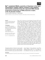
Fig. 1. Identification of ferritin in pea stem mitochond ria. (A) SDS/
PAGE(12%)analysisofproteins(25lg) from crude mitochondria
(CMt), purified mitochondria (PMt), matrix from pea stem purified
mitochondria (MP), and to tal pea seed proteins (CP , control p roteins);
molecular mass of protein standards is indicated in kDa ( Std). (B)
Immunoblotting of the same proteins with polyclonal antibod y against
ferritin (PAAF). (C) Immunoblotting of recomb inant h uman mito-
chondrial ferritin (rHuMt, 10 ng), protein extract from HeLa cells
overexpressing human mitoc hondrial ferritin ( MtF-HeLa, 30 lg) and
matrix proteins from pea stem purified mito chondria (MP, 35 lg) with
antihuman mitochondrial ferritin polyclon al antibody after native 6%
PAGE. (D) SDS/PAGE (12%) of the 25–26 kDa protein purified by
immunoprecipitation.
Table 2. Sequence coverage by trypsin digestion peptide mass fingerprint of the pea ferritin, purified from mitochondria, with t he translated sequence
precursor of a pea ferritin (SwissProt accession number P19975). In bold are reported the protein regions covered in the mass fingerprint. Peptide
sequences confirmed by fragmentation analysis by post source decay (PSD) are underlined. The putative peptide leader sequence located at the
N-terminus is highlighted in black.
1
MALSSSKFSS FSGFSLSPVS GNGVQKPCFC DLRVGEKWGS RKFRVSATTA
51 PLTGVIFEPF EEVKKDYLAV PSVPLVSLAR QNFADECESV INEQINV EYN
101 ASYVYHSLFA YFDRDNVALK GFAKFFKESS EEHREHAEKL MKYQNTRGGR
151 VVLHPIKDVP SEFEHVEKGD ALYAMELALS LEKLTNEKLL NVHSVAERNN
201 DLEMTHFIEG EYLAEQVEAI KKISEYVAQL RRVGKGHGVW HFDQRLLHGV
251 HGA
Ó FEBS 2004 Plant mitochondrial ferritin (Eur. J. Biochem. 271) 3659
Protein assay
Protein concentration was determined by the method of
Bradford [26], using bovine s erum albumin as a s tandard.
Results
Purified pea stem mitochondria, particularly when com-
pared with CMt, were almost devoid o f contamination from
different types of cellular components (Table 1). ATPase
activity of this fraction was, indeed, uninhibited or only very
slightly inhibited by vanadate (plasmalemma ATPase
inhibitor), bafilomycin A
1
(tonoplast ATPase inhibitor)
and molybdate (soluble phosphatase inhibitor), but strongly
inhibited by oligomycin (mitochondrial ATPase inhibitor).
In addition, PMt showed a low level of latent IDPase ( Golgi
membrane marker enzyme). Cytochrome c reductase activ-
ity was assayed in the presence and absence of antimycin A
to assess the contamination from endoplasmic reticulum. In
purified mitochondria (in the presence of antimy cin A), the
activity was 4.35 times and almost six-fold lower than that
recovered in c ontrol mitochondria and microsomes, respect-
ively. On the other hand, the activity of cytochrome c
reductase, still detected in the presence of antimycin A,
could depend on the presence of a similar enzyme on the
outer membrane of plant mitochondria [27]. Finally, this
preparation exhibited a negligible glucose-6-phosphate
dehydrogenase activity (plastid marker enzyme), partic-
ularly when compared to that of a sample of etioplasts
isolated from the same plant material.
The proteins of CMt, PMt, and the relative matrix
components were subjected to SDS/PAGE, in compari-
son with a pea seed protein extract containing ferritin
Fig. 3. Immunocytological l ocalization of ferritin in etiolated pea stem.
(A) Cross-section of etiolated pea stem; cw, cell wall, v, vacuole, m,
mitochondria. (B) and (C) Higher magnification of t he same electron
micrograph showing labeled mitochondria. Arrows in dicate electron-
dense particles after immunolabeling with PAAF followed by gold-
conjugated se condary a ntibo dy. Bars co rres pond to 30 0 lm.
Fig. 2. Localization of ferritin in pea stem purified mitochondria. (A)
Immunoblotting with P AAF of PMt (25 lg) incubated (+) or not ( –)
with 0.5% (w/v) Triton X-100 for 10 min, then subjected to proteolysis
with 125 lgÆmL
)1
trypsinfor30minat25°C and stopped by the
addition of 1 m
M
PMSF. (B) Imm unoblotting of PMt (25 lg) and
Mpl (25 lg) with monoclonal antibody raised against cytochrome
c (Cyt c), polyclonal antibody raised a gainst the a/b-subunit of mito-
chondrial ATPase (a/b-subunit) or PAAF (ferritin).
3660 M. Zancani et al. (Eur. J. Biochem. 271) Ó FEBS 2004
(Fig. 1A). Proteins, thus separated, were then subjected to
an immunoblot assay by using PAAF (Fig. 1B). The results
show that this antibody cross-reacted with a protein
exhibiting an apparent molecular mass of approximately
25–26 kDa, a v alue similar to that o f ferritins. This
reactivity w as achieved in all types of preparations. In
particular, in some cases two very close bands were evident.
As already suggested, they represent ferritin and a p roduct
of degradation of the same protein [14]. The same cross-
reactivity was a lso detected for a protein of twofold purified
pea stem mitochondria (result not shown). In addition, the
pea stem mitochondrial protein, after nondenaturing elct-
rophoresis and blotting, cross-reacted with the polyclonal
antibody anti-human mitochondrial ferritin [10] (Fig. 1C).
The matrix proteins from purified pea mitochondria were
also subjected to purification by immunoprecipitation. SDS/
PAGE analysis of the immunoprecipitated revealed, after
Coomassie staining, a protein band at 25–26 kDa (Fig. 1D).
The tryptic peptides of this band were analyzed with a
MALDI-TOF m ass s pectrometer and the monoisotopic
masses of each singly charge species were annotated with
their intensities. These data were fitted on the NCBI non-
redundant Viridiplantae database returning, with a confidence
score greater than 95% (P<0.05) accuracy, the pea ferritin
1 chloroplast precursor (NCBI accession gi/417006; Swiss-
Prot accession P19975). This assignment was confirmed by
fragmentation analysis employing a MALDI-TOF post
source decay experiment, selecting the ion species at 1078.52
amu (MH
+
) with a time gate ion selector. The resulting
fragmentation pattern was characteristic the y and b ion
series of the sequence ISEYVAQLR (223–231). The PSD
fragments were fitted on the NCBI nonredundant Viridi-
plantae d atabase returning again the ferritin sequence
ISEYVAQLR (223–231). The overall mass fingerprint data
cover about the 30% of the assigned sequence and details are
reported in Table 2. The theoric molecular mass, 23.6 kDa,
calculated from the database sequence after removal of the
N-terminus signal peptide, is in agreement with the value of
25–26 kDa estimated from the SDS/PAGE.
The localization of ferritin in pea stem purified mito-
chondria was investigated (Fig. 2). Figure 2A shows an
immunoblot of ferritin in PMt, treated (lane +) or
untreated (lane –) with Triton X -100, which were then
subjected to trypsin d igestion. The intensity of the immuno-
labeled band was lower in the presence of the detergent,
demonstrating that ferritin is localized inside the mito-
chondrial membranes. Furthermore, Mpl were obtained
by osmotic shock of PMt to remove the outer mito-
chondrial membrane. Mitoplast and PMt proteins were
then cross-reacted with monoclonal antibodies raised
Fig. 4. Ultrastructural localization of ferritin in pea stem mitochondria. Electron micrograph from fixed Percoll-purified pea st em mitochondria,
subjected to immunogold decoration in th e presence (A and B) or absence (C) of PAAF and at lower magnification (D). Arrows indicate ele ctron-
dense particles after immunolabeling with PAAF followe d by gold-conj ugated secondary ant ibody. Bars correspond to 300 lm.
Ó FEBS 2004 Plant mitochondrial ferritin (Eur. J. Biochem. 271) 3661
against cytochrome c, polyclonal antibodies raised against
the a/b-subunit of mitochondrial A TPase and PAAF,
respectively (Fig. 2B). These results show that Mpl partially
lost the cytochrome c; the densitometric a nalysis of the
immunodecoration show a d ecrease of approximately 50%
in the Mpl proteins. On the other hand, the immunodec-
oration of PMt and Mpl proteins with antibodies against
the a/b-subunit and PAAF was comparable (Fig. 2B). This
indicates that both the a/b-subunit and the ferritin are still
retained in Mpl, thus confirming the localization of ferritin
in the mitochondrial matrix.
The ultrastructural localization of the pea stem mito-
chondrial ferritin was further confirmed in ultra-thin
sections of pea s tem c ross-sections (Fig. 3) and fixed
mitochondria (Fig. 4), both immunolabeled with PAAF,
and followed by gold-conjugated secondary antibody.
Figure 3 shows that mitochondria, selected from a trans-
mission electron microscopic micrograph of pea stem cells
(Fig. 3 A), have some electron-dense particles (Fig. 3B,C;
arrows). In agreement, such particles were also detected in
isolated mitochondria (Fig. 4A,B). The electron-dense par-
ticles were not detected when PAAF was omitted in both
cross-sections (result not shown ) and isolated mitochondria
(Fig. 4C). According to the low level of plastidial enzymatic
marker, detected in PMt (Table 1), the elec tron micro-
graphs show that the purified mitochondrial fraction was
almost free from etioplast contamination (Fig. 4D).
Figure 5A shows the protein patterns of control proteins
and o f CMt and PMt from A. thaliana.Whenthese
separated proteins were subjected to cross-reaction with
PAAF (Fig. 5B), a gain a band with an apparent molecular
mass of 25– 26 kDa w as revealed, thus s uggesting the
presence of this iron-storage protein also in mitochondria
from this type of plant cells. Furthermore, preliminary
results indicate that ferritin was also present in Percoll-
purified mitochondria isolated from soybean hypocotyls
(results not shown).
The genome of A. thaliana contains a family of nuclear
genes for ferritins (AtFer1–4) [28]. These genes encode the
ferritin subunit precursors, each containing a transit peptide.
The structural analysis of the presequences of the corres-
ponding polypeptides suggests that all are targeted to plastids
[28]. Table 3 shows the scores for t he mitochondrial/plasti-
dial localization of some plant ferritins from P. sativum
(SwissProt accession P19975), cowpea (Vigna unguiculata,
SwissProt accession T08124), soybean (Glycine max,
SwissProt accession BAB64536) and AtFer1 and AtFer4
from A. thaliana. While it is clear that AtFer1 is a poor
candidate for a mitochondrial localization, for the other
proteins significant scores were found. In particular,
PSORT
and
IPSORT
programs predicted high probability for the
presence of a mitochondrial target peptide in pea ferritin.
Remarkably, the ferritins from cowpea, soybean and AtFer4
exhibit values corresponding to a high probability for a
mitochondrial targeting from at least three programs.
Discussion
Animal and plant ferritins are encoded by nuclear gene
families, which diverge in their exon/intron organization
[13]. This suggests that they derive from a common ancestor,
albeit animal ferritins display a cytoplasmic localization,
whereas the plant ones are plastidic [8,15]. However, as seen,
an unusual intronless gene on human chromosome 5q23.1
encodes a 242 amino acid precursor of a ferritin H-like
Fig. 5. Identification of ferritin in A. thaliana mitochondria. (A) SDS/
PAGE (12%) of proteins (25 lg) from crude mitochondria (CMt),
purified mitochondria (PMt), and total pea seed proteins (CP, control
proteins). (B) Immunoblotting of the same proteins with PAAF.
Molecular mass of protein standards is indicated in kDa (Std).
Table 3. Calculated values for prediction of mitochondrial targeting for some plant ferritins. Scores were obtaine d from different pro grams available
on the net; values higher than 0.6 are highlighted in bold; the output for
IPSORT
is given as mitochondrial target peptide (mTP) or chloroplast transit
peptide (cTP).
Plant species
Prediction programs
SwissProt accession
PREDOTAR MITOPROT
II
PSORT IPSORT
Pisum sativum P19975 0.031 0.3601 0.611 mTP
Vigna unguiculata T08124 0.783 0.9613 0.473 mTP
Glycine max BAB64536 0.648 0.8187 0.617 mTP
Arabidopsis thaliana Q39101 (AtFer1) 0.020 0.5482 0.360 cTP
Q9S756 (AtFer4) 0.974 0.8137 0.694 mTP
3662 M. Zancani et al. (Eur. J. Biochem. 271) Ó FEBS 2004
protein [10]. This 30 kDa protein is targeted to mitochon-
dria and processed to a 22 kDa subunit. This ferritin,
expressed in HeLa cells, is available to exogenous iron,
similarly to t he cytosolic ferritin, suggesting that t his
mitochondrial protein may have profound consequences
on c ellular i ron homeostasis [11]. A s shown in this p aper, a
ferritin has also been identified in higher plant mitochon-
dria. This evidence arises from the following findings. First,
a mitochondrial matrix protein of 25–26 kDa, cross-reacted
with a polyclonal antibody of pea seed ferritin in b oth pea
stem (Fig. 1) and A. thaliana (Fig. 5) mitochondria. Such
organelles were highly purified by discontinuous Percoll
gradient, providing a v ery low interference in the immuno-
decoration from other cellular c omponents, especially from
etioplast contamination. On the basis of densitometric
analysis of immunoblots obtained with etioplasts isolated
from pea stem (results not shown), we calculated that if
PAAF detects j ust etioplast ferritin, these organelles h ave to
be present, in purified mitochondrial fractions, as a heavy
contamination (estimated to be approximately 25% of the
total protein). The results shown here demonstrate that this
is not the case, because the low enzymatic activity of
glucose-6-phosphate dehydrogenase in PMt (Table 1) con-
firms that the purified mitochondrial fractions possess a
maximum of 2 .5% of plastid proteins and, in addition , the
electron micrographs (Fig. 4) clearly show a very limited
contamination of P Mt from other o rganelles. Second,
ferritin was immunocytochemically identified in etiolated
pea stem cross-sections (Fig. 3) and in isolated pea mito-
chondria (Fig. 4). The pea stem mitochondrial ferritin is
present in the mitochondrial matrix as demonstrated by its
colocalization in Mpl with the a/b-subunit of mitochondrial
ATPase (Fig. 2). Finally, the 25–26 kDa soluble protein
was purified by immunoprecipitation (Fig. 1D); the primary
structure o f the polypeptide chain, inferred by Mass Finger
Print experiments on MALDI-TOF mass spectrometry, fits
to a high degree with the sequence of the ferritin from
P. sativum (SwissProt accession P19975, Table 2).
In A. thaliana, four ferritin genes (AtFer1–4) have been
reported and it has been suggested that the proteins AtFer1–
4 possess at the N-terminus the typical presequences of the
chloroplastic protein transit peptide [28], similarly to what
reported for pea ferritin [29,30]. On the other hand, the
analysis of the presequence of AtFer4 reveals a high score
for its mitochondrial localization, especially when co mpared
with AtFer1 (Table 3). The same analysis for pea ferritin
shows that the programs
PSORT
and
IPSORT
give a high
probability for this protein to be targeted to mitochondria
(Table 3).
The data presented in this paper strongly indicate a
mitochondrial localization for ferritins in P. sativum and
A. thaliana and could be rationalized as follows: the protein
may be targeted to both plastids and mitochondria,
similarly to what shown for several plant p roteins [31]. This
feature can be accomplished by alternative transcription,
alternative translation starts, alternative exon splicing (or
a combination of the above), or t he presence in the
N-terminus of an ambiguous presequence [31]; prediction
programs could just be unable to detect such dual targeting.
On the other hand, a similar situation has been described
for ferrochelatase-I, an enzyme involved in heme biosyn-
thesis and, probably, in protection against o xidative stress in
A. thaliana [32]. This enzyme has been recently rep orted to
be present also in pea mitochondria [33]. The presence of
ferrochelatase-I and -III in A. thaliana mitochondria has
been recently questioned, while their presence in pea
mitochondria has been related to the fact that the latter
organelles import a variety of (but not all) chloroplastic
proteins [34].
Plant mitochondria possess an electron transport chain
where superoxide anion may be generated by univalent
reactions at the level of complex I or III [35]. For this
reason, mitochondria have evolved systems to scavenge
ROS, or to prevent their formation [7,9], but sequestration
of potential harmful ferrous ions has not yet been
described.
Metal tolerance and homeostasis in plant cells is accom-
plished by different mechanisms [36]. In this context, the
main role of ferritins could concern iron sequestration.
Overexpression of this protein, in either the cytoplasm or
plastids of transgenic tobacco, leads to an increase of iron
sequestration that induces an activation of the iron trans-
port systems [37]. Therefore, they are crucial in controlling
iron storage and homeostasis in the plant cells. Other
functions of plant ferritins are, on the other hand, still
obscure. It has been suggested that sequestering of intracel-
lular iron m ay protect from oxidative damage induced by a
wide range of stresses [38]. Indeed, an increase of ferritin
mRNA has been observed in A. thaliana leaves photo-
inhibited w ith high light or fumigated with ozone [39].
Therefore, the sequestration of iron by ferritins in chloro-
plasts and mitochondria, two of the major sites of ROS
generation in plant cells [6,7], can constitute an additional
strategy to prevent th is damage.
Acknowledgements
We thank Dr J.F. Briat, Centre National d e l a R echerche S cientifique,
Montpellier, France, for a gene rous gift of pea s eed ferritin antibody.
We also thank very much Dr Sonia Levi, Istituto di Ricovero e Cura a
Carattere Scientifico (IRCCS), H. San Raffaele, Milan, Italy, for the
cross-reactivity analysis with human mitochondrial ferritin antibo dy.
Thanks are also due to Dr Sonia Patui for her help during Percoll-
purified mito chondria preparation and to Mr Claudio Gamboz for his
help with electron microscopy analysis. This research was s upported by
‘Ministero dell’Universita
`
e della Ricerca Scientifica e Tecnologica’
(Cofin 2000–01) i n the fr ame of the program entitled: Nitric Oxide an d
Plant Resistance to Pathogens.
References
1. Briat, J F., Fo bis-Loisy, I., Grignon, N., Lobre
´
aux, S., Pascal, N.,
Savino, G., Thoiron, S., von Wire
`
n, N . & Van Wuytswinkel, O.
(1995) Cellular and molecular aspects of iron metabolism in
plants. Biol. Cell. 84, 69–81.
2. Halliwell, B. & Gutteridge, J.M.C. (1999) Free Radicals in Biology
and Medicine, 3rd edn. Oxford University Press, Oxford.
3. Elstner, E.F. & Osswald, W. (1994) Mechanisms of oxygen
activation during plant stress. Proc. Royal Soc. Edinburgh 102B,
131–154.
4. Mehdy, M.C., Sharma, Y.K., Sathasivan, K. & Bays, N.W. (1996)
The role of activated oxygen species in plant disease resistance.
Physiol. Plant. 98, 3 65–374.
5. Briat, J F. & Lobre
´
aux, S. (1997) Iron transport and s torage in
plants. Trends Plant Sci. 2, 187–192.
Ó FEBS 2004 Plant mitochondrial ferritin (Eur. J. Biochem. 271) 3663
6. Alscher, R.G., Donahue, J.N. & Cramer, C.L. (1997) Reactive
oxygen species and antioxidants: relationship in green cells. Phy-
siol. Plant. 100, 224–233.
7. Møller, I.M. (2001) Plant mitochondria and oxidative stress:
electron transport, NADPH turnover, and me tabolism of reactive
oxygen species. Annu. Rev. Plant Physiol. Plant Mol. Biol. 52 , 561–
591.
8. Seckbach, S. (1982) Ferreting out the secrets of plant ferritin.
J. Plant. Nutr. 5, 369–394.
9. Casolo, V., Braidot, E., Chiandussi, E ., Macrı
`
,F.&Vianello,A.
(2000) The role of mild uncoupling and non-coupled respiration in
the regulation of hydrogen peroxide generation by plant
mitochondria. FEBS Lett. 474, 53–57.
10. Levi, S., Corsi, B., Bosisio, M., Invernizzi, R., Volz, A., Sanford,
D., Arosio, P. & Drysdale, J. (2001) A human mitochondrial
ferritin encoded by an introneless gene. J. Biol. Chem. 270,
24437–24440
11. Corsi, B., Cozzi, A., Arosio, P., Drysdale, J., Santambrogio, P.,
Campanella, A., Biasiotto, G., Alberini, A. & Levi, S. (2002)
Human mitochondrial fe rrit in expressed in HeLa cells
incorporates iron and affects cellular iron metabolism. J. Biol.
Chem. 277, 22430–22437.
12. Theil, E.C. (1987) Ferritin: structure, gene regulation, and cellular
function in animals, plants, a nd microorganisms. Ann. Rev.
Biochem. 56, 289–315.
13. Harrison, P.M. & Arosio, P. (1996) The ferritins: molecular
properties, iron storage function and cellular regulation. Biochim.
Biophys. Acta 1275, 161–203.
14. Laulhere, J.P., Laboure, A M. & Briat, J F. (1989) Mechansim
of the transition from plant ferritin to phytosiderin. J. Biol. Chem.
264, 3629–3635.
15. Proudhon, D., Wei, J., Briat, J.F. & Theil, E.C. (1996) Ferritin
gene organization: differenc es between plants and animals s uggest
possible kingdom-specific selective constraints. J. Mol. Evol. 42,
325–336.
16. Zancani, M., Casolo, V., Vianello, A. & Macrı
`
,F.(1998)H
+
/PP
i
stoichiometry of a membrane-bound pyrophosphatase of plant
mitochondria. Physiol. Plant. 103, 304–311.
17. Petrussa,E.,Casolo,V.,Braidot,E.,Chiandussi,E.,Macrı
`
,F.&
Vianello, A. (2001) Cyclosporin A induces the opening of a
potassium-selective channel in higher plant mitochondria.
J. Bioenerg. Biomembr. 33, 107–117.
18. Macrı
`
, F., Braidot, E., Petrussa, E. & Vianello, A. (1994) L ipoxy-
genase activity associated to isolated soybean plasma membranes.
Biochim. Biophys. Acta 1215, 109–114.
19. Green, J.R. (1983) The Golgi apparatus. In Isolation of Mem-
branes and Organelles from Plant Cells (Hall, J.L. & Moore, A.L.,
eds), pp. 135–152. Acad emic Press Inc., London.
20. Lord, J.M. (1983) Endoplasmic reticulum and ribosomes. In
Isolation of Membranes and O rganelles from Plant Cells (Hall, J.L.
& Moore, A.L., eds), pp. 119–134. Academic Press Inc., London.
21. Bergmeyer, H.U., Gawehn, K. & Grassl, M. (1974) Enzymes as
biochemical reagents; glucose-6-phosphate dehydrogenase. In
Methods of Enzymatic Analysis,Vol.1(Bergmeyer,H.U.,ed.),pp.
458–459. Academic Press Inc., London.
22. Laulhere, J.P., Lescure, A.M. & Briat, J.F. (1988) Purification a nd
characterization of ferritins from seed of maize, pea and soya
bean: distribution in various pea organs. J. Biol. Chem. 262,
10289–10294.
23. Laemmli, U.K. (1970) Cleavage of structural proteins during the
assembly of the head of bacteriophage T4. Nature 277, 680–685.
24. Tomasetig, L., Di P ancrazio, F., Harris, D.A., Mavelli, I. & Lippe,
G. (2002) Dimerization of F
o
F
1
ATP synthase from bovine heart is
independent from the binding of the inhibitor p rotein IF
1
.
Biochim. Biophys. Acta 1556, 133–141.
25. Shevchenko, A. & Shevchenko, A. (2001) Evaluation of the effi-
ciency of in-gel digestion of p roteins by peptide isotopic labelling
and MALDI ma ss spe ctro metry . Anal. Bio chem. 296 , 279–283.
26. Bradford, M.M. (1976) A rapid and sensitive method for the
quantitation of microgram quantities of protein utilizing the
principle of protein-dye bindin g. Anal. Biochem. 72, 248–254.
27. Møller, I.M. & Lin, W. (1986) Membrane-bound NAD(P)H
dehydrogenases in higher plant c ells. Ann. Rev. Plant Physiol. 37,
309–334.
28. Petit, J.M., Briat, J F. & Lobreaux, S. (2001) Structure and dif-
ferential expression of the four members of the Arabidopsis thali-
ana ferritin gene family. Biochem. J. 359, 575–582.
29. Lobre
´
aux, S., Yewdall, S.J., Briat, J F. & Harrison, P.M. (1992)
Amino-acid sequence and predicted three-dim ensional structure of
pea seed (Pisum sativum)ferritin.Biochem. J. 288, 931–939.
30. Van Wuytswinkel, O., Savino & Briat, J F. (1995) Purification
and characterization o f re combinant pe a-seed ferritins expressed
in Escherichia coli: in fluence of N-terminus deletions on protein
solubility and c ore formation in vitro. Biochem. J . 305 , 253–261.
31. Peeters, N. & Small, I. (2001) Dual targeting to mitochondria and
chloroplasts. Biochim. Biophys. Acta 1541, 54–63.
32. Chow,K.S.,Singh,D.P.,Roper,J.M.&Smith,A.G.(1997)A
single precursor protein for ferrochelatase-I from Arabidopsis is
imported in vitro into both chloroplasts and mitochondria. J. Biol.
Chem. 272, 27565–27571.
33. Cornah, J.E., Roper, J.M., Pal Singh, D. & Smith, A.G. (2002)
Measurement of ferrochelatase activity using a novel assay sug-
gests that plastids a re the major site of haem biosynthesis in both
photosynthetic and non-photosynthetic cells of pea (Pisum sati-
vum L.). Biochem. J. 362, 423–432.
34. Lister, R., Chew, O., Rudhe, C., Lee, M N. & Whelan, J. (2001)
Arabidopsis thaliana ferrochelatase-I an d -II are not impo rted into
Arabidopsis mitochon dria. FEBS Lett. 506, 291–295.
35. Braidot, E., Petrussa, E., Vianello, A. & Macrı
`
, F. (1999)
Hydrogen peroxide generation by higher plant mitochondria
oxidizing complex I or complex II substrates. FEBS Lett. 451,
347–350.
36. Clemens, S. (2001) Molecular mechanisms of plant metal tolerance
and homeostasis. Planta 212, 475–486.
37. Van Wuytswinkel, O., Vansuyt, G., Grignon , N., Fourcroy, P. &
Briat, J F. (1998) Iron homeostasis alteration in transgenic
tobacco overexpressin g ferritin. Plant J. 17, 93–97.
38. Dea
´
k, M., Horva
´
th, G.V., Davletova, S., To
¨
ro
¨
k, K., Sass, L.,
Vass, I., Barna, B., Kira
´
ly,Z.&Dudits,D.(1999)Plantsectopi-
cally expressing the iron-binding p rotein, ferritin, are tolerant t o
oxidative damage and pathogens. Nature Biotechnol. 17, 192–196.
39. Murgia, I., Briat, J F., Tarantino, D. & Soave, C. (2001) Plant
ferritin accumulates in response to photoinhibition but its ectopic
overespression does not protect against photoinhibition. Plant
Physiol. Biochem. 39, 797–705.
40. Vianello, A., Dell’Antone, P. & Macrı
`
, F. (1982) ATP-dependent
and ionophore-ind uced pr oton translo cation in pe a stem micr o-
somal vesicles. Biochim. Biophys. Acta 689, 89–96.
41. Eichacker, L.A., Helfrich, M., Ru
¨
diger, W. & Mu
¨
ller, B. (1996)
Stabilization of chlorophyll a-binding apoprotein s P700, CP47,
CP43, D2, and D1 by chlorophyll a or Zn-pheophytin a. J. Biol.
Chem. 271, 32174–32179.
3664 M. Zancani et al. (Eur. J. Biochem. 271) Ó FEBS 2004