Báo cáo khoa học: Probing the unfolding region of ribonuclease A by site-directed mutagenesis potx
Bạn đang xem bản rút gọn của tài liệu. Xem và tải ngay bản đầy đủ của tài liệu tại đây (334.47 KB, 10 trang )
Probing the unfolding region of ribonuclease A by site-directed
mutagenesis
Jens Ko¨ ditz, Renate Ulbrich-Hofmann and Ulrich Arnold
Department of Biochemistry and Biotechnology, Martin-Luther University Halle-Wittenberg, Halle, Germany
Ribonuclease A contains two exposed loop regions, around
Ala20 and Asn34. Only the loop around Ala20 is sufficiently
flexible even under native conditions to allow cleavage by
nonspecific proteases. In contrast, the loop around Asn34
(together with the adjacent b-sheet around Thr45) is the first
region of the ribonuclease A molecule that becomes sus-
ceptible to thermolysin and trypsin under unfolding condi-
tions. This s econd region therefore has been sugges ted to b e
involved in early steps of unfolding and was designated as
the unfolding region of the ribonuclease A molecule. Con-
sequently, modifications in this region should have a great
impact on the unfolding and, thus, on the thermodynamic
stability. Also, i f the Ala20 loop contributes to the stability o f
the ribonuclease A molecule, rigidification of this flexible
region should stabilize the entire protein molecule. We sub-
stituted several residues in both regions without any dra-
matic effects on the native conformation and catalytic
activity. As a result of their remarkably differing stability, the
variants fell into two groups carrying the mutations: (a)
A20P, S21P, A20P/S21P, S21L, or N34 D; (b) L35S, L35 A,
F46Y, K31A/R33S, L35S/F46Y, L35A/F46Y, or K31A/
R33S/F46Y. The first group showed a thermodynamic and
kinetic stability similar to wild-type ribonuclease A, whereas
both stabilities of the variants in the second group were
greatly decreased, suggesting that the decrease in DG can be
mainly attributed to an increased unfolding rate. Although
rigidification of the Ala20 loop by introduction of proline
did not result in stabilization, disturbance of the network of
hydrogen bonds and hydrophobic interactions that interlock
the proposed unfolding region dramatically destabiliz ed the
ribonuclease A molecule.
Keywords: limited proteolysis; local unfo lding; protein
engineering; ribonuclease A; stability.
In contrast with the ensemble of conformational species in
the unfolded state [1], the native state of proteins is generally
characterized by a uniform overall global conformation [2].
Whereas larger proteins t hat consist of structural subunits
or domains often behave very complexly during the
processes of unfolding and refolding, most small proteins
can be considered as a single unit [3]. Apart from local
fluctuations of the p rotein str ucture in t he native state,
the protein molecule unfolds highly co-operatively when
exposed to denaturing conditions. The stability of the
natively folded protein molecule is not determined by a
single feature, but a number of internal and external factors
contribute to the formation and stabilization of the native
protein structure [4]. Studies on a large variety of proteins
led t o t he assumption that confined regions of the 3 D
protein s tructure are crucial for the conservation o f its
folded, native state. A local disruption of the most labile
region, which was referred to as unfolding region, was
postulated to initiate the co-operative unfolding of the
protein molecule [5,6]. This assumption was supported by
the identification of a region in the neutral protease from
Bacillus stearothermophilus that responds most sensitively
to changes in the amino-acid composition by site-directed
mutagenesis [7–9]. Consistent with this Ôcritical regionÕ,a
Ôweak pointÕ in Arthrobacter
D
-xylose isomerase was postu-
lated based on results from proteolysis experiments under
thermal denaturation [10]. More recently, Machius et al.
[11] deduced a Ôweak regionÕ in a-amylase and Gaseidnes
et al. [12] identified a Ôweak spotÕ or a Ônucleation site for
unfoldingÕ in chitinase by mutational analysis. Because of
the decreased number of hydrogen bonds, loo p regions at
the surface of the protein molecule are candidates for such
Ôu nfolding regionsÕ. In fact, loops that are tethered by
irregular hydrogen bonds or hydrophobic patches were
found to be crucial for either the folding or unfolding of
proteins (for a review see [13]).
Ribonuclease A (RNase A) is a small, compact, and
rather stable enzyme which is cross-linked by four disulfide
bonds [14]. Nevertheless, even under native conditions the
loop region around Ala20 is highly flexible [15], which leads
to proteolytic cleavage by non-specific proteases. In con-
trast, in spite of increased mobility detected for residues
37–42 by NMR [ 16], the fl exibility of the loop around
Asn34, which contains potential cleavage sites for trypsin
and thermolysin, is obviously not sufficient to allow
Correspondence to U. Arnold, Department of Biochemistry and Bio-
technology, Martin-Luther University Halle-Wittenberg, Kurt-
Mothes Strasse 3, 06120 Halle, Germany. Fax: +49 3 455527303,
Tel.: +49 3455524865, E-mail:
Abbreviations: 6-FAM-dArU(dA)
2
-6-TAMRA, 6-carboxyfluorescein-
dArU(dA)
2
-6-carboxytetramethylrhodamine; GdnHCl, guanidine
hydrochloride; RNase A, ribonuclease A.
Enzymes: bovine pancreatic ribonuc lease A (EC 3.1.27.5); thermolysin
(EC 3.4.24.27)
Note: a website is available at h ttp://www.biochemtech.uni-halle.de/
biotech
(Received 30 June 2004, revised 27 August 2004,
accepted 3 September 2004)
Eur. J. Biochem. 271, 4147–4156 (2004) Ó FEBS 2004 doi:10.1111/j.1432-1033.2004.04355.x
cleavage by these proteases. As soon as the protein molecule
starts to unfold globally, however, the RNase A molecule
becomes susceptible to these proteases, too. The primary
cleavage sites were found in the loop region around Asn34
and the adjacent b-strand around Thr45, suggesting this
section as the unfolding region [17].
To investigate the contribution of the two loop regions to
the stability of the entire RNase A molecule, we replaced
several amino-acid residues b y site-directed mutagenesis
(A20P, S21P, A20P/S21P, S21L, N34D, L35S, L35A,
F46Y, K31A/R33S, L35A/F46Y, L35S/F46Y, and K31A/
R33S/F46Y) and studied the effect of the mutations on the
thermodynamic and kinetic stability. The similarity of the
impact on both the thermodynamic and kinetic stabilities
suggests a predominant effect on the native state by these
mutations.
Experimental procedures
Proteins and chemicals
RNase A from Sigma (St Louis, MO, USA) was purified on
a Mono S FPLC column (Amersham Biosciences, Uppsala,
Sweden). Thermolysin (from Calbiochem, Schwalbach,
Germany) was used without further purification. Oligonu-
cleotides were from MWG Biotech (Ebersberg, Germany)
and restriction enzymes AvaI, BsmI, EcoRI, HindIII, NdeI,
and SacI w ere f rom N ew England Biolabs (Frankfurt/
Main, Germany). Growth media were from Difco
Laboratories (Detroit, MI, USA). Escherichia coli
strains XL-1 Blue and BL21(DE3) were from Stratagene
(Heidelberg, Germany). 6-Carboxyfluorescein-dArU(dA)
2
-
6-carboxytetramethylrhodamine (6-FAM-dArU(dA)
2
-6-
TAMRA) was purchased from Integrated DNA
Technologies (Coralville, IA, USA). All other chemicals
were of purest grade commercially available.
Site-directed mutagenesis
The RNase A variants A20P, S21P, and A20P/S21P had
been produced previously [18]. For other variants, the
rnase A gene in the plasmid pBXR [19], a gift from
R. T. Raines (University of Wisconsin, Madison, WI,
USA), was modified by use of the Q uikChange
TM
site-directed mutagenesis kit (Stratagene) to obtain the
mutations N34D, K31A/R33S, L35A, L35S, and F46Y.
For the mutations K31A/R33S/F46Y, L35A/F46Y, a nd
L35S/F46Y, site-directed mutagenesis was started from the
rnase A genes that carry the mutations for K31A/R33S,
L35A, or L35S using the oligonucleotides for the F46Y
mutation. The oligonucleotides and the introduced restric-
tion sites to facilitate the selection of positive clones are
shown in Table 1. The mutations were verified by DNA
sequencing as described by Sanger et al. [20] (SequiTherm-
Excel
TM
LongRead
TM
DNA sequen cing kit, Biozym, Hess,
Oldendorf, Germany, and Li-COR 4000 DNA-sequencer,
MWG Biotech, Ebersberg, Germany). The p lasmids
carrying the correct DNA sequence were each transformed
into E. coli expression host strain BL21(DE3).
Expression, renaturation, and purification
of the enzyme variants
The experimental procedure was performed as described
previously [18]. Briefly, cultures of E. coli strain
BL21(DE3) that had b een t ransformed with a plasmid
directing the expression of the corresponding RNase A
variant were g rown in terrific broth containing
50 lgÆmL
)1
kanamycin [variants A 20P, S 21P, and
A20P/S21P in vector pET 26b(+)] or 400 lgÆmL
)1
ampicillin [the other variants i n vector pET 22b(+)] at
37 °CtoanA
600
of 2. Gene expression was induced by
1m
M
isopropyl thio-a-
D
-galactoside, and cells were
grown a dditionally for 4 h before being harvested. Cell
lysis was performed by treatment with lysozyme and
homogenization with a Gaulin homogenizer. T he inclu-
sion bodies were isolated by centrifugation followed by
resolubilization ( 20 m
M
Tris/HCl, 7
M
guanidine h ydro-
chloride (GdnHCl), 100 m
M
dithiothreitol, 1 0 m
M
EDTA, pH 8.0) and dialysis of the protein solution
against 20 m
M
acetic acid. Precipitates formed during
dialysis were removed by centrifugation. After renatura-
tion of the protein [100 m
M
Tris/HCl, pH 8.5,
100 m
M
NaCl, 1 m
M
glutathione (reduced), 0.2 m
M
glutathione ( oxidized), 10 m
M
EDTA at room tempera-
ture for 24 h], it was purified on a Mono S column
(50 m
M
Tris/HCl, pH 7.5, with a linear gradient of
0–500 m
M
NaCl).
Table 1. Oligonucleotides for site-directed mutagenesis. The replaced nucleotides are bold-face and the introduced restriction sites are underlined.
Mutation Oligonucleotides Restriction site
S21L fw 5¢-C TCC AGC ACT TCC GCC GCC CT
G AGC TCC AAC TAC TG3¢ SacI
rev 5¢-CA GTA GTT G
GA GCT CAG GGC GGC GGA AGT GCT GGA G-3¢
N34D fw 5¢-C CAG ATG ATG AAG AGC CGG GAC CTG ACC AAA GAT CGA TGC-3¢ No restriction site
rev 5¢-GCA TCG ATC TTT GGT CAG GTC CCG GCT CTT CAT CAT CTG G-3¢
K31A/R33S fw 5¢-C TGT AAC CAG ATG ATG GC
G AGC TCG AAC CTG ACC AAA GAT C3¢ SacI
rev 5¢-G ATC TTT GGT CAG GTT C
GA GCT CGC CAT CAT CTG GTT ACA G-3¢
L35A fw 5¢-G ATG ATG AAG AGC CG
GAAT GCC ACC AAA GAT CGA TGC AAG C-3¢ BsmI
rev 5¢-G CTT GCA TCG ATC TTT GGT G
GC ATT CCG GCT CTT CAT CAT C-3¢
L35S fw 5¢-G ATG ATG AAG AGC CG
GAAT TCC ACC AAA GAT CGA TGC AAG C-3¢ EcoRI
rev 5¢-G CTT GCA TCG ATC TTT GGT G
GA ATT CCG GCT CTT CAT CAT C-3¢
F46Y fw 5¢-GC AAG CCA GTG AAC A
CA TAT GTG CAC GAG TCC CTG G-3¢ NdeI
rev 5¢-C CAG GGA CTC GTG CA
CATA TGT GTT CAC TGG CTT GC-3¢
4148 J. Ko
¨
ditz et al.(Eur. J. Biochem. 271) Ó FEBS 2004
Determination of the protein concentration
The prote in concentration of RNase A and the F46Y-free
variants was determined using the molar absorption coef-
ficient of 9800
M
)1
Æcm
)1
at 278 nm [21]. For the
F46Y-containing RNase A variants, a molar absorption
coefficient of 11 300
M
)1
Æcm
)1
at 278 nm, determined as
described by Thannhauser et al.[22],wasused.
For activity measurements, the concentration of the
RNase stock solutions was determined by use of the BCA
protein assay kit (Pierce, Bonn, Germany) with BSA as
calibration standard according to the instructions of the
manufacturer. The absorbance of the samples was measured
at 560 nm after incubation at 37 °C for 30 min using a
micro plate reader MR 7000 (Dynatech, Denkendorf,
Germany).
Activity assay
Values of k
cat
/K
m
of wild-type RNase A and its variants
were determined at 25 °Cin100m
M
Mes/NaOH, pH 6.0,
containing 100 m
M
NaCl, 50 n
M
6-FAM-dArU(dA)
2
-
6-TAMRA, and 0.2 5–0.5 ngÆmL
)1
RNase A as described
by Kelemen et al. [23]. The increase in fluorescence emission
at 515 nm (band width 10 nm), on excitation at 490 nm
(bandwidth1nm),wasfollowedina1· 0.4 cm fluores-
cence cuvette using a Fluoro-Max-2 spectrometer (Jobin
Yvon, Grasbrunn, Germany).
The values of k
cat
/K
m
were determined using the follow-
ing equation:
k
cat
=K
m
¼
m
v
ðF
end
À F
start
Þ½E
where m
v
is the initial velocity calculated from the linear
increase in the flu orescence signal, F
start
is the signal of the
substrate before the addition of enzyme, F
end
is the signal
after cleavage of all substrate, and [E] is the concentration of
RNase A .
CD spectroscopy
CD spectra of RNase A and its variants were recorded in
10 m
M
Tris/HCl, pH 8.0, containing 1–2 mgÆmL
)1
RNase
on a CD s pectrometer 62 A DS (Aviv, Piscat away, NJ,
USA) at 25 °C. Cuvettes of 1 cm and 0.01 cm path length
were used for CD spectroscopy in the near-UV region (250–
340 nm) and in the far-UV region (200–260 nm), respect-
ively.
GdnHCl-induced transition curves
GdnHCl-induced transition curves of RNase A and its
variants were obtained by fluorescence spectroscopy on a
Fluoro-Max-2 spectrometer (Jobin Yvon) at 25 °Cusing
a c uvette of 1 · 0.4 cm. Protein concentration was
50 lgÆmL
)1
in 50 m
M
Tris/HCl, pH 8.0, containing 0–6
M
GdnHCl. After equilibration, the fluorescence signal was
recorded at 303 nm and averaged over 40 s. The band width
was 1 nm for excitation at 278 nm and 10 nm for emission.
To calculate values of [D]
50%
(the concentration of
denaturant [D] at which 50% of the protein is denatured)
and m
DG
(the measure of the dependence of t he free energy
on denaturant concentration) the linear function,
DG
½D
¼ DG
water
À m
DG
½D
was used where DG
[D]
is the free energy of unfolding at a
given denaturant c oncentration, and DG
water
is the free
energy of unfolding in the absence of denaturant [24].
The fluorescence signals y were fitted by nonlinear
regression using the program
SIGMA PLOT
as described
by Santoro & Bolen [25] with the modification by Clarke
& Fersht [26],
DG
½D
¼ m
DG
ð½D
50%
À½DÞ
leading to the equation;
y ¼
ðy
N
0
þ m
N
½DÞ þ ðy
D
0
þ m
D
½DÞ exp
ðÀm
DG
ð½D
50%
À½DÞ
RT
1 þ expð
Àm
DG
ð½D
50%
À½D
RT
Þ
where y
N
0
and y
D
0
are the intercepts, and m
N
and m
D
the
slopes in the pre-transition and post-transition region,
respectively, in the y vs. [D] graph. The fraction of native
protein (f
N
) was calculated from the fi tted values using
equation;
f
N
¼
y
D
À y
y
D
À y
N
with y
D
¼ y
D
0
+ m
D
[D]andy
N
¼ y
N
0
+ m
N
[D], where
y
N
and y
D
are the signals of the native and the denatured
protein as a function of the denaturant concentration. The
effect of mutations on the free energy was calculated as
described by Clarke & Fersht [26],
DDG
½D
¼ DG
½D
À DG
0½D
¼ m
DG
ð½D
50%
À½DÞ À m
0
DG
ð½D
0
50%
À½DÞ
where DDG
[D]
is the change i n the free energy on
mutation at a defined concentration of denaturant, DG
[D]
,
m
DG
,and[D]
50%
are the values for wild-type RNase A,
and DG¢
[D]
, m¢
DG
,and[D¢]
50%
are the values of the
respective variant.
Thermal transition curves
Thermal transition c urves of wild-type, F46Y-RNase A,
and L35A/F46Y-RNase A were obtained by measuring the
absorbance at 287 nm and 25–80 °C a fter equilibration
using a U-2000 spectrophotometer (Hitachi, Tokyo, Japan)
and a water-jacketed cuvette (1 cm) connected to a W K14
thermostat (Colora, Lorch, Germany). The protein concen-
tration was 0.5–1.0 mgÆmL
)1
in 50 m
M
Tris/HCl, pH 8.0,
containing 100 m
M
NaCl.
The signal of absorbance w as fitted a s described by
Santoro & Bolen [25] to obtain the transition midpoint T
m
.
By use of the van’t Hoff equation,
dðln K
D
Þ
dð1=TÞ
¼À
DH
R
DH
m
, the free enthalpy at T
m
, was calculated.
Usingavalueof9.4kJÆK
)1
Æmol
)1
for DC
p
of wild-type
RNase A [27], DG
T
can be calculated by the Gibbs–
Helmholtz equation;
Ó FEBS 2004 Unfolding region of ribonuclease A ( Eur. J. Biochem. 271) 4149
DG
T
¼ DH
m
1 À
T
T
m
À DC
p
ðT
m
À TÞþT ln
T
T
m
Values of DDG
25°C
were estimated by the relation
DDG
T
¼ DG
T
À DG
0T
where DG
T
and DG¢
T
are the values of wild-type RNase A
and its variant, respectively.
Proteolysis
Proteolysis was carried out at 35.0–57.5 °C with final
concentrations of 0.1 mgÆmL
)1
wild-type RNase A or its
variants and 0.2 mgÆmL
)1
thermolysin in 50 m
M
Tris/
HCl, pH 8.0, containing 1 m
M
CaCl
2
.Inatypical
experiment, 20 lL thermolysin solution (2 mgÆmL
)1
in
50 m
M
Tris/HCl, pH 8.0, containing 10 m
M
CaCl
2
)were
mixedwith160lL50m
M
Tris/HCl, pH 8.0, and
equilibrated at a defined t emperature. The reaction was
started by addition of 20 lL RNase solution (1 mgÆmL
)1
in 50 m
M
Tris/HCl, pH 8.0). After defined time intervals,
samples of 20 lL were withdrawn, mixed immediately
with 5 lL50m
M
EDTA, dried under nitrogen, and
analyzed by SDS/PAGE.
SDS/PAGE and determination of the rate constants
of unfolding (
k
U
)
Electrophoresis was carried out on a Midget Electro-
phoresis Unit (Hoefer, San Francisco, CA, USA) as
described by L aemmli [28] using 10% and 15% acryl-
amide for stacking and separating gels, respectively. The
gels we re stained with Coomassie B rillant Blue R 250.
After b eing s tained, the gels w ere evaluated at 5 60 nm
using a densitometer CD 60 (Desaga, Heidelberg,
Germany).
The rate constants o f proteolysis (k
p
) were calculated
from the decrease in the peak areas of the intact RNase
band as a function of time of proteolysis, which followed
pseudo-first-order kinetics. The determ ination of k
p
was
performed at least twice. If the protease can degrade
the unfolded protein only and the unfolding reaction is the
rate-limiting step for proteolysis, as was the case in our
experiments, k
p
corresponds to the rate constant of unfold-
ing, k
U
[27,29].
From the linear function ln(k
U
/T)vs.1/T in the Eyring
plot, the k
U
values at 25 °C were calculated. On the basis of
the Eyring equation,
DG
#
U
¼ RT ln
K
h
À ln
k
U
T
where DG
#
U
, K, h,andR are free activation e nergy for the
unfolding reaction, Boltzmann’s, Planck’s, and the gas
constant, t he change in activation e nergy for unfolding
DDG
#
U
on mutation is given by,
DDG
#
U
¼ DG
#
U
À DG
0#
U
¼ RT ln
k
0
U
k
U
where k¢
U
is the rate constant for the variant and k
U
the rate
constant of the wild-type enzyme [26].
Results
Design of the RNase A variants
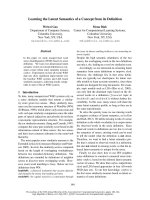
Analysis by use o f t he program
FIRST
[30] (http://
firstweb.asu.edu/) indicates the highest flexibility of the
peptide backbone of native RNase A at the N-terminus
and in the loop region between helices I and II (around
Ala20), followed by the region from the end of helix II
spanning the loop to the adjacent b-strand (Lys31–Phe46;
Fig. 1), which had a lso shown low stability i n both
refolding [31] and unfolding [17] experiments. We replaced
various amino-acid residues as both single and multiple
mutations in the two loop regions to investigate their
contribution to the overall stability of the RNase A
molecule. To maintain RNase A folding and activity, we
refrained from dramatic interference with the protein
structure such as charge-reversal mutations or the intro-
duction or deletion of disulfide bonds.
In the A la20 loop region the exposed, proteolytically
sensitive residues A la20 and/or Ser21 were replaced b y
proline t o rigidify the flexible loop. As a control, Ser21 was
replaced by leucine to introduce a cleavage site for
thermolysin, which was also used to determine the unfolding
rate constants of RNase A. Thus, both the local unfolding
of this loop (vi a the cleavage a t Ala20–Leu21) and t he
global unfolding of the RNase A molecule (via the cleavage
at Asn34–Leu35/Thr45–Phe46) can be detected. In the
proposed unfolding region, we selected residues with side
chains found to be involved in intramolecular interactions
(analysis using t he program
WHAT IF
[32]) ( Table 2). Lys31,
Arg33, Leu35, and Phe46 (Fig. 1) were replaced as both
single and multiple mutations (K31A/R33S, L35S, L35A,
F46Y, L35S/F46Y , L35A/ F46Y, and K31A/R33S/F46Y).
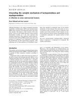
As the crystal structure reveals, the side chains of these
residues a re involved in intramolecular interactions that
form either a hydrophobic patch (Leu35 and Phe46 with
Met29 and Met30; Fig. 2A) or a hydrogen bond network
(Arg33, Fig. 2B). Furthermore, these residues had proven
to be crucial in the proteolytic degradation of the RNase A
molecule on unfolding [17]. A s a control for replacing
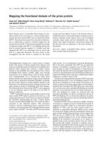
Fig. 1. Tertiary structure of RNase A. The model (7rsa) was taken
from the Brookhaven protein databank and drawn with Swiss
PDB
-
VIEWER
v3.7. The replaced residues are marked in red for the region
around Ala20 and green for the proposed unfolding region (Lys31–
Phe46).
4150 J. Ko
¨
ditz et al.(Eur. J. Biochem. 271) Ó FEBS 2004
solvent-exposed amino-acid residues, Asn34 was replaced
by aspartate.
Expression, renaturation, and purification
All RNase A variants were expressed as inclusion bodies.
Even though they differed in their tendency to form
aggregates during renaturation, all variants could be
obtained in sufficient amounts (up to 30 mgÆL
)1
culture
medium). The purified proteins proved to be homogeneous
by SDS/PAGE and rechromatography on a Mono S
column.
CD spectra
As detected by CD spectroscopy, a ll RNase A variants
revealed a tertiary and secondary structure comparable to
that of wild-type RNase A (not shown) with a marginal
disturbance of the secondary structure in A20P/S21P-
RNase A. An increased signal in the CD spectra in the
near-UV region of F46Y containing RNase A variants is
attributed to the introduction of the additional tyrosine.
Activity
Enzymatic activity provides a sensitive measure of the
impact of modifications on the native structure of an
enzyme [33]. The k
cat
/K
m
values for RNase A and its
variants, determined w ith 6-FAM-dArU(dA)
2
-6-TAMRA
as substrate [23], revealed that all RNase A variants are
active (Table 3). However, while the RNase A variants with
mutations in the Ala20 loop region a s well as N 34D-
RNase A and L35A-RNase A showed an activity compar-
able to that of wild-type RNase A, the variants with
mutations in the unfolding region (except for N34D and
L35A) showed a more s ignificant decrease in the k
cat
/K
m
values, with the lowest activity (% 20%) for L35S/F46Y-
RNase A and L35A/F46Y-RNase A.
Thermodynamic stability
To study the effect of the mutations on the thermodynamic
stability o f t he RNase A molecule, G dnHCl-induced
Table 2. Relative solvent accessibility o f amino acid residues of wild-type RNase A. The relative accessibility was calculated using the program
WHAT IF
[32]andrelatestheaccessibilityofthesidechainoftheresidueintheprotein to the accessibility in a G ly-XXX-Gly peptide in vacuu m w hich
is a good approximation for the accessibility in the unfolded state of the protein.
Residue
Relative
accessibility
(%) Known side chain interactions and effects by modification
Met29 18 Hydrophobic core with Met30, Leu35, and Phe46
Met30 0 Hydrophobic core with Met29, Leu35, and Phe46
Lys31 76 No interactions; K31C slightly decreases T
m
[44]
Ser32 66 No interactions; S32C slightly decreases T
m
[44]
Arg33 23 H bonds with the backbone of Arg10 and Met13
Asn34 48 No interactions; attached carbohydrate moiety in the related RNase B increases T
m
by 1.5 °C [27]
Leu35 9 Hydrophobic core with Met29, Met30, and Phe46
Thr36 12 No interactions but in proximity to Met30, Tyr97, and the disulfide bond Cys40–Cys95
Lys37 60 No interactions
Asp38 68 No interactions; D38R decreases T
m
by 4 °C [52]
Arg39 69 No interactions
Cys40 11 Disulfid bond with Cys95; C40A/C95A decreases T
m
by 20 °C [53]
Lys41 21 P1 subsite; H bond to the side chain of Asn44; K41R strongly decreases the activity but does not affect T
m
[54];
a chemical crosslink K7–K41 increases both DG
(H2O)
and DG
#
U
by about 12 kJ mol
)1
[51]
Pro42 43 No interactions; P42A does not affect the thermodynamic stability [55]
Val43 44 No interactions
Asn44 3 H bond with Gln11 and Lys41
Thr45 8 B1 subsite; T45G decreases T
m
by 10 °C [56]
Phe46 0 Hydrophobic core with Met29, Met30, and Leu35; exchange by Leu, Val, Glu, Lys, or Ala greatly decreases
DG
(H2O)
[42,43]
Fig. 2. Tertiary structure of the unfolding region of wild-type RNase A.
The model (7rsa) was taken from the Br ookhaven protein databank
and drawn with Swiss
PDB
-
VIEWER
v3.7. (A) Hydrophobic cluster
formed by residues Phe46, Leu35, Met29 and Met30. The ribbon at
positions Phe46 and Leu35 is marked in green. (B) Hydroge n b onds
between the side ch ains o f Arg33 and the backbone of Met13 and
Arg10 and the hyd rogen bo nd be tween t he side ch ain of Arg10 and the
backbone of Arg33. The hydrophobic residues are marked as green
balls.
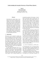
Ó FEBS 2004 Unfolding region of ribonuclease A ( Eur. J. Biochem. 271) 4151
transition curves were recorded (Fig. 3). The values for
[D]
50%
and m
DG
as well as the change in free energy by the
mutation at the transition midpoint of wild-type RNase A
DDG
[D]50%
were determined (Table 3).
All mutations in the A la20 loop as well as the control
N34D did not significantly affect the GdnHCl-induced
unfolding so that the transition c urves of t hose
RNase A variants resemble t hat of w ild-type RNase A
(group I; Fig. 3) with a mean value for [D]
50%
of
2.85 ± 0.10
M
(Table 3). In contrast, a ll other mutations
in the proposed unfolding region resulted in a considerable
decrease in the transition midpoint (group II). Interestingly,
the variants K31A/R33S, L35S, L35A, and F46Y show
a remarkably coincident decrease in the thermodynamic
stability ([D]
50%
¼ 1.8 5 ± 0.10
M
; Table 3, Fig. 3) whereas
the variants obtained by the combination of destabilizing
mutations in the unfolding region (K31A/R33S/F46Y,
L35S/F46Y, and L35A/F46Y) are characterized by a
further slight but uniform decrease in stability ([D]
50%
¼
1.59 ± 0.10
M
; Table 3, Fig. 3).
A similar destabilizing effect by the mutations was
observed in thermal tran sition curves determined for wild-
type RNase A and the variants F46Y and L35S/F46Y (not
shown). Values of T
m
were 62.0 ± 0.1 °C, 53.0 ± 0.1 °C,
and 48.0 ± 0.1 °C, respectively, corresponding to values
of DDG
25°C
of 10.4 ± 1.5 kJÆmol
)1
and 23.6 ± 1.4 kJÆ
mol
)1
caused by the mutations F4 6Y and L35A/F36Y
(calculat ed with DC
p
¼ 9.4 kJÆK
)1
Æmol
)1
for wild-type
RNase A [27] and the DH
m
values of 544 ± 14 kJÆmol
)1
,
481±8kJÆmol
)1
,and341±5kJÆmol
)1
for wild-type
RNase A and the variants F46Y and L35S/F46Y, respect-
ively, obtained from the van’t Hoff plot).
Kinetic stability
The decreased thermodynamic s tability of the RNase A
variants with mutations in the proposed unfolding region
could arise from faster unfolding or slower refolding (or
both). To dissect the effect of the mutations, rate constants
of unfolding of wild-type RNase A and its variants were
determined. Owing to the isomerization of natively cis
proline peptide bonds in the unfolded state [34,35] refolding
of RNase A is known to be rather complex [36] and the
introduction of further proline residues at positions 20 and
21 is expected to further increase this complexity, as
indicated by the decreased m
DG
values f or the A20P and
A20P/S21P variants (Table 3). Hence, we refrained from
refolding experiments.
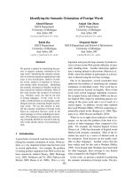
Unfolding rate c onstants w ere d etermined b y limited
proteolysis with thermo lysin at three different temperatures
between 35.0 °C and 57.5 °C (Fig. 4). By linear extrapola-
tion of the Eyring plots [29], v alues of k
U
at 25 °Cwere
obtained which were used to calculate values of DDG
#
U
at
25 °C (Table 3). Wild-type RNase A and all variants of
group I with respect to their thermodynamic stability unfold
with the same rate constants at 47.5–57.5 °C (Fig. 4),
Table 3. Activity and thermodynamic and kinetic parameters of wild-type RNase A and its variants at 25 °C. Values of k
cat
/K
m
were determined as
described in E xperimental Procedures with 6-FAM-dArU(dA)
2
-6-TAMRA as substrate i n 100 m
M
Mes/NaOH, pH 6.0, containing 100 m
M
NaCl.
The thermodynamic parameters were determined from the GdnHCl-induced transition curves at 25 °C as described in Experimental Procedures.
Values of DDG
#
U
(25 °C) were calculated from parameters obtained from the Eyring plot (Fig. 4) as described in Experimental Procedures.
RNase A
variant
10
)7
· k
cat
/K
m
(s
)1
Æ
M
)1
)
[D]
50%
(
M
)
m
DG
(kJÆmol
)1
Æ
M
)1
)
DDG
[D]50%
(kJÆmol
)1
)
DDG
#
U
25
C
(kJÆmol
)1
)
Wild-type 3.9 ± 0.7 2.79 ± 0.03 13.7 ± 1.6 – –
A20P 2.4 ± 0.4 2.89 ± 0.03 10.2 ± 0.9 ) 1.0 ± 0.5 0.3 ± 1.0
S21P 3.5 ± 0.3 2.90 ± 0.02 13.0 ± 0.8 ) 1.4 ± 0.5 ) 0.6 ± 0.4
S21L 2.5 ± 0.2 2.78 ± 0.02 16.4 ± 1.8 0.2 ± 0.5 1.8 ± 0.4
A20P/S21P 2.8 ± 0.4 2.82 ± 0.02 11.1 ± 0.8 ) 0.3 ± 0.5 ) 1.0 ± 1.6
N34D 2.9 ± 0.1 2.86 ± 0.02 13.1 ± 1.2 ) 0.9 ± 0.5 ) 0.9 ± 0.6
L35A 3.1 ± 0.3 1.93 ± 0.02 10.8 ± 0.6 9.3 ± 0.7 5.9 ± 0.5
L35S 0.9 ± 0.2 1.84 ± 0.02 16.1 ± 1.9 15.3 ± 1.9 10.2 ± 0.4
F
46Y
1.8 ± 0.1 1.88 ± 0.02 11.9 ± 0.9 10.8 ± 0.9 7.1 ± 0.4
K31A/R33S 1.2 ± 0.2 1.92 ± 0.02 13.5 ± 1.3 11.7 ± 1.2 13.1 ± 0.2
L35A/F
46Y
0.8 ± 0.1 1.59 ± 0.01 14.1 ± 0.6 16.9 ± 0.8 11.7 ± 0.4
L35S/F
46Y
0.8 ± 0.2 1.51 ± 0.02 12.8 ± 1.1 16.4 ± 1.5 12.6 ± 0.2
K31A/R33S/F
46Y
1.2 ± 0.3 1.64 ± 0.02 13.4 ± 0.9 15.4 ± 1.1 16.3 ± 0.3
[GdnHCl] (M)
012345
f
N
0.0
0.5
1.0
Fig. 3. GdnHCl-induced transition curves. The transition curves of
wild-type RNase A (teal) and its variants A20P (black), S21P (grey),
S21L (bright green), A20P/S21P (blue), N34D (red), L35A (cyan),
L35S (green), F46Y (dark red), K31A/R33S (pink), L35A/F46Y (dark
yellow), L35S/F46Y (dark blue), and K31A/R33S/F46Y (violet) were
determined by fluorescence spectroscopy in 50 m
M
Tris/ H Cl , pH 8 .0 ,
at 25 °C.
4152 J. Ko
¨
ditz et al.(Eur. J. Biochem. 271) Ó FEBS 2004
indicating that the kinetic stability is also not affected by
these mutations. All the thermodynamically less stable
RNase A variants also show a large increase in k
U
.Even
though the effects are not as uniform as for the thermo-
dynamic stability, the comparison of DDG
[D]50%
and
DDG
#
U
25
C
shows that the decrease in the thermodynamic
stability is mainly caused by an increase in the unfolding rate
constant, i.e. a decrease in the kinetic stability. Whereas the
introduction of a cleavage site for thermolysin in the
control variant S21L-RNase A facilitated degradation of
the RNase A molecule under native conditions (not
shown), this variant was degraded like wild-type RNase A
under denaturing conditions.
Discussion
As in the folding of proteins, confined regions of the protein
structure have a crucial role in the unfolding process and
are, thus, particularly important for kinetic stability [8,9,12].
These r egions are m ostly located on the surface of the
protein molecule, and loops in particular often represent
critical spots [8,11,17].
RNase A possesses two structural sections that might
function as such a critical region (Fig. 1): (a) the loop region
around Ala20, which is highly flexible under native condi-
tions [14,15] as reflected in efficient proteolytic attack by
nonspecific proteases such as proteinase K and subtilisin
Carlsberg [37–39]; (b) the region from the end of helix II to
the a djacent a-sheet (Lys31–Ph e46), which becomes access-
ible to an H–D exchange [40] and to t he proteases
thermolysin and trypsin when the molecule starts to unfold
[17]. Furthermore, this region (residues 31–39) is the last one
that becomes protected against tryptic attack during
RNase A folding [31].
RNase A variants with amino-acid substitutions in the
two regions fell into two classes w ith respect to thermo-
dynamic stability (Fig. 3). The RNase A variants with
similar unfolding transition curves to wild-type RNase A
(group I) are obtained by mutations in the loop region
around Ala20 or by the control mutation N34D. These
amino-acid residues are not involved in interactions like
hydrogen bonds, salt b ridges or hydrophobic clusters, as
reflected in great flexibility of the loop region around
Ala20 [15,16]. So even the replacement of two adjoined
residues i n this r egion by proline (A20P/S21P) was
tolerated. On the other hand, the introduction of the
proline residues, i.e. the decrease in loop flexibility, did not
increase the global stability of the RNase A molecule.
By introducing a cleavage site for thermolysin (S21L-
RNase A), the flexibility of the Ala20 loop became traceable
for this protease. Nevertheless, the unfolding rate constants
of this RNase A variant correspond to those of wild-type
RNase A (Fig. 4), indicating that the local unfolding of the
Ala20 loop is independent of the global unfolding of the
RNase A molecule. In the control variant Asn34-RNase A,
a s olvent-exposed residue that belongs to the unfolding
region (Lys31–Phe46) and serves as anchor for the stabil-
izing carbohydrate moiety in the related RNase B [41], was
replaced. As expected, the mimicked deamidation does not
affect interactions essential for stability.
In contrast, the less stab le RNase A variants of group II
(L35S, L35A, F46Y, K31A/R33S, L35S/F46Y, L35A/
F46Y, and K31A/R33S/F46Y) all of which were obtained
by mutations in the region Lys31–Phe46 indicate a consid-
erable contribution of this region to the thermodynamic
stability of the entire RNase A molecule. The coincidence of
the degree o f destabilization in these variants points to an
effect on the stability of the entire region rather than on a
particular interaction. A similar d estabilization was also
found by Chatani et al. [42] and Kadonosono et al. [43] by
replacement of Phe46 with Leu, Val, Ala, Lys, or Glu. The
authors concluded that Phe46 has a n important role in the
folding reaction through hydrophobic interactions and by
the correct packing of the amino-acid side ch ains between
two structural domains [42]. However, from the rate of
oxidative protein folding, they concluded t hat there was a
decreased k
U
, i.e. kinetic stabilization of the F46L, F46V,
and F46A variants. In contrast, we found an acceleration
of the unfolding reaction, i.e. kinetic destabilization, for
the variants and the similarity of DDG
[D]50%
and DDG
#
U
indicates that the decrease in the thermodynamic stability is
mainly caused by an in crease in k
U
. Furthermore, our
results suggest that Leu35, the side chain of which is buried
in the interior of the molecule like that of Phe46 (Table 2),
is involved in the formation of a hydrophobic cluster
with Phe46, Met29 and Met30 (Fig. 2A) and consequently
plays a similar role to Phe46. Molecular modeling revealed
that any mutation in position 35 d estabilizes the e ntire
molecule by disturbing these complex hydrophobic inter-
actions (G. Vriend, University of Nijmegen, personal
communication).
In addition to these hydrophobic interactions, this region
is stabilized by a network of hydrogen bonds between the
side chain of Arg33 and the backbone of Met13 and Arg10
(three hydrogen bonds) and between the side chain of Arg10
and the backbone of Arg33 (one hydrogen bond; Fig. 2B).
Because no hydrogen bonds were identified for the side
chain of Lys31 of RNase A (analysis using the program
WHAT IF
[32]) and its exchange with Cys results in only a
1000 / T (K
-1
)
3.00 3.05 3.10 3.15 3.20 3.25
ln (k
U
/ T)
-14
-12
-10
Fig. 4. Eyring plot for the unfolding of wild-type RNase A and its vari-
ants. Values for k
U
of wild-type RNase A (teal) and its variants A20P
(black), S21P (grey), S21L (bright green), A20P/S21P (blue), N34D
red), L35A (cyan), L35S (green), F46Y (dark red), K31A/R33S (pink),
L35A/F46Y (dark yellow), L35S/F46Y (dark blue), and K31A/R33S/
F46Y (violet) were determined by limited proteolysis with thermolysin
in 50 m
M
Tris/HCl, pH 8.0, at 35.0–57.5 °C as described in Experi-
mental procedures.
Ó FEBS 2004 Unfolding region of ribonuclease A ( Eur. J. Biochem. 271) 4153
slight decrease in the stability [44], the d estabilizing effect
of the mutation K31A/R33S is probably caused by t he
mutation of Arg33.
Generally, changes in the thermodynamic s tability by
mutations can be caused by effects on t he native and/or the
unfolded state, whereas changes in the kinetic stability are
due to a change in the native and/or transition state. The
determination of the unfolding rate constants of wild-type
RNase A and its variants (Fig. 4) allowed differentiation
between the several possibilities. The RNase A variants
with GdnHCl-induced transition curves similar to that of
wild-type R Nase A, i.e. t he members of g roup I, also show
thermal unfoldin g rate constants and consequently DG
#
U
values comparable to that of wild-type RNase A (Table 3),
indicating that the native state, relative to the transition
state, is not affected by the mutations. The labile RNase A
variants show a large increase in the unfolding rate
constants. For the variants K31A/R33S and K31A/R33S/
F46Y, a value of DDG
#
U
was obtained that corresponds
very well to that of DDG
[D]50%
(Table 3), indicating that the
decrease in the thermodynamic stability is caused by
destabilization of the native state relative to th e unfolded
state. The decrease in the thermodynamic s tability of the
other less s table v ariants, all of which were exclusively
obtained by exchanges of the hydrophobic residues Leu35
and/or Phe46, is not solely attributable to faster unfolding.
The differences between DDG
[D]50%
and DDG
#
U
also point
to slower refolding, e.g. by disturbance of the formation of a
hydrophobic cluster [42]. Nevertheless, the decrease in the
thermodynamic stability is m ainly c ause d by t he faster
unfolding resulting from destabilization of the native state
relative to the transition state, underlining the predominant
importance of this region for maintaining the natively
folded structure of the RNase A molecule.
Interestingly, the hydrophobic nature of residues 29, 30,
35, and 46 is conserved throughout the members of the
ribonuclease A superfamily (Fig. 5). While Phe46 and
Met30 (numbered by the RNase A sequence) are found in
all members, Met29 and Leu35 can be occupied by Met, Ile,
or Ala and Leu, Met, or Ile, respectively (Fig. 5, cf [45]).
Furthermore, with the exception of mammalian ribonuc-
leases 2 and frog ribonucleases, the charged residue Arg33 is
conserved (Fig. 5).
Altogether, whereas the loop region between helices I and
II, i.e. around Ala20, does not contribute to the stability of
the RNase A molecule and local flexibility does not lead to
global unfolding, the interface between helix II and the
adjacent a-sheet is stabilized by a multitude of interactio ns
and is very sensitive to mutations. Connecting regions
between different folding motifs have also been found to be
crucial for the stability of other proteins [46–49]. Despite a
vast number o f RNase A variants p roduced by protein
engineering (for a review, see [50]), only two variants
concerning this region are more stable than wild-type
RNase A: the naturally occurring glycosylated RNase B (at
Asn34 [41]) and the chemically cross-linked RNase A
(Lys7–Lys41) [51]. F or both variants, the thermodynamic
stabilization is comparable to the kinetic stabilization
[27,51]. Also the effect of the mutations reported here on
the thermodynamic stability can mainly be attributed to
effects o n the kinetic stability o f the protein, providing
further evidence for the validity of the concept of t he
unfolding region.
Acknowledgements
We are grateful to Professor R. T. Raines (University of Wisconsin,
Madison, WI, USA) for the gift of the plasmid pBXR, to Professor
G. Vriend (University of Nijmegen, the Netherlands) for molecular
modeling, and to Y. Markert for providing the plasmids for the variants
A20P, S21P, and A20P/S21P. J. K. was supported by a grant from the
Max-Buchner-Forschungsstiftung, Frankfurt, Germany.
References
1. Shortle, D. (1996) The denatu red state (the oth er half of the folding
equation) and its role in protein stability. FASEB J. 10, 27–34.
2. Daggett, V. & Fersht, A.R. (2003) Is there a unifying mechanism
for protein folding? Trends Biochem. Sci. 28, 18–25.
3. Jackson, S.E. (1998) How do small single-d omain prot eins fold?
Fold. Des. 3, R81–R91.
4. Jaenicke, R. (1999) Stability and folding of domain proteins. Prog.
Biophys. Mol. Biol. 71, 155–241.
5. Schellenberger, A. & Ulbrich, R. (1989) Protein stabilization by
blocking the native unfolding nucleus. Biomed. Biochim. Acta 48,
63–67.
6. Ulbrich-Hofmann, R., Arnold, U. & Mansfeld, J. (1999) The
concept of the un folding region for approaching the mechanisms
of enzyme stabilization. J. Mol. Catal. B Enzym. 7, 125–131.
7. Vriend, G. & Eijsink, V. (1993) Prediction and analysis of struc-
ture, stability and unfolding of thermolysin-like proteases.
J. Comput. Aided Mol. Des. 7, 367–396.
8. Eijsink, V.G., Veltman, O.R., Aukema, W., Vriend, G. &
Venema, G. (1995) Structural determinants of the stability of
thermolysin-like proteinases. Nat. Struct. Biol. 2, 374–379.
Fig. 5. Alignment of the amino-acid sequence (residues 1–58) for members of the RNase A superfamily. RNase A (P00656) from bovine pancreas,
RNase 1 (P07998) from human pancreas, and bs RNase (P00669) from bovine semen belong to the mammalian ribonucleases 1 (pancreatic type).
Human EDN (P10153, eosinophil-derived neurotoxin) belongs to the mammalian (nonsecretory or neurotoxin-type) ribonucleases 2, human
RNase 4 (P34096) belongs to the mammalian ribonucleases 4, human angiogenin (P03950) belongs to the mammalian angiogenins, and onconase
(P22069) from Rana pipiens oocytes belongs to frog ribonucleases [45]. Residues are numbered according to RNase A. Ident ical residues and sim ilar
residues are u nderlaid in black and grey, respectively. Deletions in the sequences are indicated by $.
4154 J. Ko
¨
ditz et al.(Eur. J. Biochem. 271) Ó FEBS 2004
9. Mansfeld, J., Vriend, G., Van den Burg, B., Eijsink, V.G. &
Ulbrich-Hofmann, R. (1999) Probing the unfolding region in
a thermolysin-like protease by site-specific immobilization.
Biochemistry 38, 8240–8245.
10. Siddiqui, K.S., Rangarajan, M., Hartley, B.S., Kitmitto, A.,
Panico, M., Blench, I.P. & Morris, H.R. (1993) Arthrobacter
D
-xylose isomerase: partial proteolysis with thermolysin. Biochem.
J. 289, 201–208.
11. Machius, M., Declerck, N., Huber, R. & Wiegand, G. (2003)
Kinetic stabilization of Bacillus licheniformis alpha-amylase
through introduction of hydrophobic residues at the surface.
J. Biol. Chem. 278, 11546–11553.
12. Gaseidnes, S., Synstad, B., Jia, X., Kjellesvik, H., Vriend, G . &
Eijsink, V.G. (2003) Stabilization of a chitinase from Serratia
marcesce ns by Gly fi Ala and Xxx fi Pro mutations. Protein
Eng. 16, 841–846.
13. Fetrow, J.S. (1995) Omega loops: nonregular secondary struc-
tures significant in protein function and stability. FASEB J. 9,
708–717.
14. Wlodawer, A., Bott, R. & Sjolin, L. (1982) The refined crystal
structure of ribonuclease A at 2.0 A
˚
resolution. J. Biol. Chem. 257,
1325–1332.
15. Hubbard, S.J., Beynon, R.J. & Thornton, J.M. (1998) Assessment
of conformational parameters as pre dictors of limited proteolytic
sites in native protein structures. Protein Eng. 11, 349–359.
16. Santoro, J., Gonzalez, C., Bruix, M., Neira, J.L., Nieto, J.L.,
Herranz, J. & Rico, M. (1993) High-resolution three-dimensional
structure o f ribonuclease A in solution by nu clear magnetic
resonance spectroscopy. J. Mol. Biol. 22 9, 722–734.
17. Arnold, U., Ru
¨
cknagel, K.P., Schierhorn, A. & U lbrich-
Hofmann, R. (1996) Thermal unfolding and proteolytic suscept-
ibility of ribonuclease A. Eur. J. Biochem. 237, 862–869.
18. Markert, Y., Ko
¨
ditz, J., Ulbrich-Hofmann, R. & Arnold, U.
(2003) Proline versus charge concept for protein stabilization
against proteolytic attack. Pr otein Eng. 16, 1041–1046.
19. delCardayre
´
, S.B., Ribo, M., Yokel, E.M., Quirk, D.J.,
Rutter, W.J. & Raines, R.T. (1995) Engineering ribonuclease A:
production, purification and characterization of wild-type enzyme
and mutants at Gln11. Protein Eng. 8, 261–273.
20. Sanger, F., Nicklen, S. & Coulson, A.R. (1977) DNA sequencing
with chain-terminating inhibitors. Proc. Natl Acad. Sci. USA 74,
5463–5467.
21. Sela, M. & Anfinsen, C.B. (1957) Some spectrophotometric and
polarimetric experiments with ribonuclease. Biochim. Biophys.
Acta 24, 229–235.
22. Thannhauser, T.W., Konishi, Y. & Scheraga, H.A. (1984) Sensi-
tive quantitative analysis of disulfide bonds in polypeptides and
proteins. Anal. Biochem. 138, 181–188.
23. Kelemen, B.R., Klink, T.A., Behlke, M.A., Euban ks, S .R.,
Leland, P.A. & Raines, R.T. (1999) Hypersensitive substrate for
ribonucleases. Nucleic Acids Res. 27, 3696–3701.
24. Pace, C.N. (1986) Determination and analysis of u rea and
guanidine hydrochloride de naturation curves. I n Methods in
Enzymology (Hirs, C.H.W. & Timasheff, S.N., eds), pp. 266–280.
Academic Press, New York.
25. Santoro, M.M. & Bolen, D.W. (1988) Unfolding free energy
changes determined by the linear extrapolation method. 1.
Unfolding of phenylmethanesulfonyl alpha-chymotrypsin using
different denaturants. Biochemistry 27, 8063–8068.
26. Clarke, J. & Fersht, A.R. (1993) Engineered disulfide bonds as
probes of the folding pathway of barnase: increasing the stability
of proteins against the rate of denaturation. Biochemistry 32,
4322–4329.
27. Arnold, U. & Ulbrich-Hofmann, R. (1997) Kinetic and thermo-
dynamic thermal stabilities of ribonuclease A and ribonuclease B.
Biochemistry 36, 2166–2172.
28. Laemmli, U.K. (1970) Cleavage of structural proteins during the
assembly of the head of bacteriophage T4. Nature (London) 227,
680–685.
29. Arnold, U. & Ulbrich-Hofmann, R. (2001) Proteolytic degrada-
tion of ribonuclease A in the pretransition region of thermally and
urea-induced unfolding. Eur. J. Biochem. 268, 93–97.
30. Jacobs,D.J.,Rader,A.J.,Kuhn,L.A.&Thorpe,M.F.(2001)
Protein flexibility predictions using graph theory. Proteins 44,
150–165.
31. Lang, K. & Schmid, F.X. (1986) Use of a trypsin-pulse method to
study the refolding pathway of ribonuclease. Eur. J. Biochem. 159,
275–281.
32. Vriend, G. (1990) WHAT IF: a molecular modeling and drug
design program. J. Mol. Graph. 8, 52–56.
33. Knowles, J.R. (1987) Tinkering with e nzymes: what a re we
learning? Science 236, 1252–1258.
34. Brandts, J.F., Halvorson, H.R. & Brennan, M. (1975) Con-
sideration of the possibility that the slow step in protein dena-
turation reactions is due to cis-trans isomerism of proline residues.
Biochemistry 14, 4953–4963.
35. Juminaga, D., Wedemeyer, W.J. & Scheraga, H.A. (1998) Proline
isomerization in bovine pancreatic ribonuclease A. 1. Unfolding
conditions. Biochemistry 37, 11614–11620.
36. Wedemeyer, W.J., Welker, E. & Scheraga, H.A. (2002) Proline
cis-trans isomerization and protein fo lding. Bioc hemistry 41,
14637–14644.
37. Rauber, N.R., Jany, K.D. & Pfleiderer, G. (1978) Ribonuclease A
digestion by proteinase K. Z. Naturforsch. [C] 33, 660–663.
38. Richards, F.M. & Vithayathil, P.J. (1959) The preparation of
subtilisin-modified ribonuclease and the separation of the peptide
and protein components. J. Biol. Chem. 234, 1459–1465.
39. Markert, Y., Ko
¨
ditz, J., Mansfeld, J., Arnold, U. &
Ulbrich-Hofmann, R. (2001) Increased proteolytic resistance
of ribonuclease A by protein engineering. Protein Eng. 14, 791–
796.
40. Loh,S.N.,Rohl,C.A.,Kiefhaber,T.&Baldwin,R.L.(1996)A
general tw o-process model d escribes the hyd rogen e x change
behavior of RNase A in unfolding conditions. Proc.NatlAcad.
Sci. USA 93, 1982–1987.
41. Plummer, T.H. Jr & Hirs, C.H. (1964) On the structure of bovine
pancreatic ribonuclease B. Isolation of a glycopeptide. J. Biol.
Chem. 239, 2530–2538.
42. Chatani, E., N onomura, K., Hayashi, R., Balny, C. & Lange, R.
(2002) Comparison of heat- and pressure-induced unfolding of
ribonuclease A: the critical role of phe46 which appears to belong
to a new hydrophobic chain-folding initiation site. Biochemistry
41, 4567–4574.
43. Kadonosono, T., Chatani, E., Hayashi, R., Moriyama, H. &
Ueki, T. (2003) Minimization of cavity size ensures protein sta-
bility and folding: structures of Phe46-replaced bovine pancreatic
RNase A. Biochemistry 42, 10651–10658.
44. Catanzano, F., Graziano, G., Cafaro, V., D’Alessio, G., Di
Donato, A. & Barone, G. (1997) From ribonuclease A toward
bovine seminal ribonuclease: a step by step thermodynamic ana-
lysis. Biochemistry 36, 14403–14408.
45. Beintema, J.J., Breukelman, H.J., Carsana, A. & Furia, A. (1997)
Evolution of vertebrate r ibonucle ases: ribonuclease A superfamily.
In Ribonucleases: Structures and Functions (D’Alessio, G. & Riordan,
J.F., eds), pp. 245–269. Academic Press, New York.
46. Ahrweiler, P.M. & Frieden, C . (1991) Effects of point mutations in
a hinge region on the stability, folding, and enzymatic activity of
Escherichia coli dihydrofolate reductase. Bioche mistr y 30, 7801–
7809.
47. Collinet, B., Garcia, P., Minard, P. & Desmadril, M. (2001) Role
of loops in the folding and stability of yeast phosphoglycerate
kinase. Eur. J. Biochem. 268, 5107–5118.
Ó FEBS 2004 Unfolding region of ribonuclease A ( Eur. J. Biochem. 271) 4155
48. Gu, H., Kim, D. & Baker, D. (1997) Contrasting roles for sym-
metrically dispo sed beta-turns in the f olding of a small protein.
J. Mol. Biol. 274, 588–596.
49. Urfer, R. & Kirschner, K. (1992) The importance of surface loops
for stabilizing a n eightfold beta alpha barrel protein. Protein Sci. 1,
31–45.
50. Chatani, E. & Hayashi, R. (2001) Functional and structural roles
of constituent a mino acid resid ues of bovine pancreatic ribo-
nuclease A. J. Biosci. Bioeng. 92, 98–107.
51. Lin, S.H., Konishi, Y., Nall, B.T. & Scheraga, H.A. (1985)
Influence of an extrinsic cross-link on the folding pathway of
ribonuclease A. Kinetics of folding-unfolding. Biochemistry 24,
2680–2686.
52. Leland,P.A.,Schultz,L.W.,Kim,B.M.&Raines,R.T.(1998)
Ribonuclease A variants with potent cytotoxic activity. Proc. Natl
Acad. Sci. USA 95, 10407–10412.
53. Klink, T.A., Woycechowsky, K.J., Taylor, K.M. & Raines, R.T.
(2000) Contribution of disulfide bonds to the conformational
stability and catalytic activity of ribonuclease A. Eur. J. Biochem.
267, 566–572.
54. Haigis, M.C., Kurten, E.L., Abel, R.L. & Raines, R.T. (2002)
KFERQ sequence in ribonuclease A-mediated cytotoxicity.
J. Biol. Chem. 277, 11576–11581.
55. Dodge, R.W. & Scheraga, H.A. (1996) Folding and unfolding
kinetics of the proline-to-alanine mutants of bovine pancreatic
ribonuclease A. Biochemistry 35, 1548–1559.
56. delCardayre
´
,S.B.&Raines,R.T.(1995)Aresiduetoresidue
hydrogen bond mediates the nucleotide specificity of ribonuclease
A. J. Mol. Biol. 252, 328–336.
4156 J. Ko
¨
ditz et al.(Eur. J. Biochem. 271) Ó FEBS 2004