Báo cáo khoa học: Alternative initiation of transcription of the humanpresenilin 1gene in SH-SY5Y and SK-N-SH cells The role of Ets factors in the regulation ofpresenilin 1 pptx
Bạn đang xem bản rút gọn của tài liệu. Xem và tải ngay bản đầy đủ của tài liệu tại đây (379.18 KB, 10 trang )
Alternative initiation of transcription of the human
presenilin 1
gene
in SH-SY5Y and SK-N-SH cells
The role of Ets factors in the regulation of
presenilin 1
Martine Pastorcic
1
and Hriday K. Das
1,2,3
1
Department of Pharmacology & Neuroscience,
2
Department of Molecular Biology & Immunology and
3
Institute of Cancer Research,
University of North Texas Health Science Center at Fort Worth, TX, USA
We have identified DNA seq uences required fo r the expres-
sion of the presenilin 1 (PS1) gene. A promoter region has
been mapped in SK-N-SH cells and includes sequences be-
tween )118 and +178 flanking the major initiation site (+1).
The PS1 ge ne is also efficiently transcribed in the SH-SY5Y
subclone of SK-N-SH cells. However the promoter appears
to be utilized in alternative ways in both cell types. Sequences
both upstream as well as downstream from the initiation
site mapped in SK-N-SH cells were shown by 5¢-and3¢-
deletion analysis to play a crucial role in both cell lines.
However, in SH-SY5Y cells either upstream or downstream
sequences are sufficient to direct transcription, whereas in
SK-N-SH cells 5¢-deletions past the +1 site eliminate over0
95% of transcription. Several Ets motifs (GGAA)
11
as well as
Sp1 motifs [(G/T)GGCGGRRY]
22
are juxtaposed both up-
stream and downstream from +1. To understand how the
promoter may be utilized alternatively in different cell types
we have examined the effect of point mutations in these
elements. Altering an Ets motif at )10 eliminates 80% of
transcription in SK-N-SH cells whereas the same mutation
has only a minor effect in SH-SY5Y cells. Conversely,
mutation of the Ets element at +90, which eliminates 70% of
transcription in SH-SY5Y cells, has a lesser effect in SK-N-
SH cells. In both cell types a promoter including mutations at
both )10 and +90 sites loses over 90% transcription activity
indicating the crucial importance of these two Ets motifs. The
effect of Sp1 mutations appears to be similar in both cell
types. Hence the differential e xpression in each cell type may
be at least partially determined by Ets factors and the )10/
+90 sites. We have identified several Ets factors that
recognize specifically the )10 Ets motif by the yeast one-
hybrid selection including avian erythroblastosis virus E
26
oncogene homologue 2, Ets-like gene 1, Ets translocation
variant 1 and Ets related molecule (ERM)
3
. We show here
that ERM specifically recognizes Ets motifs on the PS1
promoter located at )10 as well as downstream a t +90,
+129 and +165 and activates PS1 transcription with pro-
moter fragments containing or not the )10 Ets site.
Presenilins (PS1 and PS2) are highly homologous multipass
transmembrane proteins [ 1–3]. T hey are required for the
protease activity of a multiprotein complex termed c-secr-
etase, which includes presenilin, nicastrin, Aph-1 and Pen-2
that are all necessary for proteolytic activity [4–7]. It appears
that presenilin acts as a catalyst or a required cofactor. c-
Secretase cleaves the amyloid precursor protein (APP),
resulting in the production of the Ab peptide which appears
to be central in the pathogenesis of Alzheimer’s disease [8–
10]. Indeed PS1 mutations
4
have been linked to many cases
of early onset familial Alzheimer’s disease [11–13]. Similarity
in the processing of APP a nd the Notch receptor protein,
which controls signaling and cell–cell communication in
development, have largely contributed to the understanding
of the role of presenilins. The Notch receptor, a type 1
membrane protein, is also cleaved by c-secretase. The
stimulation of Notch by its ligand leads to the intramem-
brane proteolysis of the receptor, freeing the Notch
intracellular domain, which translocates to the nucleus
and regulates gene expression [14–16]. Increasing evidence
indicates that p resenilin and c-secretase cleave a variety of
Type 1 transmembrane proteins which all release intracel-
lular fragments with the ability to interact with transcription
coactivators [17]. CD44, a ubiquitous cell adhesion protein,
is cleaved by c-secretase and releases an intracellular domain
that activates CBP/p300 [18]. Neuronal cadherin (N-cadh-
erin), mediates Ca
2+
-dependent cell–cell adhesion and
recognition, and plays a crucial role in neurogenesis, tissue
development a nd homeostasis. The proteolytic cleavage of
N-cadherin releases an intracellular C -termin al fragment
that suppresses CRE-dependant activation of transcription
by promoting CBP degradation by the proteasome [19].
Hence it appears that presenilins may affect the expression
of many genes through intramembrane proteolysis. This
mechanism appears to be modulated by neuronal activity
[19] and may represent an important aspect in the pathology
of Alzheimer’s disease. Furthermore, PS1 appears to play a
crucial role in the normal metabolism of APP as well as in
the pathological increase of the Ab42 [20]. APP is a
Correspondence to H. K Das, University of North Texas Health Sci-
ence Center at Fort Worth, 3500 Camp Bowie Boulevard, Fort Worth,
TX 76107, USA. Fax: +1 817 735 2091, Tel.: +1 817 735 5448,
E-mail: .edu
Abbreviations: APP, amyloid precursor protein; CAT, chloramphen-
icol acetyltransferase; Elk1, Ets-like gene 1; ERM, Ets related mole-
cule; ER81, alias for ETV1 (Ets translocation variant 1); Ets2, avian
erythroblastosis vi rus E
26
oncogene homologue 2; PS1, presenilin 1.
(Received 30 July 2004, accepted 30 September 2004)
Eur. J. Biochem. 271, 4485–4494 (2004) Ó FEBS 2004 doi:10.1111/j.1432-1033.2004.04453.x
ubiquitously expressed cell surface protein and its process-
ing by c-secretase cleavage generates not only Ab but also
an intracellular domain of APP [21] that may function in
nuclear signaling both in normal as well as pathological
signal transduction [22–24].
We have studied the transcriptional regulation of PS1 in
different cell types and in particular the role of Ets and Sp1
sequence motifs flanking the major initiation site mapped in
SK-N-SH cells [25]. A crucial Ets element located at )10 has
been used as bait in yeast one-hybrid screening of a human
brain cDNA library to isolate Ets factors that may control
PS1 expression in neu ronal cells [26,27]. The specificity of
Ets elements is specified by four factors: their site of
synthesis and chemical modification, the presence of specific
Ets promoter motifs and the set of nuclear factors with
which they interact in the tissue being considered [28,29].
Hence they should be a powerful determinant in the degree
of activation, the initiation site, as well as the regulation of
the PS1 gene.
Materials and methods
Transfection assays
SK-N-SH and SH-SY5Y cells were transfected with
PS1CAT fusion genes containing various fragments of PS1
sequences flankin g the transcription initiation site [25]. Cells
were seeded at a density of 10
4
Æcm
)2
2 days before transfec-
tion. On the day of transfection, medium was replaced with
serum-free Dulbecco’s modified Eagle’s medium 2 h p rior
the addition of DNA. Usually 6 lg of PS1CAT and 4 lgof
pSV-b-galactosidase control vector (Promega, Madison,
WI)
5
were mixed in 112 lLH
2
O. CaCl
2
(12.5 lL) was then
added to the DNA mixture which was slowly added to
125 lLof2· Hank’s buffered salt solution
6
[25]. Precipitate
wasallowedtoformat37°C for 40 min and added to the
cells (on 5 cm plates). After 4 –5 h i ncubation at 37 °Ca
glycerol shock was performed. The medium was removed
and 1.5 mL of 12.5% (v/v) glycerol was added for 90 s on
SK-N-SH cells and 60 s on SH-SY5Y cells. Glycerol was
removed after dilution with 2 m L NaCl/P
i
and the plates
were washed three times with 3 mL NaCl/P
i
. Dulbecco’s
modified Eagle’s medium containing 10% (v/v) fetal bovine
serum was finally added and the cells were returned to the
incubator for 16–18 h before harvesting.
7
Again, the medium
was removed, cells were washed three times with 3 mL
NaCl/P
i
and harvested in 500 lLNaCl/P
i
and pelleted at
1000 g for 4 min. The pellets were resuspended into 100 lL
of 250 m
M
Tris/HCl pH 7.5 and lysed by three cycle s of
freeze/thaw and cellular debris were pelleted at 12 000 g for
4 min. Supernatants were stored at )70 °C. For chloram-
phenicol acetyltransferase
8
(CAT) assays, 50 lgproteinwas
heat-treated at 60 °C for 10 min, centrifuged for 4 min at
12 000 g, incubated in 100 lL reaction containing
0.25 mgÆmL
)1
n-butyrylCoA and 0.1 lCi [
14
C]chloram-
phenicol at 37 °C [25]. CAT assays were incubated for 3–
4 h and extracted with 250 lL mixed xylenes. The xylene
phase was t hen back extracted twice with 2 50 m
M
Tris
pH 7.5 and the
14
C in the xylene phase was then counted.
For b-galactosidase assays 12–20 lg o f p rotein extract
(non heat-treated) was incubated in 0.1
M
sodium phos-
phate pH 7.5, 1 m
M
MgCl
2
,50m
M
2-mercaptoethanol,
and 3 m
M
2-nitrophenyl-b-
D
-galactopyranoside
9
. Reaction s
were stopped with 800 lLof1
M
Na
2
CO
3
and A
420
was
measured in the visible spectrum.
Promoter activity in different samples was usually
compared using b-galactosidase activity as an internal
control, except in the experiments testing the activity of Ets
related molecule (ERM)
10
cDNA to limit possible competi-
tion between promoters. Each experiment was r epeated
three times, with a minimum o f triplicate te sts of each
construct and treatment.
Point mutagenesis of the
PS1
promoter
Promoter mutants were generated in the context of the
()118/+178)PS1CAT constr uct by PCR-based site-direc-
ted mutagenesis, using the QuickChange k it from Strata-
gene (La Jolla, CA)
11
and t he primers listed b elow. The
following point mutations were generated with the primers
listed and each primer was used together with its re verse
complementary strand. The point mutation at the )70 Sp1
motif, m()70), was obtained with p)70F (5¢-GGCCGGA
GGCCTCGAAGCCTTCCTCCTGG-3¢) and its reverse
complement p)70R. Mutation m()50) was obtained with
p)50F (5¢-CTCCTGGCTCCTCAAGTCCTCCGTGG-3¢)
and the corresponding p)50R; m()30) was obtained
with p)30F (5¢-CCCTCCTCCGTGATGAGGCCGCC
AACGACG-3¢)andp)30R; m(+20) with p+20F
(5¢-GTGAGGGTTCTCGGGCTCATCCTGGGACAG
GCAGCTC-3¢) and p+20R; m(+65) with p+65F
(5¢-GCGGTTTCACATCCTAGACAAAACAGCG-3¢)and
p+65R; m(+90) with p+90F (5¢-GGCTGGTCTGTGA
CTAACCTGAGCTACG-3¢) and p +90R; m(+129) with
p+129F (5¢-CGGCGGCAGCGGGGCGGCGACTAA
GCGTATGTGCGTGATG-3¢); and p+129R using
p()118, +178)CAT as the template.
Rapid amplification of cDNA ends (RACE)
12
in SH-SY5Y
and SK-N-SH cells transfected with the
PS1
promoter
Total RNA was prepared from SK-N-SH cells and
SH-SY5Y cells transfected with ()119, +178)PS1CAT by
the guanidinium/cesium chloride method [25]. Rapid ampli-
fication of cDNA ends (RACE) was performed using the
BD Bioscie nces (Clontech, P alo Alto, CA)
13
Smart R ace
cDNA amplification kit with the primer (5¢-CCGGAT
GAGCATTCATCAGGCGG-3¢) specific for the CAT
gene in order to detect RNA encoded from the transfected
plasmid. Amplification products were gel purified, inserted
into KS bluescript vector (Stratagene) and sequenced.
Cloning of ERM
Two independent clones containing the entire ERM cDNA
were obtained by yeast one-hybrid screening of a human
brain cDNA library from Clontech [26]. The cDNA was
then inserted into pC1 (Promega) by generating a subclone
using Pfu DNA polymerase and the following forward (F)
and reverse (R) primers: ERM-F: 5¢-gatcacgcgtCTCAGG
AGGATCCCTTTTC-3¢ and E RM-R: 5¢-gatcgtcgacGCG
GGTACTAACCTGAACAAGA-3¢.
PCR conditions were according to the manufacturer’s
recommendations at 94 °C for denaturation, 62 °Cfor
4486 M. Pastorcic and H. K. Das (Eur. J. Biochem. 271) Ó FEBS 2004
annealing and 72 °C for extension,
14
for 30, 30 and 60 s,
respectively. Primers were designed to incorporate sites
for the restriction enzymes MluI (forward primer) and
SalI ( reverse primer), to direct integration into the
cloning vector (restriction sites 5¢ flanking sequence
shown in lowercase)
15
. PCR products were purified,
digested with MluIandSalI and in serted into the
corresponding sites of pCI.
Nuclear extracts
Nuclear extracts from SH-SY5Y cells were prepared from
cells growing exponentially as described previously for SK-
N-SH [25]. Compacted nuclei were extracted with buffer C
[20 m
M
Hepes pH 7.9, 600 m
M
NaCl, 1.5 m
M
MgCl
2
,
0.2 m
M
EDTA, 0.5 m
M
dithiothreitol, and 25% (v/v)
glycerol]. The volume of buffer C was adjusted to obtain
an ionic strength equivalent to 300 m
M
NaCl in the
homogenate. Protein concentration of the nuclear extracts
was 5–6 mgÆmL
)1
.
Analysis of specific DNA binding by electrophoretic gel
mobility shift assays (EMSAs)
To assay the binding of Ets factors to the PS1 promoter,
the proteins were synthesized from the corresponding pCI-
based vectors by in vitro transcription–translation as
recommended by the manufacturer (Promega). For e lec-
trophoretic gel mobility shift assays (EMSAs)
16
, aliquots
(2 lL) from in vitro translation reactions were added to
20 lL of DNA binding mixtures including 12 m
M
Hepes
pH 7 .5, 50 m
M
NaCl, 1 m
M
dithiothreitol, 0.1 m
M
EDTA,
1% (w/v) Igepal CA-630 (Sigma, S t Louis, MO)
17
, 12% (v/v)
glycerol, 10–50 pg of end-labeled DNA probe, and 1 lgof
poly(dI-dC)Æpoly(d I-dC). Reactions were incubated at
24 °C for 30 min and analyzed by electrophoresis on
native 4.5% (w/v) polyacrylamide gels containing 0.1% (w/
v) Igepal at 4 °C a s described previously [25]. Nuclear
extracts from SK-N-SH or SH-SY5Y cells were prepared
as described above an d binding reactions were carried out
by incubating 0.1–0.2 ng of probe with 5 lg of nuclear
extracts in the presence of 2 lg of poly(dI-dC)Æpoly(dI-dC)
18
in 10 m
M
Hepes pH 7.9, 50 m
M
NaCl, 0 .7 5 m
M
MgCl
2
,
0.1 m
M
EDTA, 1 m
M
dithiothreitol, 1% (w/v) Igepal CA-
630 (Sigma) and 10% (v/v) glycerol for 30 min at 4 °C.
The goat polyclonal antibody sc-1955X (Santa Cruz
Biotechnology, Santa Cruz, CA) raised against a 20 amino
acid C-terminal peptide of the human ETV5/ERM was
used in supershift assays, and was preincubated with either
the protein extracts for 60 min at 4 °C. EMSAs included
32
P-labeled p robes c ontaining either wild type or mutant
promoter sequences generated by PCR as described
previously [25]. The +50/+107 probe was synthesized
using primers p19 and labeled p 26 [25], digesting the )22/
+107 fragment wit h SacII and purifying t he +50/+107
fragment by e lectrophoresis on 12% (w/v) polyacrylamide
gels. The +107/+178 probe was generated with the p19
and labeled p27 primer p air, digestion with SacII followed
by gel purification of the + 107/+178 fra gment. Mutant
probes were obtained by substituting mutant templates
instead o f w ild typ e. The probe containing a mutation
at +165 was generated by substituting p27m (5¢-gat
ctctagaCGGTGCCTGACTGGCTTGC-3¢) instead of
p27.
Results
Differential effect of 5¢-and 3¢ -deletions in SH-SY5Y cells
as compared to SK-N-SH cells
The )118/+178 promoter fragment (Fig. 1) produced
the maximum level of expression in SK-N-SH a s well as
SH-SY5Y cells (Fig. 2). In both cell types deletions of
sequences upstream from )22 had little effect (Fig. 2;
[25]). The minor effects observed f rom )687 to )22 in
SK-N-SH c ells have been discussed previously [25].
Deletions of the )22/)6 region reduced transcription
drastically in SK-N-SH, however, in SH-SY5Y it a ffected
transcription by less than 50% and l arger deletions to
+2 did not result in further decrease in PS1 expres sion.
Hence in SH-SY5Y cells the )10 Ets site does not
appear to be crucial for transcription. It is probable t hat
in SH-SY5Y c ells elements downstre am from +1 play a
major role in directing transcription and that initiation is
also shifted further downstream.
The pattern of the effects of 3¢-dele tions presents similar-
ities in both cell types. Independently from the 5¢-end point
of the fragment tested, 3¢ deletions from +178 to +107
markedly decreased transcription by about fivefold in
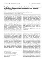
Fig. 1. Th e PS1 promoter. The sequences from )119 to +178 flanking
the major transcription initiation site (+1) in SK-N-SH cells included
in the PS1CAT reporter fusion vector are shown. The binding sites for
known transcription factors Sp1 and Ets are indicated with brackets
defined by footprinting with SK-N-SH cells (25) as well as site C which
corresponds to an unknown binding protein. Arrowheads indicate the
position of DNase I hypersensitive sites obser ved in footpr inting
experiments. The Ets and Sp1 consensus motifs are underlined. Arrows
indicate the end points of 5¢ or 3¢-deletions. SacII restriction enzyme
sites use d in the pre paration of the probes used i n EMSAs are boxed.
Ó FEBS 2004 Regulation of the presenilin 1 gene (Eur. J. Biochem. 271) 4487
SH-SY5Y cells and similarly by about tenfold in SK-N-SH
and cells. F urther deletion to +42 increased transcription in
both cells lines: about fivefold in S H-SY5Y cells and
twofold in SK-N-SH cells. Further deletions to +6 did not
result in any change in expression in any cell type. Hence
element(s) required for transc ription in both cell types are
located between +178 and +107. Element(s) repressing
transcription, at least in the context of a promoter truncated
to +107, are contained between +107 and +42. In
SH-SY5Y cells alternative initiation mechanisms are clearly
indicated by the significant level of promoter activity
conferred by either of the two fragments ()118 to +6) or
(+ 2 to +178).
Effects of point mutations at Ets and Sp1 motifs
on
PS1
transcription
The effects of point mutations in Ets motifs at positions
)10, +65, +90 and +129 as well as Sp1 motifs at )70,
)50 and +20 have been examined in both SK-N-SH
and SH-SY5Y cells (Fig. 3A). Altering an Ets motif at
)10 eliminated 80% of transcription i n S K-N-SH ce lls
whereas the mutation had only a minor effect (30%) in
SH-SY5Y cells. Conversely, the Ets element at +90
which eliminated 70% of transcription i n SH-SY5Y cells
had a lesser e ffect (40%) in SK-N-SH cells. In both cell
types a prom oter including mutations at both )10 and
+90 Ets sit es lost o ver 9 0% transcription a ctivity
indicating the crucial importance of these two Ets motifs.
Mutations at +65 and +129 resulted in a mild 25–30%
decrease in SH-SY5Y cells and 40–50% decrease in
SK-N-SH cells. T he double m utation at +65 an d +129
actually increased transcription markedly in SH-SY5Y
cells to 180% of the wild type promoter. Similarly it is
possible that the double mutant
19
at +90 and +129
resulted in activity comparable to wild type promoter,
hence the double mutation appears to reverse the effect
of either of the single mutations alone. These results
considered toge ther with the effect of 3¢-deletions to
+107 and +42 suggest that deletion of sequences
downstream from +107 or mutations at the +129 site
in particular does not just result in loss of function, but
rather produces a cis-acting n egative effect, such as an
abortive protein complex. In S K-N-SH cells the effect of
the double mutants was not as striking and showed the
same activity as each of the single mutations independ-
ently. However, the absence of an additive effect between
each mutation may suggest some interaction also
between each pair of sites in SK-N-SH cells.
The effect of Sp1 mutations appeared similar in both cell
types. The mutation at )70 had the most deleterious effect
decreasing transcription by 65–70%. The m()50) mutation
affected transcription by 45% and 60% in SK-N-SH and
SH-SY5Y c ells, respectively. It is interesting t o note that
)70 had more effect than )10 in SH-SY5Y cells, suggesting
that )70 is also required for )10 independent initiation. The
same case may apply t o )50. Furthermore the double
mutant )70,)10 appears to result i n a more than additive
effect, in contrast with the double mutant )50,)10 in
SH-SY5Y cells. This may reflect a cis -negative effect in
addition to simply the loss of function, su ch as squelching
20
of
a factor in limiting amount or steric hindrance of an
abortive complex.
Mutation at the Sp1 site at +20 did not alter transcription
and the )10/+20 double mutant appeared to reflect the
effect of the )10 mutation alone (Fig. 3A). The double
mutant )10/+65 appears to reflect also a simple additive
effect. The )30 site appears to bind a nuclear factor that
has not been identified [25]. Mutation at this site alone
or together with the )10 mutation affected transcription
only mildly as compared to the wild type or the )10 mutant
alone.
The higher requirement for the +90 Ets site would be
consistent with the ability of SH-SY5Y cells to direct
transcription from the +2/+178 promoter fragment.
Hence the differential expression in each cell type may be
at least partially determined by Ets factors and the )10 and
+90 sites. Efficient initiation requires a t least one of the two
sites. It appears that the )70 Sp1 motif is required whether
initiation is directed by either of th e )10 or +90 Ets site.
Fig. 2. D eletion mapping of the PS1 promoter.
The positions of the 5¢-and3¢-ends of each
deletion fragment are indicated on the left
(5¢D) and on the right (3¢D). PS1CAT plasmids
(6 lg) were cotransfected with 3 lg b-galac-
tosidase expression vector. Promoter activity
was expressed as the ratio of CAT to b-ga-
lactosidase activity for each transfected plate.
The mean values for each construct (n ¼ 3or
4) are indicated. SD values were 10–20% in all
cases. All constructs were tested in at least
three different e xperiments. The )118/+178
construct was taken as 100%.
4488 M. Pastorcic and H. K. Das (Eur. J. Biochem. 271) Ó FEBS 2004
The effect of the point mutation at )70 appears in contrast
with the effect of the 5¢-deletion to )35 or )22, which do not
decrease transcription in either cell type (Fig. 2). We have
not examined which sequences in the )118/)35 fragment
aside from the )70 motif account for this difference.
In addition, in both cell types the requirement for the )10
Etssiteishigherinshorterpromoterfragments
21
(
21
Table 1). In
the )22/+178 promoter fragment a mutation altering the
)10 Ets site eliminated over 90% transcription in SK-N-SH
cells [25]. In the context of the +118/+178 fragment the
same mutation was slightly less deleterious, reducing
transcription by a bout 80% (Table 1). In SH-SY5Y cells
the )10 mutation showed a 66% reduction in the )22/+178
context whereas in the )118/+178 context the same
mutation only had a minor effect, reducing transcription
by 30%. This suggests complex interactions between
upstream and downstream sequences and the )10 and
+90 Ets sites. In SH-SY5Y cells the requirement for the )10
Ets element appears most strongly in very short promoter
fragment )22/+42. This is consistent with the greater
importance of +90 in SH-SY5Y cells.
A
B
Fig. 3. Effect of PS1 promoter point mutations
on the efficiency of transcription initiation and
the position of the start site(s)
23
. (A) Effect of
point mutations in Ets and Sp1 motifs on PS1
promoter activity. The positions of Ets (d)
and Sp1 (h)motifsinthe)119/+178 region
of the PS1 promoter are indic ated. Th e p osi-
tions relative to the +1 site of point mutations
(X) eliminating Ets or Sp1 consensus in each
motif are indicated on the right together with
the corresponding promoter activity in SK-
N-SH or SH-SY5Y cells. (The mutant DNA
sequences are reported in Experimental pro-
cedures.) b-Galactosidase activity was used as
an internal standard. The mean values for
each construct (n ¼ 3 or 4) are indicated and
SD values were 10–20% in all cases. All con-
structs were tested in at least three different
experiments. The )119/+178 construct was
taken as 100%. (B) RACE in SH-SY5Y and
SK-N-SH cells transfected with the ()199/
+178) PS1 promoter. RACE was performed
as described above using the CAT4 primer (5¢-
CCGGATGAGCATTCATCAGGCGG-3¢)
specific for the CAT gene (solid box). RNA
was prepared from SH-SY5Y cells transfected
with wild type PS1()119/+178) CAT (lane 1)
(open box) o r the same CAT reporter con-
taining the )10Etsmutation(lanes2)orSK-
N-SHcellstransfectedwithwildtypePS1
()119/+178) CAT (lane 3). PCR products
were analyzed by electrophoresis on 2%
agarose gels. The size of molecular mass
markers (lane 4) is indicated in bp.
Table 1. Effects of )10 Ets GGAA fi TTAA mutation. The activity of
PS1CAT constructs including the promoter fragments shown on the
left containing (m) or not (wt) the GGAA to TTAA mutation at the
)10 Ets site was assayed by transfection in SK-N-SH and SH-SY5Y
cells. Each construct was tested in three transfections, each with n ¼ 3.
Promoter fragment
SK-N-SH cells SH-SY5Y cells
wt m wt m
)119/+178 100 ± 12 21 ± 5 100 ± 5 70 ± 20
)119/+143 73 ± 10 10 ± 4 133 ± 30 70 ± 6
)22/+178 91 ± 10 3.4 ± 2.6 100 ± 20 28 ± 7
)22/+4 23 ± 5 150 ± 20 9 ± 2
Ó FEBS 2004 Regulation of the presenilin 1 gene (Eur. J. Biochem. 271) 4489
RACE in SH-SY5Y and SK-N-SH cells transfected
with the
PS1
promoter
RACE PCR w ith RNA from SK-N-SH cells transfected
with the wild type [)119, +178] PS1 promoter yielded a
single band of about 450 bp (Fig. 3B). RACE with the
RNA from SH-SY5Y cells transfected with the wild type
PS1 promoter produced two bands. The larger product
appeared to a have a size similar to that obtained in SK-N-
SH cells. The smaller product appeared to be about 50–
80 bp shorter. RNA from SH-SY5Y cells transfected with
the PS1 promoter containing a mutated Ets site at )10
produced only the shorter RACE product. Sequencing of
RACE PCR products from the lower band showed the
5¢-end points at +63 (Fig. 1). Hence sizing and sequencing
of the RACE P CR products from SH-SY5Y cells is
consistent with the g reater importance of s equences down-
stream from the +1 start site originally mapped in SK-N-
SH cells [25] and with a higher frequency of downstream
(+63) initiation events in the )10 mutant.
Nuclear factors in SH-SY5Y and SK-N-SH cells specifically
recognize Ets motifs on the
PS1
promoter upstream
and downstream from the +1 site
The binding of nuclear factors present in SK-N-SH and
SH-SY5Y cells to various fra gments of the PS1 promoter
was examined by EMSAs. We have compared binding
A
B
Fig. 4. Nuclear factors from SK-N-SH and SH-SY5Y cells recognize specifically Ets motifs flanking the +1 site. (A) The binding of nuclear factors is
eliminated by mutations in Ets motifs. The binding of nuclear factors present in SK-N-SH (K) or SH-SY5Y (H) nuclear extracts over the PS1
promoter was assayed by EMSAs.
32
P-labeled probes included sequences )22/+6 (lanes 1–6), +50/+107 (lanes 7–15) and +107/+178 (lanes
16–24). Either the wild type probes or the same fragments containing mutant Ets sites at positions )10 (lanes 4–6), +90 (lanes 10–12), +65 (lanes
13–15), +129 (lanes 19–21) and +165 (lanes 22–24) were incubated with 5 lg nuclear extracts or in absence of extract (O). The position of
complexes t hat a re signific antly affected by mutatio ns is indicated by d ots. (B) The binding of nuclear factors is specific ally co mpete d b y a
heterologous Elk1 binding site. The specific binding in the p romoter regions )22/+6 (lanes 1–6), +50/+107 (lanes 7–18), +107/+178 (lanes 19–
30) was tested by competition with the cold oligonucleotide E74 containing the Drosophila Elk1 binding motif (25) (+) or E74 containing a mutated
Ets consensus (m). The position of c omplexe s define d in (A) is indicated by arrows or lines.
4490 M. Pastorcic and H. K. Das (Eur. J. Biochem. 271) Ó FEBS 2004
between wild type fragments a nd fragments containing the
point mutations at the )10, +65, +129 and +165 Ets
motifs that have been tested in transfection experiments
(Fig. 4A). The binding of several nuclear factors to the
probe including sequences )22/+6 appears to be eliminated
by the )10 Ets mutation: complexes at levels A, B, C, D and
E are decreased in lanes 5 and 6 as compared to lanes 2 and
3. With the +50/+107 probe, complex F d ecreases with
m(+90) (lanes 11 and 12) whereas G decreases w ith
m(+65) (lanes 14 and 15) as compared to binding to the
wild type probe (lanes 8 and 9), suggesting that A and B
may specifically recognize the +90 and +65 E ts sites,
respectively. With the +107/+178 probe, c omplex I i s
abolished by m(+129) (lanes 20 and 21) as compared to the
wild type probe (lanes 17 and 18) and may represent specific
recognition of this site. The specificity of the binding was
assessed further by competition assays using E74 (a
heterologous DNA sequence containing a known Ets-like
gene 1 [Elk1] binding site [25]) as competitor. Among
complexes formed with )22/+6 (Fig. 4B), B and E were
eliminated by competition with wild type E74 competitor
(lanes 2 a nd 5) but were unaffected by E74 competitor
containing a mutated Ets motif (lanes 3 and 6). With the
+50/+107 m(+65) probe, the complex(s) in region F are
decreased selectively with the wild type E74 (lanes 8 and 11)
as compared with the mutant E74 (lanes 9 and 12). This
confirms the specificity of complex F and indicates that the
+90 site is recognized by a nuclear factor related to Ets.
With the +50/+107 m(+90) probe none of the complexes
appear selectively affected by competition with the wild type
E74 (lanes 14 and 17) as compared with the mutant E74
(lanes 15 and 18). Hence complex G is not competed
although it is eliminated by the +65 mutation. We do not
know whether G represents Ets specific binding to the +65
site that does not bind stably to the Ets com petitor used
here, or if it represents specific recognition by nuclear
factor(s) different from Ets. With the +107/+178 m(129)
probe no competition is observed (Fig. 4B lanes 19–24) and
this is consistent with the absence of an effect of m(+165)
on the binding pattern (Fig. 4A, lanes 23 and 24 compared
to lanes 17 and 18). This suggests that the +165 site does
not bind any Ets related protein. With the +107/+178
m(+165) probe the complex in region H is efficiently
competed by both wild type (lanes 26 and 29) as well as
mutant E74 (lanes 27 and 30). Hence the competition data is
inconclusive for complex H because it is possible that the
factor may have a strong affinity for DNA ends present in
large excess with both competitors.
ERM transactvates
PS1
and does not require
the )10 Ets element
We have begun to identify the Ets factors controlling the
expression of PS1 using the yeast one-hybrid selection and
the )10 Ets site as bait. We have found that avian
erythroblastosis virus E
26
oncogene homologue 2 (Ets2),
Elk1 and Ets translocation variant 1 (ER81) recognize PS1
and act as trans-effectors [26–28]. ERM was also identified
by one-hybrid selection and we have examined its effects on
PS1 transcription. Cotransfection of SK-N-SH cells with a
)119/+178 promoter fragment fused to a CAT r eporter
revealed that ERM acts as an activator of PS1 (Fig. 5A).
Fig. 5. Ac tivation of PS1 transcription by ERM and the effect of
promoter sequences on PS1 transcription
24
. (A) ERM activates the PS1
promoter in SK-N-SH cells. The ()119/+178) PS1CAT reporter
(6 lg) was cotransfecte d with various amounts of ERM expression
vector integrated in pC1 (ERM) or the same amount of empty vector
(pC1).Theamountofproteinineachassaywasusedasaninternal
standard. (B) PS1 sequences upstream or downstream from the +1 site
confer transactivation by ERM. PS1CAT constructs (6 lg) containing
various fragment o f the PS1 promoter w ere cotransfected with 3 lg
pC1 expression vector including or not ERM. Promoter activities were
compared using the c oncen tration of protein in the extract as an
internal standard. The activity of ()119/+178) PS1CAT in the pres-
ence of the empty vector was taken as 100%. The values represented in
the graph are indicated below. In both A and B each data po int cor-
responds to the average from threeplatesineachexperiment.Each
data point was retested in three independent experiments.
Ó FEBS 2004 Regulation of the presenilin 1 gene (Eur. J. Biochem. 271) 4491
We also asked which PS1 sequences are required for
activation by ERM. Various promoter fragments were
cotransfected with ERM (Fig. 5B). All the PS1 fragments
assayed were transactivated by ERM, including )22/+178
containing a mutated )10 Ets, the +2/+178 3¢-deletion
eliminating the major initiation site, and the )22 /+42
where all downstream Ets sites are absent. H ence it
appears that either sequences upstream or downstream
from the +1 site are sufficient to confer transactivation by
ERM.
ERM specifically recognizes Ets motifs upstream and
downstream from the +1 site
We first detected specific interactions between various
portions of the PS1 promoter and ERM by supershifting
the DNA–protein complexes appearing i n EMSAs using an
anti-ERM Ig (Fig. 6A). In vitro translated ERM was
preincubated with anti-ERM Ig. PS1 promoter probe was
then combined with the protein mix and the protein–DNA
interactions were analyzed by EMSAs. All portions of the
PS1 promoter tested ()22/+6) ( lanes 1–4), (+50/+107)
(lanes 5–7), (+107/+178) (lanes 8–10) formed complexes
with in vitro translated ERM which could be supershifted by
the antibody, indicating the presence of ERM binding site(s)
in each of the three probes. We also examined the pattern of
complexes formed o n the same promoter fragments inclu-
ding individual point mutations at +65, +90, +129 and
+165, with in vitro translated ERM as compared to t he
control t ranslation mix containing the empty pC1 vector
(Fig. 6B). All ERM-containing lanes (M) showed com-
plexes that were absent in the control reactions (O) (dots)
with the +50/+107 probe (lanes 1–6), suggesting the
binding of ERM to the Ets sites present on this fragment.
The binding profile on the +107/+178 probe (lanes 7–12)
was not clearly modifed b y the addition of in vitro translated
ERM (lanes M) as compared to control binding reactions
(lanes O). The amount of complex formed may be small
and/or not form a sharp band due to a lack of stability or a
change in conformation during electrophoresis. Specific
ERM–DNA complexes can, however, be captured a s
supershifted complexes with this probe (Fig. 6A, lane 9)
indicating that ERM may indeed recognize Ets sites +129
and/or +165.
Discussion
The PS1 promoter appears to be used alternatively in SK-
N-SH cells and its SH-SY5Y subclone. Two Ets elements at
)10 and +90 app ear to play an essential role although they
are required to different extents in each cell type. Hence it is
likely that Ets elements play a major role in determining
both the level of expression and the location of the start of
transcription of PS1 . In the Ets family of transcription
AB
Fig. 6. ERM and nuclear factors recognize specifically several Ets elements on the PS1 promoter
25
. (A) Supershift of specific DNA–protein complexes
by anti-ERM Ig. In vitro translated ERM was preincubated with anti-ERM Ig. PS1 promoter probe fragments were combined with the protein mix
and the protein–DNA complexes formed were analyzed by EMSA. Various portions of the PS1 promoter were tested for the presence of ERM
binding site(s): )22/+6 (lanes 1–4), +50/+107 (lanes 5–7), +110/+178 (lanes 8–10). Lanes 1, 4 and 7 contained no antibody or antibody buffer,
lanes 2, 5 and 8 (aEtv5) contained 2 lL ERM antibody (Santa Cruz Biotech sc-1955X), lanes 3, 6 and 9 (C) contained control antibody buffer. An
arrow marks the position of supershifted complexes. (B) The binding of in vitro translated ERM is eliminated by mutations in Ets motifs. The
binding of in vitro translated ER M o ver t he PS1 promoter was a ssayed by E MSAs inc luding the
32
P-labeled probes +50/+107 (lanes 1–6) and
+107/+178 (lanes 7–12). Either the wild type probes or the same fragments containing mutant Ets sites at positions +65 (lanes 3, 4), +90 (lanes 5,
6), +129 (lanes 9, 10), +165 (lanes 11, 12) were incubated with 2 lL ERM translation mixture (M) or in absence of ERM protein (O). The position
of complexes that are present only w ith t ranslation reactions containing ERM expression vector is indicated by dots.
4492 M. Pastorcic and H. K. Das (Eur. J. Biochem. 271) Ó FEBS 2004
factors a highly conserved 85 amino acid DNA binding
domain called the ETS domain determines the specific
recognition of the sequence (GGAA/T). However, se-
quences immediately flanking this core element determine
the recognition of target sites in different genes. In addition
promoter sequences flanking the Ets motif determine
activity t hrough protein–protein interactions between Ets
factors and factors binding at adjacent sites, as w ell as
cofactors that do not bind DNA themselves [29,30]. Hence
the specific set of factors present in a given cellular context
probably determines th e recognition a nd activation of
promoter sequences by Ets f actors. Binding sites for S p1
factors are interdigitated with Ets m otifs on the PS1
promoter. The sites at )70 a nd )50 appear to have some
degree of activity in both cell types, whereas the point
mutation at )20 did not indicate any importance for the
downstream motif. Sp1 factors have been known to activate
transcription and to form synergistic i nteraction with Ets
factors b inding at adjacent sites [31]. The specific factors
that transactivate PS1 through binding at the Sp1 motifs
remain to be identified.
We do not know what major d ifferences between
SH-SY5Y and SK-N-SH affect PS1. The results mostly
demonstrate the potential for flexibility in the utilization of
the promote r. Ana lysis of the start sites of the major species
of PS1 mRNAs present in brain and human placenta
revealed a set of 5¢ ends, all present between positions +1
and +90 defined in SK-N-SH ce lls [32]. H ence initiation
within the first exon may indeed occur in vivo.Inthehuman
presenilin 1 gene the 5¢-UTR is encoded by exons 1–4. Most
full length cDNAs characterized and discussed above were
initiated in exon 1 and directly spliced to exon 3 [32]. The
absence of a 3¢ splice sequence in exon 2 suggests that its
presence on a minority of mRNAs results from alternative
initiation site(s) at e xon 2 rather than alternative splicing
[32]. Such flexibility in the mechanism of initiation presents
several advantages. First it is consistent with the ubiquitous
expression o f the gene and increases its potential for
expression in a variety of cellular systems. Secon d it
increases the potential for regulation by various signal
transduction cascades. Third alternative initiation of shorter
transcripts may have implications for additional regulation
at the level of translation. The 5¢-UTR of PS1 mRNA
appears to be naturally unfavorable for translation, due to a
number of AUG codons
22
present upstream from the actual
initiation site and the potential for secondary structure of
the RNA [32,33]. The relatively low translatability of the
message may provide potential mechanisms for the rapid
induction of PS1. A shift in the transcription initiation site
and a shorter 5¢-UTR may increase translation by simply
reducing the path of ribosome scanning. Alternatively it
may alter secondar y structure and/or regulatory pr otein
binding sites on the 5¢-UTR, which are likely to result in
changes in the affinity of the cap binding the translation
initiation complex. Transiently produced shorter RNAs
may have a crucial r egulatory role during differentiation.
Indeed promoter activity from cryptic promoters is found in
long 5¢-UTRs of genes with a crucial role in the regulation
of other genes [34].
Ets factors are likely to be important determinants in the
choice of initiation site in a particular cellular context. The
specificity of Ets factors is determined by their site of
expression, their selective target sequence recognition, their
selective abilities to interact with other transcription factor
coactivators, and their selec tive modification by s ignal
transduction kinases [29,30]. We have s hown here that
ERM transactivates the PS1 promoter. In our assay system
Ets elements either upstream o r downstream from + 1
appear to confer response t o ERM and downstream
sequences retain a significant fourfold activation. Hence it
is possible that in a particular cellular context ERM may
also direct a significant level of initiation of shorter
transcripts in vivo . ERM belongs to the PEA3 subfamily
of Ets transcription factors that includes only three mem-
bers (ERM, ER81 and PEA3), ER81 and ERM being the
most related [35]. PEA3 members all contain the highly
conserved ETS domain. ERM contains two DNA binding
inhibitory domains flanking the Ets domain: the central 87
amino acid inhibitory domain is poorly conserved a nd
virtually specific to ERM, and the C-terminal domain that is
present in all three PEA3 members. Toge ther with a 32
residue N-terminal acidic stretch which is also conserved
within the PEA3 group the C-terminal domain contains
transactivation properties of the P EA3 factors [35]. There-
fore, among the Ets factors that transactivate PS1 [27,28]
ERM should confer certain specific regulatory properties.
Although ERM mRNA is found in many tissues, its
expression is remarkably high in the brain [35] and t his also
suggests its importance for the regulation of PS1 i n vivo in
this tissue.
Acknowledgements
This research was supp orted by a grant f rom the National I nstitutes of
Health (AG 18452).
References
1. Rogaev, E.I., Sherrington, R., Rogaeva, E.A., Levesque, G.,
Ikeda, M., Liang, Y., Chi, H., Lin, C., Holman, K., Tsuda, T.,
Mar., L., Sorbi, S., Nacmias, B., Piacentini, S., Chumakov, I.,
Cohen, D., Lannfelt, L., Fraser, P.E., Rommens, J.M. & St
George-Hyslop, P.H. (1995) Cloning of a gene bearing missense
mutations in early-onset familial Alzheimer’s disease. Nature 376,
775–778.
2. Li, X. & Greenwald, I. (1998) Additional evidence for an eight-
transmembrane-domain topology for Caenorhabditis elegans and
human presenilins. Proc.NatlAcad.Sci.USA95, 7109–7114.
3. Dewji, N.N., Valdez, D. & Singer, S.J. (2004) The presenilins
turned inside out: implications for their structures and functions.
Proc.NatlAcad.Sci.USA101, 1057–1062.
4. De Strooper, B. (2003) Aph-1, Pen-2, and nicastrin with presenilin
generate an active c-secretase complex. Neuron 38, 9–12.
5. Kimberly, W.T., LaVoie, M.J., Ostaszewski, B.L.YeW., Wolfe,
M.S. & Selkoe, D.J. (2003) Gamm a-secre tase is a me mbra ne
protein complex comprised of pre senilin, nicast rin, Aph-1, and
Pen-2. Proc. Natl Acad. Sci. USA 100, 6382–6387.
6. Edbauer, D., Winkler, E., Regula, J.T., Pesold, B., Steiner, H. &
Haass, C. (2003) Reconstitution of gamma-secretase activity. Na t.
Cell Biol. 5, 486–488.
7. Takasugi, N., Tomita, T., Hayashi, I., Tsuruoka, M., Niimura,
M., Takahashi, Y ., Thinakaran,G.&Iwatsubo,T.(2003)The
role of presenilin cofactors in the c-secretase complex. Nature 422,
438–441.
8. De Strooper, B., Saftig, P., Craessaerts, K., Vanderstichele, H.,
Guhde, G., Annaert, W., Von Figura, K. & Van Leuven, F. (1998)
Ó FEBS 2004 Regulation of the presenilin 1 gene (Eur. J. Biochem. 271) 4493
Deficiency of presenilin-1 inhibits the normal cleavage of amyloid
precursor protein. Nature 391, 387–390.
9. Price, D.L. & Sisodia, S.S. (1998) Mutant genes in familial Alz-
heimer’s disease and transgenic models. Annu. Rev. Neurosci. 21,
479–505.
10. Levy-Lahad, E., Wasco, W., Poo rkaj, P., Ro mano, D.M., Oshima ,
J. & Pettingell, W.H., YuC.E., Jondro, P.D., Schmidt, S.D. &
Wang, K. (1995) Candidate gene for the chromosome 1 familial
Alzheimer’s disease locus. Science 269, 973–977.
11. Tanzi, R.E., Kowacs, D.M., Kim, T W., Moir, R.D., Guenette,
S.Y. & Wasco, W. (1996) The gene defects responsible for familial
Alzheimer’s disease. Neurobiol. Dis. 3, 159–168.
12. Sherrington, R., Rogaev, E.I., Liang, Y., Rogaeva, E.A.,
Levesque,G.,Ikeda,M.,Chi,H.,Lin,C.,Li,G.,Holman,K.,
Tsuda, T., Mar., L ., Foncin, J F., Bruni, A.C., Mo ntesi, M.P.,
Sorbi,S.,Rainero,I.,Pinessi,L.,Nee,L.,Chumakov,I.,Pollen,
D., Brookes, A., Sanseau, P., Polinsky, R.J., Wasco, W., Da Silva,
H.A.R.,Haines,J.L.,Pericak-Vance,M.A.,Tanzi,R.E.,Roses,
A.D., Fraser, P.E., Rommens, J.M. & St George-Hyslop, P.H.
(1995) Cloning of a gene bearing missense mutations in early-onset
familial Alzheimer’s disease. Nature 375, 754–760.
13. Goate, A., Chartier-Harlin, M.C., Mullan, M., Brown, J., Craw-
ford,F.,Fidani,L.,Giuffra,L.,Haynes,A.,Irving,N.,James,L.,
Mant, R., Newton, P., Rooke, K., Roques, P., Talbot, C., Pericak-
Vance, M., Roses, A., Williamson, R., Rossor, M., Owen, M. &
Hardy, J. (1991) Segregation of a missense mutation in the amy-
loid precursor protein gene with familial Alzheimer’s disease.
Nature 349, 704–706.
14. Fortini, M.E. (2001) Notch and Presenilin: a proteolytic mech-
anism emerges. Curr. Opin. Cell Biol. 13, 627–634.
15. Kopan, R. & Goate, A. (2000) A common enzyme connects Notch
signaling and Alzheimer’s disease. Genes Dev. 14, 2799–2806.
16. Weinmaster, G. (2000) Notch signal transduction: a real rip and
more. Curr. Opin. Genet. Dev. 10, 363–369.
17. Fortini, M.E. (2002) G amma-secretase-mediated prote olysis in
cell-surface-receptor signalling. Nat. Rev. Mol. Cell Biol. 9, 673–
684.
18. Okamoto, I., Kawano, Y., Murakami, D., Sasayama, T., Araki,
N., Miki, T., Wong, A.J. & Saya, H. (2001) Proteolytic release of
CD44 intracellular domain and its role in the CD44 signaling
pathway. J. Cell Biol. 155, 755–761.
19. Marambaud,P.,Wen,P.H.,Dutt,A.,Shioi,J.,Takashima,A.,
Siman, A. & Robakis, N.K. (2003) A CBP binding transcrip-
tional repressor produced by the PS1/-cleavage of N-cadherin is
inhibited by P S1 F AD mu tations. Cell 114, 635–645.
20. De Strooper, B., Saftig, P., Craessaerts, K., Vanderstichele, H.,
Guhde, G., Annaert, W., Von Figura, K. & Van Leuven, F. (1998)
Deficiency of presenilin-1 inhibits the normal cleavage of amyloid
precursor protein. Nature 391, 387–390.
21. Passer, B., Pellegrini, L., Russo, C., Siegel, R.M., Lenardo, M.J.,
Schettini, G., B achmann, M ., T abaton, M . & D’Adamio, L.
(2000) Generation of an apo ptotic i ntracellular peptide by c-
secretase cleavage of Alzheimer’s amyloid b protein precursor.
J. Alzheimers Dis. 2, 289–301.
22. Cao, X. & Sudhof, T.C. (2001) A transcriptionally active complex
of APP with Fe65 and histone acetyltransferase Tip60. Science
293, 115–120.
23. Baek, S.H., Ohgi, K .A., Rose, D.W., Koo, E.H., Christopher, K.,
Glass, C.K. & Rosenfeld, M.G. (2002) Exchange of N-CoR cor-
epressor and Tip60 coactivator complexes links gene expression by
NF-jBandb-amyloid precursor protein. Cell 110, 55–67.
24. Scheinfeld, M.H., Ghersi, E., Laky, K ., Fowlkes, B.J. &
D’Adamio, L. (2002) P rocessing o f b-amyloid precur sor-like
protein-1 and -2 by c-secretase regulates transcription. J. Biol.
Chem. 277, 44195–44201.
25. Pastorcic, M. & Das, H.K. (1999) An upstream element contain-
ing an ETS binding site is crucial for transcription of the human
presenilin-1 gene. J. Biol. Chem. 274, 24297–24307.
26. Pastorcic, M. & Das, H.K. (2003) An abbreviated procedure for
the cloning and identification of Ets t ranscription factors r eg-
ulating the expression of the human presenilin 1 gene. Brain Res.
Brain Res. Protoc. 12, 35–40.
27. Pastorcic, M. & Das, H.K. (2003) Ets transcription factors ER81
and Elk1 regulate the transcription of the human presenilin 1 gene
promoter. Brain Res. Mol. Brain Re s. 113, 57–66.
28. Pastorcic, M. & Das, H.K. (2000) Regulation of transcription of
the human presenilin-1 gene by ets transcription factors and the
p53 protooncogene . J. Biol. Chem. 27 5, 34938–34945.
29. Sharrocks, A.D. (2001) The ETS-domain transcription factor
family. Nat. Rev. Mol. Cell Biol. 11, 827–837.
30. Verger, A. & Duterque-Coquillaud, M. (2002) When Ets tran-
scription factors meet their partners. Bioessays 24, 362–370.
31. Gegonne, A., Bosselut, R., Bailly, R.A. & Ghysdael, J. (1993)
Synergistic activation of the HTLV1 LTR Ets-responsive region
by transcription factors Ets1 and Sp1. EMBO J. 12 , 1169–
1178.
32. Rogaev, E.I., Sherrington, R., Wu, C., Levesque, G., Liang, Y.,
Rogaeva,E.A.,Ikeda,M.,Holman, K., Lin, C., Lukiw, W.J., de
Jong, P.J., Fraser, P .E., Rommens, J.M. & St George-Hyslop, P.
(1997) Analysis of the 5¢ sequence, genomic structure, and alter-
native splicing of the presenilin-1 gene (PSEN1) associated with
early onset A lzheimer disease. Genomics 40, 415–424.
33. Kozak, M. (2000) Do the 5¢untranslate d domains of human
cDNAs challenge the rules for initiation of translation (or is it vice
versa). Genomics 70, 396–406.
34. Han, H. & Zhang, J T. (2002) Regulation of gene expression by
internal ribosom e entry sites or cryptic p romoters: the eIF4G
story. Mol. Cell. Biol. 22, 7372–7384.
35. DeLaunoit, Y., Baert, J.L., Chotteau, A., Mont e, D., Defossez,
P.A., Coutte, L., Pelczar, H. & Leenders, F. (1997) Structure-
function relationships of the PEA3 group of Ets-related tran-
scription factors. Biochem. Mol. Med. 61, 127–135.
4494 M. Pastorcic and H. K. Das (Eur. J. Biochem. 271) Ó FEBS 2004