Báo cáo khoa học: Protective effect of active oxygen scavengers on protein degradation and photochemical function in photosystem I submembrane fractions during light stress pdf
Bạn đang xem bản rút gọn của tài liệu. Xem và tải ngay bản đầy đủ của tài liệu tại đây (264.26 KB, 11 trang )
Protective effect of active oxygen scavengers on protein
degradation and photochemical function in photosystem I
submembrane fractions during light stress
Subramanyam Rajagopal*, David Joly, Alain Gauthier, Marc Beauregard and Robert Carpentier
Groupe de Recherche en Biologie Ve
´
ge
´
tale, Universite
´
du Que
´
bec a
`
Trois-Rivie
`
res, Que
´
bec, Canada
Excessive light causes the inactivation of photosyn-
thesis. The detailed mechanism of PSII inactivation
has been extensively characterized both in vivo and
in vitro [1–5] and it has been suggested that the inac-
tivation develops at either acceptor or donor side of
the photosystem [2,6]. PSI was first believed to be
tolerant to strong light. However, several in vitro
studies suggested that PSI photochemical activity
could be inhibited under strong illumination [7,8]. In
these early experiments, photoinactivation of PSI was
not observed in the absence of oxygen. Later, Satoh
& Fork [9] demonstrated the inactivation of PSI in
intact Bryopsis chloroplasts under strongly reducing
conditions. The authors suggested the photoinactiva-
tion site to be either P700 itself [9] or close to the
PSI reaction center [10] under both aerobic and
anaerobic conditions. The main site of PSI photo-
inhibition under aerobic conditions was then shown
to be the inactivation of iron–sulfur centers [11],
whereas anaerobic photoinhibition resulted in the
blocking of electron transfer between A
0
and F
X
[12].
Keywords
photosystem I; submembrane fraction;
active oxygen; P700; light stress
Correspondence
R Carpentier, Groupe de Recherche en
Biologie Ve
´
ge
´
tale, Universite
´
du Que
´
bec a
`
Trois-Rivie
`
res, C.P. 500, Trois-Rivie
`
res,
Que
´
bec, Canada, G9A 5H7
Fax: 1 819 376 5057
E-mail:
*Present address
School of Life Sciences and Center for the
Study of Early Events in Photosynthesis,
Arizona State University, Tempe, Arizona
85287, USA
(Received 17 August 2004, revised 23
November 2004, accepted 2 December
2004)
doi:10.1111/j.1742-4658.2004.04512.x
The protective role of reactive oxygen scavengers against photodamage
was studied in isolated photosystem (PS) I submembrane fractions illumin-
ated (2000 lEÆm
)2
Æs
)1
) for various periods at 4 °C. The photochemical
activity of the submembrane fractions measured as P700 photooxidation
was significantly protected in the presence of histidine or n-propyl gallate.
Chlorophyll photobleaching resulting in a decrease of absorbance and
fluorescence, and a blue-shift of both absorbance and fluorescence maxi-
mum in the red region, was also greatly delayed in the presence of these
scavengers. Western blot analysis revealed the light harvesting antenna
complexes of PSI, Lhca2 and Lhca1, were more susceptible to strong light
when compared to Lhca3 and Lhca4. The reaction-center proteins PsaB,
PsaC, and PsaE were most sensitive to strong illumination while other
polypeptides were less affected. Addition of histidine or n-propyl gallate
lead to significant protection of reaction-center proteins as well as Lhca
against strong illumination. Circular dichroism (CD) spectra revealed that
the a-helix content decreased with increasing period of light exposure,
whereas b-strands, turns, and unordered structure increased. This unfolding
was prevented with the addition of histidine or n-propyl gallate even after
10 h of strong illumination. Catalase or superoxide dismutase could not
minimize the alteration of PSI photochemical activity and structure due to
photodamage. The specific action of histidine and n-propyl gallate indicates
that
1
O
2
was the main form of reactive oxygen species responsible for
strong light-induced damage in PSI submembrane fractions.
Abbreviations
Chl, chlorophyll; LHC I, light-harvesting complex I; P700, primary electron donor; PSI and II, photosystem I and II; SOD, superoxide
dismutase.
892 FEBS Journal 272 (2005) 892–902 ª 2005 FEBS
Recently, Velitchkova et al. [13] reported that PSI-
mediated electron transport, P700 content, and reac-
tion-center proteins, PsaA ⁄ B, were altered by strong
light illumination at room or low temperatures in
isolated spinach thylakoid membranes. Also, these
authors observed that under these conditions toxic
hydroxyl radicals were generated. Inactivation of PSI-
mediated electron flow was also reported in isolated
PSI core particles such as spinach PSI-180 and PSI-
100, as well as cyanobacterial PSI membranes exposed
to strong light [14]. In PSI core particles illuminated
with strong light, damage to the light-harvesting com-
plex (LHC) and degradation of reaction-center pro-
teins as well as acceptor side proteins were observed
[15]. We have recently shown that exposure of PSI
submembrane fractions to strong light under low tem-
perature altered the structure of chlorophyll (Chl)–pro-
tein (CP) complexes and decreased the photochemical
activity and the efficiency of excitation energy migra-
tion [16–18]. The damages were associated with the
formation of reactive oxygen species. It was also found
that the above photoinactivation of PSI was retarded
by glycinebetaine and sucrose [18].
In intact leaves, PSI was also found to be inacti-
vated by light. In Cucumis sativus leaves, weak light
induced the photoinactivation of PSI at chilling tem-
peratures, while practically no damage to PSII was
reported [19,20]. Sonoike et al. [20] demonstrated that
electron carriers located on the acceptor side of PSI
(A
1
, F
X
, F
A
, and F
B
) were damaged during the photo-
inhibition of PSI. The loss of PSI activity in thylakoids
isolated from spinach leaves exposed to weak light illu-
mination at room temperature is believed to be associ-
ated with the degradation of PsaB protein, one of the
PSI reaction-center subunits [21]. In barley and cucum-
ber leaves exposed to weak light at chilling temper-
ature, photoinhibition of PSI coincided with the
damage of both reaction-center proteins, PsaA and
PsaB, and the proteins PsaD and PsaE on the acceptor
side of the photosystem [22].
In plants, reactive oxygen species (ROS) are associ-
ated with normal physiological processes as well as with
responses to adverse conditions. ROS are implicated in
many ways with stressful conditions: as primary elici-
tors, as products and propagators of oxidative damage,
or as signal molecules initiating defense or adaptation
[23–25]. Several authors proposed that oxidative mecha-
nisms are at the basis of PSI photodamage in intact
leaves [19,22], isolated chloroplasts [7,8,11], and isolated
PSI fragments [15–18]. Two types of active oxygen spe-
cies with their derivative products are proposed to be
involved in photooxidative damage, superoxide anion
radicals, ÆO
2
–
, and singlet oxygen,
1
O
2
[26–29]. Hence,
addition of glucose oxidase, glucose, and catalase to
scavenge dissolved oxygen suppressed the photodamage
of PSI submembrane fractions [17].
However, the protective role of specific active oxy-
gen scavengers against photo-induced changes in the
photosystems was discussed only in a limited number
of reports. LHCII protein degradation was analyzed
during strong light illumination of isolated LHCII or
BBY PSII subcomplexes [28]. Random cleavage, start-
ing in the NH
2
terminal region resulted in the
complete degradation of the antenna proteins. The
addition of scavengers such as histidine, DABCO, and
n-propyl gallate, retarded the above damages to the
antenna proteins indicating mainly
1
O
2
was involved
[28]. In spinach thylakoids, illumination at low light
intensity resulted in the degradation of PsaB gene
product into two fragments of 51 and 45 kDa [30].
These fragments were absent with added n-propyl gal-
late, which removes hydroxyl radicals [30]. There are
no reports regarding the protective role of oxygen
scavengers against reactive oxygen species under strong
light in PSI submembrane fractions.
We used PSI submembrane particles as model sys-
tem to obtain a better insight into the photo-induced
changes in PSI. The submembrane particles used con-
tain all the components of PSI, including the cyto-
chrome b6 ⁄ f complex and plastocyanin [31,32]. In the
present study, the degradation of each polypeptide and
the protein conformation changes are assessed in con-
nection with alterations in photochemical activity dur-
ing strong light illumination of the PSI submembrane
fractions. It is concluded from the specificity of the
various active oxygen scavengers used that only the
generation of
1
O
2
is responsible for the photooxidation
effects. The role of active oxygen scavengers in the
protection of the structure and function of the photo-
systems against excess light indicates they should also
be beneficial under natural conditions.
Results
PSI submembrane fractions were exposed to strong
light (2000 lEÆm
)2
Æs
)1
) for various time durations at
4 °C. The action of several active oxygen scavengers
was analyzed for all the parameters studied in order to
emphasize the role of reactive oxygen species in the
photooxydative damage.
Changes in DA
830
Exposure of isolated PSI submembrane fractions to
strong white light at 4 °C resulted in the loss of ability
of P700 to undergo reversible redox changes. Similar
S. Rajagopal et al. Photoprotective effect of active oxygen scavengers
FEBS Journal 272 (2005) 892–902 ª 2005 FEBS 893
results were obtained at room temperature (data not
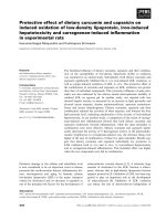
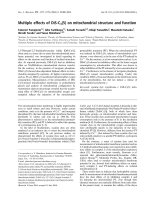
shown). Figure 1 shows the magnitude of DA
830
signal
remained stable during the first 4 h of strong light
treatment of PSI submembrane fractions in the
absence of active oxygen scavengers. It indicated an
unchanged amount of photooxidizable P700 during
this period. However, further irradiation caused a
rapid decline in DA
830
. The addition of histidine and
n-propyl gallate to the submembrane fractions signifi-
cantly prevented the decline of absorbance changes
(DA
830
). The magnitude of DA
830
was almost constant
until 10 h of light exposure with the above reactive
oxygen scavengers. In contrast, in presence of cata-
lase or superoxide dismutase, or both, the amplitude
of DA
830
was similar to control values (data not
shown).
Absorption changes
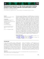
Figure 2A illustrates the changes in the room tempera-
ture absorption spectra of PSI submembrane fractions
exposed to various periods of strong white light illumin-
ation at 4 °C. The spectra measured in untreated sub-
membrane fractions showed typical absorption peaks at
680 and 440 nm corresponding to Chl a and shoulders
at 650 and 470 nm originating from Chl b [16]. As the
period of illumination increased, the magnitude of the
absorption peak at 680 nm declined (Fig. 2). In addi-
tion, the position of that peak was finally shifted by
6 nm towards shorter wavelengths after a 6-h illumin-
Fig. 1. The changes in the magnitude of light-induced absorbance
at 830 nm during strong light illumination in PSI submembrane frac-
tions. Results are means ± SE. n ¼ 3. All experimental conditions
are given in Experimental procedures.
Fig. 2. Room temperature absorption spectra of the isolated PSI
submembrane fractions illuminated for various periods of time with
strong WL (2000 lmolÆm
)2
Æs
)1
)at4°C. (A) Control (no addition of
any additives), (B) in the presence of histidine, and (C) in the pres-
ence of n-propyl gallate. These experiments were repeated three
times and yielded identical spectra; a typical spectrum is presented.
Insets: the first derivative of the absorption maxima in the red is
presented to show peak position.
Photoprotective effect of active oxygen scavengers S. Rajagopal et al.
894 FEBS Journal 272 (2005) 892–902 ª 2005 FEBS
ation. This becomes particularly clear from the changes
in first derivative spectra that further demonstrate the
gradual time-dependent alteration in the position of the
major peak in the red region (Fig. 2, insert). On the
other hand, in the presence of the scavengers histidine
or n-propyl gallate, the absorption maximum at 680 nm
was less affected by the strong illumination and even
after a 10-h exposure this absorption was higher than in
control (6 h). Consequently, the blue shift of the maxi-
mum absorption peak at 680 nm was also less pro-
nounced (Fig. 2B,C). In the presence of histidine the
peak shift was about 5 nm after 6 h, while in n-propyl
gallate this peak shift was only 3 nm after 10 h.
Fluorescence emission changes
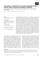
The 77 K fluorescence emission spectra and their first
derivatives measured in PSI submembrane fractions
illuminated for various times at 4 °C are shown in
Fig. 3. The intensity of the peak at 736 nm associated
to the PSI complex decreased by about 24% after
1 h of strong light illumination. It declined with
further light exposure, with only 30% of its initial
magnitude remaining after a 3-h illumination. Similar
to the absorbance spectra, a blue shift of the position
of the major fluorescence peak was observed. The
extent of that shift was larger in the fluorescence
emission spectra and reached 15–18 nm after 6 h
(Fig. 3A,C). The shift in fluorescence maximum was
evident from both absolute (Fig. 3A) and first deriv-
ative (Fig. 3C) spectra. The latter also demonstrates
that the half-width of the emission band peaking at
736 nm in untreated preparations was not affected by
light exposure despite the marked changes in the
peak position.
In the presence of histidine or n-propyl gallate the
changes in the fluorescence maximum at 736 nm were
delayed. In the presence of histidine, the fluorescence
maximum declined by 80% after 10 h of illumination.
In the case of propyl gallate this fluorescence was
reduced by 70% (Fig. 3B). Also, the peak shift was
delayed by these reactive oxygen scavengers. The fluor-
escence maximum peak shifts were 18 and 11 nm in
the presence of histidine and n-propyl gallate, respect-
ively, after 10 h of illumination (Fig. 3C). In the pres-
ence of catalase and SOD the changes in fluorescence
and peak shift resembled the control experiment (data
not shown).
Changes of polypeptide composition
Specific antibodies of PSI polypeptides were used to
study the degradation of proteins in PSI submembrane
Fig. 3. (A) Low temperature fluorescence emission spectra of the
isolated PSI submembrane fractions illuminated for various periods
of time with strong WL (2000 lmolÆm
)2
Æs
)1
)at4°C. Inset: the first
derivative of the absorption maxima in the red is presented to
show peak position. (B) Changes in fluorescence intensity meas-
ured at the maximum in the presence or absence of scavengers.
Results are means ± SD. n ¼ 3. (C) Changes in the position of the
fluorescence maximum obtained from the first derivative of the
fluorescence spectra of PSI complexes. Control (s), histidine (h),
and n-propyl gallate (,). For further details see Experimental proce-
dures. These experiments were repeated three times and yielded
identical spectra; a typical spectrum is presented.
S. Rajagopal et al. Photoprotective effect of active oxygen scavengers
FEBS Journal 272 (2005) 892–902 ª 2005 FEBS 895
particles during strong illumination. The reaction-
center protein PsaA was stable until 4 h and then
decreased very slightly with further illumination. How-
ever, PsaB protein degradation started after a 1-h
exposure and a degradation product appeared, whereas
after a 5-h exposure this protein mostly disappeared
(Fig. 4). These reaction-center proteins were protected
in the presence of histidine and n-propyl gallate (data
not shown). The stromal ridge constituted of PsaC,
PsaD, and PsaE polypetides provides the docking site
for the soluble electron acceptors ferredoxin and flavo-
doxin [33,34]. Among these polypeptides, PsaC was
relatively sensitive to strong light and this protein star-
ted to degrade even after 1 h of illumination and fur-
ther illumination accelerated the degradation (Fig. 4,
Table 1). PsaD was less sensitive (Fig. 4) and after 6 h
of exposure this polypeptide degraded only by 30%
(Table 1). PsaE was the most sensitive, after a 3-h
exposure more than 60% of this protein content was
decreased (Table 1). In comparison, the integral mem-
brane protein PsaF was more stable (Fig. 4). Addition
of reactive oxygen scavengers significantly protected
the above proteins against photodegradation (Fig. 5,
Table 1).
The outer antenna system of higher plant photo-
system I, LHCI, is composed of four proteins with
molecular masses of 20–24 kDa, the products of the
genes Lhca1–4, which are associated with the PSI core
[33–35]. We have analyzed the photodegradation pro-
file of each polypeptide in Fig. 4. Lhca1 is degraded
linearly with 30% loss of this protein after 3 h, and it
completely disappeared after a 5-h exposure (see also
Table 1). However, with the addition of histidine, this
degradation was negligible and after 6 h only 20% of
this subunit was lost (Fig. 5, Table 1). Surprisingly,
this protein was not degraded in the presence of n-pro-
pyl gallate (Fig. 5, Table 1). Lhca2 was more sensitive
to strong light and started to degrade after 1 h and
completely vanished after 3 h. With histidine, this pro-
tein declined by 45 and 60% after 3 and 6 h of illu-
mination, respectively, while with n-propyl gallate, the
degradation of this submit was only about 40% after 3
and 6 h illumination. The Lhca3 subunit linearly
degraded with the illumination period. After 3 h of
illumination, this protein was lost by 40% and almost
completely disappeared after 6 h. In the presence of
histidine this protein declined by only 15 and 60%
after 3 and 6 h of illumination, respectively, whereas,
in presence of n-propyl gallate the protein was stable
after 3 h and 20% loss was noticed after 6 h of illu-
mination. Lhca4 was the most stable subunit, after 3 h
this subunit was altered by 25 and by 50% after 6 h of
Table 1. Quantification of photosystem I polypeptides from immuno-
blots obtained from Fig. 5.
Control (%)
Histidine
(%)
n-Propyl
gallate (%)
0h 3h 6h 3h 6h 3h 6h
PsaC 100 68 0 105 109 55 45
PsaD 100 106 72 103 103 106 109
PsaE 100 36 0 73 55 95 86
Lhca1 100 71 0 91 77 97 106
Lhca2 100 45 0 54 38 63 58
Lhca3 100 62 9 83 37 93 77
Lhca4 100 74 52 83 65 91 76
Fig. 4. Quantitative immunoblot analysis of PSI submembrane frac-
tions illuminated with strong light at 4 °C for different time dur-
ation. All experimental conditions are given in Experimental
procedures.
Fig. 5. Quantitative immunoblot analysis of PSI submembrane frac-
tions illuminated with strong light at 4 °C for different time duration
in the presence of histidine or n-propyl gallate. All experimental
conditions are given in Experimental procedures.
Photoprotective effect of active oxygen scavengers S. Rajagopal et al.
896 FEBS Journal 272 (2005) 892–902 ª 2005 FEBS
illumination. Histidine and n-propyl gallate retarded
the photodegradation. The sensitivity of LCHI pro-
teins to strong illumination was Lhca2 > Lhca1 >
Lhca3 > Lhca4 (Fig. 4).
Analysis of CD changes
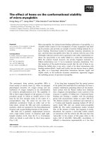
As shown in Fig. 6A, strong light induced significant
alterations in the secondary structure of proteins from
PSI submembrane fractions. After a 6-h exposure, a
clear decrease in elipticity measured at 222 and 190 nm
in the CD spectra was observed, which is typical of a
decrease in helical content. In the presence of histidine
and n-propyl gallate the above elipticities were not
affected after a 6-h illumination, and after a 10-h expo-
sure these changes were still minor (not shown).
The CD curve-analyzing algorithm (see Experimen-
tal procedures) revealed that untreated (0 h) PSI sub-
membrane fractions were composed of a-helix (24%),
b-sheet (25%), turns (22%), and unordered (29%)
structures (calculated from Fig. 6A). Performing the
same spectral analysis after various treatments allowed
quantifying the change in secondary structures in PSI
fractions. The proportion of a-helix decreased by 33%
after 6 h of exposure to strong light (Fig. 6B).
However, there was no change in a-helix even after a
6-h illumination in the presence of histidine or n-pro-
pyl gallate. After 10 h in the presence of both agents,
the a-helix helix percentage dropped slightly, although
the amplitude of this decrease was comparable to the
error margin of the method used for its computation
(Fig. 6B). These results clearly reveal that protein con-
formation is protected against photodamage in the
presence of histidine and n-propyl gallate.
Discussion
The primary objective of this work was to evaluate the
protective role of reactive oxygen scavengers against
strong light in PSI submembrane fractions. Our data
clearly demonstrated that some specific scavengers sig-
nificantly protected both the structure and function
of PSI.
Several authors postulated that oxidative mecha-
nisms are the basis of PSI photodamage in intact
leaves [19,20,22], isolated chloroplasts [7,8,11], or iso-
lated PSI fragments [15–18]. The selective action of
reactive oxygen scavengers used here indicated that the
species involved in the photodamage of proteins and
photochemical functions in the PSI submembrane frac-
tions were
1
O
2
, OH, and alkoxyl radicals. However,
H
2
O
2
and
•
O
2
–
were apparently not implicated in the
damaging processes. Addition of n-propyl gallate pro-
vided more protection than histidine (Figs 1–3). It is
known that n-propyl gallate protects against OH and
alkoxyl radicals, and histidine scavenges
1
O
2
[28,29].
Generally,
•
OH radicals are formed by the reaction
between hydrogen peroxide and reduced metal ions
during the so-called Fenton reaction. In the present
case, addition of SOD and ⁄ or catalase did not show
any protection against strong light. Nonetheless, this
was not due to a temperature-dependent inhibition of
SOD or catalase as similar changes of absorption,
fluorescence, and P700 oxidation under strong illumin-
Fig. 6. (A) Room temperature CD spectra of PSI submembrane
fractions illuminated for various time duration in control PSI sub-
membrane particles illuminated without active oxygen scavengers.
(B) Content in a-helical structures calculated from CD spectra
obtained from experiments such as shown in Fig. 6 A with no
additive (s) or with histidine (,), and n-propyl gallate (h). Results
are means ± SD. n ¼ 3. All experimental conditions are given in
Experimental procedures.
S. Rajagopal et al. Photoprotective effect of active oxygen scavengers
FEBS Journal 272 (2005) 892–902 ª 2005 FEBS 897
ation were observed at both room and low tempera-
ture in the PSI submembrane fractions (results not
shown). Thus, OH and alkoxyl radicals originated
from
1
O
2
. It is well known that
•
OH and alkoxyl radi-
cals can be produced from reactions of
1
O
2
or
•
OH
with organic molecules [36,37].
Generation of singlet oxygen by Chl is expected dur-
ing strong illumination as it depends on the population
of excited Chls molecules and is formed by energy
transfer from Chl molecules in triplet state to oxygen
[27 and references therein, 38]. The involvement of
P700 triplet states appearing as a consequence of
charge recombination between P700
+
and reduced A
1
or A
0
[39] could not represent a significant source for
1
O
2
production in isolated submembrane fractions
because of the low rates of electron flow through PSI
in the absence of an electron donor system (Fig. 1). As
P700
+
is known to be a very efficient quencher of exci-
ted singlet states of Chls close to the PSI reaction cen-
ter [40], excited states available for oxygen must have
appear mostly in the light-harvesting complexes, not in
the PSI core. In such case,
1
O
2
derivatives had first to
attack antenna Chls [41]. This might explain the faster
Chl breakdown compared to the loss of PSI photo-
chemical activity (Figs 1 and 2).
The native PSI complex from higher plants contains
about 200 Chls per P700. Among them, LHCI binds
about 70–110 Chl a + b molecules and serves as an
accessory antenna to harvest light and funnel its energy
to the reaction center, P700. The latter is located in the
PsaA ⁄ B proteins of the core component (CCI), where
96 Chls are bound [33,34]. The Chl a ⁄ b binding peri-
pheral antenna of plants PSI (LHCI) is composed of
four nuclear gene products, Lhca1–4, with molecular
masses of 20–24 kDa [33,34]. Each Lhca protein binds
approximately 10 Chls [42–44]. A total of eight Lhca
proteins are thought to be present per PSI: Lhca1 and
Lhca4 are present as heterodimers, whereas Lhca2 and
Lhca3 are likely present as homodimers [33,34]. Immu-
noblot analysis showed that among the Lhca proteins,
Lhca2 is more sensitive and Lhca4 is stable to strong
light. The sequence of the changes observed in Lhca
proteins was: Lhc2 > Lhca1 > Lhca3 > Lhca4
(Fig. 4).
As in the PSI core antenna, excitonically coupled
dimers or trimers of Chl a or b in the Lhca were also
suggested to form a pool of red pigments of low-
energy [45–47], more specifically in the Lhca4 subunit
of the LHCI-730 complex [45,48,49]. Recent findings
confirmed that Lhca2 and Lhca3 are also having low-
energy red pigments [44] but this is more pronounced
in Lhca3 [35]. If the absorbed energy migrates towards
PSI holochromes with higher-absorption wavelengths
[27], then, the Chl molecules with an absorption max-
ima in the red located at a relatively long wavelength
in Lhca3 and Lhca4 should be bleached first. A blue
shift in absorption and fluorescence maximum in the
red was clearly observed in the PSI submembrane frac-
tions (Figs 2 and 3, see also [16–18]). Thus, the pig-
ment aggregates absorbing at these long-wavelengths
could be involved in photoprotection [16,17,27]. This
phenomenon is in agreement with our results showing
that Lhca4 and Lhca3, which have red pigments with
higher absorption wavelength, are more stable. More-
over, Lhca1 and Lhca2 have a higher content in bulk
Chl. Thus, the fast degradation of Lhca1 and Lhca2 is
due to generation of reactive oxygen species during
strong light illumination which leads to some alter-
ation in the Chl–protein interaction. Hui et al. [15]
reported in PSI core complexes that LHCI-680 is more
sensitive due to decreased interaction between Chl–Chl
or Chl–protein. The discrepancy in sensitivity to strong
light between LHCI-680 and LHCI-730 could be
explained by the organization of Chl–Chl or Chl–pro-
tein in the Lhca subunits. The degradation of Lhca
protein subunits was reduced with the addition of his-
tidine or n-propyl gallate in PSI submembrane parti-
cles exposed to strong light even after 10 h, with a
more pronounced action of n-propyl gallate (Fig. 5,
Table 1). The above results are consistent with previ-
ous reports of strong light-induced degradation of
LHCII proteins where histidine and n-propyl gallate
also retarded the photooxidative effects with a more
efficient action of n-propyl gallate [28,29].
The PSI core is a large pigment–protein complex
composed of 11–13 protein subunits, the largest two
of which, PsaA and PsaB, comprises the molecular
masses of 83.2 and 83.4 kDa [33,34]. This is a het-
erodimer to which the majority of the core antenna
pigments, as well as most of the reaction center
cofactors, are bound. In PSI core preparations, strong
light exposure induced changes to both reaction-cen-
ter proteins [14]. In the present study using submem-
brane fractions, PsaB was more sensitive to strong
light and also produced degradation products
(Fig. 4). Similar degradation products of PsaB were
observed with no apparent modification of PsaA in
spinach thylakoids or cucumber leaves illuminated
with weak light at low or room temperature [21,29].
These authors proposed that the degradation of PsaB
was mainly due to formation of superoxide and
hydroxyl radicals and this was protected by n-propyl
gallate. In another report using barley leaves illumin-
ated with weak light at low temperatures, damage to
both reaction-center proteins of PsaA and PsaB was
reported [23]. The involvement of reactive oxygen
Photoprotective effect of active oxygen scavengers S. Rajagopal et al.
898 FEBS Journal 272 (2005) 892–902 ª 2005 FEBS
species such as superoxide, hydrogen peroxide, and
hydroxyl radicals was suggested. They also showed
that
1
O
2
was involved in the damage of reaction-
center proteins. In vivo, the photooxidative damages
in PSI may occur with a similar pattern as in PSII
where reactive oxygen species are thought to initiate
protein degradation that is completed by chloroplast
proteases [21,30]. It was recently shown that the deg-
radation of PsaA ⁄ B proteins and alteration of photo-
chemical activity in spinach thylakoids are more
intensive at room temperature than at low tempera-
ture under strong light [13], which suggested that the
photoinactivation may involve an enzymatic contribu-
tion to the phenomenon.
As mentioned above, in PSI submembrane fractions,
PsaA was more resistant to strong light compare to
PsaB. PsaA was stable until 4 h of illumination and
then started degradation (Fig. 4). The PSI core is
mainly composed of bulk antenna Chl that absorbs in a
broad band with a maximum at 680 nm, but also con-
tains 3–10% of low-energy Chl (red). The quenching of
excitations located on the red Chls by P700
+
could pro-
vide a pathway to prevent the generation of Chl triplet
states, which can lead to the formation of harmful
1
O
2
under strong light illumination [46]. The lower sensitiv-
ity of PsaA to photooxidative damage compare to PsaB
may indicate that it contains less low energy Chl aggre-
gates. Alternatively, PsaA may be less closely associated
with the antenna Chl of the light harvesting complexes
and thus receive less excess energy that leads to the
generation of
1
O
2
. Addition of histidine and n-propyl
gallate protected these two reaction-center proteins
even after 10 h of illumination (data not shown), which
supports the involvement of
1
O
2
.
Apart from PsaA and PsaB, other smaller polypep-
tides of PSI were degraded during strong light illumin-
ation. PsaC together with PsaD and E comprises the
stromal side of PSI. PsaC anchors the two Fe
4
S
4
clus-
ters F
A
and F
B
, which are needed to carry out the elec-
tron transfer from F
x
. The order of sensitivity to
strong light illumination was PsaE > PsaC > PsaD
(Fig. 5, Table 1). Similar results were observed in PSI
core particles [15]. This order closely corresponds with
the degradation profile observed during disassembly
studies using urea treatment [50]. The crystal structure
of plant and cyanobacterium PSI revealed that loops
at the stromal surface of PsaA ⁄ B partly contribute the
binding interface to the PsaC, D and E subunits
[51,52]. These observations are in agreement with our
data showing that the degradation of PsaC subunit
ensue almost simultaneously the degradation of Lhca2
and PsaB subunits (Figs 4 and 5). PsaD is stable even
after 5 h of illumination, whereas, PsaE is more
susceptible to strong light. The structural model of
Thermosynechococcus PSI showed that part of PsaE is
sandwiched between the PsaA ⁄ B heterodimer and
PsaC [33,53]. The higher plant reaction-center structure
retains the same organization as the cyanobacterial
complex [52]. Thus, it seems possible that PsaE affects
the intracomplex electron transport under some condi-
tions. From our data it is clear that the degradation of
the PSI stromal ridge starts at PsaE. Most likely,
in plants PsaE would determine the susceptibility to
photodamage of the PSI complex. Interestingly, the
present data also indicate that, even though PsaC
seems to be required during the assembly of PsaD and
PsaE with the PSI complex [50], PsaC can be lost inde-
pendently from the two other extrinsic proteins (Figs 4
and 5).
Excess light clearly altered the secondary protein
structure of the PSI submembrane fractions. With
increased time of exposure a significant portion of the
a-helices was lost (Fig. 6). The protein unfolding was
significantly prevented by histidine and n-propyl gallate
(Fig. 6). The alteration of protein secondary structure
components as revealed from the CD spectral studies is
likely to play a central role in the active oxygen-depend-
ent photodegradation processes of the PSI complex. It is
possible that the observed structural perturbations
involved altered pigment–protein and protein–protein
interaction, contributing to the observed decreased
efficiency of energy migration and capture, and leading
to Chl degradation.
Experimental procedures
Isolation of photosystem I submembrane
fractions
PSI submembrane particles were isolated from fresh spin-
ach leaves obtained from the local market, according to the
procedure of Peters et al. [31] with some modifications [32].
The isolated preparations with Chl content of 1–2 mg
ChlÆmL
)1
were suspended in a medium containing 20 mm
Tricine ⁄ KOH buffer (pH 7.8), 10 mm NaCl, 10 mm KCl,
and 5 mm MgCl
2
, and stored at )80 °C until its use. The
Chl a ⁄ b ratio was found to be higher than 6.0 in isolated
PSI submembrane fractions. Chl was determined in 80%
acetone according to Porra et al. [54].
Light treatment
The PSI preparations (500 lg ChlÆmL
)1
) were illuminated
for 5 h with continuous stirring at 4 °C using strong white
light (WL) (2000 lEÆm
)2
Æs
)1
) from a 150 W quartz ⁄ halogen
projector lamp. WL was passed through a 5-cm layer of
S. Rajagopal et al. Photoprotective effect of active oxygen scavengers
FEBS Journal 272 (2005) 892–902 ª 2005 FEBS 899
water containing CuSO
4
to cut the infrared radiation. The
samples were illuminated in the presence of various active
oxygen scavengers to selectively remove the reactive oxygen
species. The scavengers used were histidine at a concentra-
tion of 25 mm for
1
O
2
, n-propyl gallate at 1 mm for -OH
and alkoxyl radicals, and SOD and catalase at 250 lgÆmL
)1
for
•
O
2
–
and H
2
O
2
, respectively [28,29].
Measurement of P700 redox state
The changes in the redox state of P700, the primary donor
of PSI, were measured by the light-induced absorbance
changes at 830 nm (DA
830
) using a dual-wavelength unit
ED-P700DW connected to a PAM fluorometer (Walz,
Effeltrich, Germany) as described by Schreiber et al. [55].
All measurements were carried out at an identical sensitiv-
ity of the PAM fluorometer. The absorbance changes at
830 nm represented only the oxidation and reduction of
P700 as no contribution to the absorbance changes due to
plastocyanin redox transformations can be detected with
the ED-P700DW unit. White actinic light was obtained
from the KL 1500 projector (Walz, Effeltrich, Germany).
The aliquots of 60 lL taken from the suspension of PSI
submembrane particles during strong light treatment were
added to 140 lL of suspension buffer containing 200 lm
ascorbate as an artificial donor. Chl concentration during
the measurements of DA
830
was 30 lgÆmL
)1
in the control
experiments (prior to the illumination).
Measurement of absorption spectra
The room temperature absorption spectra and their first
derivatives were recorded using a PerkinElmer Lambda 40
spectrophotometer (Wellesley, MA, USA). Ten microlitres
of the suspension of submembrane particles were repeatedly
taken during the illumination. Such aliquots taken from the
suspension of untreated particles contained 5 lg of Chl,
whereas the Chl content decreased gradually in the samples
taken during strong illumination.
Fluorescence spectroscopy
Low temperature (77 K) spectra of fluorescence emission
excited at 436 nm were measured as reported previously
[16] using a PerkinElmer LS55 spectrofluorometer. The Chl
content of the samples was adjusted to 5 lgÆmL
)1
. The
excitation and emission slit width were set at 5 and 2.5 nm,
respectively.
Circular dichroism spectroscopic analysis
CD spectra were measured in a Jasco-720 spectropolari-
meter (Easton, MD, USA) in a cell with a 0.1 mm optical
path length over a wavelength range of 190–260 at a
temperature of 20 °C. Each CD spectrum was the average
of five accumulations at a scanning speed of 20 nmÆmin
)1
and a 1 nm spectral band width. The base line was correc-
ted with blank buffer for each spectrum. The secondary
structure of PSI proteins was determined from the CD
spectra using the cd pro program as described previously
[56]. Sample concentration was 50 lgÆmL
)1
Chl in the con-
trol measurements (prior to the illumination).
Immunoblot analysis
PSI submembrane polypeptides were separated by poly-
acrylamide gel electrophoresis (PAGE) according to Raja-
gopal et al. [32]. Electrophoresis was performed on a 13%
separating and 4% stacking gel of polyacrylamide. The sus-
pension (10 lL aliquots) of submembrane fractions were
repeatedly taken during the course of photoinhibition. Such
aliquots contained 5 l g Chl obtained from the suspension
of untreated preparations. An equal volume of 2· buffer
was added to the aliquots.
To identify and quantify the PSI polypeptides, immuno-
blotting was carried out essentially as described by Towbin
et al. [57]. Western blotting was performed by electropho-
retic transfer of proteins to nitrocellulose membranes
(0.45 lm, Millipore, Billerica, MA, USA). The membrane
was incubated with polyclonal antibodies raised in rabbits
against PSI complex. Subsequently, secondary antibodies
ligated to alkaline phosphate were applied. Bromo-chloro-
indolyl-phosphate and tetrazolium blue were used for the
coloring reaction. The developed membranes were analyzed
by using Bio-Rad Gel-Doc 2000 system (Hercules, CA,
USA).
Acknowledgements
The authors wish to thank Drs J.H. Golbeck, K.
Sonoike and P. Chitnis for antibodies and J. Harnois
for helpful professional assistance. This research was
supported by the Natural Sciences and Engineering
Research Council of Canada and by Fonds Que
´
be
´
cois
de la Recherche sur la Nature et les Technologies.
References
1 Powles SB (1984) Photoinhibition of photosynthesis
induced by visible light. Annu Rev Plant Physiol 35, 15–44.
2 Aro EM, Virgin I & Andersson B (1993) Photoinhibi-
tion of photosystem II. Inactivation, protein damage
and turnover. Biochim Biophys Acta 1143, 113–134.
3 Prasil O, Adir N & Ohad I (1992) Dynamics of photo-
system II: Mechanism of photoinhibition and recovery
process. In Topics in Photosynthesis, the Photosystem
Structure, Function and Molecular Biology, Vol. 11
(Barber J, ed.), pp. 295–384. Elsevier, Amsterdam.
Photoprotective effect of active oxygen scavengers S. Rajagopal et al.
900 FEBS Journal 272 (2005) 892–902 ª 2005 FEBS
4 Andersson B & Barber J (1996) Mechanisms of photo-
damage and protein degradation during photoinhibition
of photosystem II. In Photosynthesis and the Environ-
ment (Baker NR, ed.), pp. 101–121. Kluwer Academic
Publishers, Dordrecht, The Netherlands.
5 Mulo P, Laakso S, Maenpaa P & Aro E (1998) Step-
wise photoinhibition of photosystem II. Plant Physiol
117, 483–490.
6 Barber J & Andersson B (1992) Too much of a good
thing: light can be bad for photosynthesis. Trends
Biochem Sci 17, 61–66.
7 Satoh K (1970) Mechanism of photoinactivation in
photosynthetic systems. I. The dark reaction in photo-
inactivation. Plant Cell Physiol 11, 15–27.
8 Satoh K (1970) Mechanism of photoinactivation in
photosynthetic systems. II. The occurrence of and
properties of two different types of photoinactivation.
Plant Cell Physiol 11, 29–38.
9 Satoh K & Fork DC (1982) Photoinhibition of reaction
centers of photosystem I and II in intact Bryopsis chlor-
oplasts under anaerobic conditions. Plant Physiol 70,
1004–1008.
10 Satoh K (1970) Mechanism of photoinactivation in
photosynthetic systems. III. Site and mode of photoacti-
vation in photosystem I. Plant Cell Physiol 11, 187–197.
11 Inoue K, Sakurai H & Hiyama T (1986) Photoinactiva-
tion sites of photosystem I in isolated chloroplasts.
Plant Cell Physiol 27, 961–968.
12 Inoue K, Fujii T, Yokoyama E, Matsuura K, Hiyama
T & Sakurai H (1989) The photoinhibition site of
photosystem I in isolated chloroplasts under extremely
reducing conditions. Plant Cell Physiol 30, 65–71.
13 Velitchkova M, Yruela I, Alfonso M & Picorel R (2003)
Different kinetics of photoinactivation of photosystem
I-mediated electron transport and P700 in isolated thy-
lakoid membranes. J Photochem Photobiol Biol B 69,
41–48.
14 Baba K, Itoh S, Hastings G & Hoshina S (1996) Photo-
inhibition of photosystem I electron transfer activity
in isolated photosystem I preparations with different
chlorophyll contents. Photosynth Res 47, 121–130.
15 Hui Y, Jie W & Carpentier R (2000) Degradation of the
photosystem I complex during photoinhibition. Photo-
chem Photobiol 72, 508–512.
16 Rajagopal S, Bukhov NG & Carpentier R (2002)
Changes in the structure of chlorophyll–protein com-
plexes and excitation energy transfer during photo-
inhibitory treatment of isolated photosystem I
submembrane particles. J Photochem Photobiol B: Biol
62, 194–200.
17 Rajagopal S, Bukhov NG & Carpentier R (2003) Photo-
inhibitory light-induced changes in the composition of
chlorophyll–protein complexes and photochemical activ-
ity of photosystem I submembrane fractions. Photochem
Photobiol 77, 284–291.
18 Rajagopal S & Carpentier R (2003) Retardation of
photoinduced changes in photosystem I submembrane
particles by glycinebetaine and sucrose. Photosynth Res
78, 77–85.
19 Terashima I, Funayama S & Sonoike K (1994) The site
of photoinhibition in leaves of Cucumis sativus L. at low
temperatures is photosystem I, not photosystem II.
Planta 193, 300–306.
20 Sonoike K, Terashima I, Iwaki M & Itoh S (1995)
Destruction of photosystem I iron–sulfur centers in
leaves of Cucumis sativus L. by weak illumination at
chilling temperatures. FEBS Lett 362, 235–238.
21 Sonoike K, Kamo M, Hihara T & Enami I (1997) The
mechanism of the degradation of PsaB gene product,
one of the photosynthetic reaction center subunits of
photosystem I, upon photoinhibition. Photosynth Res
53, 55–63.
22 Tjus SE, Moller BL & Scheller HV (1999) Photoinhibi-
tion of photosystem I damages both reaction centre pro-
teins PSI-A and PSI-B and acceptor-side located small
photosystem I polypeptides. Photosynth Res 60, 75–86.
23 Allen RD (1995) Dissection of oxidative stress tolerance
using transgenic plants. Plant Physiol 107, 1049–1054.
24 Foyer CH, Lelandais M & Kunert KJ (1994) Photooxi-
dative stress in plants. Physiol Plant 92, 696–717.
25 Vass I (1997) Adverse effects of UV-B light on the
structure and function of the photosynthetic apparatus.
In Handbook of Photosynthesis (Pessarakli M, ed.),
pp. 931–949. Marcel Dekker, New York.
26 Demmig-Adams B & AdamsWW III (1992) Photopro-
tection and other responses of plants to high light stress.
Annu Rev Plant Physiol Plant Mol Biol 43, 599–626.
27 Carpentier R (1997) Influence of high light intensity on
photosynthesis: photoinhibition and energy dissipation.
In Hand Book of Photosynthesis (Pessarakli M, ed.), pp.
443–449. Marcel Dekker, New York.
28 Zolla L & Rinalducci S (2002) Involvement of active
oxygen species in degradation of light-harvesting pro-
teins under light stresses. Biochemistry 41, 14391–14402.
29 Rinalducci S, Pedersen JZ & Zolla L (2004) Formation
of radicals from singlet oxygen produced during photo-
inhibition of isolated light-harvesting proteins of photo-
system II. Biochim Biophys Acta 1608, 63–73.
30 Sonoike K (1996) Degradation of psaB gene product,
the reaction center subunit of photosystem I, is
caused during photoinhibition of phtosystem I:
possible involvment of active oxygen species. Plant Sci
115, 157–164.
31 Peters FALJ, Van Wielink JE, Wong Fong Sang HW,
De Vries S & Kraayenhof R (1983) Studies on well
coupled photosystem I-enriched subchloroplast vesicles:
content and redox properties of electron-transfer com-
ponents. Biochim Biophys Acta 724, 159–165.
32 Rajagopal S, Bukov NG & Carpentier R (2003) Control
of energy dissipation and photochemical activity in
S. Rajagopal et al. Photoprotective effect of active oxygen scavengers
FEBS Journal 272 (2005) 892–902 ª 2005 FEBS 901
photosystem I by NADP-dependent reversible confor-
mational changes. Biochemistry 42, 11839–11845.
33 Scheller HV, Jensen PE, Haldrup A, Lunde C & Knoet-
zel J (2001) Role of subunits in eukaryotic photosystem
I. Biochim Biophys Acta 1507, 41–60.
34 Jensen PE, Haldrup A, Rosgaard L & Scheller HV
(2003) Molecular dissection of photosystem I in higher
plants: topology, structure and function. Physiol Plant
119, 1–9.
35 Castellatti S, Morosinotto T, Robert B, Caffarri S,
Bassi R & Croce R (2003) Recombinant Lhca2 and
Lhca3 subunits of the photosystem I antenna system.
Biochemistry 42, 4226–4234.
36 Elstner EF (1982) Oxygen activation and oxygen toxic-
ity. Annu Rev Plant Physiol 33, 73–96.
37 Halliwell B & Gutteride S (1984) Oxygen toxicity, oxy-
gen radicals, transition metals and disease. Biochem J
219, 1–14.
38 Siefermann-Harms D (1987) The light-harvesting and
protective functions of carotenoids in photosynthetic
membranes. Physiol Plant 69, 561–568.
39 Warren PV, Golbeck JH & Warden JT (1993) Charge
recombination between P700
+
and A
À
1
occurs directly
to the ground state of P700 in a photosystem I core
devoid of F
X
,F
B
and F
A
. Biochemistry 32, 849–857.
40 Nuijs AM, Shuvalov VA, van Gorkom HJ, Plijter JJ &
Duysens LNM (1986) Picosecond absorbance difference
spectroscopy on the primary reaction and the antenna-
excited states in photosystem I particles. Biochim Bio-
phys Acta 850, 310–318.
41 Clarke RH, Jagannathan SP & Leenstra WR (1980)
Optical-microwave double resonance spectroscopy of
in vivo chlorophyll. Photochem Photobiol 32, 805–808.
42 Jansson S, Andersson B & Scheller HV (1996) Nearest
neighbor analysis of higher-plant photosystem I holo-
complex. Plant Physiol 12, 409–420.
43 Ganateg U, Strand A, Gustafsson P & Jansson S (2001)
The properties of the chlorophyll a ⁄ b-binding proteins
Lhca2 and Lhca3 studied in vivo using antisense inhibi-
tion. Plant Physiol 127, 150–158.
44 Schmid VHR, Potthast S, Wiener M, Bergauer V, Paul-
sen H & Storf S (2002) Pigment binding of photosytem
I light harvesting proteins. J Biol Chem 277, 37307–
37314.
45 Melkozernov AN (2001) Excitation energy transfer in
photosystem I from oxygenic organisms. Photosynth Res
70, 129–153.
46 Gobet B & van Grondelle R (2001) Energy transfer and
trapping in photosystem I. Biochim Biophys Acta 1507,
80–99.
47 Schmid VHR, Thome P, Ruhle W, Paulsen H, Kuhl-
bandt W & Rogle H (2001) Chlorophyll b is involved in
long-wavelength spectral properties of light-harvesting
complexes LHCI and LHCII. FEBS Lett 499, 27–31.
48 Melkozernov AN & Blankenship R (2003) Structural
modeling of the Lhca4 subunit of LHCI-730 peripheral
antenna in photosystem I based on similarity with
LHCII. J Biol Chem 278, 44542–44551.
49 Melkozernov AN, Lin S, Schmid VH, Paulsen H,
Schmidt GW & Blankenship RE (2000) Ultrafast excita-
tion dynamics of low energy pigments in reconstituted
peripheral light-harvesting complexes of photosystem I.
FEBS Lett 471, 89–92.
50 Antonkine ML, Jordan P, Fromme P, Kraub N,
Golbeck JH & Stehlik D (2003) Assembly of protein
subunits within the stromal ridge of photosystem I.
Structural changes between unbound and sequentially
PSI-bound polypeptides and correlated changes of mag-
netic properties of the terminal iron sulfur clusters.
J Mol Biol 327, 671–697.
51 Jordan P, Fromme P, Witt HT, Klukas O, Saenger W
& Krauss N (2001) Three-dimensional structure of
cyanobacterial photosystem I at 2.5 angstrom
resolution. Nature 411, 909–917.
52 Ben-Shem A, Frolow F & Nelson N (2003) Crystal
structure of plant photosystem I. Nature 426, 630–635.
53 Klukas O, Schubert WD, Jordan P, Krauss N, Fromme
P, Witt HT & Saenger W (1999) Photosystem I, an
improved model of the stromal subunits PsaC, PsaD,
and PsaE. J Biol Chem 274, 7351–7360.
54 Porra RJ, Thompson WA & Kriedemann PE (1989)
Determination of accurate extinction coefficients and
simultaneous equations for assaying chlorophylls a and
b extracted with four different solvents: verification of
the concentration of chlorophyll standards by atomic
absorption spectroscopy. Biochim Biophys Acta 975,
384–394.
55 Schreiber U, Klughammer C & Neubauer C (1988)
Measuring P700 absorbance changes around 830 nm
with a new type of pulse modulated system. Z Natur-
forsch 43c, 686–698.
56 Sreerama N & Woody RW (2000) Estimation of protein
secondary structrure from circular dichroism spectra:
comparison of CONTIN, SELCON, and CDSSTR
methods with an expanded reference set. Anal Biochem
287, 252–260.
57 Towbin H, Stahelin T & Gordon J (1979) Electrophor-
esis transfer of proteins from polyacrylamide gels to
nitrocellulose sheets: procedure and some application.
Proc Natl Acad Sci USA 76, 43450–43454.
Photoprotective effect of active oxygen scavengers S. Rajagopal et al.
902 FEBS Journal 272 (2005) 892–902 ª 2005 FEBS