Báo cáo khoa học: Syndecan-4 is a signaling molecule for stromal cell-derived factor-1 (SDF-1)/ CXCL12 pptx
Bạn đang xem bản rút gọn của tài liệu. Xem và tải ngay bản đầy đủ của tài liệu tại đây (525.18 KB, 15 trang )
Syndecan-4 is a signaling molecule for stromal cell-derived
factor-1 (SDF-1)
/
CXCL12
Nathalie Charnaux
1,2
*, Se
´
verine Brule
1,2
*, Morgan Hamon
1
, Thomas Chaigneau
1
, Line Saffar
1
,
Catherine Prost
1
, Nicole Lievre
1
and Liliane Gattegno
1,2
1 Laboratoire de Biologie Cellulaire, Biothe
´
rapies Be
´
ne
´
fices et Risques, UPRES 3410 Universite
´
Paris XIII, Bobigny, France
2Ho
ˆ
pital Jean Verdier, Bondy, France
Chemokines are low molecular mass proteins mediating
several functions such as hematopoiesis regulation, leu-
kocyte maturation, angiogenesis, T and B lymphocytes
trafficking, homing and lymphoid tissues development
[1–3]. Stromal cell-derived factor-1 (SDF-1) ⁄ recently
renamed CXCL12 [4], is the only known ligand for
CXCR4 [5,6]. SDF-1 and CXCR4 are constitutively
expressed in various tissues [7] and are implicated in
several diseases. CXCR4 is involved in HIV infection
and pathogenesis [5,8]. SDF-1 and CXCR4 also regulate
Keywords
CXCR4; proteoglycan; SDF-1 ⁄ CXCL12;
syndecan-4
Correspondence
L. Gattegno, Laboratoire de Biologie
Cellulaire, Biothe
´
rapies Be
´
ne
´
fices et
Risques, UPRES 3410 Universite
´
Paris XIII,
74, rue Marcel Cachin, 93017, Bobigny,
France, Ho
ˆ
pital Jean Verdier, 93017, Bondy,
France
Fax: +33 1 48026503
Tel: +33 1 48387752
E-mail:
*These authors contributed equally to this
work.
(Received 18 January 2005, accepted 21
February 2005)
doi:10.1111/j.1742-4658.2005.04624.x
Stromal cell-derived factor-1 (SDF-1) ⁄ CXCL12, the ligand for CXCR4,
induces signal transduction. We previously showed that CXCL12 binds to
high- and low-affinity sites expressed by primary cells and cell lines, and
forms complexes with CXCR4 as expected and also with a proteoglycan,
syndecan-4, but does not form complexes with syndecan-1, syndecan-2,
CD44 or beta-glycan. We also demonstrated the occurrence of a CXCL12-
independent heteromeric complex between CXCR4 and syndecan-4.
However, our data ruled out the glycosaminoglycan-dependent binding of
CXCL12 to HeLa cells facilitating the binding of this chemokine to
CXCR4. Here, we demonstrate that CXCL12 directly binds to syndecan-4
in a glycosaminoglycan-dependent manner. We show that upon stimulation
of HeLa cells by CXCL12, CXCR4 becomes tyrosine phosphorylated as
expected, while syndecan-4 (but not syndecan-1, syndecan-2 or beta-glycan)
also undergoes such tyrosine phosphorylation. Moreover, tyrosine-phos-
phorylated syndecan-4 from CXCL12-stimulated HeLa cells physically
coassociates with tyrosine phosphorylated CXCR4. Pretreatment of the
cells with heparitinases I and III prevented the tyrosine phosphorylation of
syndecan-4, which suggests that the heparan sulfate-dependent binding of
SDF-1 to this proteoglycan is involved. Finally, by reducing syndecan-4
expression using RNA interference or by pretreating the cells with hepari-
tinase I and III mixture, we suggest the involvement of syndecan-4 and
heparan sulfate in p44 ⁄ p42 mitogen-activated protein kinase and Jun N-ter-
minal ⁄ stress-activated protein kinase activation by action of CXCL12 on
HeLa cells. However, these treatments did not modify the calcium mobil-
ization induced by CXCL12 in these cells. Therefore, syndecan-4 behaves
as a CXCL12 receptor, selectively involved in some transduction pathways
induced by SDF-1, and heparan sulfate plays a role in these events.
Abbreviations
dsRNA, double-stranded RNA; FBS, fetal bovine serum; GAG, glycosaminoglycan; HS, heparan sulfate; JNK ⁄ SAPK, Jun N-terminal ⁄ stress-
activated protein kinase; MAPK, mitogen-activated-protein kinase; PFA, paraformaldehyde; PMA, phorbol 12-myristate-13-acetate; PG,
proteoglycan; Ptyr, tyrosine phosphorylated; SDF, stromal cell-derived factor; SD, syndecan.
FEBS Journal 272 (2005) 1937–1951 ª 2005 FEBS 1937
embryonic development [9]. Much of the heparan
sulfate (HS) at the cell surface is derived from the
syndecan (SD) family of transmembrane proteoglycan
(PG) [10]. The SDs bind a variety of growth factors,
cytokines, proteases, antiproteases and cell adhesion
molecules [10,11]; they are individually expressed in dis-
tinct cell-, tissue-, and development-specific patterns
[12], and show cell-specific variations in the structure of
their HS chains [13]. SDs may regulate ligand-depend-
ent activation of cell surface growth factor receptors by
several potential mechanisms [10,11,14]. SD-4 is one
of the principal HS carrying protein on cell surfaces
[15,16]. We recently showed that SDF-1 binds to
high- and low-affinity sites on HeLa cells and forms
complexes on these cells and on human primary lympho-
cytes and macrophages, which comprise CXCR4, as
expected, and also SD-4 [17], but not SD-1, SD-2, beta-
glycan or CD44 ([17] and unpublished data). Moreover,
we recently demonstrated the occurrence of an SDF-1-
independent heteromeric complex on the plasma mem-
brane of these cells, which comprises CXCR4 and SD-4
but not SD-1, SD-2, CD44 or beta-glycan [17]. This
suggested that SDF-1 may bind both the PG SD-4 and
its G-protein-coupled receptor (GPCR), CXCR4. How-
ever, our previous data have shown that while glycos-
aminidases pretreatment of primary macrophages
decreases the binding of SDF-1 to CXCR4, such treat-
ment had no effect on the chemokine binding to
CXCR4 expressed by the HeLa cell line [17]. This has
suggested that while SD-4 may serve as a binding
anchor for SDF-1 on primary macrophages to enable
the chemokine to interact with CXCR4, this was not
true if HeLa cells were tested.
The present study was designed to test whether
SD-4 functions as a specific SDF-1 signaling molecule.
Therefore, we first determined whether SDF-1 directly
binds SD-4 and the glycosaminoglycan (GAG)-
dependency of this binding. Because protein phos-
phorylation plays a critical role in the generation of
intracellular signals in response to external stimuli,
we then investigated whether SD-4 becomes tyrosine
phosphorylated (Ptyr) upon SDF-1 stimulation of
HeLa cells, and whether, in these conditions, tyrosine-
phosphorylated SD-4 is physically coassociated with
tyrosine-phosphorylated CXCR4, and what the GAG-
dependency of these events is. Finally, we asked whe-
ther SD-4 is involved in other biochemical signals
induced by SDF-1. By specifically reducing SD-4
expression using RNA interference, or by reducing the
HS expressed at the plasma membranes of HeLa cells
by the use of heparitinases I and III, we analyzed the
respective roles of SD-4 and HS in transduction path-
ways induced by SDF-1 on these cells.
Results
SDF-1 directly binds to SD-4
The HeLa cells used in the present study express
CXCR4, SD-2, beta-glycan (data not shown) [17,18],
CD44, SD-1 and SD-4 (Fig. 1A), as assessed by flow
cytometry analysis after indirect immunofluorescence
A
B
ab
c
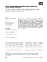
Fig. 1. PGs on HeLa cells. (A) Cell surface expression of SD-1,
SD-4 and CD44 on HeLa cells. HeLa cells (5 · 10
5
) were stained
for FACS analysis with anti-(SD-1) DL-101 mAb (a), anti-(SD-4)
5G9 mAb (b) or anti-CD44 mAb (c) (thick lines). Reactivity was
compared to an isotype-matched control monoclonal antibody
(a,b,c, dotted lines). (B) Immunoblot analysis of PGs from HeLa
cells. HeLa cells were lysed in the presence of Triton X-100 and
urea. PGs (from 2 · 10
6
cells per lane) were enriched by DEAE
Sephacel anion exchange chromatography and then treated with
heparitinases I, III, and chondroitinase ABC mixture, electroblotted
and revealed with 3G10 mAb (lane 1) or the isotype, IgG2b (lane
2). The respective immunoreactivity with anti-(SD-1) DL-101, anti-
(SD-4) 5G9, anti-CD44 mAbs, or anti-(SD-2) Igs are represented by
arrows. Data are representative of three individual experiments.
Syndecan-4 is an auxiliary receptor for SDF-1/CXCL12 N. Charnaux et al.
1938 FEBS Journal 272 (2005) 1937–1951 ª 2005 FEBS
labeling. The core proteins of most PGs enriched from
HeLa cells lysates were analyzed [17] after heparitinase
I and III and chondroitinase ABC treatment to detect
their apparent molecular masses. Proteins of 32 kDa
and 50–58 kDa, immunoreactive with anti-SD-4 5G9
and 3G10 mAbs, were observed (Fig. 1B). The 50–
58 kDa proteins may represent, in accordance with
other studies, homo- or hetero-oligomers of the SD-4
core protein, which is a 32 kDa protein [19]. Other
PGs were also detected: 34 kDa proteins immunoreac-
tive with both anti-SD-2 mAbs and mAb 3G10,
45- and 90 kDa proteins immunoreactive with anti-
SD-1DL-101 and 3G10 mAbs (the 90 kDa ones
probably being dimers of the 45 kDa ones), and
60 kDa proteins immunoreactive with anti-CD44 and
3G10 mAbs (Fig. 1B). All these apparent molecular
masses are close to the predicted ones [9]. These PGs
were glycanated, as mAb 3G10 reacts with an epitope
including a terminal unsaturated uronic acid residue,
which is unmasked after HS removal by heparitinases
treatment [20].
Native PGs may migrate in a diffuse high molecular
mass distribution on SDS ⁄ PAGE. Using the respective
specific Abs, glycanated PGs migrate as follows: SD-4
as a 100–250 kDa broad smear, SD-1 as a single
98 kDa band, CD44 as a 110 kDa band, SD-2 as a
50 kDa protein. Beta-glycan migrates as two broad
bands of 55 and 100 kDa, respectively (Fig. 2, lanes
1–5). No immunoreactivity was detected with the iso-
types (data not shown). The fact that all these PGs
were also immunoreactive with anti-HS mAb 10E4,
but not with its isotype, demonstrates their glycana-
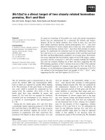
tion (Fig. 2, lane 6 and data not shown). Biotinylated
SDF-1a bound to the broad smear of 100–250 kDa,
characterized as glycanated SD-4, but did not bind to
SD-1, SD-2, beta-glycan or CD44 (Fig. 2, lane 7 vs.
1–5). Heparitinase I and III, and chondroitinase ABC
pretreatment of the strips abolished the binding of
electroblotted PGs to anti-HS mAb 10E4 (Fig. 2, lane
8 vs. 6), and strongly decreased that of biotinylated
SDF-1a to SD-4 (Fig. 2, lane 9 vs. 7), but did not
change SD-4 binding to anti-SD-4 mAb 5G9 (specific
for the core protein of SD-4)(data not shown). This
demonstrated that (a) the heparitinases treatment was
efficient; (b) SD-4 was still present on the polyvinylid-
ene difluoride membrane (data not shown); and (c)
the direct binding of SDF-1 to SD-4 was GAG
dependent.
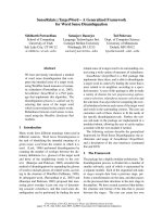
Confocal microscopy analysis showed that fluores-
cently labeled biotinylated SDF-1a colocalizes with
SD-4 on the plasma membranes of these cells, as
assessed by the yellow (red-green colocalization) stain-
ing (Fig. 3A, and data not shown). This association
was further analyzed by electron microscopy (Fig. 3B).
Beads at the cell surface were counted and considered
as associated when the distance between them was less
than 15 nm. Forty per cent of the beads that labeled
SD-4 were associated with 45% of the beads that labe-
led SDF-1a, while no association of SDF-1a with
SD-1 was detected. Controls, run without biotinylated
SDF-1a or with the isotypes, were not stained (data
not shown).
Fig. 2. SDF-1 binds to SD-4. HeLa cells were lysed in the presence of Triton X-100 and urea. PGs were enriched by DEAE Sephacel anion
exchange chromatography, electroblotted and revealed with anti-SD-4 5G9 mAb (lane 1), anti-(SD-1) DL-101 mAb (lane 2), anti-CD44 mAb
(lane 3), anti-(SD-2) Igs (lane 4), anti-(beta-glycan) Igs (lane 5), anti-HS 10E4 mAb (lane 6), biotinylated SDF-1a (lane 7). Alternatively, strips
were treated with heparitinases I, III mixture and revealed with anti-HS mAb 10E4 (lane 8) or biotinylated SDF-1a (lane 9). Data are represen-
tative of three individual experiments.
N. Charnaux et al. Syndecan-4 is an auxiliary receptor for SDF-1/CXCL12
FEBS Journal 272 (2005) 1937–1951 ª 2005 FEBS 1939
SDF-1 induces the tyrosine phosphorylation of
CXCR4 and the homo- or hetero-oligomerization
of this GPCR on HeLa cells
SDF-1-activated or nonactivated HeLa cell lysates
were either immunoprecipitated with anti-CXCR4 mAb
G19 then blots were developed with anti-Ptyr mAb
4G10, or immunoprecipitated with anti-Ptyr mAb 4G10
then blots were developed with anti-CXCR4 mAb
12G5. A protein was observed at 48 kDa, and several
others of apparent molecular masses > 48 kDa. All
amounts of these tyrosine-phosphorylated proteins
were significantly increased upon SDF-1 stimulation
of the cells (Fig. 4A, lane 2 vs. 1, and lane 4 vs. 3).
These increases were not significant if the cells were
stimulated with 3 nm SDF-1, and strongly significant
for a cell stimulation with 125 nm SDF-1. These
increases were marginally observed if the cells were
stimulated for 2 min or 30 min in the presence of
125 nm SDF-1 and were strongly significant after
10 min of incubation of the cells with 125 nm of the
chemokine (Fig. 4A and data not shown). Among
these tyrosine phosphorylated proteins, those immuno-
reactive with anti-CXCR4 mAb 12G5 probably repre-
sent, respectively, CXCR4 monomers and homo- or
hetero-oligomers (Fig. 4A, lane 4). Residual phos-
phorylation of CXCR4 in unstimulated cells was
detected (Fig. 4A, lanes 1 and 3), as reported previously
[21–23].
SDF-1 also induces the tyrosine phosphorylation
of SD-4 on HeLa cells and tyrosine phosphory-
lated SD-4 is physically associated to tyrosine
phosphorylated CXCR4
The anti-CXCR4 G19 IP of SDF-1-stimulated cell
lysates revealed with anti-Ptyr mAb 4G10, just des-
cribed, was also characterized by a 110–200 kDa
broad smear, which was marginally revealed if the cells
were not stimulated (Fig. 4A, lane 2 vs. 1) and was
not detected if the anti-Ptyr 4G10 IP was revealed with
anti-CXCR4 mAb 12G5 (Fig. 4A, lane 4 vs. 2). This
suggests that it represents proteins which are physically
associated to CXCR4 and are tyrosine phosphorylated
when the cells are stimulated by SDF-1.
To characterize these proteins, the SDF-1-unactivated-
and SDF-1-activated HeLa cell lysates were immuno-
precipitated in parallel with anti-Ptyr mAb 4G10 and
blots were developed with several different anti-PG Abs:
anti-SD-4 mAb 5G9, anti-SD-1 mAb DL-101, anti-SD-
2 Abs or anti-beta-glycan Abs (Fig. 4B and data not
shown). The tyrosine phosphorylated smear described
above was only significantly observed when the anti-
Ptyr 4G10 IP from SDF-1-activated HeLa cell lysates
Syndecan-4 SDF-1α
Merged
A
B
Fig. 3. SDF-1 colocalizes with SD-4 on HeLa
cells. (A) HeLa cells were double stained
with fluorescently labeled biotinylated
SDF-1a (green) and anti-(SD-4) mAb 5G9
(red). Confocal microscopy analysis shows
the colocalization of biotinylated SDF-1a
with SD-4, as assessed by the yellow
(red-green) colocalization, suggesting the
clustering of SDF-1 and SD-4. Data are
representative of three individual experi-
ments. Bar ¼ 5 lm. (B) HeLa cells were
double-stained with biotinylated SDF-1a and
with anti-(SD-4) mAb. Stainings were
revealed with streptavidin-15 nm colloidal
gold particles or anti-mouse Ig bound to
6 nm colloidal gold particles, respectively.
Black arrows show colocalization of 6- and
15-nm colloidal gold particles. Bar ¼ 100 nm
(initial magnification · 27 500). Data are rep-
resentative of three individual experiments.
Syndecan-4 is an auxiliary receptor for SDF-1/CXCL12 N. Charnaux et al.
1940 FEBS Journal 272 (2005) 1937–1951 ª 2005 FEBS
was revealed with anti-SD-4 mAb 5G9 (Fig. 4B, lane 3
vs. 6, 8 and data not shown); it was marginally observed
if the cells were not stimulated (Fig. 4B lane 1). This
increase of the tyrosine-phosphorylation of SD-4
induced by SDF-1 on HeLa cells is time and concentra-
tion-dependent: it was marginal if the cells were incuba-
ted for 2 min or 30 min with 3, 50 or 125 nm of SDF-1,
and significant if the cells were incubated for 10 min
with 125 nm SDF-1 (Fig. 4B, lanes 3 vs. 2, 4 and data
not shown). These latter conditions were therefore used
for the following IPs. To further demonstrate the occur-
rence of tyrosine-phosphorylated SD-4, the SDF-1-
unactivated- and SDF-1-activated HeLa cell lysates
were precipitated with anti-SD-4 mAb 5G9 and devel-
oped with anti-Ptyr mAb 4G10 (Fig. 5A, lanes 1 and 2).
To confirm equal loading of the samples, the 5G9 IPs
were stripped and reprobed with anti-SD-4 mAb 5G9
(Fig. 5A, lanes 5 and 6). The phosphorylated 110–
200 kDa smear was revealed with anti-Ptyr mAb 4G10
in the electroblotted IP of the SDF-1-stimulated cell
lysates (Fig. 5A, lane 2). This smear was marginally
revealed in the unstimulated cells (Fig. 5A, lane 1).
These data strongly indicate that SDF-1 induces a rapid
and significant increase in the tyrosine phosphorylation
of SD-4 on HeLa cells and that a physical association of
tyrosine phosphorylated CXCR4 with tyrosine phos-
phorylated SD-4 occurs.
The protein core of tyrosine phosphorylated SD-4
was examined in parallel after digestion of the GAGs
chains (Fig. 5B). For this purpose, the anti-Ptyr 4G10
IPs and the anti-SD-4 5G9 IPs of the SDF-1-unstimu-
lated and stimulated HeLa cells were treated with a
AB
D
C
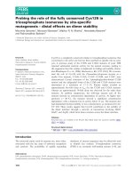
Fig. 4. SDF-1 induces the tyrosine-phosphorylation of SD-4 on HeLa cells. Confluent serum-starved HeLa cells were either stimulated (+) or
not (–) with SDF-1a. Equal amounts of proteins from whole cell extracts were immunoprecipitated with the indicated antibodies and equival-
ent amounts of IP samples were separated on 12% SDS ⁄ PAGE and immunoblotted using the indicated mAb or polyclonal antibodies. (A)
HeLa cells were stimulated (+) (lanes 2,4) or not (–) (lanes 1,3) for 10 min with 125 n
M SDF-1a. Cell lysates were immunoprecipitated either
with anti-CXCR4 Igs G19 (lanes 1,2) or anti-Ptyr mAb 4G10 (lanes 3,4). Western blots were developed, respectively, with anti-Ptyr mAb
4G10 (lanes 1,2) or anti-CXCR4 mAb 12G5 (lanes 3,4). (B) HeLa cells were stimulated (+) (lanes 2,3,4,6,8) or not (–) (lanes 1,5,7) with
125 n
M SDF-1a for the indicated time. Cell lysates were immunoprecipitated with anti-Ptyr mAb 4G10 (lanes 1–8). Western blots were
developed with anti-(SD-4) mAb 5G9 (lanes 1–4), anti-b-glycan Abs (lanes 5,6) or anti-(SD-2) Igs (lanes 7,8). (C,D) The intensities of the phos-
phorylated bands shown in A and B (lanes 1–4) were quantified in absorbance units by densitometric scanning and analyzed with
SCION IMAG-
ER
. They were expressed as ratios of the data observed for the SDF-1 stimulated cells relative to the untreated control cells. Each bar
represents the mean ± SE of triplicate determinations of an individual experiment. The significance of the differences as compared with
untreated control cells was assessed using Student’s t-test: **P < 0.05. The position of immunoglobulin chains is indicated by a star. Pro-
tein bands with changes in tyrosine phosphorylation state are indicated by arrows.
N. Charnaux et al. Syndecan-4 is an auxiliary receptor for SDF-1/CXCL12
FEBS Journal 272 (2005) 1937–1951 ª 2005 FEBS 1941
B
A
C
Fig. 5. Heparan sulfate is involved in the tyrosine phosphorylation of SD-4 induced by SDF-1 on HeLa cells. (A) Upper panel: HeLa cells were
either stimulated (+) (lanes 2 and 4) or not (–) (lanes 1 and 3) for 10 min with 125 n
M SDF-1a. In some experiments, cells were pretreated in
parallel with heparitinases I and III mixture (lanes 3 and 4). Lysates were then immunoprecipitated with anti-(SD-4) mAb 5G9. Western blots
were developed with anti-Ptyr mAb 4G10 (lanes 1–4). Lanes 5 and 6 confirm the equal loading of samples by reprobing the polyvinylidene
difluoride membrane with anti-SD-4 5G9 mAb. The position of the immunoglobulin chains is indicated by a star. (B) HeLa cells were stimula-
ted (+) (lanes 2, 4, 6 and 8) or not (–) (lanes 1, 3, 5 and 7) for 10 min with 125 n
M SDF-1a. Cells lysates were immunoprecipitated with anti-
Ptyr 4G10 mAb (lanes 1–4) or with anti-SD-4 5G9 mAbs (lanes 5–8). The IPs were treated with heparitinases I, III, and chondroitinase ABC.
Western blots were developed, respectively, with anti-SD-4 5G9 mAb (lanes 1 and 2), anti-(SD-1) DL-101 mAb (lanes 3 and 4), anti-Ptyr
4G10 mAb (lanes 5 and 6) or the isotype IgG2b (lanes 7 and 8). (C) Upper panel: HeLa cells were stimulated (+) (lanes 2 and 3) or not (–)
(lane 1) for 10 min with 125 n
M SDF-1a. In some experiments, cells were pretreated, in parallel, with heparitinases I and III (lane 3). Lysates
were then immunoprecipitated with anti-(SD-4) mAb 5G9. Western blots were developed with anti-CXCR4 mAb 12G5. Lower panels in (A)
and (C): The data shown in (A) (lanes 1–4) and in (C) were quantified in absorbance units by densitometric scanning and analyzed with
SCION
IMAGER
. They were expressed as the ratios of the data observed for the SDF-1 stimulated cells relative to those observed for the correspond-
ing unstimulated, control cells. Each bar represents the mean ± SE of triplicate determinations of an individual experiment. The significance
of the differences as compared either with controls or with heparitinase-treated cells was assessed using a t-test. **P < 0.05.
Syndecan-4 is an auxiliary receptor for SDF-1/CXCL12 N. Charnaux et al.
1942 FEBS Journal 272 (2005) 1937–1951 ª 2005 FEBS
mixture of heparitinases I and III, and chondroitinase
ABC, and then eluted from the beads. The eluates
were electroblotted and revealed, respectively, with
anti-SD-4 mAb 5G9 and anti-Ptyr mAb 4G10. Pro-
teins of 50–55 kDa which increased significantly after
stimulation of the cells by SDF-1 were revealed. No
immunoreactivity was detected using either the isotype
or anti-SD-1 mAb DL-101, anti-SD-2 and anti-(beta-
glycan) Igs (Fig. 5B and data not shown).
We then used coimmunoprecipitation experiments to
further analyse the physical association of CXCR4 and
SD-4. The anti-SD-4 5G9 IPs of unstimulated as well
as SDF-1-stimulated HeLa cells lysates, respectively,
were characterized by the presence of 48 kDa proteins
and of several other minor proteins of apparent
molecular masses > 48 kDa, all immunoreactive with
12G5 (Fig. 5C, lanes 1 and 2). Therefore, the SDF-1-
independent heteromeric complex between CXCR4 and
SD-4 (Fig. 5C, lane 1) is still present if the cells are sti-
mulated by the chemokine (Fig. 5C, lane 2 vs. 1).
The tyrosine phosphorylation of SD-4 induced
by SDF-1 on HeLa cells depends on the HS chains
of this PG
To examine whether the tyrosine phosphorylation of
SD-4 induced by SDF-1 on HeLa cells depends on
HS, we treated these cells with mixtures of heparitinase
I and III prior to their stimulation by SDF-1. To pre-
serve cell viability, concentrations of heparitinases were
lower than those used to treat the IPs. The efficiency
of the enzymes was investigated: if the cells were incu-
bated in enzyme-free medium and then stimulated with
SDF-1, the 5G9 IPs revealed with 10E4 showed, as
expected, the 110–200 kDa broad smear, described
above; however, if the cells were pretreated with hepari-
tinases I and III, this smear was no longer present
(data not shown). Moreover, this heparitinases pre-
treatment of the cells prevented in a significant manner
the tyrosine-phosphorylation of SD-4 induced by
SDF-1, as assessed by the anti-SD-4 5G9 IPs revealed
with anti-Ptyr 4G10 mAb (Fig. 5A, lane 4 vs. 3). In
these experiments, the apparent relative molecular
masses of most tyrosine-phosphorylated SD-4 mole-
cules were also decreased, as expected [17] (Fig. 5A,
lanes 3 and 4 vs. 2).
The homo- or hetero-oligomerization of CXCR4
induced by SDF-1 on HeLa cells is prevented by
heparitinases I and III pretreatment of these cells
Heparitinases I and III pretreatment of the HeLa cells
also significantly prevented the homo- or hetero- oligo-
merization of CXCR4 induced by SDF-1 on HeLa cells,
as assessed by the anti-SD-4 5G9 IP of the cell lysates
revealed with anti-CXCR4 mAb 12G5 (Fig. 5C, lane 3
vs. 2). This indicates that the HS-dependent binding of
SDF-1 to SD-4 enables the chemokine to induce the
homo- or hetero-oligomerization of its GPCR.
The physical association of tyrosine-phosphoryl-
ated SD-4 with tyrosine phosphorylated CXCR4
does not depend on GAGs chains
When anti-CXCR4 G19 IPs of the SDF-1 stimulated
cell lysates were treated with heparitinases I and III,
and chondroitinase ABC mixture, both SD-4 and
CXCR4 remained on the beads, as assessed by their
respective revelation with 12G5 and 5G9 (data not
shown). This suggests that GAG-dependent interac-
tions are not involved in these physical associations.
Finally, in all the experiments described above,
results of immunoprecipitation of cell lysates with iso-
type-matched control antibodies (data not shown) rule
out nonspecific protein association with membrane
components under our experimental conditions.
The activation of p44
/
p42 MAPK and JNK
/
SAP
kinase by SDF-1 on HeLa cells is HS- and
SD-4- dependent
To analyze some of the transduction pathways induced
by SDF-1 on HeLa cells, whole cell extracts from
either unstimulated or stimulated HeLa cells were elec-
troblotted and revealed using phospho-specific anti-
p44 ⁄ p42 mitogen-activated protein kinase (MAPK) or
anti-p46 ⁄ p54-Jun N-terminal ⁄ stress-activated protein
kinase (JNK ⁄ SAP kinase) Abs, respectively. Parallel
immunoblottings with anti-total polyclonal Abs
confirmed equal loading of the samples (Fig. 6). As
expected [24–26], SDF-1a and phorbol 12-myristate-
13-acetate (PMA) induced a rapid activation of p44 ⁄ 42
MAPK and JNK ⁄ SAP kinase signaling in HeLa cells
by increasing phosphorylations of the respective pro-
teins (Fig. 6). This effect was time and concentration-
dependent: It rose from 3 nm up to 125 nm SDF-1a
and if the time of incubation with the chemokine was
enhanced from 5 to 15 min. On the contrary, these
phosphorylations decreased if the time of incubation
with the chemokine was enhanced up to 30 min
(Fig. 6A). According to these results, the cells were
incubated for 15 min in the presence of 125 nm of
SDF-1 in the following experiments (Fig. 6B). In these
conditions, pretreating these cells with heparitinases I
and III significantly decreased these SDF-1-induced
phosphorylations (Fig. 6B) (P<0.05).
N. Charnaux et al. Syndecan-4 is an auxiliary receptor for SDF-1/CXCL12
FEBS Journal 272 (2005) 1937–1951 ª 2005 FEBS 1943
As expected [21,26,27], SDF-1 also stimulates intra-
cellular calcium mobilization in HeLa cells (Fig. 7A).
However, enzymatic removal of HS from the surface
of these cells did not affect this increased fluorescence
intensity observed in dye-loaded cells (mean ±
SE ¼ 101 ± 21, n ¼ 30) as compared to untreated
control cells (mean ± SE ¼ 97 ± 16, n ¼ 32), or the
percentage of SDF-1 responding cells (Fig. 7A,B and
data not shown).
Transfection of HeLa cells with SD-4 double-stran-
ded RNA (SD-4 dsRNA) resulted, as expected, in a
SD-4 mRNA downregulation reaching 80% reduc-
tion on day 3, while the mRNAs of SD-1, SD-2 and
CXCR4 were not changed (Fig. 8A). Moreover, when
measuring the expressions of these proteins by FACS in
these transfected cells, we found a 65% downregulation
of SD-4 expression, while SD-1, SD-2, beta-glycan or
CXCR4 expressions remained unchanged as expected
400
300
200
100
300
200
100
0
phosphorylation level
(% versus control)
phosphorylation level
(% versus control)
**
**
A
B
Fig. 6. Heparan sulfate is involved in the activation of MAPK induced by SDF-1 stimulation of HeLa cells. (A) Serum-starved HeLa cells were
either stimulated or not with 3 n
M or 125 nM SDF-1a for 5, 15 and 30 min, and then analyzed for p44 ⁄ p42 MAPK and JUN ⁄ SAPK activations.
(B) Upper panel: Untreated (–) or heparitinases I- and III-treated (+) HeLa cells were either stimulated or not for 10 min with PMA (0.5 l
M)or
SDF-1a (125 n
M).Whole cell extracts were separated on 12% SDS ⁄ PAGE and immunoblotted using either phosphospecific anti-(p44 ⁄ p42
MAPK) or phosphospecific p46 ⁄ p54-SAPK ⁄ JNK rabbit polyclonal antibodies. Parallel immunoblottings with anti-(total p44 ⁄ p42 MAPK) or anti-
(total p46 ⁄ p54-SAPK ⁄ JNK) polyclonal antibodies, respectively, confirmed equal loading of samples. Lower panel in (B): The results were
quantified by densitometric scanning and analyzed with
SCION IMAGER. For each lane, data were expressed as p44 ⁄ p42 or SAPK ⁄ JNK phos-
phorylated proteins over total proteins in absorbance units. The amount of MAPK (p44 ⁄ p42 or SAPK ⁄ JNK) phosphorylation in the SDF-1-sti-
mulated cells was calculated according to the level of phosphorylated MAPK proteins in unstimulated control cells, which was considered as
100%. Each bar represents the mean ± SE of triplicate determination of an individual experiment. The significance of the differences
between the SDF-1-stimulated cells and the corresponding heparitinases treated cells was assessed using a t -test. **P < 0.05.
Syndecan-4 is an auxiliary receptor for SDF-1/CXCL12 N. Charnaux et al.
1944 FEBS Journal 272 (2005) 1937–1951 ª 2005 FEBS
(Fig. 8B–D and data not shown). To monitor
the sequence specificity for SD-4 RNA interference,
mutSD-4 dsRNAs was used as a control. The mutSD-
4 dsRNA construct caused no significant reduction of
SD-4 mRNA and protein expressions, concordant with
previous reports on RNA interference methodology
[28,29] (Fig. 8A and data not shown) (P ¼ 0.11). We
then observed that both p44 ⁄ p42 MAPK and
JNK ⁄ SAP kinase activations were significantly reduced
after the knockdown of SD-4 upon SDF-1a stimula-
tion, as compared with the data observed in mock-
transfected cells and in cells transfected with mutSD-4
dsRNA, respectively (Fig. 8E) (P<0.05). By contrast,
under the same conditions, no change in the Ca
2+
mobilization induced by SDF-1 after the knockdown
of SD-4 on HeLa cells was observed (Fig. 7C,D).
Discussion
CXCR4 and SDF-1 play pivotal roles in many diseases
[5–8,30–32]. SDF-1 binding to GAGs, especially HS,
has been demonstrated [17,33,34]. Moreover, SDF-1
forms complexes on CXCR4-positive cells with
CXCR4 as expected and also with SD-4, but not with
SD-1, SD-2, beta-glycan or CD44 [17]. Furthermore,
an SDF-1-independent heteromeric complex between
CXCR4 and SD-4 occurs on these cells, but not with
SD-1, SD-2, beta-glycan or CD44 [17]. Therefore,
SDF-1 may bind both its GPCR CXCR4 and SD-4.
However, whether SDF-1 directly binds SD-4 has not
been demonstrated previously. We show here a direct
binding of SDF-1 to electroblotted SD-4 enriched from
HeLa cell lysates. The fact that no binding of the
chemokine to SD-1, SD-2, beta-glycan or CD44 was
detected strongly argues for the selectivity of this bind-
ing. We then examined whether SDF-1 is associated
with SD-4 at the plasma membranes of intact HeLa
cells. By using both confocal and electron microscopy
analysis, we show strong evidence for the occurrence
of a colocalization between SDF-1 and SD-4 at the
HeLa cell plasma membrane. The fact that in the same
conditions, no colocalization of SDF-1 with another
PG, SD-1, was observed, argues further for the selec-
tivity of this association. Therefore, our findings
observed at the molecular level were strengthened by
experiments performed at the cellular level.
Thereafter, we asked whether GAGs are involved in
SDF-1 binding to SD-4. By pretreating the electroblot-
ted PGs from the HeLa cells with heparitinase I and
III and chondroitinase ABC mixture, we demonstrate
the strong GAG dependency of this binding. However,
our data do not rule out the additional involvement of
protein–protein interactions between SDF-1 and the
SD-4 core protein. Indeed, while the SD core proteins
share a high degree of conservation in their short cyto-
plasmic and transmembrane domains, in contrast their
120
60
0
120
60
0
050100
0 50 100
time (sec)
50 100
time (sec)
time (sec)
050100
time (sec)
fluorescence intensity
fluorescence intensity
120
60
0
fluorescence intensity
120
60
0
fluorescence intensity
heparitinases I and III treated HeLa cell
untreated HeLa cell
SD-4 dsRNA HeLa cell
mock-transfected
HeLa cell
AB
CD
Fig. 7. Heparan sulfate is not involved in int-
racellular Ca
2+
mobilization induced by SDF-
1 on HeLa cells. Untreated HeLa cells (A),
heparitinases I- and III-treated HeLa cells
(B), mock-transfected HeLa cells (C), or
SD-4 dsRNA transfected HeLa cells (D) were
loaded for 30 min with Fluo-3 and then
stimulated with SDF-1a (125 n
M), as indica-
ted by black arrows. The plots show the
variations of the fluorescence intensity
(expressed in arbitrary units), measured
overtime within the analyzed cells. Data
are representative of three individual
experiments.
N. Charnaux et al. Syndecan-4 is an auxiliary receptor for SDF-1/CXCL12
FEBS Journal 272 (2005) 1937–1951 ª 2005 FEBS 1945
extracellular domains are divergent with the exception
of consensus sites for GAG attachment [15,35].
The participation of the SD-4 ectoplasmic domain in
SDF-1 binding raises the question whether this binding
is accompanied by intracellular modifications of SD-4
such as tyrosine phosphorylation, which plays critical
role in a variety of cellular processes. We have therefore
asked whether SD-4 functions as an SDF-1 signaling
molecule. For this purpose, we investigated whether
SDF-1 stimulation of HeLa cells induces an increase in
the tyrosine phosphorylation of SD-4, besides that of
CXCR4 which has already been reported [21–23]. The
SD cytoplasmic domains contain four conserved tyro-
sine residues, two of which are in favorable sequences
for phosphorylation [36]. Endogenous tyrosine phos-
phorylation of SDs has already been detected while
most cell surface SDs are phosphorylated following
treatment with the tyrosine phosphatase inhibitor per-
vanadate [37]. Tyrosine phosphorylation of the SD
cytoplasmic domain may be a common mechanism for
regulating SD activity. In this study, immunoprecipita-
tion experiments using anti-Ptyr, anti-CXCR4 and anti-
(SD-4) mAbs show for the first time that besides the
tyrosine phosphorylation of CXCR4, tyrosine phos-
phorylation of SD-4 occurs in response to SDF-1 sti-
mulation of HeLa cells. This tyrosine phosphorylation
depends on the time of incubation of the cells with the
chemokine: marginal for 2-min incubation, significant
A
D
E
BC
10
0
0
32
10
1
10
2
10
3
10
4
10
0
0
32
0
32
10
1
10
2
10
3
10
0
10
1
10
2
10
3
10
4
IgGl
IgGl
(SD-4ds RNA)
SD-1 (SD-4 ds RNA)
SD-1 (mocktransfected)
(mocktransfected)
Fig. 8. SD-4 is involved in SDF-1 activation of MAPK pathways. HeLa cells were transfected with either SD-4 dsRNAs or MutSD-4 dsRNA or
were mock-transfected. (A) Left panel: HeLa cells were analyzed for SD-4, SD-1, SD-2, CXCR4 specific mRNA, by semiquantitative RT-PCR,
3 days post transfection. To normalize for input of total RNA, GAPDH mRNA was also determined. Right panel: SD-4 mRNA levels were
quantified by densitometric scanning and analyzed with
SCION IMAGER. Results are depicted relative to mock-transfected control. Each bar rep-
resents the mean ± SE of triplicate determination of an individual experiment. The significance of the differences as compared to mock-
transfected control cells was assessed using a t-test. **P < 0.05. (B, C, D) HeLa cells were analyzed for (B) SD-4 (C) SD-1 and (D) CXCR4
protein expressions by FACS analysis, 3 days post transfection. Reactivity was compared to an isotype-matched control mAb. (E) Upper
panel: HeLa cells were treated for 15 min with 125 n
M SDF-1a, 3 days post-transfection. Whole cell extracts were separated on 12%
SDS ⁄ PAGE and analyzed by immunoblot using phosphospecific anti-(p44 ⁄ p42 MAPK) or phosphospecific p46 ⁄ p54-SAPK ⁄ JNK polyclonal rab-
bit antibodies, respectively. Parallel immunoblotting with anti-(total p44 ⁄ p42 MAPK) or anti-(total p46 ⁄ p54-SAPK ⁄ JNK) polyclonal antibodies
was performed to confirm equal loading of samples. Lower panel: The results were quantified by densitometric scanning and analyzed with
SCION IMAGER. For each lane, data were expressed as p44 ⁄ p42 MAPK or SAPK ⁄ JNK phosphorylated proteins over total proteins in absorbance
units. The amount of MAPK (p44 ⁄ p42 or SAPK ⁄ JNK) phosphorylations in the SDF-1-stimulated cells was calculated according to the level of
phosphorylated MAPK proteins in untreated control cells, which was considered as 100%. Each bar represents the mean ± SE of triplicate
determination of an individual experiment. The significance of the differences between the phosphorylation states of the SDF-1a-stimulated,
SD-4dsRNA- transfected cells and those of the SDF-1a-stimulated, mock-transfected cells was assessed using a t-test. **P < 0.05.
Syndecan-4 is an auxiliary receptor for SDF-1/CXCL12 N. Charnaux et al.
1946 FEBS Journal 272 (2005) 1937–1951 ª 2005 FEBS
for 10-min incubation in the presence of 125 nm SDF-
1. It also depends on the concentration of the chemo-
kine and is highly significant when 125 nm SDF-1 is
used, and marginal for 3 and 50 nm concentrations of
the chemokine. As SDF-1 stimulation of HeLa cells did
not induce the increase in the tyrosine phosphorylation
of other PGs, such as SD-1, SD-2 or beta-glycan, this
indicates the selectivity of this process. However, in
agreement with other results, marginal endogenous
tyrosine phosphorylation of SD-4 was observed [36]. In
addition, the data reported in these experiments indi-
cate that tyrosine-phosphorylated SD-4 coassociates
with tyrosine phosphorylated CXCR4, and suggest
GAG to be independent of this association.
As the tyrosine phosphorylation of intact SD core
proteins is not easily detected, we examined here the
protein core of tyrosine phosphorylated SD-4 after
digestion of the GAG chains with heparitinases I and
III and chondroitinase ABC. The 50–55 kDa proteins
which were revealed with anti-SD-4 mAb 5G9 and
with anti-Ptyr mAb 4G10 in the respective glycosami-
nidases-treated anti-Ptyr IP and anti-SD-4 IP probably
represent dimers of tyrosine-phosphorylated SD-4.
Similar apparent relative molecular masses of the SD-4
protein core were observed in the enriched PGs from
glycosaminidases-treated cell lysates.
We then observed firstly an increase in SD-4 tyro-
sine phosphorylation, and secondly that homo- or
hetero-oligomerization of CXCR4, induced by SDF-1
on HeLa cells, was prevented if the cells were pre-
treated with heparitinases I and III. This indicates the
involvement of HS in these two events.
In this study, in parallel experiments, either the cells
were treated with heparitinases I and III or the IPs
were treated with three glycosaminidases, heparitinases
I and III and chondroitinase ABC. To preserve cell
viability, lower concentrations of heparitinases were
used to treat the cells than the IPs. According to these
different conditions, GAGs, especially chondroitin sul-
fates, were still present on SD-4, if the enzyme treat-
ment was performed on the cells. This explains why
incomplete deglycanation of SD-4 was observed if the
cells were treated with heparitinases.
Finally, we asked whether HS and SD-4 were
involved in other SDF-1-induced cellular activation
signals. As SDF-1 binding to CXCR4 activates
p44 ⁄ p42 MAPK and JNK ⁄ SAP kinases and calcium
mobilization [21,24–27], we compared the activation
on either untreated or heparitinase I and III-treated
HeLa cells. In parallel, we investigated whether the
reduction of expression of SD-4 on HeLa cells by the
use of RNA interference prevented these activations.
HS removal from HeLa cells or decreasing endogenous
SD-4 significantly reduced the phosphorylations of
p44 ⁄ p42 MAPK and JNK ⁄ SAP kinases induced by
SDF-1. By contrast, these treatments did not change
the calcium mobilization triggered by the chemokine.
These data indicate that HS and SD-4 are selectively
required, at least partly and either directly or indi-
rectly, for the activation of p44 ⁄ p42 MAPK and
JNK ⁄ SAP kinases by SDF-1 on HeLa cells.
In conclusion, this study strongly suggests that
1-SD-4 behaves as an SDF-1 receptor selectively
involved in transduction pathways induced by SDF-1
on HeLa cells and 2-HS play a role in these events.
Whether these observations correlate with a biological
activity of SDF-1 deserves further study.
Experimental procedures
Cell culture
HeLa cells were cultured in DMEM (Invitrogen Corp.,
Paris, France) containing 10% fetal bovine serum (FBS;
Biowhittaker, Paris, France) and l-glutamine (2 mm; Invi-
trogen Corp.), and split twice a week.
Flow cytometry
Flow cytometry was performed as described [17,38,39],
using anti-SD-1 mAb DL-101 (mouse IgG-1; clone DL-101;
specific for the ectodomain of SD-1 of human origin), anti-
(SD-4) mAb 5G9 (mouse IgG2a; clone 5G9; specific for the
ectodomain of SD-4 of human origin); anti-(SD-2) (goat
IgG; specific for the C-terminal domain of syndecan-2 of
human origin) (all from Santa Cruz Biotechnology Inc,
Santa Cruz, CA, USA) or anti-(beta-glycan) Igs (goat IgG;
R & D systems, Abingdon, UK), anti CD44 mAb (mouse
IgG2a; Serotec, Oxford, UK), anti-CXCR4 mAb 12G5
(mouse IgG2a; specific for the second extracellular domain
of CXCR4; BD Bioscience Pharmingen, San Diego, USA),
or their isotypes (mouse IgG1, IgG2a or goat IgG, Jackson
Immunoresearch, Laboratories Inc. (Baltimore, MD, USA)
or BD Bioscience Pharmingen (San Diego, CA, USA), all
at 10 lg Æ mL
)1
.
Preparation of PGs
The PGs from HeLa cells lysates were enriched by anion
exchange chromatographies, as described previously [38].
Binding of biotinylated SDF-1 to electroblotted
PGs
Enriched PGs were loaded onto 12% SDS ⁄ polyacrylamide
gels (Invitrogen Corp.) under non reducing conditions
and blotted onto polyvinylidene difluoride membranes
N. Charnaux et al. Syndecan-4 is an auxiliary receptor for SDF-1/CXCL12
FEBS Journal 272 (2005) 1937–1951 ª 2005 FEBS 1947
(Amersham Pharmacia Biotech., Little Chalfont, Bucks,
UK) as described [39].
After blocking, strips were incubated for 1 h at room
temperature with biotinylated SDF-1a (6.25 nm; synthes-
ized by F. Baleux, Institut Pasteur, Paris, France; it was
verified that biotin incorporation did not modify the chem-
okine behavior). After washing, strips were reacted with
streptavidin-peroxidase (1.5 lgÆmL
)1
, Sigma-Aldrich, St
Louis, MO, USA) for 60 min at room temperature and
revealed by enhanced chemoluminescence (ECL) detection
(Amersham Pharmacia Biotech; or Supersignal West Dura
Extended, Pierce, Perbio Science, Brebie
`
res, France). Alter-
natively, strips were incubated for 1 h at room temperature
with anti-(SD-1) DL-101, anti-(SD-4) 5G9, anti-HS 10E4
or 3G10 mAbs (the latter two from Seikagaku, Tokyo,
Japan), anti-SD-2 or anti-beta-glycan Abs or their isotypes
(mouse IgG1, IgG2a, IgM, IgG2b or goat IgG). After
washing, strips were incubated with HRP-labeled anti-
mouse Ig (dilution of 1 : 5000; Amersham Pharmacia
Biotech) and developed. In some experiments, before the
binding assay, electroblotted PGs were digested for 18 h at
37 °C with 10 mUÆmL
)1
heparitinase III (heparin lyase; EC
4.2.2.7), 20 mUÆmL
)1
heparitinase I (heparan sulfate lyase;
EC 4.2.2.8) and 33 mUÆmL
)1
chondroitinase ABC (chon-
droitin ABC lyase; EC 4.2.2.4) (all from Sigma–Aldrich) as
described previously [39].
Immunofluorescence staining and confocal
microscopic analysis of the cells
To determine whether SDF-1 colocalizes with SD-4, HeLa
cells were incubated with anti-(SD-4) mAb 5G9, which was
revealed by Cy-3 donkey anti-mouse Igs (1 : 400; Jackson
Immunoresearch, West Grove, PA, USA). Cells were then
subsequently incubated for 1 h at 4 °C with 1-biotinylated
SDF-1a (10 lgÆmL
)1
). Cells were then labeled for 30 min at
4 °C with a streptavidin-Alexa Fluor 488 complex (1 : 100,
Molecular Probe, Inc., Eugene, OR, USA) and fixed with
paraformaldehyde (Sigma-Aldrich). As controls, cells were
incubated with the isotypes or biotinylated SDF-1a was
omitted. Cells were mounted and observed using a Zeiss
microscope (Axiovert 135 m; Carl Zeiss AG, Go
¨
ttingen,
Germany) in conjunction with a confocal laser scanning
unit (Zeiss LSM 410).
Immunoelectron microscopy
The HeLa cells were grown until 80% confluence in multi-
well chambers. After washes with phosphate buffered saline
(NaCl ⁄ P
i
), cells were incubated for 1 h at 4 °C with anti-
(SD-4) mAb 5G9 (20 lgÆmL
)1
) or anti-(SD-1) mAb DL-
101 (20 lgÆmL
)1
), which was followed by an incubation for
30 min at 4 ° C with a donkey anti-(mouse IgG) Ig linked
to 6-nm colloidal gold particles (Aurion, AA Wageningen,
the Netherlands). The cells were then incubated for 1 h at
4 °C with 1-biotinylated SDF-1a (20 lgÆmL
)1
), which was
followed by an incubation with streptavidin linked to
15 nm colloidal gold particles (Aurion). Cells were then
post-fixed with 2.5% (v ⁄ v) glutaraldehyde (Sigma-Aldrich),
dehydrated in graded ethanol series, and embedded in
epoxy resin. Ultra-thin sections (100 nm) were performed
and observed in transmission electron microscopy (CM-10,
Philips Medical Systems, Suresne, France) at high magnifi-
cation (· 27 500).
Immunoprecipitation and western blot analysis
HeLa cells were washed with NaCl ⁄ P
i
and cultured for
48 h in DMEM supplemented with 0.1% (v ⁄ v) FBS and
incubated for 0, 2, 10, 30 min at 37 °C with SDF-1a (0 up
to 125 nm). In some experiments, cells were pretreated for
2 h at 37 °C with heparitinase I (0.1 UÆmL
)1
) and hepari-
tinase III (0.2 UÆmL
)1
) mixture. It was verified that these
enzymes treatment had no effect on cell viability, as
assessed by Trypan blue exclusion dye. After washing the
cells with NaCl ⁄ P
i
supplemented with orthovanadate
(1 mm, Sigma-Aldrich), whole-cell extracts were prepared
by lysis of the cells in 20 mm Tris, 150 mm NaCl, 1 mm
orthovanadate, 1% (v ⁄ v) NP-40, 10 mm phenylmethylsulfo-
nyl fluoride, 5 mm iodoacetate, 25 mm phenanthrolin and
20 lgÆmL
)1
aprotinin (all from Sigma-Aldrich), The protein
concentration in whole-cell extracts was determined by the
BCA protein assay (Pierce). These extracts were then sup-
plemented with 10 mm dithiothreitol (Sigma-Aldrich).
Thereafter, equal amounts of proteins from these extracts
were incubated for 18 h at 4 °C with 100 lL of Protein G-
Sepharose beads (Amersham Pharmacia Biotech), precoated
either by anti-Ptyr mAb 4G10 (mouse IgG2b; Upstate Bio-
technology, Inc, Lake Placid, NY, USA), anti-SD-4 mAb
5G9, or anti-CXCR4 Abs G19 (goat IgG; specific for the
first extracellular domain of CXCR4; Santa Cruz Biotech-
nology) (each at 2 lg), as described previously [33,40,41].
To release bound components, beads were then boiled for
10 min with 300 lLof2· sample buffer for SDS ⁄ PAGE
and centrifuged (400 g; 5 min at 15 °C). Cell lysates, eluates
or eluted proteins were submitted to 12% SDS ⁄ PAGE
under non reducing conditions and then transferred onto
polyvinylidene difluoride membranes. Complexes were
revealed by incubation for 1 h at room temperature with
either anti-SD-4 5G9, anti-Ptyr 4G10, anti-CXCR4 12G5,
anti-HS 10E4 mAbs, anti-SD-2 Igs or anti-(beta-glycan) Igs
or their isotypes (all at 1 : 1000–1 : 2000). After washing,
strips were incubated with HRP-labeled anti-(mouse Ig) (at
1 : 5000) and revealed by ECL reagent. In some experi-
ments, the immunocomplexes immobilized on the beads
were treated by heparitinase I (1 UÆ mL
)1
), heparitinase III
(15 UÆmL
)1
) and chondroitinase ABC (5 UÆmL
)1
) mixture.
Beads were washed. Bound components were then eluted as
just described and then electroblotted. Results were quanti-
fied by scanning the exposed X-ray film with an Agfa scanner
Syndecan-4 is an auxiliary receptor for SDF-1/CXCL12 N. Charnaux et al.
1948 FEBS Journal 272 (2005) 1937–1951 ª 2005 FEBS
and analyzed using an area measurement from scion
imager. They were expressed as the ratios of the data
observed for the SDF-1 stimulated cells relative to those
observed for the corresponding untreated cells. The signifi-
cance of the differences was assessed with a t-test.
Activation of p44
/
p42 MAPK and JNK
/
SAP
kinases by SDF-1
HeLa cells were washed with NaCl ⁄ P
i
and cultured for
48 h in DMEM supplemented with 0.1% (v ⁄ v) FBS. In
some experiments, cells were pretreated for 2 h at 37 °C
with heparitinases I and III mixture, as just described. It
was verified that these enzymes treatment had no effect on
cell viability, as assessed by Trypan blue exclusion dye.
Thereafter, cells were incubated for 0–30 min at 37 °C with
SDF-1a (at 0–125 nm). After washing with NaCl ⁄ P
i
-ortho-
vanadate (1 mm), whole cell extracts were prepared [39].
The amount of protein of these extracts was controlled by
using a protein detection kit (Pierce). Equal amounts of
total proteins from these extracts were then submitted
to 10% SDS ⁄ PAGE and transferred to nitrocellulose
membrane (Amersham Pharmacia Biotech). MAPKs were
detected using polyclonal Abs, respectively, specific for
phospho-p44 ⁄ p42 [Thr202 ⁄ Tyr204], phospho-SAPK ⁄ JNK
[Thr183 ⁄ Tyr185], total p44 ⁄ p42 or total SAPK-JNK (rabbit
IgG; all from Cell Signaling Technology). Revelation was
performed as described [39]. Quantification of p44 ⁄ p42
MAPKs- and of SAPK ⁄ JNK phosphorylations was per-
formed by using the scion imager after autoradiography
scanning. For each sample, data were expressed as a ratio
of p44 ⁄ p42 MAPKs- or SAPK ⁄ JNK-phosphorylated pro-
teins over total proteins, in absorbance units. The
mean ± SE of triplicate determinations of individuals
experiments was calculated and the statistical significance
of the differences was evaluated using the Student’s t-test.
RNA interference
SD-4 gene-specific sense and antisense 21 nt single-stranded
RNAs (ssRNAs) with symmetric 2 nt-3¢(2¢-deoxy) thymi-
dine overhangs, were chemically synthesized, HPLC puri-
fied (Eurogentec, Seraing, Belgium) and used. RNA
sequences corresponding to SD-4 double strand RNA (SD-4
dsRNA) were: sense 5¢-GUU-GUC-CAU- CCC-UUG-GUG-
CdTdT-3¢; antisense 5¢-GCA-CCA-AGG-GAU-GGA-CAA-
CdTdT-3¢. To verify the sequence specificity of the RNA
interference, a SD-4 double-stranded RNA with one mis-
match (mutSD-4 dsRNA) was used as negative control
as described [28,29]: sense 5¢-GUU-GUC-GAU-CCC-
UUG-GUG-CdTdT-3¢; antisense 5¢-GCA-CCA-AGG-GAU-
CGA-CAA-CdTdT-3¢. For RNA interference experiments,
double-stranded RNAs were generated by mixing equi-
molar amounts (50 lm ) of sense and antisense ssRNAs in
annealing buffer (50 mm Tris, pH 7.5–8.0, 100 mm NaCl in
DEPC-treated water) for 1 min at 94 °C, followed by 60-min
incubation at 37 °C.
HeLa cells were tranfected with 300 n m dsRNA in
serum-free medium using Jetsi tranfectant reagent (Euro-
gentec) following the manufacturer’s instructions. Mock
cells were cultured in parallel and transfected with the
transfection mixture lacking dsRNA. Cells transfected with
SD-4 dsRNA or mutSD-4 dsRNA were used 3 days post-
tranfection for further analysis. The efficiency of the RNA
interference experiments was assayed by analyzing the
respective expressions of the mRNAs from SD-4, SD-2,
SD-1 and CXCR4. In parallel, the protein expressions of
SD-4, SD-1, SD-2, beta-glycan, CXCR4 were analyzed by
indirect immunofluorescence and FACS analysis. SD-4
mRNA, SD-1 mRNA, SD-2 mRNA and CXCR4 mRNA
and, to normalize for input of total RNA, glyceraldehyde
3-phosphodehydrogenase (GAPDH) mRNA were quanti-
fied by RT-PCR. Total cellular RNA was extracted, using
a Qiagen RNA ⁄ DNA Mini Kit (Qiagen S.A., Cortaboeuf,
France). For this purpose, confluent monolayers of mock-
transfected HeLa cells, mutSD-4 dsRNA-transfected HeLa
cells and from SD-4 dsRNA transfected HeLa cells were
previously grown in a six-well tissue culture. Reverse tran-
scription was performed using a Advantage RT-for-PCR
Kit (BD Biosciences Clontech, Le Pont-de-Claix, France).
The following synthetic SD-4 primers were used: – upper
primer CGA GAG ACT GAG GTC ATC GAC; lower pri-
mer: CGC GTA GAA CTC ATT GGT GG. These primers
were designed to amplify a 531 bp coding sequence of SD-
4. The following SD-1 primers were used: sense primer,
5¢-TCTGACAACTTCTCCGGCTC-3¢; antisense primer:
5¢-CCACTTCTGGCAGGACTACA-3¢; these primers were
designed to amplify a 211 bp coding sequence of SD-1. The
following synthetic SD-2 primers were used: sense primer
5¢-GGGAGCTGATGAGGATGTAG-3¢; antisense primer
5¢-CACTGGATGGTTTGCGTTCT-3¢. These primers were
designed to amplify a 394 bp coding sequence of SD-2. The
following synthetic CXCR4 primers were used: sense pri-
mer: 5¢-ATCTTTGCCAACGTCAGT-3¢; antisense primer:
5¢-TCACACCCTTGCTTGATG-3¢. These primers were
designed to amplify a 308 bp coding sequence of CXCR-4.
Optimum semiquantitative RT-PCR conditions were estab-
lished to remain within the exponential phase of amplifica-
tion curve. After 23 cycles of amplification, 30 lL were
electrophoresed in 2% agarose and analyzed.
Intracellular Ca
2+
mobilization
Possible changes in intracellular calcium concentration were
monitored using the fluorescent probe Fluo-3⁄ AM
(Molecular Probes). HeLa cells were washed in phenol red-
and sodium bicarbonate-free RPMI 1640 (Invitrogen Cor-
poration), supplemented by 25 mm Hepes (Sigma-Aldrich).
They were then incubated in the dark for 30 min at 37 °C,
with 2 lm Fluo-3 acetoxymethyl ester (Fluo-3 ⁄ AM) which
N. Charnaux et al. Syndecan-4 is an auxiliary receptor for SDF-1/CXCL12
FEBS Journal 272 (2005) 1937–1951 ª 2005 FEBS 1949
has been previously solubilized in dimethylsulfoxide (Sig-
ma-Aldrich), supplemented by Pluronic F-127 (20%)
(Molecular Probes). Cells were then washed with RPMI
1640 and maintained in this buffer at 20 °C in the dark for
5 min before their analysis. They were then incubated at
34 °C and stimulated or not by SDF-1a (125 nm). Fluores-
cence measurement was performed with a Biorad MRC 600
confocal laser scanning imaging system and its time course
ratiometric measurement software (TCSM), interfaced with
a Nikon Diaphot inverted microscope. Results are shown
as plots of the relative pixel intensities, measured over time
in each tested cell.
Acknowledgements
This work was supported by the Direction de la
Recherche et des Enseignements Doctoraux (Ministe
`
re
de l’Enseignement Superieur et de la Recherche), Uni-
versite
´
Paris XIII. We thank R. Fagard for his techni-
cal advices. We are grateful to J. Vaysse for her
suggestions.
References
1 Aiuti A, Webb IJ, Bleul C, Springer T & Gutierrez-
Ramos JC (1997) The chemokine SDF-1 is a chemoat-
tractant for human CD34+ hematopoietic progenitor
cells and provides a new mechanism to explain the
mobilization of CD34+ progenitors to peripheral
blood. J Exp Med 185, 111–120.
2 Voermans C, Von Gerritsen WR, dem Borne AE &
Van der Schoot CE (1999) Increased migration of cord
blood-derived CD34+ cells, as compared to bone mar-
row and mobilized peripheral blood CD34 cells across
uncoated or fibronectin-coated filters. Exp Hematol 27,
1806–1814.
3 Cinamon G, Grabovsky V, Winter E, Franitza S, Fei-
gelson S, Shamri R, Dwir O & Alon R (2001) Novel
chemokine functions in lymphocyte migration through
vascular endothelium under shear flow. J Leukoc Biol
69, 860–866.
4 Zlotnik A & Yoshie O (2000) Chemokines: a new classi-
fication system and their role in immunity. Immunity 12,
121–127.
5 Bleul CC, Farzan M, Choe H, Parolin C, Clark-Lewis
I, Sodroski J & Springer TA (1996) The lymphocyte
chemoattractant SDF-1 is a ligand for LESTR ⁄ fusin
and blocks HIV-1 entry. Nature 382, 829–833.
6 Oberlin E, Amara A, Bachelerie F, Bessia C, Virelizier
JL, Arenzana-Seisdedos F, Schwartz O, Heard JM,
Clark-Lewis I, Legler DF, Loetscher M, Baggiolini M
& Moser B (1996) The CXC chemokine SDF-1 is the
ligand for LESTR ⁄ fusin and prevents infection by
T-cell-line-adapted HIV-1. Nature 382, 833–835.
7 Pablos JL, Amara A, Bouloc A, Santiago B, Caruz A,
Galindo M, Delaunay T, Virelizier JL & Arenzana-Seis-
dedos F (1999) Stromal-derived factor is expressed by
dendritic cells and endotheliums in human skin. Am J
Pathol 155, 1577–1586.
8 Amara A, Gall SL, Schwartz O, Salamero J, Montes
M, Loetscher P, Baggiolini M, Virelizier JL & Are-
nzana-Seisdedos F (1997) HIV coreceptor downregula-
tion as antiviral principle: SDF-1alpha-dependent
internalization of the chemokine receptor CXCR4 con-
tributes to inhibition of HIV replication. J Exp Med
186, 139–146.
9 McGrath KE, Koniski AD, Maltby KM, McGann JK
& Palis J (1999) Embryonic expression and function of
the chemokine SDF-1 and its receptor, CXCR4. Dev
Biol 213, 442–456.
10 Bernfield M, Gotte M, Park PW, Reizes O, Fitzgerald
ML, Lincecum J & Zako M (1999) Functions of cell
surface heparan sulfate proteoglycans. Annu Rev Bio-
chem 68, 729–777.
11 Zimmermann P & David G (1999) The syndecans,
tuners of transmembrane signaling. FASEB J 13
(Suppl.), S91–S100.
12 Kim CW, Goldberger OA, Gallo RL & Bernfield M
(1994) Members of the syndecan family of heparan sul-
fate proteoglycans are expressed in distinct cell-, tissue-,
and development-specific patterns. Mol Biol Cell 5, 797–
805.
13 Sanderson RD, Turnbull JE, Gallagher JT & Lander
AD (1994) Fine structure of heparan sulfate regulates
syndecan-1 function and cell behavior. J Biol Chem 269,
13100–13106.
14 Clasper S, Vekemans S, Fiore M, Plebanski M, Words-
worth P, David G & Jackson DG (1999) Inducible
expression of the cell surface heparan sulfate proteogly-
can syndecan-2 (fibroglycan) on human activated
macrophages can regulate fibroblast growth factor
action. J Biol Chem 274, 24113–24123.
15 Carey DJ (1997) Syndecans: multifunctional cell-surface
co-receptors. Biochem J 327, 1–16.
16 Couchman JR, Chen L & Woods A (2001) Syndecans
and cell adhesion. Int Rev Cytol 207, 113–150.
17 Hamon M, Mbemba E, Charnaux N, Slimani H, Brule
S, Saffar L, Vassy R, Prost C, Lievre N, Starzec A &
Gattegno L (2004) A Syndecan-4 ⁄ CXCR4 complex
expressed on human primary lymphocytes and macr-
ophages, and HeLa cell line binds the CXC chemokine,
stromal cell derived factor-1 (SDF-1). Glycobiology 14,
311–323.
18 Chang TL, Gordon CJ, Roscic-Mrkic B, Power C,
Proudfoot AE, Moore JP & Trkola A (2002) Interaction
of the CC-chemokine RANTES with glycosaminogly-
cans activates a p44 ⁄ p42 mitogen-activated protein kin-
ase-dependent signaling pathway and enhances human
Syndecan-4 is an auxiliary receptor for SDF-1/CXCL12 N. Charnaux et al.
1950 FEBS Journal 272 (2005) 1937–1951 ª 2005 FEBS
immunodeficiency virus type 1 infectivity. J Virol 76,
2245–2254.
19 Oh ES, Woods A & Couchman JR (1997) Multimeriza-
tion of the cytoplasmic domain of syndecan-4 is
required for its ability to activate protein kinase C.
J Biol Chem 272, 11805–11811.
20 David G, Bai XM, Van der Schueren B, Cassiman JJ &
Van den Berghe H (1992) Developmental changes in
heparan sulfate expression: in situ detection with mAbs.
J Cell Biol 119, 961–975.
21 Mitra PA, Ethier MF, Mimori K, Kodys K, Shibuta K,
Mori M, Madison JM, Miller-Graziano C & Barnard
GF (2001) Loss of chemokine SDF-1a-mediated
CXCR4 signalling and receptor internalization in
human hepatoma cell line HepG2. Cell Signal 13, 311–
319.
22 Vila-Coro AJ, Rodriguez-Frade JM, Martin De Ana A,
Moreno-Ortiz MC, Martinez AC & Mellado M (1999)
The chemokine SDF-1a triggers CXCR4 receptor dime-
rization and activates the JAK ⁄ STAT pathway. Faseb J
13, 1699–1710.
23 Haribabu B, Richardson RM, Fisher I, Sozzani S, Pei-
per SC, Horuk R, Ali H & Snyderman R (1997) Regu-
lation of human chemokine receptors CXCR4. J Biol
Chem 272, 28728–28731.
24 Ganju RK, Brubaker SA, Meyer J, Dutt P, Yang Y,
Qin S, Newman W & Groopman JE (1998) The
a-chemokine, stromal cell-derived factor-1a , binds to
the transmembrane G-protein-coupled CXCR-4 receptor
and activates multiple signal transduction pathways.
J Biol Chem 273, 23169–23175.
25 Roland J, Murphy BJ, Ahr B, Robert-Hebmann V,
Delauzun V, Nye KE, Devaux C & Biard-Piechaczyk M
(2003) Role of the intracellular domains of CXCR4 in
SDF-1-mediated signaling. Blood 101, 399–406.
26 Eddleston J, Christiansen SC & Zuraw BL (2002) Func-
tional expression of the C-X-C chemokine receptor
CXCR4 by human bronchial epithelial cells: regulation
by proinflammatory mediators. J Immunol 169, 6445–
6451.
27 Amara A, Lorthioir O, Valenzuana A, Magerus A, The-
len M, Montes M, Virelizier JL, Delepierre M, Baleux
F, Lortat-Jacob H & Arenzana-Seisdedos F (1999)
Stromal cell-derived factor-1a associates with heparan
sulfates through the first beta-strand of the chemokine.
J Biol Chem 274, 23916–23925.
28 Takei Y, Kadomatsu K, Yuzawa Y, Matsuo S &
Muramatsu T (2004) A small interfering RNA targeting
vascular endothelial growth factor as cancer therapeu-
tics. Cancer Res 64, 3365–3370.
29 Elbashir SM, Martinez J, Patkaniowska A, Lendeckel
W & Tuschl T (2001) Functional anatomy of siRNAs
for mediating efficient RNAi in Drosophila melanogaster
embryo lysate. EMBO J 20, 6877–6888.
30 Premack BA & Schall TJ (1996) Chemokine receptors:
gateways to inflammation and infection. Nat Med 2,
1174–1178.
31 Murphy PM (2001) Chemokines and the molecular
basis of cancer metastasis. N Engl J Med 345, 833–835.
32 Nanki T, Hayashida K, El-Gabalawy HS, Suson S, Shi
K, Girschick HJ, Yavuz S & Lipsky PE (2000) Stromal
cell-derived factor-1-CXC chemokine receptor 4 interac-
tions plays a central role in CD4+ T cell accumulation
in rheumatoid arthritis synovium. J Immunol 165, 6590–
6598.
33 Mbemba E, Gluckman JC & Gattegno L (2000) Glycan
and glycosaminoglycan binding properties of stromal
cell-derived factor (SDF) -1alpha. Glycobiology 10, 21–
29.
34 Valenzuela-Fernandez A, Palanche T, Amara A, Mage-
rus A, Altmeyer R, Delaunay T, Virelizier JL, Baleux
F, Galzi JL & Arenzana-Seisdedos F (2001) Optimal
inhibition of X4 HIV isolates by the CXC chemokine
stromal cell-derived factor 1 alpha requires interaction
with cell surface heparan sulfate proteoglycans. J Biol
Chem 276, 26550–26558.
35 Beauvais DM & Rapraeger AC (2004) Syndecans in
tumor adhesion and signalling. Reprod Biol Endocrinol
2,3.
36 Ott VL & Rapraeger AC (1998) Tyrosine phosphoryla-
tion of syndecan-1 and -4 cytoplasmic domains in
adherent B82 fibroblasts. J Biol Chem 273, 35291–
35298.
37 Reiland J, Ott VL, Lebakken CS, Yeaman C, McCarthy
J & Rapraeger AC (1996) Pervanadate activation of
intracellular kinases leads to tyrosine phosphorylation
and shedding of syndecan-1. Biochem J 319, 39–47.
38 Slimani H, Charnaux N, Mbemba E, Saffar L, Vassy R,
Vita C & Gattegno L (2003) Binding of the CC-chemo-
kine RANTES to syndecan-1 and syndecan-4 expressed
on HeLa cells. Glycobiology 13, 623–634.
39 Charnaux N, Brule S, Chaigneau T, Saffar L, Sutton A,
Hamon M, Prost C, Lievre N, Vita C & Gattegno L
(2005) RANTES (CCL5) induces a CCR5-dependent
accelerated shedding of syndecan-1 (CD138) and synde-
can-4 from HeLa cells and forms complexes with the
shed ectodomains of these proteoglycans as well as with
those of CD44. Glycobiology 15, 119–130.
40 Mbemba E, Benjouad A, Saffar L & Gattegno L
(1999) Glycans and proteoglycans are involved in the
interactions of human immunodeficiency virus type 1
envelope glycoprotein and of SDF-1a with membrane
ligands of CD4 (+) CXCR4 (+) cells. Virology 265,
354–364.
41 Mbemba E, Saffar L & Gattegno L (2002) Role of
N-glycans and SDF)1a on the association of CD4 with
CXCR4 at the plasma membrane of monocytic cells
and blood lymphocytes. FEBS Lett 514, 209–213.
N. Charnaux et al. Syndecan-4 is an auxiliary receptor for SDF-1/CXCL12
FEBS Journal 272 (2005) 1937–1951 ª 2005 FEBS 1951