Báo cáo khoa học: Signal peptide hydrophobicity is critical for early stages in protein export by Bacillus subtilis ppt
Bạn đang xem bản rút gọn của tài liệu. Xem và tải ngay bản đầy đủ của tài liệu tại đây (339.73 KB, 14 trang )
Signal peptide hydrophobicity is critical for early stages in
protein export by Bacillus subtilis
Geeske Zanen
1
, Edith N. G. Houben
2
, Rob Meima
2,
*, Harold Tjalsma
3,
†, Jan D. H. Jongbloed
3,
‡,
Helga Westers
1,3
, Bauke Oudega
2
, Joen Luirink
2
, Jan Maarten van Dijl
1,
§ and Wim J. Quax
1
1 Department of Pharmaceutical Biology, University of Groningen, the Netherlands
2 Department of Molecular Microbiology, Vrije Universiteit, Amsterdam, the Netherlands
3 Department of Genetics, Groningen Biomolecular Sciences and Biotechnology Institute, University of Groningen, the Netherlands
Bacillus subtilis and Escherichia coli are used as proto-
type models for studies on protein translocation and
secretion in Gram-positive and Gram-negative bac-
teria, respectively. The absence or presence of a hydro-
phobic export signal, called signal peptide, determines
whether newly synthesized proteins are retained in the
cytoplasm or exported to other cellular compartments.
Signal peptides and their recognition by cytoplasmic
chaperones play a key role in membrane insertion of
membrane proteins and in targeting of secretory
Keywords
SRP; signal peptide; protein targeting;
protein translocation; trigger factor
Correspondence
W. J. Quax, Department of Pharmaceutical
Biology, University of Groningen, Antonius
Deusinglaan 1, 9713 AV Groningen,
the Netherlands
Fax: +31 50 3633000
Tel: +31 50 3632558
E-mail:
Present addresses
*DSM Food Specialties, Postbus 1, 2600
MA Delft, the Netherlands; †Department of
Clinical Chemistry, University Medical
Centre Nijmegen, PO Box 9101, 6500 HB
Nijmegen, the Netherlands; ‡Department of
Clinical Genetics, University Medical Center
of Groningen, P.O box 30001, 9700 RB,
Groningen, the Netherlands; §Laboratory of
Molecular Bacteriology, Department of
Medical Microbiology, University Medical
Center of Groningen and University of
Groningen, Hanzeplein 1, PO Box 30001,
9700 RB Groningen, the Netherlands
(Received 29 March 2005, revised 03 May
2005, accepted 18 May 2005)
doi:10.1111/j.1742-4658.2005.04777.x
Signal peptides that direct protein export in Bacillus subtilis are overall
more hydrophobic than signal peptides in Escherichia coli. To study the
importance of signal peptide hydrophobicity for protein export in both
organisms, the a-amylase AmyQ was provided with leucine-rich (high
hydrophobicity) or alanine-rich (low hydrophobicity) signal peptides.
AmyQ export was most efficiently directed by the authentic signal peptide,
both in E. coli and B. subtilis. The leucine-rich signal peptide directed
AmyQ export less efficiently in both organisms, as judged from pulse-chase
labelling experiments. Remarkably, the alanine-rich signal peptide was
functional in protein translocation only in E. coli. Cross-linking of in vitro
synthesized ribosome nascent chain complexes (RNCs) to cytoplasmic pro-
teins showed that signal peptide hydrophobicity is a critical determinant
for signal peptide binding to the Ffh component of the signal recognition
particle (SRP) or to trigger factor, not only in E. coli, but also in B. subtilis.
The results show that B. subtilis SRP can discriminate between signal
peptides with relatively high hydrophobicities. Interestingly, the B. subtilis
protein export machinery seems to be poorly adapted to handle alanine-
rich signal peptides with a low hydrophobicity. Thus, signal peptide hydro-
phobicity appears to be more critical for the efficiency of early stages in
protein export in B. subtilis than in E. coli.
Abbreviations
DSS, disuccinimidyl suberate; RNCs, ribosome nascent chain complexes; SRP, signal recognition particle; TF, trigger factor.
FEBS Journal 272 (2005) 4617–4630 ª 2005 FEBS 4617
proteins to the translocation machinery in the mem-
brane, the so-called Sec machinery in particular [1–4].
Signal peptides are usually sophisticated N-terminal
extensions, containing multipurpose functional infor-
mation. A signal peptide can be divided into three
distinct domains; the N-, H-, and C-domains [5,6]. The
N-domain interacts with the translocation machinery
and the negatively charged phospholipids in the lipid
bilayer of the membrane [7,8]. The H-domain can adopt
an a-helical conformation in the membrane due to a
stretch of hydrophobic residues [9]. To allow the forma-
tion of a hairpin-like structure that can insert into the
membrane, helix-breaking glycine or proline residues are
often present in the middle of the hydrophobic stretch.
Unlooping of this hairpin might result in the insertion
of the complete signal peptide into the membrane [8].
Analyses of the H-domain show that the hydrophobic
core is the dominant structure in determining signal
peptide function [10–12]. The C-domain contains the
cleavage site for specific signal peptidases that remove
signal peptides from the mature part of the exported
protein during or shortly after translocation [13,14].
Although the overall structure of signal peptides is
quite similar, small variations can result in export via
different targeting pathways [15–17]. Signal peptides
directing proteins into the signal recognition particle
(SRP)-dependent pathway have a significantly more
hydrophobic H-domain than those mediating SRP-
independent targeting, at least in E. coli [18,19].
Reduction of the net positive charge or the hydro-
phobicity of certain signal peptides decreases the
effectiveness of SRP recognition. However, in E. coli a
high degree of H-domain hydrophobicity can compen-
sate for the loss of basic residues in the N-domain and
restore SRP binding [20]. Signal peptides containing an
(S ⁄ T)RRXFLK motif in E. coli or an RRXFF motif
in B. subtilis (F is a hydrophobic residue, X can be
any residue) are candidates to be translocated via the
twin arginine translocation (Tat) pathway [15,21]. In
general, Tat-targeting signal peptides have H-domains
which are less hydrophobic than signal peptides that
target proteins to the Sec machinery [22]. Upon emer-
gence from the ribosome, the signal peptide of a
nascent secretory protein can be recognized by several
cytoplasmic chaperones and ⁄ or targeting factors, such
as Ffh or trigger factor (TF) [23]. In contrast to Ffh,
which is required for cotranslational protein export in
E. coli, the cytoplasmic chaperone SecB has mainly
been implicated in post-translational protein targeting.
For E. coli it has been shown that by increasing the
hydrophobicity of signal peptides, exported proteins
can be re-routed from SecB into the SRP pathway
[19,24,25]. Altogether, this means that different specifi-
city determinants are involved in early stages of pro-
tein export from the cytoplasm.
Most research on the interactions between signal pep-
tides and cytoplasmic chaperones has so far been per-
formed in E. coli. However, as shown by Collier, signal
peptides can behave differently in different hosts [26].
Notably, B. subtilis lacks a SecB homologue, the chaper-
one that is involved in post-translational targeting of the
secretory proteins in E. coli [2]. Moreover, signal pep-
tides of Gram-positive organisms are usually longer and
more hydrophobic than those of Gram-negative organ-
isms [2,27,28]. Until now, it is not known whether this
difference in hydrophobicity and length of signal pep-
tides represents a functional difference in these species.
In the present studies, we have addressed the effects
of major variations in signal peptide hydrophobicity
on translocation, processing, and signal peptide inter-
action with cytoplasmic chaperones using a combined
in vivo and in vitro approach in both E. coli and
B. subtilis. The results show interesting differences for
the translocation of an a-amylase of B. amyloliquefac-
iens (AmyQ) with altered signal peptides in these
organisms. Whereas E. coli translocates AmyQ with a
less hydrophobic alanine-rich signal peptide, even in a
secB mutant, B. subtilis accumulates the respective pre-
cursor intracellularly. Cross-linking studies show that
TF of B. subtilis interacts with the authentic signal
peptide of AmyQ, whereas Ffh and TF of E. coli com-
pete to interact with this signal peptide. Remarkably, a
more hydrophobic leucine-rich signal peptide resulted
in reduced AmyQ translocation efficiencies, both in
B. subtilis and E. coli . Taken together, these findings
suggest that the hydrophobicity of signal peptides is
more critical for early stages in protein translocation
in B. subtilis than in E. coli.
Results
Changing the hydrophobicity of the AmyQ signal
peptide
To study the effects of signal peptide hydrophobicity on
the export of the a-amylase AmyQ of B. amylolique-
faciens by E. coli or B. subtilis, plasmids were construc-
ted encoding AmyQ precursors with signal peptides of
distinct hydrophobicity. Specifically, an Ala-rich signal
peptide (MIQKRKRTVSLAAAAACAAAALQPITK
TSAVN) and a Leu-rich signal peptide (MIQKRKR
TVSLLLLLLCLLLLLQPITKTSAVN) were designed.
Hereafter, these mutant signal peptides are referred to
as Ala or Leu signal peptides. These signal peptides
have grand average of hydropathicity (Gravy) values
that are significantly lower (0.341 for the Ala signal
Secretory protein targeting in Bacillus subtilis G. Zanen et al.
4618 FEBS Journal 272 (2005) 4617–4630 ª 2005 FEBS
peptide) or higher (0.903 for the Leu signal peptide)
than that of the authentic AmyQ signal peptide (MIQK
RKRTVSFRLVLMCTLLFVSLPITKTSAVN; Gravy
value 0.591).
In vivo translocation and processing of a-amylase
in E. coli and B. subtilis
To study the effects of the different signal peptides on
in vivo translocation of AmyQ, E. coli TG90 and
B. subtilis 168 were transformed with the E. coli–
B. subtilis shuttle vectors pKTHM10, pKTHM101 or
pKTHM102. These vectors encode the authentic pre-
AmyQ, pre-AmyQ with the Ala signal peptide, and
pre-AmyQ with the Leu signal peptide, respectively.
Cells were grown overnight and samples were prepared
for western blotting experiments and immunodetection
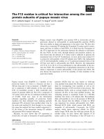
with specific antibodies against AmyQ. As shown in
Fig. 1A, mature AmyQ was detectable in cellular sam-
ples of E. coli, irrespective of the signal peptide used.
Pre-AmyQ was only detectable in significant amounts
when the Ala signal peptide was used, and it was
barely detectable when the Leu signal peptide was
used. When expressed in B. subtilis, mature AmyQ
was secreted into the growth medium when synthesized
with the authentic or Leu signal peptide. In contrast,
no mature AmyQ was secreted when this protein was
synthesized with the Ala signal peptide (Fig. 1B). To
verify whether the AmyQ secreted by B. subtilis was
active, an activity assay was performed that is based
on the degradation of starch in agar plates. As reflec-
ted by the formation of halos upon staining with
iodine vapour, active AmyQ was secreted when this
protein was provided with the authentic or Leu signal
peptide, but not when the Ala signal peptide was pre-
sent (Fig. 1C).
To examine the effects of signal peptide hydropho-
bicity on the kinetics of pre-AmyQ processing, pulse-
chase labelling experiments were performed with
B. subtilis 168 or E. coli TG90 cells producing AmyQ
with the authentic, Ala, or Leu signal peptides. After
pulse labelling of newly synthesized proteins with
[
35
S]methionine for 1 min, excess nonradioactive
methionine (chase) was added (t ¼ 0). After different
periods of chase, samples were taken from which
AmyQ was precipitated with specific antibodies. As
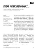
shown in Fig. 2A, the authentic pre-AmyQ was almost
completely processed after 5 min of chase when pro-
duced in E. coli. In contrast, processing of AmyQ pre-
cursors with the Leu or Ala signal peptides was
significantly less efficient. After 5 min chase, 46% or
53% of the AmyQ molecules synthesized with the Leu
or Ala signal peptides, respectively, were still in the
precursor form (note that pre-AmyQ with the Ala sig-
nal peptide has a lower mobility on SDS ⁄ PAGE than
pre-AmyQ with the authentic or Leu signal peptides).
In contrast, 45% of the authentic pre-AmyQ molecules
was processed to the mature form within 1 min of
chase. Processing of AmyQ with the Ala signal peptide
was so slow, that even after a chase of 30 min precur-
sor molecules were still detectable (data not shown).
The observation that, in E. coli, AmyQ precursors with
the Leu signal peptide were processed less efficiently
was unexpected, since Doud and coworkers have previ-
ously shown that signal peptides with increased hydro-
phobicity improved the export efficiency for PhoA in
this organism [29]. Also in B. subtilis, the processing of
AmyQ with the Leu signal peptide occurred at a lower
rate than that of AmyQ with the authentic signal pep-
tide (Fig. 2B). After 2 min of chase 53% of the AmyQ
with the Leu signal peptide was processed to the
mature form, whereas 71% of the AmyQ with the
authentic signal peptide was processed within this time
of chase. About 68% of the AmyQ molecules synthes-
ized with the Leu signal peptide were mature after
5 min of chase. A completely different result was
obtained for AmyQ synthesized with the Ala signal
peptide. While AmyQ precursors with this signal pep-
tide were processed in E. coli, no processing of these
precursors could be observed in B. subtilis (Fig. 2B)
and even after a chase of 60 min no mature AmyQ
was detected (data not shown). Notably, AmyQ mole-
cules with the authentic signal peptide were processed
A
B
C
Fig. 1. AmyQ production and secretion. (A) AmyQ production in
cells of E. coli as determined by western blotting using the proteins
from total cell extracts separated by SDS ⁄ PAGE. (B) AmyQ secre-
tion into the growth medium of B. subtilis as determined by west-
ern blotting using the proteins from culture supernatants separated
by SDS ⁄ PAGE. The images in A and B relate to equal numbers of
E. coli or B. subtilis cells, respectively. (C) Plate assay for AmyQ
secretion by B. subtilis. The signal peptides fused to AmyQ are
indicated. p, Pre-AmyQ; m, mature AmyQ.
G. Zanen et al. Secretory protein targeting in Bacillus subtilis
FEBS Journal 272 (2005) 4617–4630 ª 2005 FEBS 4619
more efficiently in B. subtilis than in E. coli, and the
same was true for AmyQ molecules with the Leu sig-
nal peptide.
The fact that no processing of AmyQ with the Ala
signal peptide could be detected in B. subtilis raised
the question whether this precursor was translocated
across the membrane. To determine the topology of
(pre)AmyQ at steady state, protoplasts of B. subtilis
cells were incubated with trypsin. In parallel, proto-
plasts were incubated without trypsin or with trypsin
plus Triton X-100. As shown in Fig. 3, cells producing
the authentic AmyQ or AmyQ with the Leu signal
peptide contained both precursor and mature forms of
AmyQ. Notably, the accumulation of pre-AmyQ in
wild-type cells of B. subtilis 168 is commonly observed,
despite the fact that this precursor is shown to be
processed efficiently in pulse-chase labelling experi-
ments [21,30,31]. In contrast to AmyQ with the
authentic or Leu signal peptides, all AmyQ synthesized
with the Ala signal peptide was present in the precur-
sor form. As previously shown, all AmyQ molecules
synthesized with the authentic signal peptide were
accessible to trypsin upon protoplasting of the cells
[31]. In contrast, the situation was slightly different for
AmyQ synthesized with the Leu signal peptide: while
all mature molecules were accessible to trypsin upon
protoplasting, a significant fraction of the pre-AmyQ
molecules remained inaccessible to trypsin. The latter
pre-AmyQ molecules were only degraded by trypsin in
the presence of Triton X-100, indicating that they were
protected against trypsin activity by the cytoplasmic
membrane. Strikingly, none of the AmyQ molecules
synthesized with the Ala signal peptide was accessible
to trypsin upon protoplasting. These precursor mole-
cules were, however, degraded by trypsin when the
protoplasts were lysed with Triton X-100. As controls
for these fractionation experiments, the lipoprotein
PrsA, which is localized at the membrane–cell wall
interface, and the cytoplasmic protein GroEL were
used. Figure 3 shows that, irrespective of the cells
used, the accessibility of PrsA and GroEL to trypsin
was consistent with the subcellular location of these
proteins. While all PrsA was accessible to trypsin upon
protoplasting, GroEL was only degraded by trypsin
when the protoplasts were lysed with Triton X-100.
Notably, microscopic inspection of the cells suggested
that none of the strains investigated contained AmyQ
inclusion bodies in the cytoplasm (data not shown).
Consistent with the fact that AmyQ molecules synthes-
ized with the authentic, Leu or Ala signal peptides
were processed in E. coli, subcellular localization
experiments in this organism revealed that all corres-
ponding precursor and mature AmyQ molecules were
accessible to trypsin upon spheroplasting (data not
shown). Taken together, these observations show that
AmyQ molecules with the Leu signal peptide are trans-
located across the cytoplasmic membranes of B. subtilis
and E. coli, but with a slightly lower efficiency than
AmyQ molecules with the authentic signal peptide. In
contrast, AmyQ molecules with the Ala signal peptide
are translocated across the cytoplasmic membrane in
E. coli, but not in B. subtilis.
Although the processing of the AmyQ precursor
containing the Leu signal peptide was slower than
that of wild type AmyQ in E. coli and in B. subtilis,
processing of the AmyQ precursor containing the Ala
signal peptide was only observed in E. coli. Since
E. coli contains the cytoplasmic chaperone SecB, which
is absent from B. subtilis, the influence of SecB on the
processing of AmyQ containing the Ala signal peptide
A
B
Fig. 2. Processing of pre-AmyQ. Processing
of AmyQ precursors with different signal
peptides in E. coli (A) or B. subtilis (B) was
analysed by pulse-chase labelling at 37 °C.
Cells were labelled with [
35
S]methionine for
1 min prior to chase with excess nonradio-
active methionine. Samples were withdrawn
at the times indicated. The presence of pre-
cursor or mature forms of AmyQ in cells
plus growth medium was visualized by
immunoprecipitation, SDS ⁄ PAGE and fluo-
rography. The percentage of processed
(mature) AmyQ relative to the total amount
of AmyQ (precursor + mature) in each lane
is indicated (%). The signal peptides fused
to AmyQ are indicated. p, Precursor;
m, mature AmyQ.
Secretory protein targeting in Bacillus subtilis G. Zanen et al.
4620 FEBS Journal 272 (2005) 4617–4630 ª 2005 FEBS
was investigated. Pulse-chase labelling experiments
with E. coli MC4100 and the corresponding secB
mutant strain were performed at 30 °C, because the
growth of both strains at 37 °C was severely impaired
when transformed with the plasmid for AmyQ-Ala
expression (note that this was not the case in E. coli
TG90). The results obtained with E. coli MC4100
showed a less efficient processing of AmyQ precursor
containing the Ala signal peptide at 30 °C, as com-
pared to the processing of this precursor in E. coli
TG90 at 37 °C (compare Fig. 2A and Fig. 4A). As
shown in Fig. 4A, the processing rate of AmyQ with
the Ala signal peptide was mildly reduced in secB
mutant cells as compared to cells of E. coli MC4100.
Compared to the SecB-dependent OmpA protein, the
effect of the absence of SecB on the processing of
AmyQ with the Ala signal peptide was less evident
(Fig. 4).
In vitro cross-linking of a-amylase nascent chains
in E. coli and B. subtilis
The influence of signal peptide hydrophobicity on its
interactions with E. coli and B. subtilis cytoplasmic
proteins was investigated by chemical cross-linking of
in vitro translated nascent chains. In this approach,
truncated mRNAs were translated in an E. coli trans-
lation lysate in the presence of [
35
S]methionine to
Fig. 3. Localization of AmyQ in B. subtilis. To analyse the subcellular localization of AmyQ molecules synthesized with different signal pep-
tides, cells of B. subtilis were grown overnight at 37 °C in TY medium, diluted 50-fold in fresh TY medium and incubated at 37 °C for 3 h
prior to protoplasting. Protoplasts were incubated for 30 min without further additions, in the presence of trypsin (T; 1 mgÆmL
)1
), or trypsin +
Triton X-100 (1%). Samples were used for SDS ⁄ PAGE and western blotting. Specific antibodies were used to detect AmyQ, PrsA, or GroEL.
The positions of (pre)AmyQ, PrsA, and GroEL (c), and degradation products of PrsA (d*) are indicated. The signal peptides fused to AmyQ
are indicated. A cartoon of the protoplasting and protease protection experiment is shown to illustrate the effects of trypsin (T) and Triton
X-100.
A
B
Fig. 4. Processing of AmyQ with the Ala
signal peptide in E. coli secB. Processing of
pre-AmyQ containing the Ala signal peptide
(A) and pro-OmpA (B) in E. coli MC4100
secB or the parental strain (wt) were ana-
lysed by pulse-chase labeling at 30 °C and
subsequent immunoprecipitation,
SDS ⁄ PAGE, and fluorography. Cells were
labelled with [
35
S]methionine for 1 min prior
to chase with excess nonradioactive methio-
nine. Samples were withdrawn at the times
indicated. The percentage of processed
(mature) AmyQ relative to the total amount
of AmyQ (precursor + mature) in each lane
is indicated (%). p, Precursor; m, mature.
G. Zanen et al. Secretory protein targeting in Bacillus subtilis
FEBS Journal 272 (2005) 4617–4630 ª 2005 FEBS 4621
generate radioactively labelled ribosome-nascent chain
complexes (RNCs). A C-terminal 4· methionine tag
was introduced into the nascent chains to increase the
labelling efficiency. The nascent chain corresponding
to the authentic preprotein comprised 101 amino
acids, while the nascent chain corresponding to the
Leu and Ala preproteins comprised 105 amino acids.
Thus, the lengths of these nascent chains allows opti-
mal cytoplasmic exposure of the signal peptides, tak-
ing into consideration that approximately 30 amino
acids will be located within the ribosome (schemati-
cally represented in Fig. 5A). The RNCs were purified
over a high-salt sucrose cushion to remove all loosely
associated E. coli components originating from the
translation lyate. Subsequently, they were either incu-
bated with crude E. coli MC4100, B. subtilis 168, or
B. subtilis DTF cell lysates. The latter strain lacks the
TF, which is known to interact with peptides emer-
ging from the ribosome [23]. The DTF strain was
used for these experiments, because no anti-
body against the B. subtilis TF is currently available.
As a negative control, the purified RNCs were incu-
bated with incubation buffer only. Interactions
between RNCs and cytoplasmic components of E. coli
or B. subtilis were fixed by adding the homobifunc-
tional lysine-lysine cross-linking reagent disuccinimidyl
suberate (DSS).
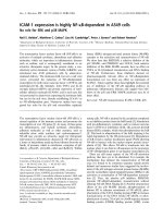
Incubation of AmyQ nascent chains containing
the authentic signal peptide with E. coli lysate in the
presence of DSS generated cross-linking adducts
of 25 kDa, 60 kDa, 68 kDa, and 80 kDa
(Fig. 5B, lane 3). The 25 kDa adduct could be immu-
noprecipitated using antiserum raised against the E. coli
ribosomal protein L23 (Fig. 5B, lane 6). In fact, cross-
linking to L23 was not only shown for RNCs with the
authentic signal peptide, but also for RNCs with the
Leu and Ala signal peptides (Fig. 5B, lanes 2–5, lanes
10–13, lanes 18–21). As shown by immunoprecipitation
A
B
Fig. 5. Cross-linking of AmyQ nascent chai-
ns to soluble E. coli and B. subtilis compo-
nents. The 101AmyQ wt, 105AmyQ Leu
and 105AmyQ Ala RNCs were synthesized
in an E. coli MC4100 translation lysate. (A)
Schematic representation of the translation
reactions. The different signal peptides used
and lysine residues (K) that may participate
in cross-linking reactions are indicated. (B)
After translation, the RNCs were purified
over a high-salt sucrose cushion, incubated
with crude E. coli MC4100, B. subtilis 168,
B. subtilis DTF cell lysates or incubation
buffer and treated with DSS. Cross-linking
was quenched by adding TCA ⁄ acetone.
Immunoprecipitations were subsequently
carried out as indicated in Experimental
procedures. IP, Immuno-precipitation; E,
crude cell lysate of E. coli MC4100; B, crude
cell lysate of B. subtilis 168; BD, crude cell
lysate of B. subtilis DTF; NC, nascent chain;
?, unknown cross-linking adducts;
*, cross-linking adducts with E. coli L23;
d, cross-linking adducts with E. coli Ffh;
s, cross-linking adducts with B. subtilis Ffh;
n
, cross-linking adducts with E. coli TF;
h, cross-linking adducts with B. subtilis TF.
Secretory protein targeting in Bacillus subtilis G. Zanen et al.
4622 FEBS Journal 272 (2005) 4617–4630 ª 2005 FEBS
with Ffh- and TF-specific antibodies, the 60 kDa
cross-linking adduct contained E. coli Ffh, while both
the 68 kDa and 80 kDa adducts contained the
E. coli TF (Fig. 5B, lanes 7, 8). Adducts of TF fre-
quently appear as a doublet [18,32], but it is not known
why the ratio between the immunoprecipitated AmyQ-
TF adducts differs from the ratio in the nonprecipitated
sample (Fig. 5B, lanes 3 and 8). Unfortunately, E. coli
SRP could not be removed completely from the ribo-
somes by high-salt treatment (Fig. 5B, lane 2 closed cir-
cle). Consequently, cross-links to E. coli Ffh were also
detected upon incubation of RNCs containing the
authentic signal peptide with B. subtilis 168 lysate in the
presence of DSS. Cross-linking of these RNCs to B. sub-
tilis Ffh could not be demonstrated by immunoprecipi-
tations using antibodies specific for B. subtilis Ffh (data
not shown). On the other hand, two dominant cross-
linking adducts of 69 kDa were detected upon incuba-
tion of authentic pre-AmyQ RNCs with B. subtilis 168
lysate (Fig. 5B, lane 4 open squares). Such adducts were
not observed after incubating these nascent chains with
B. subtilis DTF lysate in the presence of DSS (Fig. 5B,
lane 5), which implies that the 69 kDa adducts
represent cross-links to TF of B. subtilis. Interestingly,
incubation with the B. subtilis DTF lysate resulted in
40-kDa cross-linking adducts that were not observed
upon incubation with the B. subtilis 168 lysate (Fig. 5B,
lane 5 question mark). Unfortunately, the B. subtilis
protein(s) in these 40-kDa adducts could not be
identified.
Nascent chains of AmyQ with the Leu signal peptide
generated 60-kDa and 70-kDa cross-linking
adducts upon incubation with E. coli lysate in the pres-
ence of DSS (Fig. 5B, lane 10 closed circles), which
both represented cross-linking to E. coli Ffh (data not
shown). Cross-linking of these RNCs to E. coli Ffh was
even detected upon incubation with B. subtilis 168 ly-
sate in the presence of DSS (Fig. 5B, lane 14). This
cross-linked E. coli Ffh was derived from the transla-
tion lysate, despite the high-salt purification. Never-
theless, as shown by immunoprecipitation with specific
antibodies, a 60-kDa cross-linking adduct containing
B. subtilis Ffh was formed upon incubation with B. sub-
tilis 168 lysate in the presence of DSS (Fig. 5B, lane
15). Note that the antibodies against Ffh of B. subtilis
do not cross-react with Ffh of E. coli (Fig. 5B, lane 16).
The 48-kDa cross-linking adduct obtained upon incu-
bation of RNCs containing the Leu signal peptide with
the B. subtilis 168 lysate was not identified (Fig. 5B,
lane 12 question mark). Interestingly, no evidence for
specific cross-links between RNCs with the Leu signal
peptide and the B. subtilis or E. coli TFs was obtained
(Fig. 5B, lanes 11–13 and data not shown).
Finally, nascent chains of AmyQ containing the Ala
signal peptide generated strong cross-links to E. coli
and B. subtilis TF (Fig. 5B, lane 19–21 open and
closed squares), while neither cross-links to E. coli Ffh
nor B. subtilis Ffh were observed (data not shown).
Incubation of these nascent chains with the B. subtilis
DTF lysate again generated unidentified 40-kDa
cross-linking adducts (Fig. 5B, lane 21 question mark).
In conclusion, these findings show that RNCs contain-
ing the authentic signal peptide can be cross-linked
with L23, Ffh, and TF of E. coli and with TF of
B. subtilis. RNCs containing the highly hydrophobic
Leu signal peptide can be cross-linked with L23 of
E. coli, Ffh of E. coli and B. subtilis, but not detecta-
bly with TF of these organisms. In contrast, RNCs
containing the mildly hydrophobic Ala signal peptide
are efficiently cross-linked with L23 of E. coli,TFof
E. coli and B. subtilis, but not detectably with Ffh of
these organisms.
Discussion
Several studies indicate that signal peptide hydrophob-
icity is an important determinant for SRP-mediated
protein targeting to the E. coli inner membrane [18,33].
Cross-linking of nascent PhoA-derivatives revealed an
almost linear correlation between hydrophobicity and
SRP cross-linking [18]. In addition, hydrophobic alter-
ations in the signal peptides of SecB-dependent pro-
teins, re-routed these proteins into the SRP pathway
[19,24,25]. Precursor proteins from Gram-positive bac-
teria contain signal peptides that are usually longer
and more hydrophobic than the signal peptides of pre-
cursor proteins from Gram-negative bacteria [2,27,28].
It was therefore hypothesized that the higher hydro-
phobicity of signal peptides in Gram-positive bacteria,
lacking SecB, has evolved as an adaptation to the
SRP-dependent translocation pathway [2].
Changes in hydrophobicity of the signal peptide of
a-amylase AmyQ seem to have different effects on
the translocation of pre-AmyQ in E. coli or B. subtilis.
For E. coli cells, changing the alanine or leucine
content and, consequently, the hydrophobicity of the
signal peptide did not lead to major translocation
defects. However, processing was less efficient for
AmyQ precursors containing the Leu or Ala signal
peptides when compared to AmyQ with the authentic
signal peptide. Importantly, significant amounts of
mature AmyQ were released into the periplasm irres-
pective of the signal peptide used. In B. subtilis,
mature AmyQ directed to and across the membrane
with help of the authentic or Leu signal peptides, was
efficiently secreted resulting in active AmyQ in the
G. Zanen et al. Secretory protein targeting in Bacillus subtilis
FEBS Journal 272 (2005) 4617–4630 ª 2005 FEBS 4623
growth medium. In contrast, AmyQ containing the
Ala signal peptide was not translocated at all. This
implies that B. subtilis is not able to translocate pre-
cursor proteins with Ala-rich signal peptides of low
overall hydrophobicity. This could be due to specific
not previously reported effects of Ala residues in a
B. subtilis signal peptide. For example, the relatively
small size of the Ala side chain might be of relevance
with respect to the recognition of the Ala signal
peptide by the secretion machinery of B. subtilis.
Nevertheless, certain B. subtilis signal peptides of
which the in vivo activity has been demonstrated con-
tain a relatively large amount of alanine. For example
the YxkA signal peptide contains 12 Ala residues
[2,4], but has a grand average of hydrophaticity of
0915. This suggests that the low hydrophobicity
rather than the high Ala content is responsible for
the observed malfunction of the Ala signal peptide.
Clearly, this malfunction cannot be explained by the
absence of a SecB homologue in B. subtilis, because
SecB contributes only to a minor extent to the export
of AmyQ with the Ala signal peptide in E. coli.
The processing of AmyQ with the authentic or Leu
signal peptides was faster in B. subtilis than in E. coli.
This is likely due to the overall characteristics of the
AmyQ signal peptide. Precursors have normally shor-
ter signal peptides in E. coli than in B. subtilis [2,4],
and thus the signal peptides used in this study are
probably suboptimal for E. coli. Nevertheless, when
produced in E. coli, most AmyQ molecules with the
authentic signal peptide are processed within 5 min of
chase. Finally, in both species the processing rate for
AmyQ precursors containing the Leu signal peptide
was lower compared to those containing the authentic
signal peptide. A possible explanation for this observa-
tion could be that the Leu-rich H-domain of high
hydrophobicity, perhaps in combination with the four
positively charged residues already present in the
N-domain, results in a tighter binding of a signalpep-
tide to SRP. This might slow down the release of the
precursor protein from SRP, which would result in
slower translocation and processing by signal pepti-
dase. Another possibility could be that the Sec trans-
locon has a lower affinity for more hydrophobic
AmyQ-derived signal peptides. However, it has been
shown that an increased leucine content of a signal
peptide increases the cross-linking to Sec of E. coli
[34]. Taken together, our observations indicate that, in
particular, a low signal peptide hydrophobicity com-
pletely impairs precursor translocation in B. subtilis,
but not in E. coli.
Together with the DnaK system, the ribosome-asso-
ciated chaperone TF promotes the folding of newly
synthesized proteins in the cytosol of E. coli [35,36].
E. coli TF interacts with virtually all nascent polypep-
tides, whereas Ffh interacts specifically with hydropho-
bic signal peptides [23]. The present studies show for
the first time that the hydrophobicity of a signal pep-
tide has a critical impact on its binding to TF or Ffh in
B. subtilis. In addition, our studies provide first support
for binding of the B. subtilis TF to nascent chains.
While the authentic AmyQ signal peptide binds both to
TF and (E. coli) Ffh, the less hydrophobic Ala signal
peptide only binds to TF, and the more hydrophobic
Leu signal peptide binds mainly to Ffh. This obser-
vation suggests that TF of B. subtilis plays also an
important role in the early stages of signal peptide
recognition. In E. coli, ribosomal protein L23 is located
near the exit site of the ribosomal tunnel that runs from
the peptidyl transferase centre to the surface of the
large ribosomal subunit [37]. Interestingly, all AmyQ
nascent chains tested were found to bind L23 present in
the E. coli lysates used for in vitro translation. Remark-
ably, nascent chains containing the authentic or Leu
signal peptides were cross-linked to E. coli Ffh, even in
the presence of a B. subtilis lysate. This implies that
E. coli Ffh could not be removed completely from the
RNCs by high salt treatment and that B. subtlis Ffh
was unable to compete efficiently with E. coli Ffh. This
is probably the reason why binding of B. subtilis Ffh to
RNCs with the authentic AmyQ signal peptide could
not be visualized in our cross-linking experiments even
though it seems most likely that this binding does occur
in vivo. As previously pointed out by Walter and Blobel
[38], such inefficient binding can be exacerbated by the
fact that the H-domains of signal peptides lack lysine
residues, which are required for cross-linking with the
lysine-specific reagent DSS. Nevertheless, binding of
B. subtilis Ffh to RNCs with the Leu signal peptide
could be demonstrated. This indicates that Ffh of
B. subtilis has a higher affinity for hydrophobic signal
peptides, such as the Leu signal peptide, and that Ffh
of B. subtilis can effectively compete with Ffh of E. coli
for the binding of this signal peptide. Alternatively,
RNCs with exposed Leu signal peptides may not be
saturated with E. coli Ffh, which would allow for more
efficient binding of B. subtilis Ffh. This latter possibil-
ity would imply that B. subtilis Ffh does not bind effi-
ciently to the RNC with the authentic AmyQ signal
peptide, which seems rather unlikely. At present it is
not clear why AmyQ with the Ala signal peptide is
translocated in E. coli, but not in B. subtilis. Import-
antly, this precursor is still translocated in a secB
mutant of E. coli, indicating that factors other than
SecB are required for this process in E. coli. This
suggests that the absence of a SecB homologue in
Secretory protein targeting in Bacillus subtilis G. Zanen et al.
4624 FEBS Journal 272 (2005) 4617–4630 ª 2005 FEBS
B. subtilis is not responsible for the lack of export of
AmyQ containing the Ala signal peptide.
In conclusion, our present observations imply that
signal peptide hydrophobicity is critical for early stage
signal peptide recognition by SRP and TF, not only in
E. coli, but also in B. subtilis. This view is supported
by the fact that the sequences of TF and L23 that are
required for ribosome docking of TF [39] are con-
served in the corresponding proteins of B. subtilis.
Even though the signal peptide of AmyQ is already
longer and more hydrophobic than the average E. coli
signal peptide, its binding to Ffh of B. subtilis can be
enhanced by further increasing its hydrophobicity.
Thus, the B. subtilis SRP system is able to disciminate
between signal peptides with relatively high hydropho-
bicities. Conversely, the B. subtilis machinery for pro-
tein export appears poorly adapted to handle signal
peptides with a low hydrophobicity. These findings are
likely to be of biological relevance since the average
hydrophobicity of B. subtilis signal peptides is signifi-
cantly higher than that of E. coli signal peptides.
Experimental procedures
Plasmids, bacterial strains and media
The plasmids and bacterial strains used are listed in
Table 1. TY medium contained Bacto tryptone (1%), Bacto
yeast extract (0.5%), and NaCl (1%). S7-MAM medium
was essentially prepared as S7 medium [40] with the differ-
ence that the MAM amino acid mixture from Becton Dick-
inson (Franklin Lakes, NJ, USA) was used instead of the
amino acid mixture normally used to supplement S7
medium [41]. If required, media for E. coli were supple-
mented with ampicillin (100 lgÆmL
)1
); or chloramphenicol
(10 lgÆmL
)1
), and media for B. subtilis with chlorampheni-
col (5 lgÆmL
)1
); or kanamycin (10 or 20 lgÆmL
)1
).
DNA techniques
Procedures for PCR, DNA purification, restriction, liga-
tion, agarose gel electrophoresis, and transformation of
E. coli were carried out as described by Sambrook et al.
[42]. Competent B. subtilis cells were transformed as
Table 1. Plasmids and bacterial strains.
Relevant properties Reference
Plasmids
pMTL23 E. coli cloning vector [54]
pMTL23Q3 pMTL23 carrying the 712 bp. EcoRV-SphI fragment of pKTH10, encompassing the
5¢-terminus of the amyQ gene
This paper
pQ1 pMTL23Q3 carrying silent mutations in the 5¢-terminus of the amyQ gene, creating
HindIII, SpeI and KpnI sites at the nucleotides that specify the signal peptidase
cleavage site
This paper
pQ10 pQ1 containing additional NdeIandHindIII restriction sites This paper
pKTH10 B. subtilis vector; encodes the a-amylase AmyQ of B. amyloliquefaciens [55]
pKTHM10 E. coli–B. subtilis shuttle vector. The EcoRV–SphI fragment of pKTH10 is replaced
by the EcoRV–PvuII fragment of pQ10
This paper
pCR2.1-TOPO E. coli cloning vector Invitrogen
pQ101 As pCR2.1-TOPO with the Leu-rich signal peptide of AmyQ This paper
pQ102 As pCR2.1-TOPO with the Ala-rich signal peptide of AmyQ This paper
pKTHM101 As pKTHM10, but with the EcoRV–SphI fragment of pQ101 This paper
pKTHM102 As pKTHM10, but with the EcoRV–SphI fragment of pQ102 This paper
pC4Meth94Bla E. coli cloning vector used for in vitro transcription-translation [18]
pC4Meth95AmyQ wt pC4Meth94Bla containing the first 95 codons of the wild type amyQ gene This paper
pC4Meth95AmyQ Ala pC4Meth94Bla containing the first 95 codons of the amyQ gene from pKTHM101 This paper
pC4Meth95AmyQ Leu pC4Meth94Bla containing the first 95 codons of the amyQ gene from pKTHM102 This paper
Strains
B. subtilis
168 trpC2 [56]
168 X like 168; amyE::X; Cm
r
[57]
DTF Originally denoted SG1; trpC2; pheA1 tig::kan [58]
E. coli
TG90 pcnB80; zad::Tn10; Tc
r
[59]
MC4100 F
–
; araD139; D(argF-lac); U169; rspL150; relA1; flbB5301; fruA25; deoC1; ptsF25 [60]
secB mutant Unpublished work P. Genevaux,
laboratory collection
G. Zanen et al. Secretory protein targeting in Bacillus subtilis
FEBS Journal 272 (2005) 4617–4630 ª 2005 FEBS 4625
previously described [30]. Restriction enzymes and the
Expand long template PCR system were obtained from
Roche Diagnostics GmbH (Mannheim, Germany). T4
DNA ligase was obtained from Epicenter Technologies
(Omaha, NE, USA).
Construction of AmyQ derivatives containing
altered signal peptides
To study the effect of the amino acid composition of the
H-domain of the signal peptide on protein export, a series
of plasmids was constructed encoding AmyQ derivatives
with modified H-domains. First, an SphI–EcoRV fragment
of pKTH10 containing the 5¢-terminus of the amyQ gene
was subcloned in the E. coli cloning vector pMTL23 result-
ing in pMTL23Q3. Using pMTL23Q3 as a template, five
silent mutations were introduced into the signal peptide-
encoding region of amyQ by two subsequent rounds of
PCR mutagenesis resulting in plasmids pQ1 and pQ10,
respectively. The primers used are listed in Table 2. Thus,
restriction sites were introduced for replacement of the sig-
nal peptide or parts thereof (e.g. the H-domain) with exist-
ing or designed amino acid sequences. To construct an
E. coli–B. subtilis shuttle vector, EcoRV-digested pQ10 was
fused to plasmid pKTH10, which was cut with EcoRV–
PvuII. This resulted in plasmid pKTHM10, which carries
a full-length amyQ gene that encodes the authentic AmyQ
precursor.
Next, two modified H-domains were introduced to
replace the original H-domain of the AmyQ signal peptide.
Firstly, complementary oligonucleotides (see Table 2) were
annealed and cloned into plasmid pCR2.1-TOPO (Invitro-
gen Life Technologies, Paisley, UK), using the overhanging
HindIII and SpeI compatible sticky ends. Secondly, the
fragments were transferred to plasmid pQ10 using the same
restriction sites resulting in plasmids pQ101 and pQ102.
Finally, the EcoRV–SphI fragments of pQ101 and pQ102
were ligated to EcoRV–SphI digested pKTHM10. This
resulted in plasmids pKTHM101 (encoding pre-AmyQ with
a Leu-rich signal sequence) and pKTHM102 (encoding pre-
AmyQ with an Ala-rich signal sequence). Though not used
in the present studies, the Cys residue in the centre of the
H-region of the authentic AmyQ signal peptide was main-
tained in the H-regions of the Ala- and Leu-rich signal pep-
tides to facilitate future cross-linking experiments.
The ‘grand average of hydrophathicity’ (Gravy) value for
the signal peptide was calculated with the protparam tool
( as the sum
of hydrophobicity values of all the amino acids, divided by
the number of residues in the sequence [43,44].
SDS/PAGE, western blotting and
immunodetection
To visualize proteins of E. coli or B. subtilis by western
blotting, cells were separated from the growth medium by
centrifugation (3 min, 12 900 g,20°C). Cellular samples of
E. coli and B. subtilis, and growth medium samples of
B. subtilis were prepared for SDS ⁄ PAGE as described pre-
viously [40,45]. After separation by SDS ⁄ PAGE, proteins
were transferred to a ProtranÒ nitrocellulose transfer mem-
brane (Schleicher and Schuell, Dassel, Germany). Western
blotting was performed as described by Kyhse-Andersen
[46]. AmyQ, GroEL, and PrsA were visualized with specific
antibodies and horseradish peroxidase-conjugated goat
anti-rabbit IgG or alkaline phosphatase-conjugated goat
Table 2. Overview of primers used in the present study. Restriction sites are indicated in bold.
Primer Sequence 5¢fi3¢ Restriction site ⁄ remark
amyQ3 CGGCGTATACCATTCAAAATACTGCATCAG
GGTACCATTTA
CGGC
ACTAGTTTTTGTAATCGGCAAGCTTACAAATAACAG
Mutagenesis primer for introduction of KpnI, SpeI, and
HindIII sites into the AmyQ signal peptide coding region
amyQ1S CCATGATTACGCCAAGCTCG Reverse primer for first and second round mutagenesis of
AmyQ signal peptide coding region
amyQ4 ACTAGTTTTTGTAATCGGC
AAGCTTACAAATAACAGCGTG
CACATAAGCACAAGTCTGAAGCTTACTGTCCGCTTTCG
TTTTTGAAT
CATATGTC
Second round mutagenesis primer (introduction of
NdeIandHindIII sites upstream of H-region)
h-ala-fwd
AGCTTGGCGGCCGCGGCTGCGTGCGCCGCGGCT
GCGCTGCAGCCGATTACAAAAA
Oligo encompassing AmyQ H-region, complementary
with h-ala-rev
h-ala-rev
CTAGTTTTTGTAATCGGCTGCAGCGCAGCCG
CGGCGCACGCAGCCGCGGCCGCCA
Oligo encompassing AmyQ H-region, complementary
with h-ala-fwd
h-leu-fwd
AGCTTGCTGCTTCTCCTTTTATGCCTGCTGTTACTCCTGC
AGCCGATTACAAAAA
Oligo encompassing AmyQ H-region, complementary
with h-leu-rev
h-leu-rev
CTAGTTTTTGTAATCGGCTGCAGGAGTAACAGCAGGCAT
AAAAGGAGAAGCAGCA
Oligo encompassing AmyQ H-region, complementary
with h-leu-fwd
amyQ_ATG CGCGAATTCTAATATGATTCAAAAACGAAAGCGGA Amplification primer for construction of truncated
AmyQ variants for synthesis of nascent chains
amyQ95 GCCGGATCCTTCTCCTAAATCATACAA Amplification primer for construction of truncated
AmyQ variants for synthesis of nascent chains
Secretory protein targeting in Bacillus subtilis G. Zanen et al.
4626 FEBS Journal 272 (2005) 4617–4630 ª 2005 FEBS
anti-rabbit IgG (Biosource International, Camarillo, CA,
USA). For visualization of the horseradish peroxidase
conjugate, the ECL
+
kit (Amersham) was used and the
signal was detected by a ChemiGenius
2
XE (Syngene,
Cambridge, UK) image acquisition system. The alkaline
phosphatase conjugate was detected using a standard Nitro
Blue Tetrazolium-5-bromo-4-chloro-3-indolyl phosphate
reaction [42].
Pulse-chase protein labelling, immuno-
precipitation, SDS ⁄ PAGE and fluorography
Pulse-chase labelling of E. coli or B. subtilis proteins with
[
35
S]methionine, immunoprecipitation, SDS ⁄ PAGE and flu-
orography were performed as described previously [40,47].
E. coli cells were grown in M9 medium at 30 °Cor37°C.
For labelling of B. subtilis, the cells were grown in S7-
MAM medium at 37 °C. AmyQ and OmpA were immuno-
precipitated with specific antibodies.
AmyQ activity assay
To monitor AmyQ secretion by B. subtilis, aliquots of
growth medium were spotted on DuraporeÒ membrane fil-
ters (Millipore, Carrigtwohill, Ireland) that were placed on
TY-agar plates containing 1% starch [48]. Diffusion of
a-amylase into an agar plate will result in starch degrada-
tion around the filter, which can be visualized by staining
of the plates with iodine vapor. The radius of the resulting
halos is indicative for the amounts of active a-amylase
secreted into the growth medium. B. subtilis cells were
grown to postexponential phase. Next, the medium was
separated from the cells by centrifugation and spotted on
the filters after a correction for D
600
. After overnight incu-
bation at 37 °C, the plates were analysed for starch degra-
dation. To preclude halo formation by the endogenous
a-amylase of B. subtilis (AmyE), the halo assays were per-
formed with amyE mutant B. subtilis 168 X strains.
Subcellular localization of proteins
The subcellular localization of proteins in E. coli was deter-
mined by spheroplasting and subsequent trypsin accessibil-
ity assays. Spheroplasts were prepared from exponentially
growing cells of E. coli. Cells were resuspended in sphero-
plast buffer (40% sucrose; 33 mm Tris pH 8.0, 1 mm
EDTA), and incubated for 15 min with lysozyme
(5 lgÆmL
)1
) on ice. Next, spheroplasts were collected by
centrifugation, resuspended in fresh spheroplast buffer, and
incubated on ice in the presence of trypsin (0.5 mgÆmL
)1
)
for 1 h. The reaction was terminated by the addition of
CompleteÒ protease inhibitors (Roche Molecular Biochem-
icals). Finally, spheroplasts were used for SDS ⁄ PAGE and
western blotting. In parallel, spheroplasts were incubated
without trypsin, or in the presence of trypsin plus 1%
Triton X-100.
The subcellular localization of proteins in B. subtilis was
determined by protoplasting and subsequent trypsin accessi-
bility assays. Protoplasts were prepared from B. subtilis
cells in the late exponential growth phase, essentially as des-
cribed by Tjalsma et al. [31]. Briefly, cells were resuspended
in protoplast buffer (20 mm potassium phosphate, pH 7.5;
15 mm MgCl
2
; 20% sucrose) and incubated for 30 min with
1mgÆmL
)1
lysozyme (37 °C). Next, protoplasts were collec-
ted by centrifugation, resuspended in fresh protoplast buf-
fer and incubated at 37 °C in the presence of 1 mgÆmL
)1
trypsin for 30 min. The reaction was terminated by the addi-
tion of CompleteÒ protease inhibitors (Roche Molecular
Biochemicals) and protoplasts were used for SDS ⁄ PAGE
and western blotting. In parallel, protoplasts were incuba-
ted without trypsin, or in the presence of trypsin plus 1%
Triton X-100.
Cross-linking of in vitro synthesized AmyQ
derivatives to cellular components of E. coli or
B. subtilis
To investigate interactions between different AmyQ nascent
chains and soluble E. coli or B. subtilis components, crude
lysates of E. coli MC4100, B. subtilis 168, and B. subtilis
SG1 (in what follows referred to as DTF) were prepared.
Cells were grown to exponential growth phase, harvested,
washed, and resuspended in lysis buffer [50 mm TEA,
pH 7.5, 50 mm KOAc, 15 mm Mg(OAc)
2
,1mm dithiothrei-
tol, CompleteÒ protease inhibitors (Roche Molecular Bio-
chemicals)]. Cells were disrupted by two passages through
a French press at 8000 p.s.i. Cell debris were removed by
low-speed centrifugation and S135 lysates were obtained by
ultra-centrifugation for 20 min at 56 000 r.p.m. (4 °C) in a
Beckman TLA100 centrifuge using a TLA100.2 rotor.
For in vitro transcription–translation and subsequent
cross-linking experiments, the 5¢ ends of the amyQ genes
specified by plasmids pKTHM10, pKTHM101 and
pKTHM102 were amplified by PCR, and cloned into
plasmid pC4Meth94Bla [18]. This resulted in plasmids
pC4Meth95AmyQ wt, pC4Meth95AmyQL and pC4Meth95-
AmyQA, respectively. The primers used are shown in
Table 2.
Truncated mRNA was prepared with the megashortscript
T7 transcription kit (Ambion Inc., Austin, TX, USA), as
described previously [49] using plasmids linearized with
HindIII (pC4Meth95AmyQ wt), or SalI (pC4Meth95Amy-
QL and pC4Meth95AmyQA). This resulted in transcripts
of 101 codons (wild-type AmyQ) or 105 codons (AmyQ
with Leu- or Ala-rich signal peptides). The truncated
AmyQ transcripts were used for cotranslational in vitro
translation reactions as previously described [36,49,50].
E. coli MC4100 was used to prepare translation lysates
G. Zanen et al. Secretory protein targeting in Bacillus subtilis
FEBS Journal 272 (2005) 4617–4630 ª 2005 FEBS 4627
essentially as described by de Vrije et al. [51]. After transla-
tion, 0.5 m KOAc was added and ribosome-nascent chain
complexes were isolated by centrifugation through a high-
salt sucrose cushion [52]. Pellet fractions were resuspended
in RN buffer [100 mm KOAc, 5 mm Mg(OAc)
2
,50mm
Hepes ⁄ KOH, pH 7.9] and incubated per 12.5 lL transla-
tion mix as starting material with 30 lg of crude lysate for
5 min at 26 °C. Bifunctional cross-linking was induced with
1mm DSS for 10 min at 26 ° C and quenched by adding
10% trichloroacetic acid, 25% acetone.
Cross-linked material was either analysed directly by
SDS ⁄ PAGE and phospho-imaging, or after immunoprecipi-
tation from two- or sixfold (B. subtilis anti-Ffh) the amount
of sample used for direct analysis as described previously
[53]. E. coli Ffh, TF, and L23 were detected by immuno-
precipitation with specific polyclonal antibodies. B. subtilis
Ffh was immunoprecipitated with rabbit antibodies raised
against the synthetic peptide AFEGLADRLQQTISKIR
(Agrisera, Umea
˚
, Sweden).
Acknowledgements
The authors thank V.P. Kontinen for providing anti-
PrsA, W. Wickner for providing anti-Trigger Factor
of E. coli, R. Brimacombe for providing anti-L23 of
E. coli, M. Marahiel for providing B. subtilis SG1, and
P. Genevaux for providing E. coli secB. Funding for
the project, of which this work is a part, was provided
by grant VBI.4837 from the ‘Stichting Technische
Wetenschappen’ and the CEU projects QLK3-CT-
1999-00413, QLK3-CT-1999-00917, QLK3-2001-00519,
LSHC-CT-2004-503468 and LSHG-CT-2004-005257.
H.T. was supported by Genencor International (Leiden,
the Netherlands).
References
1 Pugsley AP (1993) The complete general secretory
pathway in Gram-negative bacteria. Microbiol Rev 57,
50–108.
2 Tjalsma H, Bolhuis A, Jongbloed JDH, Bron S &
van Dijl JM (2000) Signal peptide-dependent protein
transport in Bacillus subtilis: a genome-based survey
of the secretome. Microbiol Mol Biol Rev 64, 515–547.
3 de Keyzer J, van der Does C & Driessen AJ (2003) The
bacterial translocase: a dynamic protein channel com-
plex. Cell Mol Life Sci 60, 2034–2052.
4 Tjalsma H, Antelmann H, Jongbloed JDH, Braun PG,
Darmon E, Dorenbos R, Dubois JY, Westers H, Zanen
G, Quax WJ, et al. (2004) Proteomics of protein secre-
tion by Bacillus subtilis: separating the ‘secrets’ of the
secretome. Microbiol Mol Biol Rev 68, 207–233.
5 von Heijne G (1990a) Protein targeting signals. Curr
Opin Cell Biol 2, 604–608.
6 von Heijne G (1990b) The signal peptide. J Membr Biol
115, 195–201.
7 Akita M, Sasaki S, Matsuyama S & Mizushima S
(1990) SecA interacts with secretory proteins by recog-
nizing the positive charge at the amino terminus of the
signal peptide in Escherichia coli. J Biol Chem 265,
8162–8169.
8 Dalbey RE & von Heijne G (1992) Signal peptidases in
prokaryotes and eukaryotes – a new protease family.
Trends Biochem Sci 17, 474–478.
9 Briggs MS, Cornell DG, Dluhy RA & Gierasch LM
(1986) Conformations of signal peptides induced by
lipids suggest initial steps in protein export. Science 233 ,
206–208.
10 Gennity JM & Inouye M (1991) Protein secretion in
bacteria. Curr Opin Biotechnol 2, 661–667.
11 Hikita C & Mizushima S (1992a) Effects of total hydro-
phobicity and length of the hydrophobic domain of a
signal peptide on in vitro translocation efficiency. J Biol
Chem 267, 4882–4888.
12 Hikita C & Mizushima S (1992b) The requirement of a
positive charge at the amino terminus can be compen-
sated for by a longer central hydrophobic stretch in the
functioning of signal peptides. J Biol Chem 267, 12375–
12379.
13 Paetzel M, Karla A, Strynadka NC & Dalbey RE
(2002) Signal peptidases. Chem Rev 102, 4549–4580.
14 van Roosmalen ML, Geukens N, Jongbloed JDH,
Tjalsma H, Dubois JYF, Bron S, van Dijl JM &
Anne
´
J (2004) Type I signal peptidases of Gram-positive
bacteria. Biochem Biophys Acta 1694 , 279–297.
15 Berks BC (1996) A common export pathway for pro-
teins binding complex redox cofactors? Mol Microbiol
22, 393–404.
16 Ng DT, Brown JD & Walter P (1996) Signal sequences
specify the targeting route to the endoplasmic reticulum
membrane. J Cell Biol 134, 269–278.
17 Weiner JH, Bilous PT, Shaw GM, Lubitz SP, Frost L,
Thomas GH, Cole JA & Turner RJ (1998) A novel and
ubiquitous system for membrane targeting and secretion
of cofactor-containing proteins. Cell 93, 93–101.
18 Valent QA, de Gier JW, von Heijne G, Kendall DA, ten
Hagen-Jongman CM, Oudega B & Luirink J (1997)
Nascent membrane and presecretory proteins synthesized
in Escherichia coli associate with signal recognition
particle and trigger factor. Mol Microbiol 25, 53–64.
19 Lee HC & Bernstein HD (2001) The targeting pathway
of Escherichia coli presecretory and integral membrane
proteins is specified by the hydrophobicity of the target-
ing signal. Proc Natl Acad Sci USA 98, 3471–3476.
20 Peterson JH, Woolhead CA & Bernstein HD (2003)
Basic amino acids in a distinct subset of signal peptides
promote interaction with the signal recognition particle.
J Biol Chem 278, 46155–46162.
Secretory protein targeting in Bacillus subtilis G. Zanen et al.
4628 FEBS Journal 272 (2005) 4617–4630 ª 2005 FEBS
21 Jongbloed JDH, Antelmann H, Hecker M, Nijland R,
Bron S, Airaksinen U, Pries F, Quax WJ, van Dijl JM
& Braun PG (2002) Selective contribution of the twin-
arginine translocation pathway to protein secretion in
Bacillus subtilis. J Biol Chem 277, 44068–44078.
22 Cristo
´
bal S, de Gier JW, Nielsen H & von Heijne G
(1999) Competition between Sec- and TAT-dependent
protein translocation in Escherichia coli. EMBO J 18,
2982–2990.
23 Koch HG, Moser M & Muller M (2003) Signal recogni-
tion particle-dependent protein targeting, universal to
all kingdoms of life. Rev Physiol Biochem Pharmacol
146, 55–94.
24 Adams H, Scotti PA, De Cock H, Luirink J &
Tommassen J (2002) The presence of a helix breaker in
the hydrophobic core of signal sequences of secretory
proteins prevents recognitionby the signal-recognition par-
ticle in Escherichia coli. Eur J Biochem 269, 5564–5571.
25 Bowers CW, Lau F & Silhavy TJ (2003) Secretion of
LamB-LacZ by the signal recognition particle pathway
of Escherichia coli. J Bacteriol 185, 5697–5705.
26 Collier DN (1994) Expression of Escherichia coli SecB
in Bacillus subtilis facilitates secretion of the SecB-
dependent maltose-binding protein of E. coli. J Bacteriol
176, 4937–4940.
27 van Dijl JM, Bolhuis A, Tjalsma H, Jongbloed JDH, de
Jong A & Bron S (2001) Protein transport pathways in
Bacillus subtilis: a genome-based road map. In Bacillus
Subtilis and its Closest Relatives: from Genes to Cells.
(Sonenshein AL, Hoch A & Losick R, eds) pp. 337–355.
ASM Press, USA.
28 von Heijne G & Abrahmse
´
n L (1989) Species-specific
variation in signal peptide design. Implications for pro-
tein secretion in foreign hosts. FEBS Lett 27, 439–446.
29 Doud SK, Chou MM & Kendall DA (1993) Titration
of protein transport activity by incremental changes in
signal peptide hydrophobicity. Biochemistry 32, 1251–
1256.
30 Tjalsma H, Bolhuis A, van Roosmalen ML, Wiegert T,
Schumann W, Broekhuizen CP, Quax WJ, Venema G,
Bron S & van Dijl JM (1998) Functional analysis of the
secretory precursor processing machinery of Bacillus
subtilis: identification of a eubacterial homolog of
archaeal and eukaryotic signal peptidases. Genes Dev
12, 2318–2331.
31 Tjalsma H, Bron S & van Dijl JM (2003) Complemen-
tary impact of paralogous Oxa1-like proteins of Bacillus
subtilis on post-translocational stages in protein secre-
tion. J Biol Chem 278, 15622–15632.
32 Eisner G, Koch HG, Beck K, Brunner J & Muller M
(2003) Ligand crowding at a nascent signal sequence.
J Cell Biol 163, 35–44.
33 de Gier JW, Scotti PA, Saaf A, Valent QA, Kuhn A,
Luirink J & von Heijne G (1998) Differential use of the
signal recognition particle translocase targeting pathway
for inner membrane protein assembly in Escherichia coli.
Proc Natl Acad Sci USA 95, 14646–146451.
34 Valent QA, Scotti PA, High S, De Gier JW, von Heijne
G, Lentzen G, Wintermeyer W, Oudega B & Luirink J
(1998) The Escherichia coli SRP and SecB targeting
pathways converge at the translocon. EMBO J 17,
2504–2512.
35 Deuerling E, Schulze-Specking A, Tomoyasu T, Mogk
A & Bukau B (1999) Trigger factor and DnaK coop-
erate in folding of newly synthesized proteins. Nature
400, 693–696.
36 Teter SA & Klionsky DJ (1999) How to get a folded
protein across a membrane. Trends Cell Biol 9, 428–431.
37 Ban N, Nissen P, Hansen J, Moore PB & Steitz TA
(2000) The complete atomic structure of the large
ribosomal subunit at 2.4 A resolution. Science 289,
905–920.
38 Walter P & Blobel G (1981) Translocation of proteins
across the endoplasmic reticulum III. Signal recognition
protein (SRP) causes signal sequence-dependent and
site-specific arrest of chain elongation that is released by
microsomal membranes. J Cell Biol 91, 557–561.
39 Ferbitz L, Maier T, Patzelt H, Bukau B, Deuerling E &
Ban N (2004) Trigger factor in complex with the ribo-
some forms a molecular cradle for nascent proteins.
Nature 431, 590–596.
40 van Dijl JM, de Jong A, Smith H, Bron S & Venema G
(1991a) Non-functional expression of Escherichia coli
signal peptidase I in Bacillus subtilis. J Gen Microbiol
137, 2073–2083.
41 Westers H, Braun PG, Westers L, Antelmann H, Hec-
ker M, Jongbloed JDH, Yoshikawa H, Tanaka T, van
Dijl JM & Quax WJ (2005) Genes Involved in SkfA
Killing Factor Production Protect a Bacillus subtilis
Lipase against Proteolysis. Accepted for publication in.
Appl Environ Microbiol 71, 1899–1908.
42 Sambrook J, Fritsch EF & Maniatis T (1989) Molecular
Cloning: a Laboratory Manual, 2nd edn. Cold Spring
Harbor Laboratories, Cold Spring Harbor, NY.
43 Kyte J & Doolittle RF (1982) A simple method for dis-
playing the hydropathic character of a protein. J Mol
Biol 157, 105–132.
44 Appel RD, Bairoch A & Hochstrasser DF (1994) A new
generation of information retrieval tools for biologists:
the example of the ExPASy WWW server. Trends Bio-
chem Sci 19, 258–260.
45 van Dijl JM, de Jong A, Smith H, Bron S & Venema G
(1991b) Signal peptidase I overproduction results in
increased efficiencies of export and maturation of hybrid
secretory proteins in Escherichia coli. Mol Gen Genet
227, 40–48.
46 Kyhse-Andersen J (1984) Electroblotting of multiple
gels: a simple apparatus without buffer tank for rapid
transfer of proteins from polyacrylamide to nitrocellu-
lose. J Biochem Biophys Methods 10, 203–209.
G. Zanen et al. Secretory protein targeting in Bacillus subtilis
FEBS Journal 272 (2005) 4617–4630 ª 2005 FEBS 4629
47 Sijbrandi R, Urbanus ML, ten Hagen-Jongman CM,
Bernstein HD, Oudega B, Otto BR & Luirink J (2003)
Signal recognition particle (SRP)-mediated targeting
and Sec-dependent translocation of an extracellular
Escherichia coli protein. J Biol Chem 278, 4654–4659.
48 Westers H, Dorenbos R, van Dijl JM, Kabel J, Flana-
gan T, Devine KM, Jude F, Seror SJ, Beekman AC,
Darmon E, et al. (2003) Genome engineering reveals
large dispensable regions in Bacillus subtilis. Mol Biol
Evol 20, 2076–2090.
49 Scotti PA, Urbanus ML, Brunner J, de Gier JWL, von
Heijne G, van der Does C, Driessen AJ, Oudega B &
Luirink J (2000) YidC, the Escherichia coli homologue
of mitochondrial Oxa1p, is a component of the Sec
translocase. EMBO J 19, 542–549.
50 Urbanus ML, Scotti PA, Froderberg L, Saaf A, de Gier
JW, Brunner J, Samuelson JC, Dalbey RE, Oudega B &
Luirink J (2001) Sec-dependent membrane protein inser-
tion: sequential interaction of nascent FtsQ with SecY
and YidC. EMBO Reports 2, 524–529.
51 de Vrije T, Tommassen J & de Kruijff B (1987) Optimal
posttranslational translocation of the precursor of PhoE
protein across Escherichia coli membrane vesicles
requires both ATP and the protonmotive force. Biochim
Biophys Acta 900, 63–72.
52 Ullers RS, Houben ENG, Raine A, ten Hagen-Jongman
CM, Ehrenberg M, Brunner J, Oudega B, Harms N &
Luirink J (2003) Interplay of signal recognition particle
and trigger factor at L23 near the nascent chain exit site
on the Escherichia coli ribosome. J Cell Biol 161, 679–
684.
53 Luirink J, High S, Wood H, Tollervey D & Dobberstein
B (1992) Signal-sequence recognition by an Escherichia
coli ribonucleoprotein complex. Nature 359, 741–743.
54 Chambers SP, Prior SE, Barstow DA & Minton NP
(1988) The pMTL nic-cloning vectors. I. Improved pUC
polylinker regions to facilitate the use of sonicated
DNA for nucleotide sequencing. Gene 68, 139–149.
55 Palva I (1982) Molecular cloning of alpha-amylase gene
from Bacillus amyloliquefaciens and its expression in
B. subtilis. Gene 19, 81–87.
56 Kunst F, Ogasawara N, Moszer I, Albertini AM, Alloni
G, Azevedo V, Bertero MG, Bessieres P, Bolotin A,
Borchert S, et al. (1997) The complete genome sequence
of the Gram-positive bacterium Bacillus subtilis. Nature
390, 249–256.
57 Westers H, Darmon E, Zanen G, Veening JW, Kuipers
OP, Bron S, Quax WJ & van Dijl JM (2004) The Bacillus
secretion stress response is an indicator for alpha-amylase
production levels. Lett Appl Microbiol 39, 65–73.
58 Gothel SF, Scholz C, Schmid FX & Marahiel MA
(1998) Cyclophilin and trigger factor from Bacillus subti-
lis catalyze in vitro protein folding and are necessary for
viability under starvation conditions. Biochemistry 37,
13392–13399.
59 Gonzy-Treboul G, Karmazyn-Campelli C & Stragier P
(1992) Developmental regulation of transcription of the
Bacillus subtilis ftsAZ operon. J Mol Biol 224, 967–979.
60 Ito K & Beckwith JR (1981) Role of the mature protein
sequence of maltose-binding protein in its secretion
across the E. coli cytoplasmic membrane. Cell 24, 707–
714.
Secretory protein targeting in Bacillus subtilis G. Zanen et al.
4630 FEBS Journal 272 (2005) 4617–4630 ª 2005 FEBS