Báo cáo khoa học: Interaction of the C1 complex of Complement with sulfated polysaccharide and DNA probed by single molecule fluorescence microscopy pdf
Bạn đang xem bản rút gọn của tài liệu. Xem và tải ngay bản đầy đủ của tài liệu tại đây (189.53 KB, 7 trang )
Interaction of the C1 complex of Complement with sulfated
polysaccharide and DNA probed by single molecule fluorescence
microscopy
Be
´
range
`
re Tissot
1
,Re
´
gis Daniel
1
and Christophe Place
2
1
Laboratoire Analyse et Environnement, Universite
´
d’Evry, France;
2
Laboratoire de Physique, Ecole Normale Supe
´
rieure de Lyon,
France
The complex C1 triggers the activation of the Complement
classical pathway through the recognition and binding of
antigen–antibody complex by its subunit C1q. The globular
region of C1q is responsible for C1 binding to the immune
complex. C1q can also bind nonimmune molecules such as
DNA and sulfated polysaccharides, leading either to the
activation or inhibition of Complement. The binding site of
these nonimmune ligands is debated in the literature, and it
has been proposed to be located either in the globular region
or in the collagen-like region of C1q, or in both. Using single
molecule fluorescence microscopy and DNA molecular
combing as reporters of interactions, we have probed the
C1q binding properties of T4 DNA and of fucoidan, an algal
sulfated fucose-based polysaccharide endowed with potent
anticomplementary activity. We have been able to visualize
the binding of C1q as well as of C1 and of the isolated
collagen-like region to individual DNA strands, indicating
that the collagen-like region is the main binding site of DNA.
From binding assays with C1r, one of the protease compo-
nents of C1, we concluded that the DNA binding site on the
collagen-like region is located within the stalk part. Com-
petition experiments between fucoidan and DNA for the
binding of C1q showed that fucoidan binds also to the col-
lagen-like region part of C1q. Unlike DNA, the binding of
fucoidan to collagen-like region involves interactions with
the hinge region that accommodate the catalytic tetramer
C1r
2
–C1s
2
of C1. This binding property of fucoidan to C1q
provides a mechanistic basis for the anticomplementary
activity of the sulfated polysaccharide.
Keywords: fucoidan; C1q; complement system; single mole-
cule fluorescence microscopy.
Studies on the interactions between carbohydrates and
proteins represent a major and challenging topic in glyco-
biology, as it is now recognized that many crucial life
processes are dependent on their specific molecular recog-
nition. Carbohydrate–protein interactions mediate funda-
mental biological mechanisms, encompassing growth
control, apoptosis, cell differentiation and proliferation, as
well as physiopathologic disorders like tumoral metastasis,
autoimmune diseases and inflammation [1,2]. However the
mechanisms of these interactions involving carbohydrates
are still poorly understood, particularly with regard to the
molecular basis of the strength and specificity of binding to
targeted proteins [3]. Difficulties mainly arise from the high
structural diversity and from the complex dynamic proper-
ties of polysaccharides [4]. A new approach based on single-
molecule detection is currently arousing great interest in
biology as it allows the direct observation and manipulation
of biomolecules [5,6]. This approach has already been
successfully applied to the study of the interaction of DNA
and proteins [7,8]. Comparatively few data have been
reported on the study of carbohydrates and their interaction
by this approach [9,10], probably because of the difficulties
in manipulation of such structurally heterogeneous and
polydisperse biopolymers at the single-molecule level. Most
of the studies provide topographic images by atomic force
microscopy of polysaccharide molecules adsorbed on a
surface [6,11]. Nevertheless, we think that new insights into
the carbohydrate–protein interactions should be obtained
by studying them at a single-molecule level in terms of the
protein partner.
We have applied this strategy to the study of the bioactive
polysaccharide fucoidan, one of the most potent inhibitors
of the human Complement system. Fucoidan is a sulfated
polysaccharide extracted from brown algae and present-
ing a structural organization based on an [fi4)-a-
L
-Fucp-(1fi3)-a-
L
-Fucp-(1fi4)-a-
L
-Fucp-(1fi3)-a-
L
-Fucp
(1fi] backbone [12,13]. It is assumed that its biological
properties are related to its capacity to achieve specific
interactions with targeted proteins. We have recently shown
that fucoidan inhibits the first steps of activation of the
Complement cascade [14]. In addition, affinity electrophor-
esis experiments indicated interaction between fucoidan and
the Complement protein C1q, and this interaction could
result in the observed inhibition [14]. The protein C1q, a
subunit of the C1 complex, is involved in the recognition
Correspondence to R. Daniel, Laboratoire Analyse et Environnement,
UMR 8587 CNRS, Universite
´
d’Evry-Val-d’Essonne, Bd. Franc¸ ois
Mitterrand, 91025 Evry cedex, France.
Fax: + 33 1 69477655, Tel.: + 33 1 69477641,
E-mail:
Abbreviations: CLR, collagen-like region; EDC, 1-ethyl-3-
(3-dimethylaminopropyl) carbodiimide hydrochloride; GR,
globular region; PMMA, polymethylmethacrylate; sulfo-NHS,
sulfo-N-hydroxysuccinimide.
(Received 23 July 2003, revised 22 September 2003,
accepted 6 October 2003)
Eur. J. Biochem. 270, 4714–4720 (2003) Ó FEBS 2003 doi:10.1046/j.1432-1033.2003.03870.x
and the binding of antigen–antibody complexes that triggers
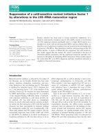
the activation of the Complement [15]. C1q (460 kDa) made
of three polypeptide chains (A, B and C) exhibits unique
structural features (Fig. 1). It consists of a C-terminus
presenting six globular head groups connected through a
hinge region to a long (approximately 11 nm) triple helical
collagen-like stalk that ends at the N-terminus [16]. Because
of this structural organization, C1q is often pictured as a
bunch of six flowers. The interaction of C1q with immune
complexes takes place at the globular region [17,18], but
C1q is known to also bind through its collagen-like region
(CLR) several nonimmune molecules [19], with conse-
quences which remain unclear. Actually the binding of the
C-reactive protein [20], of the serum amyloid protein [21]
and of DNA [22] to C1q leads to an activation of
Complement. On the other hand, the binding of sulfated
glycosaminoglycans [23–25], of proteoglycan dermatan
sulfate decorin [26], and of chondroitin 4-sulfate (i.e. the
C1q inhibitor) results in the inhibition of the classical
pathway [27].
Our goal in this study is to ascertain the binding of
fucoidan to C1q and to determine the site of interaction on
the protein. For this purpose we took advantage of the
binding properties of C1q toward DNA and of an emerging
technique allowing the molecular combing of DNA strands
and its observation by single-molecule spectroscopy. Dou-
ble- and single-stranded DNA has been demonstrated to
bind preferentially to the collagen-like region of C1q under
physiological saline conditions [22,28,29]. A cationic peptide
sequence on the A chain of C1q has been identified as the
major binding site of DNA [30]. We have analyzed herein
the binding of the human of the C1 complex and of its
subunit C1q to T4 DNA by molecular combing which
results in a large array of DNA strands individually
observed by fluorescence microscopy [8]. This approach
allowed us to implement an analytical tool useful to
investigate the binding of fucoidan to C1q through compe-
tition experiments between DNA and the polysaccharide.
We have addressed the question of whether a C1q inhibitor
(fucoidan) and a C1q activator (DNA) are able to bind to
the same region of the protein by using not only native C1q
but also the C1q isolated domains CLR and the globular
region (GR).
Materials and methods
Buffers
The following buffers were used: 250 m
M
Bis/Tris, pH 6.47;
0.1
M
Mes buffer, pH 6, containing 0.5
M
NaCl; and 1
M
sodium hydrogen carbonate (NaHCO
3
) buffer, pH 8.4. All
buffers were prepared with ultrapure water (milliQ, Milli-
pore).
Reagents and proteins
The Complement proteins C1r, C1q and C1 as well as the
depleted sera and the specific antibodies were obtained from
VWR (Fontenay-sous-Bois, France). C1q designed as
purified C1q in this study, and the derived collagen-like
region CLR and globular heads region GR were a generous
gift from G.J. Arlaud (IBS, Grenoble, France). The CLR
and GR were prepared as previously described [28]. Double-
stranded DNA from salmon testes (type III), T4 DNA and
2-mercaptoethanol were purchased from Sigma (Saint-
Quentin Fallavier, France). 1-Ethyl-3-(3-dimethylamino-
propyl) carbodiimide hydrochloride (EDC) and sulfo-NHS
(sulfo-N-hydroxysuccinimide) were purchased from Pierce
(Rockford, IL, USA). The fluorescent intercalating agent
YOYO-1 (dimer yellow oxozalone) and the Alexa 488-
fluorescent beads (27 nm) were obtained from Molecular
Probes (Eugene, OR, USA). Fucoidan from the brown
algae Ascophyllum nodosum was prepared as previously
described [31]. The fucoidan fraction used for the study
herein was of low molecular weight (8000 gÆmol
)1
as
determined by high performance size exclusion chromato-
graphy using heparin standards [32]) and of high sulfate
content (30% w/w), and was endowed with a high anti-
complementary activity as we have previously reported [14].
Fig. 1. Schematic representation of the structural organization of human
C1. (A) Model of the C1 complex showing the catalytic tetramer C1r
2
–
C1s
2
interacting with C1q (adapted from [37]). (B) Representation of
the association between the A, B and C chains constituting the six ABC
heterotrimeric triple helices of C1q. The cationic sequence 14–26 of the
A chain, as well as the sequence responsible for the kink of the colla-
gen-like region are shown.
Ó FEBS 2003 Complement C1 complex interactions (Eur. J. Biochem. 270) 4715
Surface treatment
Glass surfaces were rendered hydrophobic by coating with
polymethylmethacrylate (PMMA, M
r
8000 gÆmol
)1
). A
droplet of PMMA in chlorobenzene (13% w/w) is put
down onto a clean glass cover-slide and spread with a spin
coater of in-house design, at 2500 r.p.m. for 3 min. Surfaces
are then baked at 165 °C for 40 min and stored at room
temperature in a dust-free environment.
DNA preparation
T4 DNA (160 kb) was labeled as follows: 7.2 lLof1.35n
M
T4 DNA were incubated with 10 l
M
YOYO-1 (Molecular
Probes) in ultrapure water. This respresents a ratio of 1
molecule of dye to 30 bases of DNA, and 150 lLBis/Tris
pH 6.47, completed with 1.5 mL of ultrapure water during
at least 1 h at room temperature.
Fluorescent beads preparation
Alexa 488-conjugated beads (Molecular Probes) were
activated following the manufacturers’ instructions. Briefly,
50 lLofbeads(3.25l
M
in ultrapure water) were mixed
with 50 lL of sulfo-NHS (0.5
M
in ultrapure water) and
10 lLofEDC(0.2
M
in ultrapure water) in 100 lLof0.1
M
Mes buffer and completed with 400 lL of ultrapure water.
After 60 min incubation at room temperature under gentle
agitation, reaction was stopped by addition of 3 lLof
2-mercaptoethanol. Elimination of the excess of reactants
was performed on P6 Biospin Columns (Bio-Rad). The
concentration of used beads solutions in 0.1
M
Mes buffer
ranged from 20 to 30 n
M
, as evaluated by spectrophoto-
metric determination at 505 nm.
Protein labeling
Proteins C1q, C1r and the CLR fragments of C1q were
labeled with fluorescent beads. The concentration of beads
was adjusted according to the type and the concentration of
protein in order to maximize the rate of the labeling reaction
without generating cross-linking of the beads. Twenty
microliters of commercial C1q (2.46 l
M
) were added to
0.5 lL of beads solution (25 n
M
)andto2.5lLNaHCO
3
buffer completed with 25 lL ultrapure water. After 45 min
incubation at 20 °C under gentle agitation, reaction was
stopped by addition of 0.6 lLNH
2
OH (3
M
in ultrapure
water). Purified C1q (2.8 l
M
;17.5 lL) were added to 0.5 lL
of beads solution (25 n
M
)andto25lLNaHCO
3
buffer
completed with 100 lL ultrapure water. After 45 min at
20 °C under gentle agitation, reaction was stopped by
addition of 2.4 lLNH
2
OH (3
M
in ultrapure water). Six
microliters of CLR (7.8 l
M
) were added to 0.5 lLofbeads
solution (25 n
M
)andto12.5lLofNaHCO
3
buffer
completed with 100 lL ultrapure water. After 45 min at
20 °C under gentle agitation, reaction was stopped by
addition of 2.4 lLofNH
2
OH (3
M
in ultrapure water).
Twenty microlitres of C1 (0.27 l
M
)wereaddedto0.5lL
of beads solution (20 n
M
)andto2.5lLNaHCO
3
buffer
completed with 25 lL ultrapure water. After 45 min at
20 °C under gentle agitation, reaction was stopped with
1 lLofNH
2
OH (3
M
in ultrapure water). Two microliters
of C1r (10.5 l
M
)wereaddedto1lL of bead solution
(25 n
M
)andto4lLNaHCO
3
buffer completed with 40 lL
ultrapure water. After 45 min at 20 °C under gentle
agitation, reaction was stopped by addition of 2 lL
NH
2
OH (3
M
in ultrapure water).
Fluid phase incubations of DNA with complement
proteins
Fluorescent T4 DNA was incubated with the labeled
proteins, either in the presence of fucoidan or not, for
45–60 min at room temperature under gentle agitation in
the following conditions: 150 lLof6.5p
M
T4 DNA were
mixedwith1lL of labeled C1q prepared as above, and
according to case with 5 lLof45l
M
fucoidan; when the
purified C1q was used, 400 lLof6.5p
M
T4 DNA were
mixedwith10 lL of labeled purified C1q. One hundred and
fifty microliters of 6.5 p
M
T4 DNA were mixed with 5 lLof
labeled CLR or 2.5 lL of labeled C1, and according to case
with 1–2 lLof45 l
M
fucoidan. Then, the treated DNA was
combed as described below. For the study of the interaction
between C1r and C1q bound to combed T4 DNA, 150 lL
of 6.5 p
M
T4 DNA were preincubated with 1 lLof2l
M
unlabeled C1q for 45 min, then 6 lL of labeled C1r were
added, and according to case with 9 lLof45l
M
fucoidan.
Molecular combing
The combing process consists in the stretching of the DNA
by the passage of an air/water meniscus [33,34]. A droplet of
6.5 p
M
T4 DNA in Bis/Tris buffer, pH 6.47, is deposed on
a hydrophobic surface, incubated for 2 min and then
removed. This procedure is sufficient to stretch the DNA
strands when DNA is brought in small droplet. The
interaction between DNA and the hydrophobic surface is
very strong, so that DNA can be considered as grafted onto
the surface.
Fluorescent microscopy
Samples were observed using an inverted microscope (Leica
DM IRBE) by epifluorescence. An X100 infinity-corrected
1.4 NA oil objectives (Leica) was used and a cooled CCD
camera (C4880 Hamamatsu, and Ixon-Andor) was moun-
ted on the microscope. For fluorescence observations, a
mercury lamp was used in combination with a filters set for
fluorescein (Leica I3). The images were acquired using the
HIPIC
software (Hamamatsu) and
IXON
software (Andor),
with an exposition time ranging from 100 ms to 1 s.
Results
Interaction between DNA and C1q
DNA is described as an activator of human complement
system [30]. The DNA-dependent activation of Comple-
ment may result from the formation of a complex between
DNA and the first Complement protein C1q as proposed in
the literature [20,22,28]. The possibility to visualize individ-
ual DNA molecules prompted us first to investigate the
binding properties between C1q and DNA through the
observation of their complex.
4716 B. Tissot et al. (Eur. J. Biochem. 270) Ó FEBS 2003
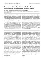
T4 DNA (160 kbps) labeled with the fluorescent inter-
calating agent YOYO-1 was combed on a PMMA surface.
Fluorescent strands were observed corresponding to indi-
vidual linear DNA chains stretched on the surface as
previously described (Fig. 2A) [8]. Fluorescent T4 DNA
was incubated in solution with labeled C1q. For that
purpose, C1q was covalently attached through its amino
groups to fluorescent beads (27 nm diameter). C1q is
estimated to have an overall size of 35 nm based on
literature data [15]. Given their similar sizes, we can
reasonably expect that one or two molecules of C1q are
bound per fluorescent bead. After incubation with such
labeled C1q (C1q*), fluorescent T4 DNA was combed as
described above. The fluorescent individual DNA molecules
then observed were clearly decorated with a succession of
fluorescent spots (Fig. 2B). The fluorescence of these spots
is easily distinguishable from the YOYO-1-induced fluores-
cence of the combed DNA, by its color and by its strong
intensity corresponding to the high density of the Alexa
fluor contained in the beads. These fluorescent spots were
then due to the presence of the beads along the DNA
strands. When the beads were noncoupled to C1q, no such
binding was observed on DNA strands (data not shown).
These results indicate that C1q mediates the attachment of
the spheres to DNA, hence evidencing interaction between
C1q and DNA.
In order to identify the preferential DNA binding region
on C1q, we have carried out experiments with the separated
domains of C1q, i.e. the collagen-like region CLR and the
globular region GR. CLR and GR were prepared by
enzymatic digestion by pepsin and collagenase, respectively,
as previously described [28]. The C-terminal domain of the
CLR, named the hinge region and joining the CLR to the
GR, is resistant to both proteases so that the conforma-
tional organization of CLR is conserved, whereas the GR
was obtained as individual globular heads [35]. In a first set
of experiments, CLR and GR were studied for their
capacity to compete with purified C1q* for the binding to
DNA. When incubation of T4 DNA and C1q* was
performed with various amounts of CLR, the analysis of
the resulting combed DNA showed that the binding of
C1q* to DNA strands started to decrease for a C1q/CLR
ratio of 1 : 10 and was totally suppressed from a 1 : 50
molar ratio. On the other hand, a higher amount of GR was
required to observe a similar inhibition as a C1q*/GR molar
ratio of 1 : 3000 was at least necessary (data not shown).
Such a large difference in the amount required to efficiently
compete for the binding of C1q* indicates that the collagen-
like region of C1q contains the preferential site for the
interaction and the binding to DNA.
In order to confirm this result, we checked for the binding
of the CLR to DNA using CLR preparation labeled with
the fluorescein-conjugated beads (CLR*). CLR* was incu-
bated with T4 DNA in fluid phase, after which the DNA
was combed on a PMMA surface. The analysis of the
images showed that CLR* was colocalized like C1q* with
the combed DNA (Fig. 2C), proving the binding of CLR
to DNA.
The C1 complex, which triggers the classical pathway of
Complement, comprises the subunit C1q and the two serine
proteases C1r and C1s associated into a C1r
2
–C1s
2
tetramer. Several lines of evidence in literature indicate that
the binding site of the tetramer on C1q is located within the
collagen-like region of C1q [36]. As our results show that
this region also contains the binding site of DNA, we
wondered whether the association of the tetramer C1r
2
–
C1s
2
to C1q could interfere with the binding of C1q to
DNA. We studied the binding to DNA of the C1 complex
labeled with the fluorescent beads (C1*). We observed that
C1* and the individual DNA strands were colocalized
(Fig. 2D), indicating the binding of C1 to DNA. Hence the
presence of the tetramer bound to the collagen-like region of
C1q does not impede the binding of C1q to DNA.
Conversely, in a subsequent experiment, we checked the
ability of C1r to associate on DNA-bound C1q. For that
purpose, T4 DNA was preincubated with nonlabeled C1q,
in order to form a DNA–C1q complex. The mixture was
then incubated with C1r labeled with fluorescent beads
(C1r*), and finally DNA was combed. We observed that
C1r* spots were aligned along the DNA strands (Fig. 2E).
Thus C1r binds to the DNA–C1q assembly, whereas C1r
does not bind to DNA strands in the absence of C1q (data
not shown). Therefore the binding of C1r is a direct
consequence of the presence of C1q on DNA strands.
Fig. 2. Molecular combing of the T4 DNA on a
PMMA surface after incubation with fluores-
cent Complement proteins and fucoidan. (A)
Individual strands of fluorescent 160 kbps T4
DNA combed on the PMMA surface. (B–D)
Molecular combing of the T4 DNA after
incubation with fluorescent (Alexa 488-fluor-
escent beads) C1q, CLR and C1 proteins,
respectively. (E) Molecular combing of the T4
DNA after incubation, first with nonlabeled
C1q and then with fluorescent C1r. (F)
Molecular combing of the T4 DNA after
incubation with fluorescent C1q in presence
of fucoidan.
Ó FEBS 2003 Complement C1 complex interactions (Eur. J. Biochem. 270) 4717
Altogether these results indicate that the collagen-like region
of C1q contains distinct sites for the binding of DNA and
for the binding of the catalytic tetramer C1r
2
–C1s
2
.
Interactions between fucoidan and C1q
We have previously reported that the sulfated polysaccha-
ride fucoidan interacts with C1q. In order to ascertain this
binding property of fucoidan, we performed competition
experiments between fucoidan and DNA for the binding to
C1q. Fluorescent T4 DNA was incubated with C1q* and
fucoidan in the C1q/fucoidan molar ratio 1 : 100 (i.e. the
amount which leads to 30% inhibition of the hemolytic
activity of C1q as we have previously reported [14]). After
combing, the individual fluorescent DNA strands clearly
appeared without any decoration by C1q* (Fig. 2F). This
result shows that fucoidan is able to compete with DNA for
the binding to C1q, suggesting that interactions between
fucoidan and C1q probably occur through the collagen-like
region. This hypothesis was confirmed by performing the
same competition experiment using labeled-CLR (CLR*).
Incubation of T4 DNA with CLR* in the presence of
fucoidan (CLR/fucoidan molar ratio of 1 : 20 and 1 : 40)
leads to an inhibition of the binding of CLR* to DNA from
the molar ratio 1 : 20. Altogether these results confirm that
fucoidan interacts with C1q through the same binding
region than DNA, i.e. the collagen-like region that includes
in our CLR preparation the stalk region and partially the
hinge region.
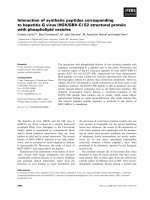
At this stage, it is worthwhile to note that, when C1
instead of C1q was used in this competition experiment with
fucoidan, a lower inhibition of the protein binding to DNA
was obtained (for the same C1/fucoidan molar ratio
1 : 100), as colocalization of C1* was still observed with
some DNA strand (Fig. 3). This lower inhibition may be
due to the presence of the catalytic tetramer C1r
2
–C1s
2
in
the C1 complex that hinders the interaction of the C1q
subunit with the polysaccharide. According to this hypo-
thesis fucoidan should then interact with the hinge region
containing the binding site of the tetramer C1r
2
–C1s
2
,in
addition to the stalk region of CLR. In order to check this
hypothesis, we tested the effect of fucoidan on the binding of
C1r to DNA-bound C1q. For this purpose, T4 DNA and
nonlabeled C1q were firstly preincubated, before the
addition of C1r* and fucoidan (molar ratio C1q/fucoidan
1 : 100). The resulting molecular combing appeared as in
Fig. 2F, exhibiting the absence of C1r* spots on the T4
strands. We have seen above that C1r is able to associate to
DNA-bound C1q. Furthermore, we have previously repor-
ted an affinity capillary electrophoresis study evidencing no
interaction between either C1r or C1s and fucoidan [14].
Hence this result proved that the binding of C1r to C1q is
inhibited by fucoidan, consistent with the interaction of the
polysaccharide with the hinge region of the collagen-like
region of C1q.
Discussion
C1q can bind several polyanionic molecules like sulfated
polysaccharides and also DNA, but with the opposite
effects of either inhibition or activation, respectively. Using
single-molecule observation of immobilized DNA strands,
we have been able to visualize the binding of C1q to
individual DNA strands. This single molecule approach
appeared as a valuable tool with which to investigate the
binding properties of fucoidan, a sulfated fucose-based
polysaccharide known as one of the most potent inhibitor of
IgG-dependent activation of Complement [14].
C1q binding to DNA was deduced in literature from data
based either on C1q–DNA precipitation experiments [22] or
on solid-phase assays with immobilized C1q [28,29]. In the
present study, we obtained the direct evidence of the DNA–
C1q binding through the observation of the colocalization
of C1q and DNA strands. It is worthwhile noting that this
interaction was performed in the fluid phase. In these
conditions, the resulting C1q-DNA complex was resistant
to the stretching and combing of DNA on PMMA surfaces,
indicating the strength of the interaction. A continuous
succession of fluorescent C1q could be observed on some
DNA strands, corresponding to a high density of binding.
Although the optical resolution does not allow the deter-
mination of this density, the obtained images are consistent
with previous estimates of one C1q per 34 nm of double-
stranded DNA for the highest density [22]. C1q is usually
described as constituted of two main domains, the
N-terminal collagen-like region CLR, and the C-terminal
globular region GR. Divergent data have been reported in
the literature concerning the binding site of DNA on C1q.
DNA has been proposed to bind to either the GR or the
CLR, or to both of the two domains, according the method
used. Here, we have individually used each of these domains
in the single molecule approach to unambiguously identify
the CLR as the main binding site of DNA. Indeed, we have
shown that CLR binds to DNA as well as C1q does, and
that, compared with GR, CLR competes much more
efficiently with C1q for binding to DNA. The dissociation
of the globular region of C1q into individual structures
could decrease the affinity of the GR, but to an extent that
could not lead to the observed difference between CLR and
GR in these competition experiments. The low competition
effect observed only when a very large excess of GR is used
could be due to non-specific interactions or to interaction
with the residual GR tail present in the GR preparation.
The collagen-like region can be divided into an
N-terminal domain organized into a triple-helical stalk,
which diverges into six arms constituting the C-terminal
Fig. 3. Molecular combing of the T4 DNA on a PMMA surface after
incubation with fluorescent C1 complex and fucoidan.
4718 B. Tissot et al. (Eur. J. Biochem. 270) Ó FEBS 2003
domain of the CLR; this has been named the hinge region
[15]. This hinge region contains the binding site for the
catalytic tetramer C1r
2
–C1s
2
, essential for the C1 activity
[35]. Our results show that C1q as well as C1 bind to DNA,
and that C1r can bind to C1q bound to DNA. Thus DNA
binding site on CLR does not overlap with the binding site
of the tetramer in the hinge region. It is likely that DNA
binds to the stalk domain of CLR, consistent with previous
findings showing that a synthetic peptide of the N-terminal
portion of the A chain (residues 14–26) binds to DNA and
inhibits its binding to C1q [20,30].
During the course of our studies of the anticomplemen-
tary activity of fucoidan, we have previously shown by
affinity electrophoresis that this sulfated polysaccharide
binds also to C1q [14]. The results obtained here clearly
showed its ability to inhibit the binding of C1q, as well as
that of CLR, to DNA. These data indicate that, like DNA,
fucoidan binds to C1q through the collagen-like region. The
C1q A chain that appeared to be essential for the binding of
nonimmune substance contained a cationic region within
residues 14–26 of stalk [20]. This positively charged sequence
contained five basic proximal residues, arginine and lysine,
that are assumed to be involved in the binding of polyanions
like DNA and fucoidan (Fig. 1B). Consistent with these
data, we have previously shown that fucoidan protects the
lysine residues of C1q from chemical modification by
specific reagent [14]. However fucoidan exhibits a major
difference with DNA in that the polysaccharide is able to
block the association of C1r to C1q. This result is in
agreement with our previous finding where the polysaccha-
ride was shown by ELISA to inhibit the reconstitution of C1
from C1r, C1s, and C1q [14]. Thus, unlike DNA, fucoidan
also interacts with the Ôarms domainÕ of CLR, which
contains the binding site of the tetramer C1r
2
–C1s
2
.Ithas
been reported that basic residues lysine and arginine in the
hinge region are involved in the assembly of C1 through
specific interaction with acidic residues of C1r [35].
Furthermore, the structural model of C1q shows a cluster
of basic residues that are located in the hinge region at the
junction between the stalk and the arms. We assumed that
fucoidan interacts with these positively charged residues in
the hinge region, leading to the observed blockage of the C1
assembly. As a consequence, the inhibiting activity of
fucoidan on the classical pathway activation should result
from this binding property to the hinge region, hampering
the activation of the two proteases C1r and C1s. This
mechanism is probably related to the inhibiting property of
endogenous C1q inhibitors of Complement, like the chon-
droitin 4-sulfate proteoglycan, which is secreted by the
human B lymphocytes. This glycosaminoglycan has been
proposed as a potential physiologic C1q inhibitor, through
the inhibition of the C1q– (C1r
2
–C1s
2
) assembly [37].
Other C1q binding substances that are not C1q inhibi-
tors, like DNA, C-reactive protein [20], and amyloid
protein, do not bind to the hinge region but rather to the
stalk domain of the CLR [38]. It has also been proposed that
the collagen-like stalk is involved in the binding of C1q to
different cell types and to liposomes [39]. Strikingly, this
binding leads to the activation of Complement, as we
observed for DNA (data not shown). The mechanism of
this activation, independent of the recognition of the
immune complex by the globular heads, remains unclear
and is debated in the literature [40]. Recently, it has been
reported from the structural model of C1r that the
activation of the C1 complex could result from mechanical
constraints upon C1q binding, which affect the flexible
hinge region [41]. Further investigations are required to
determine whether the binding to the stalk region also
results in such mechanical stresses that could be transmitted
to the hinge region.
Acknowledgements
We thank J. Ratiskol and C. Sinquin for the extraction and the
preparation of the fucoidan fraction and for their experimental advices.
We are grateful to Professor G. J. Arlaud for his generous gift of
purified C1q and of GR and CLR preparations. This work was
supported by CNRS and the county Pays de La Loire, as well as the
program Physique et Chimie du Vivant, from CNRS and the Ministe
`
re
de la Recherche, France.
References
1. Williams, S.J. & Davies, G.J. (2001) Protein–carbohydrate inter-
actions: learning from nature. Trends Biotechnol. 19, 356–362.
2. Lee, Y.C. & Lee, R.T. (1995) Carbohydrate–protein interactions:
basis of glycobiology. Acc. Chem. Res. 28, 321–327.
3. Spillmann, D. & Lindahl, U. (1994) Glycosaminoglycan–protein
interactions: a question of specificity. Curr. Opin. Struct. Biol. 4,
677–682.
4. Imberty, A. & Perez, S. (2000) Structure, conformation, and
dynamics of bioactive oligosaccharides: theoretical approaches
and experimental validations. Chem. Rev. 100, 4567–4588.
5. Mehta, A.D., Rief, M., Spudlich, J.A., Smith, D.A. & Simmons,
R.M. (1999) Single-molecule biomechanics with optical methods.
Science 238, 1689–1695.
6. Ludwig, M., Rief, M., Schmidt, L., Li, H., Oesterhelt, F. &
Gautel, M. (1999) AFM, a tool for single-molecule experiments.
Appl.Phys.Mater.Sci.Process.68, 173–176.
7. Ishijima, A. & Yanagida, T. (2001) Single molecule nano-
bioscience. Trends Biochem. Sci. 26, 438–444.
8. Gueroui, Z., Place, C., Freyssingeas, E. & Berge, B. (2002)
Observation by fluorescence microscopy of transcription on single
combed DNA. Proc. Natl Acad. Sci. USA 99, 6005–6010.
9. Brant, D.A. (1999) Novel approaches to the analysis of polysac-
charide structures. Curr. Opin. Struct. Biol. 9, 556–562.
10. Marzalek, P., Li, H. & Fernandez, J. (2001) Fingerprinting poly-
saccharides with single-molecule atomic force microscopy. Nat.
Biotechnol. 19, 258–262.
11. Camesano, T. & Wilkinson, K. (2001) Single molecule study
of xanthan conformation using atomic force microscopy.
Biomacromolecules 2, 1184–1191.
12. Daniel,R.,Berteau,O.,Jozefonvicz,J.&Goasdoue,N.(1999)
Degradation of algal (Ascophyllum nodosum) fucoidan by an
enzymatic activity contained in digestive glands of the marine
mollusk Pecten maximus. Carbohydr. Res. 322, 291–297.
13. Chevolot, L., Mulloy, B., Ratiskol, J., Foucault, A. & Colliec-
Jouault, S. (2001) A disaccharide repeat unit is the major structure
in fucoidans from two species of brown algae. Carbohydr. Res.
330, 529–535.
14. Tissot, B., Montdargent, B., Chevolot, L., Varenne, A., Descroix,
S., Gareil, P. & Daniel, R. (2003) Interaction of fucoidan with the
complement proteins of the classical pathway. Biochem. Biophys.
Acta 1651, 5–16.
15. Cooper, N.R. (1985) The classical complement pathway: activa-
tion and regulation of the first complement component. Adv.
Immunol. 37, 151–216.
Ó FEBS 2003 Complement C1 complex interactions (Eur. J. Biochem. 270) 4719
16. Kishore, U. & Reid, K.B. (2000) C1q: structure, function, and
receptors. Immunopharmacology. 49, 159–170.
17.Marques,G.,Anton,L.C.,Barrio,E.,Sanchez,A.,Ruiz,S.,
Gavilanes, F. & Vivanco, F. (1993) Arginine residues of the
globular regions of human C1q involved in the interaction with
Immunoglobulin G. J. Biol. Chem. 268, 10393–10402.
18. Kaul, M. & Loos, M. (1997) Dissection of C1q capability of
interacting with IgG. J. Biol. Chem. 272, 33234–33244.
19. Gewurz, H., Ying, S.C., Jiang, H. & Lint, T.F. (1993) Non
immune activators of the classical complement pathway. Behring
Institute Mitt. 93, 138–147.
20. Jiang, H., Robey, F. & Gewurz, H. (1992) Localization of sites
through which C-reactive protein binds and activates complement
to residues 14–26 and 76–92 of the human C1q A chain. J. Exp.
Med. 175, 1373–1379.
21. Ying, S Y., Gewurz, A.T., Jiang, H. & Gewurz, H. (1993) Human
serum amyloid P component oligomers bind and activate the
classical complement pathway via residues 14–26 and 76–92 of the
A chain collagen-like region of C1q. J. Immunol. 150, 169–176.
22. Schravendijk, M.R. & Dwek, R.A. (1982) Interaction of C1q with
DNA. Mol. Immunol. 19, 1179–1187.
23. Calabrese, G., Recondo, M., Fernandez de Recondo, M. &
Recondo, E. (1997) The first component of the human comple-
ment system recognizes the active fraction of heparin. Cell. Mol.
Biol. 43, 237–242.
24. Weiler, J. & Linhardt, R. (1989) Comparison of the activity of
polyanions and polycations on the classical and the alternative
pathways of complement. Immunopharmacology 17, 65–72.
25. Sahu, A. & Pangburn, M.K. (1993) Identification of multiple sites
of interaction between heparin and the complement system. Mol.
Immunol. 30, 679–684.
26. Krumdieck, R., Hook, M., Rosenberg, L. & Volanakis, J. (1992)
The proteoglycan decorin binds C1q and inhibits the activity of
the C1 complex. J. Immunol. 149, 3695–3701.
27. Silvestri, L., Baker, J., Roden, L. & Stroud, R. (1981) The C1q
inhibitor in serum is a chondroitin 4-sulfate proteoglycan. J. Biol.
Chem. 256, 7383–7387.
28. Rosenberg, A.M., Prokopchuk, P.A. & Lee, J.S. (1988) The
binding of native DNA to the collagen-like segment of C1q.
J. Rheumatol. 15, 1091–1096.
29. Uwatoko, S. & Mannik, M. (1990) The location of binding sites
on C1q for DNA. J. Immunol. 144, 3484–3488.
30. Jiang, H., Cooper, B., Robey, F.A. & Gewurz, H. (1992) DNA
binds and activates complement via residues 14–26 of the C1q A
chain. J. Biol. Chem. 267, 25597–25601.
31. Colliec, S., Boisson-Vidal, C. & Jozefonvicz, J. (1994) A low
molecular weight fucoidan fraction from the brown seaweed
Pelvetia canaliculata. Phytochemistry 35, 697–700.
32. Mulloy, B., Gee, C., Wheeler, S.F., Wait, R., Gray, E. & Bar-
rowcliffe, T.W. (1997) Molecular weight measurements of low
molecular weight heparins by gel permeation chromatography.
Thromb. Haemost. 77, 668–674.
33. Bensimon, A., Simon, A., Chiffaudel, A., Croquette, V., Heslot, F.
& Bensimon, D. (1994) Alignment and sensitive detection of DNA
by a moving interface. Science. 265, 2096–2098.
34. Bensimon, D., Simon, A., Croquette, V. & Bensimon, A. (1995)
Stretching DNA with a receding meniscus: experiments and
models. Phys. Rev. Lett. 74, 4754–4757.
35. Illy, C., Thielens, N.M. & Arlaud, G.J. (1993) Chemical char-
acterization and location of ionic interactions involved in the
assembly of the C1 complex of human complement. J. Prot. Chem.
12, 771–781.
36. Arlaud, G.J., Gaboriaud, C., Thielens, N.M., Budayova-Spano,
M., Rossi, V. & Fontecilla-Camps, J.C. (2002) Structural
biology of the C1 complex of complement unveils the mechanisms
of its activation and proteolytic activity. Mol. Immunol. 39,
383–394.
37. Kirschfink,M.,Blase,L.,Engelmann,S.&Schwartz-Albeiz,R.
(1997) Secreted chondroitin sulfate proteoglycan of the human B
cell lines binds to the complement protein C1q and inhibits com-
plex formation of C1. J. Immunol. 158, 1324–1331.
38. Jiang, H., Burdick, D., Glabe, C.G., Cotman, C.W. & Tenner,
A.J. (1994) b-Amyloid activates complement by binding to a
specific region of the collagen-like domain of the C1q A chain.
J. Immunol. 152, 5050–5059.
39. Bradley, A., Brooks, D., Norris-Jones, R. & Devine, D. (1999)
C1q binding to liposomes is surface charge dependent and is
inhibited by peptides consisting of residues 14–26 of the human
C1qA chain in a sequence independent manner. Biochem. Biophys.
Acta 1418, 19–30.
40. Tacnet-Delorme, P., Chevallier, S. & Arlaud, G.J. (2001)
b-Amyloid fibrils activate the C1 complex of complement under
physiological conditions: evidence for a site for Ab on the C1q
globular regions. J. Immunol. 167, 6374–6381.
41. Budayova-Spano, M., Lacroix, M., Thielens, N.M., Arlaud, G.J.,
Fontecilla-Camps, J.C. & Gaboriaud, C. (2002) The crystal
structure of the zymogen catalytic domain of complement C1r
reveals that a disruptive mechanical stress is required to trigger
activation of the C1 complex. EMBO J. 21, 231–239.
4720 B. Tissot et al. (Eur. J. Biochem. 270) Ó FEBS 2003