Báo cáo khoa học: Hydrogen independent expression of hupSL genes in Thiocapsa roseopersicina BBS pot
Bạn đang xem bản rút gọn của tài liệu. Xem và tải ngay bản đầy đủ của tài liệu tại đây (217.65 KB, 10 trang )
Hydrogen independent expression of hupSL genes
in Thiocapsa roseopersicina BBS
A
´
kos T. Kova
´
cs
1
,Ga
´
bor Ra
´
khely
1
, Judit Balogh
1
, Gergely Maro
´
ti
1
, Laurent Cournac
2
,
Patrick Carrier
2
,Lı
´
via S. Me
´
sza
´
ros
1
, Gilles Peltier
2
and Korne
´
l L. Kova
´
cs
1
1 Institute of Biophysics, Biological Research Center, Hungarian Academy of Sciences, and Department of Biotechnology,
University of Szeged, Hungary
2 CEA Cadarache, DSV, De
´
partement d’Ecophysiologie Ve
´
ge
´
tale et de Microbiologie, Laboratoire d’Ecophysiologie de la Photosynthe
`
se,
CNRS CEA, Saint Paul-Lez Durance, France
The presence of the substrate molecule of hydrogenases,
H
2
, triggers the expression of some hydrogenases through
a hydrogen-sensing regulatory hydrogenase (HupUV ⁄
HoxBC) and a two-component signal transduction
system (HupT ⁄ HoxJ and HupR ⁄ HoxA) as described
mainly in Rhodobacter capsulatus [1] and Ralstonia
eutropha [2]. In the presence of H
2
, the expression of
the membrane bound HupSL (in R. capsulatus)or
HoxKG (in Ra. eutropha) and soluble HoxFUYH
(in Ra. eutropha) hydrogenases is initiated, while the
gene products are not formed in the absence of H
2
.
HupUV and ⁄ or HoxBC are members of the regulatory
[NiFe] hydrogenases (RH) [3]. They show a predicted
structure that is similar to the typical [NiFe] hydro-
genases, possessing the small and the large subunits
and the well known [NiFe] active site with two CN
and one CO ligand [4]. RH is a soluble protein in line
with the absence of an N-terminal translocation
signal sequence on the small subunit polypeptide.
Interestingly, the large subunit proteins of the sensor
Keywords
hydrogen sensor; [NiFe] hydrogenase;
transcriptional regulation; Thiocapsa
roseopersicina
Correspondence
K. L. Kova
´
cs, Department of Biotechnology,
University of Szeged, H-6726 Szeged,
Temesva
´
ri krt. 62, Hungary
Fax: +36 62 544352
Tel: +36 62 544351
E-mail:
(Received 16 June 2005, accepted 3 August
2005)
doi:10.1111/j.1742-4658.2005.04896.x
The expression of many membrane bound [NiFe] hydrogenases is regulated
by their substrate molecule, hydrogen. The HupSL hydrogenase, encoded
in the hupSLCDHIR operon, probably plays a role in hydrogen recycling
in the phototrophic purple bacterium, Thiocapsa roseopersicina BBS.
RpoN, coding for sigma factor 54, was shown to be important for expres-
sion, suggesting a regulated biosynthsis from the hup gene cluster. The
response regulator gene, hupR, has been identified in the hup operon and
expression of hupSL was reduced in a chromosomal hupR mutant, which
indicated that HupR was implicated in the activation process. The hupT
and hupUV genes were isolated, and show similarity to the histidine kinase
element of the H
2
-driven signal transduction system and to the regulatory
hydrogenases of Ralstonia eutropha and Rhodobacter capsulatus, respect-
ively. Although the genes of the entire H
2
sensing and regulation system
were present, the expression of the hupSL genes was not affected by the
presence or absence of H
2
. Using reverse transcription PCR, we could not
detect any mRNA specific to the hupTUV genes in cells grown under
diverse conditions. The hupT and hupUV mutant strains had the same phe-
notype as the wild-type strains. The hupT gene product, expressed from a
plasmid, repressed HupSL synthesis as expected while introduction of act-
ively expressed hupTUV genes together derepressed the HupSL activity in
T. roseopersicina. The gene product of hupUV behaves similarly to other
regulatory hydrogenases and shows H–D exchange activity.
Abbreviations
IHF, integration host factor; RH, regulatory hydrogenase; RT, reverse transcription.
FEBS Journal 272 (2005) 4807–4816 ª 2005 FEBS 4807
hydrogenases terminate at a histidine residue and lack
the commonly occurring C-terminal extension that is
proteolytically processed during the last step of post-
translational maturation in energy transducing [NiFe]
hydrogenases. Some of the pleiotropic accessory pro-
teins (Hyp) are required for the proper assembly of
the H
2
-activating [NiFe] site in RH [5]. The catalytic
activity of RH is low, but the activity is insensitive to
oxygen [4]. It has been purified as a tetramer with an
a
2
b
2
structure. This tetramer forms a complex with
the HupT ⁄ HoxJ kinase in vitro [4]. The role of the
N-terminal part of the kinase, containing a PAS
domain, was established in signal transduction
between the RH and the kinase [6,7]. Addition of H
2
to HupUV before or during the incubation with HupT
rendered the complex unstable [6]. The transmission of
H
2
-induced changes from the RH to the histidine kin-
ase in vivo inhibits phosphorylation of the response
regulator. Therefore the DNA-binding positive regula-
tor remains unphosphorylated and binds to its target
site and activates the expression of the hupSL (hoxKG
and hoxFUYH) hydrogenase genes. In the absence of
molecular hydrogen the kinase phosphorylates the
HupR ⁄ HoxA regulator, which therefore looses its
activity and stops the transcription of the hydrogenase
structural genes [1]. The main difference in the signal
transduction between R. capsulatus and Ra. eutropha
is displayed by the phenotype of hupT ⁄ hoxJ and
hupUV ⁄ hoxBC mutants, respectively. R. capsulatus
hupT and hupUV mutants show a high level of hy-
drogenase activity in the absence of H
2
. Thus both the
HupT and the HupUV proteins exert a negative con-
trol on hydrogenase gene expression [8]. Phenotypic
analysis of Ra. eutropha hoxJ and hoxBC mutants
revealed that the H
2
sensing HoxBC protein counter-
acts the negative role of the HoxJ kinase [4].
Thiocapsa roseopersicina BBS is a purple sulphur
photosynthetic c proteobacterium belonging to the
Chromatiaceae family. Two sets of genes coding for
membrane bound [NiFe] hydrogenases ) the hynS-
isp1-isp2-hynL (formerly hydS and hydL) [9] and
hupSLCDHIR [10] – and a third, soluble hydrogenase
(hoxEFUYH) [11], together with other components
that are necessary for hydrogenase maturation [12,13]
were cloned and characterized. Thiocapsa roseoper-
sicina provides an attractive model system for com-
parative studies of the structure–function–stability
relationships of different hydrogenase isoenzymes [14].
Transcriptional regulation of the T. roseopersicina hyn
operon was demonstrated recently. The expression of
the hyn genes was induced under anaerobic conditions
by an FNR homologue, FnrT, and it was unaffected
by H
2
[15].
We now report that transcription of T. roseopersicina
hupSL hydrogenase genes is regulated through an
RpoN dependent promoter. The elements (hupR,
hupTUV) of a typical signal transduction system are
present and HupR is functionally active. The hupT and
hupUV genes are apparently intact, yet the hydrogen
sensing system is not functional in T. roseopersicina
BBS.
Results
Hydrogen independent hupSL expression
The HupSL enzyme of T. roseopersicina is a member
of the Group 1 uptake [NiFe]-H
2
ases [16]. Many
members of this group are expressed only in the pres-
ence of hydrogen. In order to study directly the H
2
dependent expression of hupSL the T. roseopersicina
GB11 strain was used because it lacks the other mem-
brane associated [NiFe] hydrogenase, HynSL, which
would interfere with the HupSL specific hydrogenase
assay of the membrane fraction. Deletion of the
hynSL genes did not affect the activity of HupSL
hydrogenase [11]. Mutation in the structural genes of
both membrane bound hydrogenases resulted in the
loss of all membrane bound hydrogenase activity
[11,12] (Table 1). Unexpectedly, the hydrogenase
activity measurements indicated a constant level of
HupSL activity, irrespective of the presence of hydro-
gen (Table 2). The effect of H
2
on the expression of
HupSL hydrogenase was examined under conditions
where nitrogenase was fully repressed and the HoxYH
soluble hydrogenase did not produce detectable
amount of H
2
(G. Ra
´
khely and K. L. Kova
´
cs,
unpublished data). A 708-bp DNA fragment contain-
ing the first 76 bp of the hupS coding sequence,
together with upstream sequences, was cloned into the
broad host-range lacZ expression vector, pFLAC, to
create an in-frame hupS::lacZ gene fusion. The result-
ing recombinant plasmid, pHUPRIP was introduced
into T. roseopersicina and b-galactosidase activities
were measured during growth under various condi-
tions. The measurements revealed similar expression
when cells were propagated in the absence or presence
of hydrogen (Table 2). Hydrogenase activity of
HupSL could not be detected in Ni-free conditions;
however, the b-galactosidase activities were unchaged
(55.6 ± 6.2 Miller units in Ni-free conditions and
57.7 ± 5.6 Miller units in the presence of 5 lmolÆl
)1
Ni). This suggests that Ni is important only for the
maturation of the HupSL hydrogenase enzyme
but not for the expression of hupSL genes. During
the experiments, cultures were grown under strictly
Transcription regulation of HupSL hydrogenase A
´
. T. Kova
´
cs et al.
4808 FEBS Journal 272 (2005) 4807–4816 ª 2005 FEBS
anaerobic conditions as the presence of trace amount
of oxygen abolished HupSL activity (J. Balogh, G.
Ra
´
khely, A
´
. T. Kova
´
cs and K. L. Kova
´
cs, unpub-
lished data).
Activation is dependent on RpoN
Inspection of the upstream sequence region of hupS
gene revealed a typical )24 ⁄ )12 promoter sequence
Table 1. Strains and plasmids.
Strain or plasmid Relevant genotype or phenotype Reference or source
Thiocapsa roseopersicina
GB11 hynSLD::Sm
r
[11]
GB1121 hynSLD::Sm
r
, hupSLD::Gm [11]
HRMG hupR::Em
r
, GB11 This work
RPON rpoN::Gm
r
, GB11 This work
HUVMG hupUVD, GB11 This work
HTMG hupTD, GB11 This work
Escherichia coli
S17-1(kpir) 294 (recA pro res mod) Tp
r
,Sm
r
(pRP4-2-Tc::Mu-Km::Tn7), kpir [35]
XL1-Blue MRF¢ D(mcrA)183, D(mcrCB-hsdSMR-mrr)173, endA1, supE44, thi-1,
recA1, gyrA96, relA1 lac [F¢ proAB lacI
q
ZDM15 Tn10 (Tet
r
)]
c
Stratagene
Plasmids
pGEM T-Easy Amp
r
, cloning vector, ColE1 Promega
pHUPU1 pGEM T-Easy, contains 272-bp fragment of hupU This work
pBluescript SK(+) Amp
r
, cloning vector, ColE1 Stratagene
pTUV2 8576-bp HindIII fragment that contains the hupTUV operon in pBluescript SK (+) This work
pAK35 4568-bp SphI fragment that contains the hupCDHI and hupR genes in pUC18 [10]
pKK23 3313-bp PstI fragment that contains the upstream region of hupS gene in pUC18 [10]
pK18mobsacB Km
r
, mob
+
, sacB
+
[28]
pLO2 Km
r
, mob
+
, sacB
+
[29]
p34S-Gm Cloning vector carrying Gm
r
[36]
pRL271 Cloning vector carrying sacB,Em
r
(ermC), Cm
r
GenBank no. L05081
pHRIMER2 Km
r
, 2833-bp region of hupR gene in pK18mobsacB carrying Em
r
cassette at BstXI site This work
pRPON2 Km
r
, 1618-bp region of rpoN gene in pK18mobsacB carrying Gm
r
cassette at SmaI site This work
pHTD2 Km
r
, in-frame up and downstream homologous regions of hupT in pK18mobsacB This work
pHUVD2 Km
r
, in-frame up and downstream homologous regions of hupUV in pLO2 This work
pBBRMCS2 Km
r
, mob
+
, broad host range vector [37]
pFLAC Gm
r
, mob
+
, pBBRMCS5 carrying the promoterless lacZ gene [15]
pHUPRIP Gm
r
, mob
+
, pFLAC carrying the promoter region of hupS gene fused to the lacZ gene This work
pBBRcrt Km
r
, mob
+
, pBBRMCS2 carrying the promoter region of crtD gene This work
pTUV
C
1Km
r
, mob
+
, hupTUV genes cloned after the promoter region of crtD gene This work
pTUV
C
2Km
r
, mob
+
, hupT gene cloned after the promoter region of crtD gene This work
pMHE6crtKm Km
r
, mob
+
, expression vector containing the promoter region of crtD gene [30]
pMHEUVC2 Km
r
, mob
+
, hupUV gene cloned after the promoter region of crtD gene This work
Table 2. HupSL specific H
2
uptake and b-galactosidase activities in different strains grown in the absence or presence of hydrogen. ND, Not
detected; NA, not adaptable (antibiotic conflict).
Strain Inactivated genes
HupSL hydrogenase activity
a
LacZ activity
b
–H
2
+H
2
–H
2
+H
2
GB11 DhynSL 100 ± 6.1 94.9 ± 15.4 57.7 ± 5.6 54.2 ± 7.9
GB1121 DhynSL, DhupSL 0 ± 0 0 ± 0 ND ND
RPON DhynSL, rpoN::Gm
r
0 ± 0 0 ± 0 NA NA
HRMG DhynSL, hupR::Em
r
0 ± 0 0 ± 0 7.5 ± 1.6 5.9 ± 1.1
HTMG DhynSL, DhupT 106.9 ± 24.1 112.8 ± 14.2 48.3 ± 8.7 59.1 ± 5.9
HUVMG DhynSL, DhupUV 89.5 ± 17.9 102.3 ± 9.9 58.9 ± 8.2 63.2 ± 4.8
a
Relative hydrogenase activities in the membrane fraction given in percentage compared to the T. roseopersicina GB11 strain grown in the
absence of H
2
.
b
Specific b-galactosidase activity (same strains containing pHUPRIP) given in micromoles of o-nitrophenol min
)1
ÆD
À1
650
.
A
´
. T. Kova
´
cs et al. Transcription regulation of HupSL hydrogenase
FEBS Journal 272 (2005) 4807–4816 ª 2005 FEBS 4809
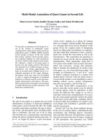
element (Fig. 1) [10]. Promoters harbouring )24 ⁄ )12
elements require the sigma factor RpoN (r
54
). Fur-
ther upstream from the r
54
element, an integration
host factor (IHF) box was recognized. The role of
IHF in transcriptional regulation will be the subject
of future studies. The rpoN gene was detected as
part of the ongoing genome project of T. roseopersi-
cina (L. S. Me
´
sza
´
ros, G. Ra
´
khely, H. P. Klenk and
K. L. Kova
´
cs, unpublished data). The sequence of
the rpoN gene was deposited in the GeneBank
(accession number: AY837592). The rpoN gene was
disrupted with a gentamycin cassette to generate
plasmid pRPON2 that was conjugated into T. roseo-
persicina. Double recombinant colonies were isolated
to yield the rpoN mutant, RPON. Southern blot ana-
lyses on genomic DNA confirmed the inactivation of
the chromosomal rpoN gene in the expected way
(data not shown). The RPON strain was unable to
grow in the absence of ammonium as a nitrogen
source indicating that the N
2
fixing ability was
impaired as well. Results in Table 2 show that
HupSL activity was also lost in the RPON mutant.
b-galactosidase activities were not measured as the
pHUPRIP vector contains a gentamycin resistance
marker and the T. roseopersicina RPON strain is also
resistant to gentamycin.
HupR activates hupSL transcription
The hupSLC structural genes are clustered with the
hupDHIR genes. blastp and clustal analyses sugges-
ted that the putative HupR protein belonged to the
family of response regulators. The translated HupR
from T. roseopersicina showed similarity to HoxA of
Ra. eutropha (53% identity and 66% similarity) and to
HupR (45% identity and 61% similarity) of R. capsul-
atus. In addition, the putative T. roseopersicina HupR
possesses a helix-turn-helix DNA binding motif (resi-
dues 434–474, with E-value of 5.4e-12) in its C-ter-
minal domain. The HupR architecture was determined
using the SMART database, revealing that T. roseo-
persicina HupR contained a response regulator receiver
domain (residues 6–125, with E-value of 6.4e-29) and a
r
54
interaction domain (residues 165–386, with E-value
of 1.2e-140).
The presence of the hupR gene in T. roseopersicina
is in apparent contradiction with the absence of a
hydrogen-dependent regulation of HupSL expres-
sion. In order to examine in detail the role of hupR
in T. roseopersicina an interposon mutant strain
(HRMG) was constructed. The mutation in hupR
affected the expression of HupSL hydrogenase dras-
tically: no hydrogenase activity could be measured in
the membrane fraction of T. roseopersicina HRMG
under any conditions compared to the wild-type
GB11 strain (Table 2). The hydrogenase activity of
HoxYH proteins in the soluble fraction was unaffec-
ted in the T. roseopersicina HRMG strain (data not
shown). Plasmid pHUPRIP carrying the hupS::lacZ
fusion was conjugated into the wild-type and HRMG
mutant T. roseopersicina strains; transconjugants were
grown in the absence and presence of hydrogen and
assayed for b-galactosidase activity. Results in Table 2
show that the expression of hupS::lacZ is dramatically
decreased in the hupR mutant independently from the
presence of hydrogen. Thus HupR is necessary for
HupSL expression, but it is not sufficient for the
H
2
-dependent regulation.
Fig. 1. Structure of the hup operon and reg-
ulatory region. The 120-bp region upstream
from hupS is presented. Hypothetical
)24 ⁄ )12 region and IHF site (on the bottom
strand) are boxed and compared to the con-
sensus RpoN and IHF sites. A vertical line
denotes residue identity. Start codon of
hupS is underlined and the first two amino
acids of HupS are indicated.
Transcription regulation of HupSL hydrogenase A
´
. T. Kova
´
cs et al.
4810 FEBS Journal 272 (2005) 4807–4816 ª 2005 FEBS
Isolation of the hydrogen sensor and sensor
kinase coding genes
Multiple alignments were performed with the known
HupUV ⁄ HoxBC protein sequences and the conserved
regions were selected. Because these proteins resemble
the regular [NiFe] hydrogenases, extreme care was
taken to avoid regions which were conserved also in
the nonregulatory hydrogenases. Finally, a 272-bp
fragment of the hupU gene was successfully amplified,
cloned and sequenced. This fragment was used to iso-
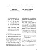
late an 8570-bp fragment carrying the hupT, hupU,
and hupV genes (Fig. 2) and flanking sequences (Gen-
Bank accession number: AY837591). The hupT and
hupUV genes encode putative proteins that are most
similar to HupT and HupUV of Azorhizobium caulino-
dans (65% similarity and 53% identity for HupT, 78%
similarity and 68% identity for HupU, 68% similarity
and 56% identity for HupV [17]). Downstream from
the hupV gene parA and orf154 were identified. The
predicted parA gene product showed similarity to the
partition protein A (57% similarity to ParA of Actino-
bacillus actinomycetemcomitans) and Orf154 showed
68% similarity to a hypothetical protein of Synecho-
cystis sp. PC6803. Upstream from the hupT gene a
truncated orf, similar to nifS gene, was identified that
lacks translational signal elements. Additionally, there
were numerous stop codons preceding this truncated
orf.
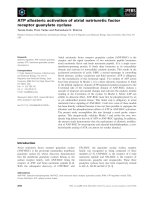
Total RNA was isolated from cells grown under var-
ious conditions (Fig. 3) and reverse transcription (RT)-
PCR was used to search for the hupTUV transcript.
No mRNA corresponding to the hupTUV genes was
found (Fig. 3). The quality of the RNA was checked
and found satisfactory using primers specific for the
coding region of Hyn hydrogenase (Fig. 3B). The
results suggest that the transcript level of the hupTUV
genes is below the detection limit or is missing in
T. roseopersicina.
Mutagenesis and homologous expression of the
hupT and hupTUV genes
In-frame deletion mutagenesis was used to characterize
the hupT and hupUV deficient phenotype. The exten-
sively truncated hupT derivative was cloned into
T. roseopersicina, resulting in HTMG. Similarly, the
HUVMG strain contained a 64-amino acid fragment
of hupUV. Both the hupT and the hupUV mutant
strains had comparable HupSL hydrogenase activities
to the control GB11 strain (Table 2). We also assayed
b-galactosidase activity in wild-type, HTMG, and
HUVMG T. roseopersicina strains carrying pHUPRIP.
Neither the hupT nor the hupUV mutation changed the
expression of hupS::lacZ (Table 2).
The hupT gene (pTrTUV
C
2), hupUV genes
(pMHEUVC2) or hupTUV genes (pTrTUV
C
1) were
cloned behind the promoter of the crtD gene and
expressed under anaerobic, phototrophic conditions.
Plasmids were transformed into T. roseopersicina, and
the transformants were grown in the presence or
absence of hydrogen and assayed for HupSL hydroge-
nase activity. Table 3 shows that HupSL hydrogenase
activity was lost in the strain, which expressed the
hupT gene. The HupT expressed from a plasmid thus
apparently performs the expected repressor function of
Fig. 2. Identified hupTUV genes. Restriction
sites used during construction of in-frame
deletion vectors are indicated. The
sequence has been deposited with Gene-
Bank Accession Number AY837591.
A
B
Fig. 3. RT-PCR analysis of T. roseopersicina hupTUV expression.
Primers TUVo24 and TUVo13 were used to detect mRNA corres-
ponding to hupU (A). Primers otsh11 and otsh14 were used to
detect mRNA corresponding to hynS (B) and used to verify the
quality of RNA prepared. PCR products were analysed on agarose
gel. Samples were loaded as follows: cells were grown in Pfennig’s
mineral medium (lanes 1, 2), and supplemented with sodium-acet-
ate (lanes 3, 4),
D-glucose (lanes 5, 6), grown in the presence of H
2
(lanes 7, 8), or ammonium chloride was omitted (lane 9, 10). In
samples loaded in lanes 1, 3, 5, 7 and 9, reverse transcription was
carried out before the PCR; in lanes 2, 4, 6, 8 and 10, reverse
transcription was omitted. M, Marker; C, control PCR made on
genomic DNA. Selected marker bands are indicated.
A
´
. T. Kova
´
cs et al. Transcription regulation of HupSL hydrogenase
FEBS Journal 272 (2005) 4807–4816 ª 2005 FEBS 4811
HupT. Production of HupTUV from a similar plasmid
construction, however, did not alter the HupSL
hydrogenase activity, i.e. HupSL was not regulated by
H
2
(Table 3). The HupSL activity was also unaltered
in strains expressing the hupUV genes only (Table 3,
pMHEUVC2). RT-PCR revealed the presence of hupT
and hupUV specific mRNA in strains expressing the
corresponding genes from the promoter of crtD gene
(data not shown), but not in strains without plasmid.
b-Galactosidase activities were not measured as the
pHUPRIP vector contains the same origin of replica-
tion as pTrTUV
C
1, pTrTUV
C
2 and pMHEUVC2.
The enzyme activities of the RH proteins measured
with various redox dyes showed very low activity com-
pared to those of energy conserving [NiFe] hydro-
genases [4]. Therefore we tested the activity of the
T. roseopersicina HupUV using the H–D exchange
reaction. H–D exchange, catalysed by the energy con-
serving hydrogenases and by the RH, can be distin-
guished on the basis of their different response to O
2
[18]. Strains lacking the HupUV expression plasmids
had no detectable H–D exchange activity in the pres-
ence of oxygen, while those expressing the HupTUV
from the promoter of crtD (pTrTUV
C
1) showed
0.19 ± 0.06 lmolÆL
)1
Æmin
)1
activity. In comparison,
the H–D exchange activity of the soluble HoxEFUYH
hydrogenase, measured in the absence of oxygen, was
23.5 ± 2.1 lmolÆL
)1
Æmin
)1
. The H–D exchange activ-
ity of the soluble hydrogenase was sensitive to oxygen
as described earlier for other hydrogenases.
Discussion
In a few organisms, e.g. methanogens, whose metabo-
lism is strictly linked to H
2
, hydrogenases are synthes-
ized constitutively [19]. In most other cases the
expression of hydrogenases is regulated by various
environmental signals. The signal may be anaerobicity
[20], Ni [21], or hydrogen itself. The signal transduc-
tion pathway that responds specifically to H
2
has been
studied in detail in Ra. eutropha [2,3,7], R. capsulatus
[1,6] and in Bradyrhizobium japonicum [21,22]. The
pathway comprises HupUV (regulatory hydrogenase),
HupT (kinase), and HupR (response regulator) in
R. capsulatus.
The genes coding for the membrane bound HupSL
hydrogenase were cloned and sequenced in T. roseo-
persicina [10]. The presence of HupR response regula-
tor downstream from the hupSLCDHI genes prompted
us to assume that hydrogen-dependent regulation may
function in T. roseopersicina by analogy to R. capsula-
tus and Ra. eutropha. The regulation of the T. roseo-
persicina HupSL hydrogenase was followed by
hydrogenase activity measurements and it was found
that hydrogen did not affect HupSL activity. This puz-
zling observation could not explain the presence of the
hupR gene and the r
54
promoter element. The r
54
spe-
cific binding site in the hupS upstream region was
investigated. Indeed, the expression of T. roseopersicina
HupSL hydrogenase depended on the presence of func-
tional RpoN protein. The expression of hydrogenase
was also RpoN-dependent in Ra. eutropha [23] and
in B. japonicum [24], while hupSL transcription is
r
70
-dependent in R. capsulatus [1]. This is in line with
the observation that the putative r
54
interaction sites
within the HupR ⁄ HoxA proteins are well conserved in
T. roseopersicina, Ra. eutropha and B. japonicum, but
not in R. capsulatus [1].
The remote possibility of the inactive hupR gene was
considered. The functional role of HupR was therefore
tested by creating a T. roseopersicina hupR mutant
strain. Results obtained with this mutant provided
straightforward evidence that HupR was essential for
the hupSL transcription under all conditions investi-
gated. The H
2
insensitive HupSL expression was there-
fore not due to an aborted hupR. The promoter region
of the T. roseopersicina hupSL genes did not reveal any
unusual feature that could be responsible for the lack
of response to the environmental signal, hydrogen.
If the presence of HupR and its effect on HupSL
expression is a sign for the biosynthesis of the enzyme
being under the H
2
control, the other elements of the
signal transduction cascade should be present in
T. roseopersicina. The clustered hupTUV genes were
identified, cloned, sequenced, and analysed. The trun-
cated HupT and HupUV proteins were most similar
to the corresponding proteins of Azorhizobium cauli-
nodans [17]. The physiological role of HupT and
HupUV in the regulation of HupSL was tested by
creating hupT and hupUV deletion mutants in T. roseo-
persicina. Hydrogenase activity measurements showed
that deletion of hupT or hupUV genes did not change
the level of HupSL hydrogenase activity, suggesting
that the putative HupT and HupUV proteins do not
Table 3. H
2
uptake activities in complementation experiments. The
results are given in percentage compared to the T. roseopersicina
grown in the absence of H
2
.
Plasmid
Complementing
gene
HupSL hydrogenase activity
–H
2
+H
2
– – 100 ± 6.1 94.9 ± 15.4
pTrTUV
C
2 hupT 0±0 0±0
pTrTUV
C
1 hupTUV 95.1 ± 22.3 104.4 ± 11.6
pMHEUVC2 hupUV 93.5 ± 9.2 107.9 ± 21.6
Transcription regulation of HupSL hydrogenase A
´
. T. Kova
´
cs et al.
4812 FEBS Journal 272 (2005) 4807–4816 ª 2005 FEBS
take part in a hydrogen sensing function and do not
regulate the HupSL formation under the growth con-
ditions examined. A possible explanation of these
data may implicate the apparently truncated nifS,
located immediately upstream from the hupT gene.
This flawed gene residue may hamper the transcrip-
tion of the hupTUV genes due to a polar effect. The
lack of expression of the HupTUV would explain the
hydrogen independent activity profiles. To confirm
this idea, RT–PCR experiments were carried out to
test the presence or absence of the hupTUV message.
RT–PCR experiments showed that no mRNA corres-
ponding to hupU gene was detected in cells grown
under various conditions. It was therefore concluded
that the hupTUV gene cluster is cryptic in T. roseo-
persicina. The question remained whether a point
mutation in the hupTUV genes or the upstream trun-
cated nif gene is responsible for the failed transcrip-
tional regulation?
Multiple alignment of T. roseopersicina HupT pro-
tein with other kinases revealed the presence of H, N,
G1, F and G2 motifs in the C-terminal region, those
necessary for kinase function. Introduction of the hupT
gene behind the promoter region of crtD gene
repressed HupSL expression in T. roseopersicina sug-
gesting that HupT can fulfil its function if expressed
behind a heterologous promoter. Thus HupT is more
similar in function to the HoxJ protein of Ra. eutro-
pha, i.e. it represses transcription of the hupSL. Thio-
capsa roseopersicina HupUV resembles typical features
of [NiFe] hydrogenases. Introduction of hupTUV genes
cloned behind the promoter region of crtD gene
restored the expression of HupSL hydrogenase. How-
ever, the expression of HupSL hydrogenase was unal-
tered by the presence of H
2
. These results suggest that
HupUV, expressed from a strong T. roseopersicina
promoter, interacts with HupT and alters its phos-
phorylation state, but the HupUV cannot change the
interaction with HupT depending on the presence of
hydrogen. Remarkably, HupUV, expressed from a
plasmid, clearly displayed catalytic activity in the H–D
exchange activity assay. When expressed, the HupUV
regulatory hydrogenase is therefore active in T. roseo-
persicina. The so-called RH
STOP
mutant protein of
Ra. eutropha lacking a C-terminal peptide of 55 amino
acids in HoxB lost its H
2
-sensing ability but still cata-
lysed the H
2
oxidation [7]. In this case the RH
STOP
was incapable of forming the (ab)
2
dimeric heterodi-
mer and the complex with HoxJ kinase, therefore the
expression of the membrane bound HoxKG hydro-
genase was repressed. Thus uncoupling of the hydro-
genase activity and the H
2
sensing ability of HupUV is
conceivable.
In summary, it can be concluded, that the expres-
sion of the hupTUV genes from a broad host range
vector could partially restore the signal transduction
cascade, although irrespective of the presence of
hydrogen. Each of the elements of the known signal
transduction (HupR and HupT) and H
2
sensing
(HupUV) system are functional, yet the expression
of HupSL does not apparently depend on the pres-
ence or absence of H
2
in the environment. The lack
of functionally active hupTUV on the chromosome is
a likely reason for the constitutive expression of the
hupSL genes in the wild type strain. At this point
one cannot exclude the possibility that additional
genetic elements are also involved in the assumed
H
2
dependent regulation of HupSL biosynthesis.
Impaired regulatory mechanisms, caused by point
mutations, have been described previously in several
cases. In Ra. eutropha H16, a mutation of HoxJ kin-
ase resulted in the loss of HoxJ protein function and
constitutive expression of hydrogenase genes [25]. In
Rhodopseudomonas palustris CGA009, the photosys-
tem is synthesized in the dark due to a single point
mutation in the helix–turn–helix DNA binding motif
of PpsR, rendering it inactive [26]. Comparison of
HupSL regulations and the functional roles of
HupTUV in other T. roseopersicina strains would
provide further insight into the understanding of the
loss of HupSL hydrogenase regulation.
Experimental procedures
Bacterial strains and plasmids
Strains and plasmids are listed in Table 1. T. roseopersicina
strains were grown in liquid cultures for 3–4 days in Pfen-
nig’s mineral medium supplemented with 0.1% NH
4
Cl [27].
Sodium acetate (2 gÆL
)1
)ord-glucose (5 gÆL
)1
) was added
when needed. NiCl was omitted only if indicated, otherwise
5 lmolÆL
)1
was used. Plates were solidified with 7 g Æ L
)1
Phytagel (Sigma, St Louis, MO, USA); when selecting for
transconjugants plates were incubated for 2 weeks in anaer-
obic jars using the GasPack (BBL, Kansas City, MI, USA)
or AnaeroCult (Merck, Rahway, NJ, USA) systems.
Escherichia coli strains were maintained on Luria–Bertani
agar. Antibiotics were used in the following concentrations
(lgÆmL
)1
): for E. coli: streptomycin (50), ampicillin (100),
kanamycin (50), gentamycin (20), erythromycin (50); for
T. roseopersicina: streptomycin (5), kanamycin (20), genta-
mycin (5) erythromycin (50).
Conjugation
Conjugation was carried out as described previously [12].
A
´
. T. Kova
´
cs et al. Transcription regulation of HupSL hydrogenase
FEBS Journal 272 (2005) 4807–4816 ª 2005 FEBS 4813
Identification of the hupU gene
A multiple alignment of the known HupU protein sequences
was performed and conserved domains were selected for
designing PCR primers. PCR was carried out using
the primers: hupUo1 (5¢-AACGAGTTCTAIGAITAIAAG
GCN-3¢) and hupUo2 (5¢-GCIACGTTCCTIGCCTTNG
GCATRTC-3¢) (where R is A or G) on T. roseopersicina
genomic DNA. The isolated PCR product of the correct
size (272 bp) was cloned into pGEM T-Easy (Promega,
Madison, WI, USA; resulting in pHUPU1) and sequenced.
Cloning of hupTUV genes from T. roseopersicina
Southern analysis was performed with the NotI fragment of
pHUPU1 as a probe. A HindIII partial genomic library
was created in pBluescript SK+ and pTUV2 was identified
by colony hybridization. The insert of the pTUV2 plasmid
was subcloned and sequenced on both strands by primer
walking. The 8576-bp sequence was deposited in the Gene-
Bank under the accession number AY837591.
Site-directed mutagenesis of hupR, rpoN, hupT
and hupUV genes
The in-frame deletion vector constructs derived from the
pK18mobsacB [28] or pLO2 [29] vectors. For insertion mut-
agenesis of the hupR gene, the 2833-bp ApaI (truncated)–
SphI fragment of pAK35 [10] was inserted into the Eco RV–
SphI site of pLO2, resulting in pHRIMER1. After digesting
the pHRIMER1 with BstXI and polishing, the truncated
SalI–EcoRI fragment (918 bp) of pRL271 (GenBank acces-
sion number L05081) containing the erythromycin resist-
ance gene was inserted (pHRIMER2).
For insertion mutagenesis of the rpoN gene, the 1618 bp
PCR fragment obtained with primers rpoN1 (5¢-GCTGC
ATCTCGACGATCTTC-3¢) and rpoN2 (5¢-ATCGCTTGC
GCTGAGCCTCT-3¢) from rpoN (GenBank Accession
Number AY837592) was inserted into the SmaI site of
pK18mobsacB, resulting in pRPON1. After digesting the
pRPON1 with SmaI, the SmaI fragment (855 bp) of p34S-
Gm (GenBank accession number AF062079) containing the
gentamycin resistance gene was inserted (pRPON2).
For removal of the hupT gene, the truncated 1379-bp
ApaI fragment of pTUV2 was inserted into the BamHI
digested and polished pK18mobsacB vector, resulting in
pHTD1. The 1311-bp SacI fragment of pTUV2 was inser-
ted into the SalI site of pHTD1 vector after polishing the
noncompatible ends, resulting in pHTD2.
For removal of the hupU and hupV gene, the 1794-bp
BamHI fragment of pTUV2 (upstream region of the hupU)
was inserted into the 5924-bp BamHI vector fragment of
pTUV2 (containing the downstream region of the hupV),
resulting in pHUVD1. The 4534-bp KpnI–XbaI fragment of
the pHUVD1 was inserted into the SacI–XbaI site of pLO2
vector after polishing the noncompatible ends, resulting in
pHUVD2.
The pHRIMER2, pRPON2, pHTD2 and pHUVD2 con-
structs were transformed into E. coli S-17(kpir), then conju-
gated into T. roseopersicina GB11 resulting HRMG
(hupR::Er), RPON (rpoN::Gm), HTMG (DhupT) and
HUVMG?(DhupUV), respectively. When creating the
hupR::Er or rpoN::Gm strain, the selection for the recombi-
nation was based on the erythromycin or gentamycin resist-
ance and then the double recombinant clones, that were
resistant to erythromycin or gentamycin and sensitive to
kanamycin, were selected. In the case of in-frame deletion
of hupT or hupUV genes, selection for the first recombina-
tion event was based on kanamycin resistance. The selec-
tion for the second recombination was based on the sacB
positive selection system [13]. The mutant clones were veri-
fied by PCR and ⁄ or Southern blotting.
Construction of hupS::lacZ fusion plasmid
The PCR fragment obtained with ohup4 (5¢-CTCGAA
ATCCGGAAAGGCTC-3¢) and )20 (5¢-GTAAAACGA
CGGCCAGT-3¢) primers on pKK23 [10] was digested with
PstI and cloned into the XbaI (polished)-PstI site of
pFLAC [15] resulting pHUPRIP1.
Construction of hupTUV expressing plasmids
The hupTUV and hupT genes of T. roseopersicina were
cloned downstream from the crtD promoter region of
T. roseopersicina as follows: the promoter region of the
crtD gene from T. roseopersicina was isolated from pRcrt4
as an XhoI–BamHI fragment and after polishing the ends it
was cloned to the SspI site of pBBRMCS2 resulting
pBBRcrt. The hupTUV genes were cloned as a HindIII–
BglII(polished) fragment from pTUV2 into the HindIII–
BstXI (polished) sites of pBBRcrt yielding pTrTUV
C
1. To
express the hupT gene only the hupUV genes were deleted
from pTrTUV
C
1 by replacing the EcoRI–StuI (polished)
fragment (containing the 3¢ region of hupT and the hupUV
genes) with the EcoRI–BamHI (polished) fragment of
pTUV2. This construct (pTrTUV
C
2) restored the whole
hupT gene, but lacked the hupUV genes. The NdeI-HindIII
digested TUVo31 (5¢-ACATATGAACCTGTTATGGCTC
CAG-3¢)–TUVo28 (5¢-AAGCTTGTGGACCGTGCAGAC
CAT-3¢) PCR fragment was cloned into the corresponding
sites of pMHE6crtKm [30] resulting in pMHEUVC2.
Isolation of total RNA and RT-PCR analysis
RNA was isolated from cells using the TRI reagent (Sigma,
St Louis, MO, USA), following the manufacturer’s recom-
mendations. Isolated total RNA was treated with RNase-free
Transcription regulation of HupSL hydrogenase A
´
. T. Kova
´
cs et al.
4814 FEBS Journal 272 (2005) 4807–4816 ª 2005 FEBS
Dnase I at 37 °C for 60 min in a total volume of 40 lL
[40 mm Tris ⁄ HCl pH 7.5, 20 mm MgCl
2
,20mm CaCl
2
,4U
of RNase-free DNase I (Promega, Madison, WI, USA)]
prior to RT-PCR. After phenol ⁄ chloroform extraction and
ethanol precipitation, the RNA was dissolved in 20 lLH
2
O.
RT–PCR was carried out as described previously [12]. The
TUVo24 primer (5¢-GAGGTTGGTGGCCAGTTC-3¢) was
used for the reverse transcription and PCR. The TUVo13
(5¢-AACGCCGTGTCGGACCATGT-3¢) served as the other
primer in PCR. Using these primers a 592-bp fragment was
expected. The quality of the RNA prepared was assayed with
primers specific for the hynS gene: otsh14 (5¢-GAT
CGCGATATTGAACATC-3¢) was used in the reverse tran-
scription and otsh11 (5¢-CTGCCCGAGCTTGACGC-3¢)
served as other primer in PCR. Using these primers a 512-bp
fragment was expected.
Enzyme assays
Hydrogenase uptake activities of membrane fractions were
determined using benzyl viologen [13]. The rates of H
2
and
HD formation, resulting from exchange between D
2
and
protons of the medium, measured at 30 °C, were monitored
continuously by MS as described in detail previously
[31,32]. For each experiment 1.5 mL (D
600
¼ 0.464 ± 0.034
of 10-times diluted cultures) culture was used. Hydrogenase
activity based on the rates of H
2
and HD formation was
calculated as described by Cournac et al. [33]. The b-galac-
tosidase activity of the toluene-permeabilized cell extracts
was assayed as described earlier for T. roseopersicina
[27,34]. Cells were assayed at the late logarithmic growth
state. One Miller unit corresponds to 1 lmol of o-nitrophe-
nyl-b-galactoside (Sigma-Aldrich) hydrolysed per minute
normalized to the optical density at 650 nm for T. roseo-
persicina.
Bioinformatics tools
Protein sequence comparisons in the various databases were
done with the blast (p, x) programs (i.
nih.nlm.gov). Multiple alignments were performed with the
clustal x program.
Acknowledgements
Supported by Hungarian Ministry of Education
(OMFB-00768 ⁄ 03) and the European Commission
(QLK5-1999-01267 and NEST STRP SOLAR-H, con-
tract 516510). We thank Dr Annette Colbeau and Dr
Sylvie Elsen (DBMS, CEA-CENG, Grenoble, France)
and Dr Douglas F. Browning (University of Biming-
ham, Birmingham, UK) for many helpful discussions.
We gratefully acknowledge Ro
´
zsa Verebe
´
ly for excel-
lent technical assistance.
References
1 Dischert W, Vignais PM & Colbeau A (1999) The
synthesis of Rhodobacter capsulatus HupSL hydrogenase
is regulated by the two-component HupT ⁄ HupR sys-
tem. Mol Microbiol 34, 995–1006.
2 Lenz O, Bernhard M, Buhrke T, Schwartz E & Frie-
drich B (2002) The hydrogen-sensing apparatus in
Ralstonia eutropha. J Mol Microbiol Biotechnol 4,
255–262.
3 Kleihues L, Lenz O, Bernhard M, Buhrke T & Friedrich
B (2000) The H
2
sensor of Ralstonia eutropha is a mem-
ber of the subclass of regulatory [NiFe] hydrogenases.
J Bacteriol 182, 2716–2724.
4 Bernhard M, Buhrke T, Bleijlevens B, De Lacey AL,
Fernandez VM, Albracht SP & Friedrich B (2001) The
H
2
sensor of Ralstonia eutropha. Biochemical character-
istics, spectroscopic properties, and its interaction with
a histidine protein kinase. J Biol Chem 276, 15592–
15597.
5 Buhrke T, Bleijlevens B, Albracht SPJ & Friedrich B
(2001) Involvement of hyp gene products in maturation
of the H
2
-sensing [NiFe] hydrogenase of Ralstonia eutro-
pha. J Bacteriol 183, 7087–7093.
6 Elsen S, Duche O & Colbeau A (2003) Interaction
between the H
2
sensor HupUV and the histidine kinase
HupT controls HupSL hydrogenase synthesis in Rhodo-
bacter capsulatus. J Bacteriol 185, 7111–7119.
7 Buhrke T, Lenz O, Porthun A & Friedrich B (2004)
The H
2
-sensing complex of Ralstonia eutropha: interac-
tion between a regulatory [NiFe] hydrogenase and a
histidine protein kinase. Mol Microbiol 51, 1677–1689.
8 Elsen S, Colbeau A, Chabert J & Vignais PM (1997)
The hupTUV operon is involved in negative control
of hydrogenase synthesis in Rhodobacter capsulatus.
J Bacteriol 178, 5174–5181.
9Ra
´
khely G, Colbeau A, Garin J, Vignais PM & Kova
´
cs
KL (1998) Unusual organization of the genes coding for
HydSL, the stable [NiFe] hydrogenase in the photosyn-
thetic bacterium Thiocapsa roseopersicina BBS. J Bacte-
riol 180, 1460–1465.
10 Colbeau A, Kova
´
cs KL, Chabert J & Vignais PM
(1994) Cloning and sequencing of the structural
(hupSLC) and accessory (hupDHI) genes for hydroge-
nase biosynthesis in Thiocapsa roseopersicina. Gene 140,
25–31.
11 Ra
´
khely G, Kova
´
cs A
´
T, Maro
´
ti G, Latinovics D,
Fodor BD, Csana
´
di G & Kova
´
cs KL (2004) A hetero-
pentameric NAD
+
reducing [NiFe] hydrogenase in the
purple sulfur photosynthetic bacterium, Thiocapsa rose-
opersicina. Appl Environ Microbiol 70, 722–728.
12 Fodor B, Ra
´
khely G, Kova
´
cs A
´
T & Kova
´
cs KL (2001)
Transposon mutagenesis in purple sulfur photosynthetic
bacteria: identification of hypF, encoding a protein cap-
able of processing [NiFe] hydrogenases in a, b and c
A
´
. T. Kova
´
cs et al. Transcription regulation of HupSL hydrogenase
FEBS Journal 272 (2005) 4807–4816 ª 2005 FEBS 4815
subdivision of the proteobacteria. Appl Environ Micro-
biol 67, 2476–2483.
13 Maro
´
ti G, Fodor BD, Ra
´
khely G, Kova
´
cs A
´
T, Arvani
S & Kova
´
cs KL (2003) Selectivity and cooperativity of
accessory proteins in the biosynthesis of [NiFe] hydro-
genases. Eur J Biochem 270, 2218–2227.
14 Kova
´
cs KL, Fodor B, Kova
´
cs A
´
T, Csana
´
di G, Maro
´
ti
G, Balogh J, Arvani S & Ra
´
khely G (2002) Hydro-
genases, accessory genes and the regulation of [NiFe]
hydrogenase biosynthesis in Thiocapsa roseopersicina.
Int J Hydrogen Energy 27, 1463–1469.
15 Kova
´
cs A
´
T, Ra
´
khely G, Browning DF, Fu
¨
lo
¨
p A, Mar-
o
´
ti G, Busby SJW & Kova
´
cs KL (2005) An FNR-type
regulator controls the anaerobic expression of Hyn
hydrogenase in Thiocapsa roseopersicina. J Bacteriol
187, 2618–2627.
16 Vignais PM & Colbeau A (2004) Molecular biology of
microbial hydrogenases. Curr Issues Mol Biol 6, 159–188.
17 Baginsky C, Palacios JM, Imperial J, Ruiz-Argueso T &
Brito B (2004) Molecular and functional characteriza-
tion of the Azorhizobium caulinodans ORS571 hydroge-
nase gene cluster. FEMS Microbiol Lett 237, 399–405.
18 Vignais PM, Dimon B, Zorin NA, Colbeau A & Elsen
S (1997) HupUV proteins of Rhodobacter capsulatus can
bind H
2
: evidence from the H-D exchange reaction.
J Bacteriol 179, 290–292.
19 Cammack R, Frey M & Robson R, eds. (2001) Hydro-
gen as a fuel. Learning from Nature. London: Taylor &
Francis.
20 Kova
´
cs A
´
T, Ra
´
khely G, Balogh J, Maro
´
ti G, Fu
¨
lo
¨
pA
& Kova
´
cs KL (2005) Anaerobic regulation of hydro-
genase transcription in different bacteria. Biochem Soc
Transact 33, 36–38.
21 Black LK & Maier RJ (1994) Sequences and characteri-
zation of hupU and hupV genes of Bradyrhizobium japo-
nicum encoding a possible nickel-sensing complex
involved in hydrogenase expression. J Bacteriol 176,
7102–7106.
22 Van Soom C, de Wilde P & Vanderleyden J (1997)
HoxA is a transcriptional regulator for expression of
the hup structural genes in free-living Bradyrhizobium
japonicum. Mol Microbiol 23, 967–977.
23 Ro
¨
mmermann D, Warrelmann J, Bender RA & Frie-
drich B (1989) An rpoN-like gene of Alcaligenes eutro-
phus and Pseudomonas facilis controls expression of
diverse metabolic pathways, including hydrogen oxida-
tion. J Bacteriol 171, 1093–1099.
24 Black LK & Maier RJ (1995) IHF- and RpoN-depen-
dent regulation of hydrogenase expression in Bradyrhi-
zobium japonicum. Mol Microbiol 16, 405–413.
25 Lenz O & Friedrich B (1998) A novel multicomponent
regulatory system mediates H
2
sensing in Alcaligenes
eutrophus. Proc Natl Acad Sci USA 95, 12474–12479.
26 Giraud E, Zappa S, Jaubert M, Hannibal L, Fardoux J,
Adriano JM, Bouyer P, Genty B, Pignol D & Verme
´
glo
A (2004) Bacteriophytochrome and regulation of the
synthesis of the photosynthetic apparatus in Rhodopseu-
domonas palustris: pitfalls of using laboratory strains.
Photochem Photobiol Sci 3, 587–591.
27 Kova
´
cs A
´
T, Ra
´
khely G & Kova
´
cs KL (2003) Genes
involved in the biosynthesis of photosynthetic pigments
in the purple sulfur photosynthetic bacterium Thiocapsa
roseopersicina. Appl Environ Microbiol 69, 3093–3102.
28 Scha
¨
fer A, Tauch A, Jager W, Kalinowski J, Thierbach
G & Puhler A (1994) Small mobilizable multi-purpose
cloning vectors derived from the Escherichia coli plas-
mids pK18 and pK19: selection of defined deletions in
the chromosome of Corynebacterium glutamicum. Gene
145, 69–73.
29 Lenz O, Schwartz E, Dernedde J, Eitinger M & Frie-
drich B (1994) The Alcaligenes eutrophus H16 hoxX
gene participates in hydrogenase regulation. J Bacteriol
176, 4385–4393.
30 Fodor B, Kova
´
cs A
´
T, Csa
´
ki R, Hunyadi-Gulya
´
sE
´
,
Klement E
´
, Maro
´
ti G, Me
´
sza
´
ros LS, Medzihradszky
KF, Ra
´
khely G & Kova
´
cs KL (2004) Modular broad-
host-range expression vectors for single-protein and pro-
tein complex purification. Appl Environ Microbiol 70,
712–721.
31 Jouanneau Y, Kelley BC, Berlier Y, Lespinat PA &
Vignais PM (1980) Continuous monitoring, by mass
spectrometry, of H2 production and recycling in Rho-
dopseudomonas capsulata. J Bacteriol 143, 628–636.
32 Vignais PM, Cournac L, Hatchikian EC, Elsen S, Sere-
bryakova L, Zorin N & Dimon B (2002) Continuous
monitoring of the activation and activity of [NiFe]-
hydrogenases by membrane-inlet mass spectrometry.
Int J Hydrogen Energy 27, 1441–1448.
33 Cournac L, Guedeney G, Peltier G & Vignais PM
(2004) Sustained photoevolution of molecular hydrogen
in a mutant of Synechocystis sp. strain PCC 6803 defi-
cient in the type I NADPH-dehydrogenase complex.
J Bacteriol 186, 1737–1746.
34 Miller J (1972) Experiments in Molecular Genetics. Cold
Spring Harbor Laboratory Press, Cold Spring Harbor,
NY.
35 Herrero M, Lorenzo V & Timmis KN (1990) Transpo-
son vectors containing non antibiotic resistance selection
markers for cloning and stable chromosomal insertion
of foreign genes in gram-negative bacteria. J Bacteriol
172, 6557–6567.
36 Dennis JJ & Zylstra GJ (1998) Plasposons: modular
self-cloning minitransposon derivatives for rapid genetic
analysis of gram-negative bacterial genomes. Appl
Environ Microbiol 64, 2710–2715.
37 Kovach ME, Elzer PH, Hill DS, Robertson GT, Farris
MA, Roop IIRM & Peterson KM (1995) Four new
derivatives of the broad-host-range cloning vector
pBBR1MCS, carrying different antibiotic-resistance
cassettes. Gene 166, 175–176.
Transcription regulation of HupSL hydrogenase A
´
. T. Kova
´
cs et al.
4816 FEBS Journal 272 (2005) 4807–4816 ª 2005 FEBS